Impact of medical practice guidelines
on the assessment of patients with acute
coronary syndrome without persistent
ST segment elevation
JEAN-BLAISE WASSERFALLEN
1, ALEXANDRE BERGER
2, PHILIPPE ECKERT
3,
JEAN-CHRISTOPHE STAUFFER
2, JÜRG SCHLAEPFER
2, DOMINIQUE GILLIS
1,2, JACQUES CORNUZ
1,
MARIE-DENISE SCHALLER
3, LUKAS KAPPENBERGER
2AND BERTRAND YERSIN
41Internal Medicine, 2Cardiology, 3Medical Intensive Care and 4Emergency Center, University Hospital (CHUV), Lausanne, VD, Switzerland
Abstract
Objective. To assess the impact of introducing clinical practice guidelines on acute coronary syndrome without persistent ST segment elevation (ACS) on patient initial assessment.
Design. Prospective before–after evaluation over a 3-month period. Setting. The emergency ward of a tertiary teaching hospital.
Patients. All consecutive patients with ACS evaluated in the emergency ward over the two 3-month periods.
Intervention. Implementation of the practice guidelines, and the addition of a cardiology consultant to the emergency team. Main outcome measures. Diagnosis, electrocardiogram interpretation, and risk stratification after the initial evaluation. Results. The clinical characteristics of the 328 and 364 patients evaluated in the emergency ward for suspicion of ACS before and after guideline implementation were similar. Significantly more patients were classified as suffering from atypical chest pain (39.6% versus 47.0%; P = 0.006) after guideline implementation. Guidelines availability was associated with significantly more formal diagnoses (79.9% versus 92.9%; P < 0.0001) and risk stratification (53.7% versus 65.4%, P < 0.0001) at the end of initial assessment.
Conclusion. Guidelines implementation, along with availability of a cardiology consultant in the emergency room had a posit-ive impact on initial assessment of patients evaluated for suspicion of ACS. It led to increased confidence in diagnosis and stratification by risk, which are the first steps in initiating effective treatment for this common condition.
Keywords: acute coronary syndrome, chest pain, clinical practice guidelines, emergency, risk
Among people with acute chest pain presenting to the emergency departments of hospitals, 15% suffer from acute coronary syndrome with persistent ST segment elevation on the electrocardiogram (ECG) [acute myocardial infarction (AMI)], 35% from acute coronary syndrome without persist-ent ST segmpersist-ent elevation (ACS) (non-ST elevation AMI and unstable angina), and 50% from other diseases [1]. The 1-year mortality of ACS is ∼12%, mainly during the acute phase of the disease [2,3]. ACS is characterized by the rupture of an atheromatous plaque in a coronary artery, leading to throm-bus formation [4,5], and can induce occlusion of the artery or distal embolization. Aggressive treatment is able to stop
this process and consequently salvage some cardiac muscle cells.
Diagnosis rests on clinical history, physical examination, and ECG, which together allow the correct identification of patients suffering from an acute ischemic event in 90% of cases [6]. Stratification into different risk categories [7,8] is useful, as prognosis is directly linked with risk categories [9] and treatment intensity.
Blood level determination of troponin is an important diagnostic aid in identifying low-risk patients [10] who can be discharged early, and high-risk patients [11] who have to be aggressively treated [12]. This test was recently introduced in
Address reprint requests to Jean-Blaise Wasserfallen, Internal Medicine, University Hospital (CHUV), CH-01011 Lausanne, VD, Switzerland. E-mail: [email protected]
the decision algorithm, in addition to clinical history, physical examination, and ECG.
Practice guidelines dedicated to the assessment of patients with ACS have existed in the United States since 1994 [13], and have recently been published in Europe [14] and revised in the US [15]. We designed this study to assess prospectively the impact of introducing practice guidelines on patient initial assessment in our institution.
Patients and methods
After a systematic review of the literature, expert physicians from the cardiology, critical care medicine, and general internal medicine staff met on several occasions to review current evidence from published randomized trials and meta-analysis of the diagnosis and treatment of ACS, as defined above. Evidence was graded according to the recommenda-tions of evidence-based medicine. A clinical practice guideline was then drafted by one of us (A.B.), and validated by internal reviewers. External reviewers, including one of the authors of the European guidelines, provided final validation.
Before guideline implementation, all patients evaluated at the emergency department of our institution over a 3-month period (1 October 2000 to 31 December 2000) with acute chest pain of <12 hours duration were considered eligible for this study. Patients suffering from chest pain of non-cardiac origin and AMI with permanent ST segment elevation were registered, but excluded from the ACS group. The medical staff at the emergency department included nine full-time res-ident positions, three senior registrars, and additional resi-dents or sturesi-dents on occasion.
For ACS patients, a research assistant (D.G.) reviewed medical charts and collected medical history, clinical charac-teristics, laboratory test results (including troponin I blood levels, measured twice, 6 hours apart), and ECG interpretation on the day following admission. Risk stratification into four categories according to the forthcoming practice guideline [16] was actively retrieved from emergency physicians, without providing them with the following precise definitions: 1. Low-risk patients: acute chest pain without modification
of the ECG or laboratory tests (troponin I blood level <0.1 µg/ml).
2. Intermediate-risk patients: acute chest pain with modifications of the ECG (down-sloped ST segment <1 mm or negative T-wave) and negative troponin I blood level (<0.1 µg/ml). 3. High-risk patients: prolonged chest pain (>20 minutes) or
modification of the ECG (down-sloped ST segment >1 mm) or at least one positive troponin I blood level.
4. Very high-risk patients: acute recurrent or refractory chest pain, or hemodynamic instability (cardiogenic shock) or rhythmic instability, and transient ST segment elevation. The high-risk group described in the European Society of Cardiol-ogy (ESC) and American Heart Association (AHA) guidelines was split into high risk and very high risk groups because different
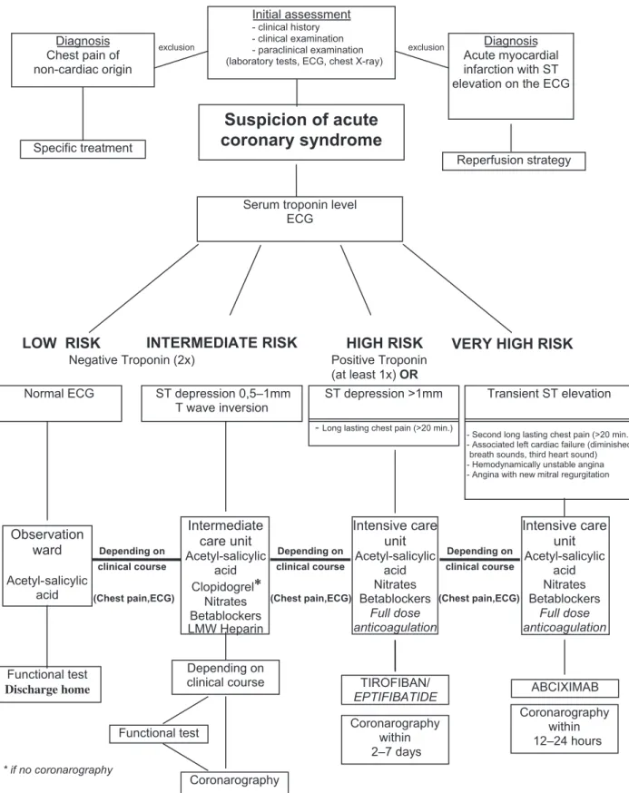
The practice guidelines were introduced in January 2001. Implementation strategy included presentation and distribution of the guidelines to all internal medicine and cardiology phy-sicians, including interns, residents, fellows, and staff physi-cians, and educational interventions by experts in the field during weekly seminars (small group meetings) and grand rounds (large group meetings). During these meetings, detailed presentation of the guidelines was carried out and all ques-tions related to the topic were answered. A comprehensive document summarizing the evidence was handed out [16], as well as a one-page algorithm (Figure 1). Both documents were also available on the intranet network. In addition, we placed reminders in the charts of all patients diagnosed with chest pain while in the emergency room, and posted general reminders on the emergency residents’ office walls. At the same time, a cardio-logy consultant, who was available during working hours 5 days a week, was added to the emergency medical staff.
For the second part of the study, conducted after introduc-tion of the guidelines, several strategies were used to minimize the change in the residents’ professional behaviour induced by the study itself (Hawthorne effect). Residents were blind to the actual aim of the trial and were only informed that a sur-vey on cardiovascular medicine would be conducted among clinical patients. Furthermore, we simply announced a train-ing program in evidence-based medicine includtrain-ing sessions on several clinical topics, such as deep venous thrombosis or community-acquired pneumonia for example.
Impact on patient initial assessment was measured following the same methodology as described above, over a 3-month period, extending from 1 February 2001 to 30 April 2001. Analysis compared patient characteristics, initial assessment characteristics, laboratory test results, ECG interpretation, and risk stratification in the two parts of the study.
Two reviewers (A.B. and J.B.W.) separately carried out independent risk assessments based on laboratory tests results and ECG interpretation for all patients. The ECG interpret-ation was based upon an independent assessment of all ECGs by two other reviewers (P.E. and J.C.S.), blinded to the clin-ical and patient’s characteristics, and ECG interpretation by emergency residents, who classified them, as did the resi-dents, into the following categories: normal, negative T-wave; down-sloped ST segment 0.5–1.0 mm; down-sloped ST segment >1.0 mm; and transient ST segment elevation, corresponding to the four categories of risk mentioned above. Disagreement between the two reviewers was resolved by a third reviewer ( J.S.). We used this final ECG analysis for the independent risk assessment. Concordance analysis was carried out only for patients with available risk stratification data and diagnosed with ACS at the end of the initial assessment.
Comparisons between the two groups were carried out using the Student’s t-test for continuous variables (after assessment of distribution normality), the Mann–Whitney U-test for ordinal variables or non-normally distributed data, and the χ2 test for distributions, as appropriate. Distribution
analyses were carried out after exclusion of missing data, and opposing missing data to available ones. All analyses were
Depending on clinical course Coronarography Functional test Intermediate care unit Acetyl-salicylic acid Clopidogrel
∗
Nitrates Betablockers LMW Heparin Depending on clinical course (Chest pain,ECG) Functional test Discharge home Observation ward Acetyl-salicylic acid Depending on clinical course (Chest pain,ECG) TIROFIBAN/ EPTIFIBATIDE Coronarography within 2–7 days Depending on clinical course (Chest pain,ECG) Intensive care unit Acetyl-salicylic acid Nitrates Betablockers Full dose anticoagulation Intensive care unit Acetyl-salicylic acid Nitrates Betablockers Full dose anticoagulation ABCIXIMAB Coronarography within 12–24 hours * if no coronarography- Long lasting chest pain (>20 min.)
- Second long lasting chest pain (>20 min.) - Associated left cardiac failure (diminished
breath sounds, third heart sound) - Hemodynamically unstable angina - Angina with new mitral regurgitation
Transient ST elevation ST depression >1mm
ST depression 0,5–1mm T wave inversion Normal ECG
Serum troponin level ECG
Positive Troponin (at least 1x) OR
HIGH RISK
VERY HIGH RISK
Negative Troponin (2x)
INTERMEDIATE RISK
LOW RISK
Suspicion of acute
coronary syndrome
exclusion exclusion Diagnosis
Acute myocardial infarction with ST elevation on the ECG Diagnosis Chest pain of non-cardiac origin Initial assessment - clinical history - clinical examination - paraclinical examination (laboratory tests, ECG, chest X-ray)
Reperfusion strategy Specific treatment
Results
During the two study periods, 3284 and 3260 patients, respec-tively, were evaluated at the Medical Emergency Department of our institution. Among them, 497 (15.1%) and 498 (15.3%) were evaluated for chest pain, by 33 and 32 different physi-cians, respectively. Residents contributed to 95% and 97% of the assessments, and only four of them treated patients dur-ing both study periods, with the majority of assessments car-ried out during one period.
Non-cardiac origin was diagnosed in 143 (28.8%) and 109 (21.9%) patients, pre- and post-guidline implementation, res-pectively. Clinical characteristics of these patients were not statistically significantly different between the two study peri-ods. One hundred and sixteen were women and 136 were men (mean age 47 ± 20 years). Fifty per cent of them suffered from osteo-articular problems, 21% from pulmonary diseases, 10%
from psychological disturbances, 5% from pericarditis and myocarditis, and 2% from gastro-intestinal disorders. Finally, unknown origin for chest pain was ascribed to 12% of them.
AMI with persistent ST elevation was diagnosed in 26 (5.2%) and 25 (5.0%) patients in the two groups, respec-tively. Clinical characteristics of these patients were again not different between the two study periods. Of these 51 patients (13 women and 38 men, mean age 47 ± 20 years), 42% of them suffered from anterior wall infarction, and 48% from inferior or posterior wall infarction, while 4% were located on the right ventricle only and 6% could not be located at all.
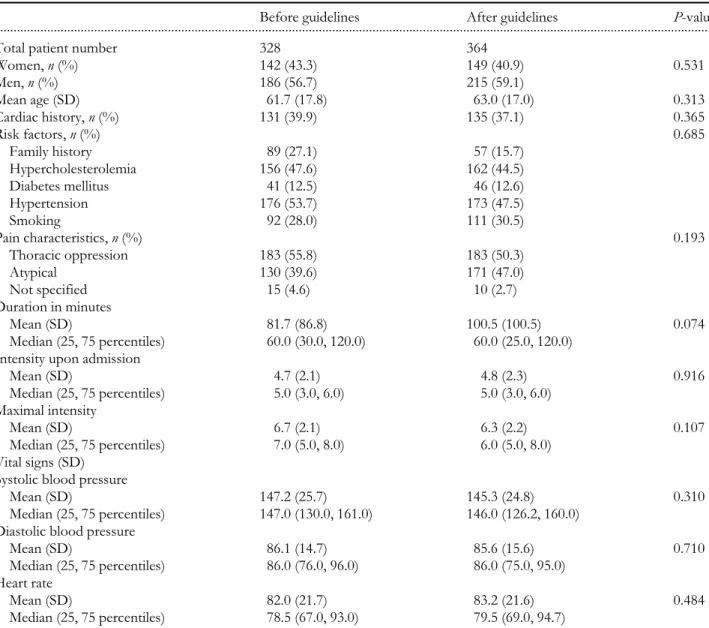
The remaining 328 (10.0% of all admissions in the first period) and 364 (11.2% of all admissions in the second period) patients were included in this study. Their main characteristics are summarized in Table 1. The two groups were not statisti-cally significantly different.
Table 1 ACS patients’ characteristics
Before guidelines After guidelines P-value
...
Total patient number 328 364
Women, n (%) 142 (43.3) 149 (40.9) 0.531 Men, n (%) 186 (56.7) 215 (59.1) Mean age (SD) 61.7 (17.8) 63.0 (17.0) 0.313 Cardiac history, n (%) 131 (39.9) 135 (37.1) 0.365 Risk factors, n (%) 0.685 Family history 89 (27.1) 57 (15.7) Hypercholesterolemia 156 (47.6) 162 (44.5) Diabetes mellitus 41 (12.5) 46 (12.6) Hypertension 176 (53.7) 173 (47.5) Smoking 92 (28.0) 111 (30.5) Pain characteristics, n (%) 0.193 Thoracic oppression 183 (55.8) 183 (50.3) Atypical 130 (39.6) 171 (47.0) Not specified 15 (4.6) 10 (2.7) Duration in minutes Mean (SD) 81.7 (86.8) 100.5 (100.5) 0.074 Median (25, 75 percentiles) 60.0 (30.0, 120.0) 60.0 (25.0, 120.0)
Intensity upon admission
Mean (SD) 4.7 (2.1) 4.8 (2.3) 0.916 Median (25, 75 percentiles) 5.0 (3.0, 6.0) 5.0 (3.0, 6.0) Maximal intensity Mean (SD) 6.7 (2.1) 6.3 (2.2) 0.107 Median (25, 75 percentiles) 7.0 (5.0, 8.0) 6.0 (5.0, 8.0) Vital signs (SD) Systolic blood pressure
Mean (SD) 147.2 (25.7) 145.3 (24.8) 0.310
Median (25, 75 percentiles) 147.0 (130.0, 161.0) 146.0 (126.2, 160.0) Diastolic blood pressure
Mean (SD) 86.1 (14.7) 85.6 (15.6) 0.710
Median (25, 75 percentiles) 86.0 (76.0, 96.0) 86.0 (75.0, 95.0)
Heart rate
Results of the initial assessment are shown in Table 2. Although vital signs, mean duration, and maximal intensity of chest pain did not change significantly, significantly more patients were considered as having atypical chest pain at the end of the initial assessment after implementation of the guidelines [120 patients (47.0%) compared with 154 patients (39.6%); P = 0.002). In addition, ECG description was more often classified as abnormal. Troponin I blood level results were less often under the detection limit. These findings sug-gest a lower diagnostic threshold, but a better targeting of the use of diagnostic tests after guideline implementation than before.
The most important changes after guideline implement-ation were the clinician’s commitment in assessing the prob-ability of coronary heart disease (94.8% compared with 82.9% pre-implementation), in stratifying patients by risk of coro-nary event (65.4% versus 53.7%), and in making a diagnosis at the end of the initial assessment (92.9% versus 79.9%). The number of missing values dropped significantly in these three variables (P < 0.0001).
Comparison of the risk stratification of patients performed by the clinicians and the reviewers at the end of the initial assessment showed significantly less differences after guide-line implementation than before (Table 3). Data were avai-lable for 87.8% of the patients before and 97.2% after
guideline implementation. Concordance was observed in 20.8% and 42.0% of cases, respectively. Discrepancies were limited to one level of risk in 59.4% of cases before and 44.2% after guideline implementation, with overestimation of risk by clinicians in 64.4% and 43.4% of cases, respectively. Conversely, underestimation of risk by clinicians was recorded in 14.8% and 14.5% of cases, respectively.
Discussion
This prospective study showed that clinical practice guide-lines for evaluation and treatment of ACS had a partial but positive impact on patient assessment. The study populations before and after guideline implementation were not statisti-cally significantly different, but their assessment at the emer-gency department was quite different. Particularly striking were the apparent decrease in relative importance of history characteristics in favour of ECG and troponin blood level results in attributing a patient to a specific risk category. This might be ascribed to the impact of the decision algorithm that summarized the clinical practice pathway.
Also striking was the increased commitment of physicians in the assessment of the probability of underlying coronary artery disease, patient risk stratification, and diagnosis at the Table 2 Results of the initial assessment
Before guidelines, n (%) After guidelines, n (%) P-value ...
Total n 328 364
Electrocardiogram 0.971
Normal 149 (45.4) 173 (47.5)
Abnormal 149 (45.4) 172 (47.4)
Assessment not available 30 (9.2) 26 (7.1)
Troponine T blood level 0.003
<0.03 µg/ml 265 (80.8) 254 (69.8)
0.03–0.09 µg/ml 28 (8.5) 67 (18.4)
>0.1 µg/ml 35 (10.7) 43 (11.8)
Presence of coronary heart disease 0.356
Yes 163 (49.7) 194 (53.3)
No 109 (33.2) 151 (41.5)
Assessment not available 56 (17.1) 19 (5.2) <0.0001
Risk of coronary event <0.0001
Small 40 (12.2) 106 (29.1)
Intermediate 50 (15.2) 55 (15.1)
High 64 (19.5) 51 (14.0)
Very high 22 (6.7) 26 (7.1)
Assessment not available 152 (46.3) 126 (34.6) 0.002
Diagnosis 0.646
Atypical chest pain 120 (36.6) 154 (42.3)
Probable unstable angina 98 (29.9) 118 (32.4)
Unstable angina 17 (5.2) 24 (6.6)
Myocardial infarction 14 (4.3) 16 (4.4)
Other diagnosis 13 (4.0) 26 (7.1)
time of the initial evaluation. This influence is likely to be ascribed to the availability of risk classification for immediate use at the bedside.
Moreover, the ACS guidelines’ positive impact on diminish-ing diagnostic uncertainty must be emphasized, as initial assess-ment is the cornerstone for allocating patients to specific treatment and monitoring. Any intervention conducted to decrease the delay in reaching the appropriate diagnosis and starting treatment is likely to have a positive impact on patient outcome. On the other hand, it should also result in a better use of scarce resources, such as expensive treatment procedures or intensive monitoring facilities, by reducing both over- and under-treatment, which both might negatively affect patient outcome. This study did, however, show important limits in compli-ance with the guidelines’ recommendations. The first one was related to the use of troponin blood level determination. Although results of the test during the second part of the study were more often located above the detection threshold, or positive, suggesting a better targeting of patients, its use was not optimal. In particular, the recommended second tro-ponin blood level determination in case of a negative first one was seldom carried out (35%). This might be explained by the fact that this kind of test had just been introduced in our insti-tution, and that physicians still lacked expertise in using it. The second limit is the relatively small improvement in accu-rate risk stratification in these patients, as demonstaccu-rated by a low level of agreement between clinicians and experts. This is possibly due to the fact that the concept of risk stratification was new for most of them. The finding that risk was over-estimated in most discrepant assessments suggests that emer-gency room physicians might be overcautious in taking care of these patients, and might prefer over-treatment and unnec-essary monitoring to under-treatment and overlooking moni-toring. Further intervention is clearly needed to improve the routine use of risk stratification and its confident assessment, and longer follow-up necessary to assess the impact of the learning curve effect linked with the residency program.
the bedside. This had already been demonstrated with the first version of the US guidelines: their introduction increased the percentage of patients treated with the recommended drugs such as aspirin or beta-blockers, and decreased the number of patients on calcium antagonists [17,18]. Another study carried out in Australia failed to show such a clear impact on drug use [19]. Altogether, a single study showed that patients’ survival improved after guideline implement-ation [20]. On the other hand, such guidelines were designed to ease the orientation of low-risk patients to outpatient care, and high-risk patients to intensive care units. With respect to this, they partly missed their goals: low-risk patients were not treated as outpatients, and hospitalizations did not decrease, but at the same time an increased demand for intensive care beds was noted, which would require additional resources for no proven survival benefit [21]. Finally, guidelines were incompletely applied to elderly patients, and quality of care varied widely between hospitals [22]. Cardiologists were more likely to apply them than general internal medicine specialists, with no differences in patient outcome [23].
These findings points out the necessity of careful assessment of both processes of care and patient outcomes when an inter-vention is implemented in the health care system, in particular the introduction of clinical practice guidelines. In addition, local adaptation of international guidelines is expensive, and not in itself a guarantee that they will be applied [24].
This study has obvious limitations: firstly, it involved only one centre; secondly, it did not extend to assessing patient outcome; thirdly, the impact of the practice guidelines was assessed shortly after their implementation, and was not repeated later; and fourthly, the emergency room teams at the times of the two study periods were different. However, the study’s findings are in perfect agreement with those described in the medical literature [17,22]. They will serve as a basis for additional interventions aimed at continuous quality improve-ment in our setting. These findings underline the importance of pre-testing guidelines by explicitly quantifying the risks and Table 3 Concordance of risk stratification between emergency room physicians and reviewers
... ER physician
... Risk before guideline implementation (n)
... Risk after guideline implementation (n) Low Moderate High Very high Total Low Moderate High Very high Total ... Reviewer Risk (n) Low 0 23 17 2 42 25 25 13 1 64 Moderate 1 8 15 2 26 6 10 9 0 25 High 1 7 13 6 27 1 4 17 12 34 Very high 0 0 6 0 6 0 4 5 6 15 Total 2 38 51 10 101 32 43 44 19 138 Kappa −0.05 0.219 Statistical significance 0.301 <0.001
between countries [1,25]. These findings will also be used to model the costs and thus the resources needed to anticipate improved compliance with the guidelines, once barriers to their implementation are addressed [26], so that patient safety can be guaranteed by appropriate monitoring when high-risk drugs and procedures are used to treat them.
References
1. Fox KAA, Goodman SG, Klein W et al. Management of acute coronary syndromes. Variations in practice and outcome. Find-ings from the Global Registry of Acute Coronary Events (GRACE). Eur Heart J 2002; 23: 1177–1189.
2. Wicox I, Freedman SB, McCredie RJ, Carter GS, Kelly DT, Harris PJ. Risk of adverse outcome in patients admitted to the coronary unit with suspected unstable angina pectoris. Am J Cardiol 1989; 64: 845–848.
3. The PURSUIT Investigators. Inhibition of platelet glycoprotein Iib/IIIa with eptifibatide in patients with acute coronary syn-dromes. N Engl J Med 1998; 339: 436–443.
4. Ross R. Atherosclerosis: an inflammatory disease. N Engl J Med 1999; 340: 115–125.
5. Libby P. Current concept of the pathogenesis of the acute coro-nary syndromes. Circulation 2001; 104: 365–372.
6. Topol EJ. Acute coronary syndromes. In Topol EJ, ed., Textbook of Cardiovascular Medicine. Philadelphia, PA: Lippincott-Raven, 1998. 7. Braunwald E. Unstable angina: a classification. Circulation 1989;
80: 410–414.
8. Rizig DG, Healy S, Margulis A et al. A new clinical classification for hospital prognosis of unstable angina pectoris. Am J Cardiol 1995; 75: 993–997.
9. Calvin JE, Klein LW, Van den Berg BJ. Risk stratification in unstable angina. Prospective validation of Braunwald classifi-cation. JAMA 1995; 273: 136–141.
10. Hamm CW, Goldmann BU, Heeschen C, Kreymann G, Berger J, Meinertz T. Emergency room triage of patients with acute chest pain by means of rapid testing for cardiac troponin T or troponin I. N Engl J Med 1997; 337: 1648–1653.
11. Antman EM, Tanaqsijevic MJ, Thompson B et al. Cardiac-specific troponin I levels to predict the risk of mortality in patients with acute coronary syndromes. N Engl J Med 1996;
335: 1342–1349.
12. Lindahl B, Venge P, Wallentin L. Troponin T identifies patients with unstable coronary artery disease who benefit from long-term antithrombotic protection. J Am Coll Cardiol 1997; 29: 43–48. 13. Braunwald E, Jones R, Mark DB. Diagnosing and managing
unstable angina. Circulation 1994; 90: 613–622.
14. Recommendations of the Task Force of the European Society of Cardiology. Management of acute coronary syndromes: acute coronary syndromes without persistent ST segment elevation. Eur Heart J 2000; 21: 1406–1432.
15. A Report of the American College of Cardiology/American Heart Association Task Force on Practice Guidelines. ACC/ AHA Guidelines for the management of patients with unstable angina and non-ST-segment elevation myocardial infarction. J Am Coll Cardiol 2000; 36: 970–1062.
16. Berger A, Eckert P, Stauffer JC, Wasserfallen JB. Le syndrome coronarien aigu. Recommandations pour la pratique clinique (in French). Genève: Editions Médecine & Hygiène, 2002.
17. Krumholz HM, Philbin DM, Wang Y et al. Trends in the quality of care for Medicare beneficiaries admitted to the hospital with unstable angina. J Am Coll Cardiol 1998; 31: 957–963.
18. Iliadis EA, Klein LW, Vandenberg BJ et al. Clinical practice guidelines in unstable angina improve clinical outcomes by assuring early intensive medical treatment. J Am Coll Cardiol 1999;
34: 1689–1695.
19. Heller RF, D’Este C, Lim LLY, O’Connell RL, Powell H. Randomised controlled trial to change hospital management of unstable angina. Med J Austr 2001; 174: 217–221.
20. Giugliano RP, Lloyd-Jones DM, Camargo CA, Makary MA, O’Donnell CJ. Association of unstable angina guideline care with improved survival. Arch Intern Med 2000; 160: 1775–1780. 21. Katz DA, Griffith JL, Beshansky JR, Selker HP. The use of
empiric clinical data in the evaluation of practice guidelines for unstable angina. JAMA 1996; 276: 1568–1574.
22. Shashi CN, Rathore SS, Wang Y et al. Quality of care among elderly patients hospitalized with unstable angina. Am Heart J 2001; 142: 263–270.
23. Reis SE, Holubkov R, Zell KA, Edmundowicz D, Shapiro AH, Feldman AM. Unstable angina: specialty-related disparities in implementation of practice guidelines. Clin Cardiol 1998; 21: 207–210.
24. Silagy CA, Weller DP, Lapsley H, Middleton P, Shelby-James T, Fazekas B. The effectiveness of local adaptation of nationally pro-duced clinical practice guidelines. Fam Pract 2002; 19: 223–230. 25. Hasdai D, Behar S, Wallentin L et al. A prospective survey of the
characteristics, treatments and outcomes of patients with acute coronary syndromes in Europe and the Mediterranean basin: the Euro Heart Survey of Acute Coronary Syndromes. Eur Heart J 2002; 23: 1190–1201.
26. Katz DA. Barriers between guidelines and improved patient care: an analysis of AHCPR’s unstable angina clinical practice guideline. Health Serv Res 1999; 34: 377–389.