JSPP © 1997
Lipids oi Ectocarpus fasciculatus (Phaeophyceae). Incorporation of
[l-
14C]Oleate and the Role of TAG and MGDG in Lipid Metabolism
Anja Makewicz, Corinne Gribi and Waldemar Eichenberger
Department of Chemistry and Biochemistry, University of Bern, Freiestrasse 3, CH-3012 Bern/Switzerland
Lipids and fatty acids of Ectocarpus fasciculatus (Ectocarpales, Phaeophyceae) were analyzed. Major polar lipids are monogalactosyldiacylglycerol (MGDG), digalacto-syldiacylglycerol (DGDG), sulfoquinovodigalacto-syldiacylglycerol (SQDG), diacylglycerylhydroxymethyl-iV.A'j.N-trimethyl-/J-alanine (DGTA), phosphatidylcholine (PC), phospha-tidylethanolamine (PE), phosphatidylglycerol (PG) and phosphatidylinositol (PI). Diphosphatidylglycerol (OPG), phosphatidic acid (PA) and phosphatidyl-O-[iV-(2-hydroxy-ethyl)glycine] (PHEG) were also present in small amounts. Nonpolar lipids mainly consist of triacylglycerol (TAG) and diacylglycerol (DAG). Major fatty acids are 16:0,18:1, al8:3, 18:4, 20:4 and 20:5. The positional distribution of fatty acids showed that molecular species of eukaryotic structure account for 99% in MGDG, 98% in DGDG, 62% in PG and 23% in SQDG. On incubation with [1-14C]18:1 for 30 min, 33% of the total label was detected in TAG, 16% in PG, 14% in PE, 10% in PC and 8% in MGDG. During 7 days of chase, the label in TAG, PG, PE and PC decreased and simultaneously increased in MGDG up to 41% of the total. In SQDG, labelled fatty acids were found in prokaryotic as well as in eukaryotic molecular species. During the experiment, the label shifted from 18:1 to 18:2, 18:3,18:4 and, to a minor extent, to 20:4 and 20:5 acids in-dicating 18:1 to be processed by elongation and/or desatu-ration. These results suggest TAG to act as a major pri-mary acceptor of exogenous oleate and to be involved in the transfer of fatty acids to MGDG and other polar lipids.
Abbreviations: Butyl-PBD, 2-(4-f-butylphenyl)-5-(4"-biphen-ylyl)-l,3,4-oxadiazol; DAG, diacylglycerol; DGDG, digalactosyl-diacylglycerol; DGTA, diacylglycerylhydroxymethyl-/V,M/v'-tri-methyl-/J-alanine; DPG, diphosphatidylglycerol; FFA, free fatty acids; FID, flame ionization detector; GLC, gas liquid chromatog-raphy; MGDG, monogalactosyldiacylglycerol; PA, phosphatidic acid; PC, phosphatidylcholine; PE, phosphatidylethanolamine; PG, phosphatidylglycerol; PHEG, phosphatidyl-O-[N-(2-hydro-xyethyl) glycine]; PI, phosphatidylinositol; RP-HPLC, reversed-phase HPLC; SQDG, sulfoquinovosyldiacylglycerol; TAG, triacylglycerol; TLC, thin-layer chromatography; 14:0, tetra-decanoic acid; 16:0, hexatetra-decanoic acid; fl6:l, 43-frww-hexadece-noic acid; cl6:l, zl9-c£s-hexadece43-frww-hexadece-noic acid; 18:0, octadeca43-frww-hexadece-noic acid; 18:1, 49-octadecenoic acid; 18:2, J9,12-octadecadienoic (linoleic) acid; j>18:3, <d6,9,12-octadecatrienoic (y-linolenic) acid; al8:3, d9,12,15-octadecatrienoic (a-linolenic) acid; 18:4, A 6,9,12,15-oc-tadecatetraenoic acid; 20:0, eicosanoic acid; 20:4, J5,8,11,14-eicosatetraenoic (arachidonic) acid; 20:5, <d5,8,ll,14,17-eicos-apentaenoic acid; 22:0, docosanoic acid.
Key words: Ectocarpus fasciculatus — Fatty acids — Lipid
metabolism — Monogalactosyldiacylglycerol — Phaeo-phyceae — Triacylglycerol.
Brown algae (Phaeophyceae) comprising around 265 genera and more than 1500 species (South and Whittick 1987) represent a considerable part of the marine flora. Many of them are used as human or animal food (Chap-man and Chap(Chap-man 1980) and are therefore of nutritional and economic significance (Jensen 1979). Compared to higher plants, brown algae like many other marine algae are exceptional in their lipid and fatty acid pattern. Like other chromatophytes, brown algae produce long-chain C20 polyenoic fatty acids mainly in the form of 20:4 and 20:5 acids (Arao and Yamada 1989, Harwood and Jones 1989, Jones and Harwood 1992, Fleurence et al. 1994, Khotimchenko 1995, Kim et al. 1996, Vaskovsky et al. 1996). Another typical feature of brown algae is the pres-ence of the betaine type lipid diacylglycerylhydroxyme-thyl-iV,iV,/v'-trimethyl-/?-alanine (DGTA) in many species (Eichenberger et al. 1993). The occurrence of this lipid and of the common phospholipid phosphatidylcholine (PC) partly reflects the brown algal taxonomy, since DGTA is either present or absent in all the members of a given order. Exceptions are the orders Ectocarpales and Chordariales which contain both DGTA-positive and DGTA-negative species. Surprisingly, in the members of the DGTA-produc-ing Dictyotales, Fucales and Durvillaeales, PC could not be detected. In addition, the phospholipid phosphatidyl-O-[7V-(2-hydroxyethyl) glycine] (PHEG) has recently been de-tected and seems to be typical of, but limited to brown algae (Eichenberger et al. 1995). A characteristic feature of brown algae is the positional distribution of fatty acids in plastidial lipids. As in other chromophyte algae, MGDG is almost exclusively of eukaryotic structure, i.e. thesn-2 posi-tion is occupied by C18 or C20 fatty acyl groups (Arao and Yamada 1989, Jones and Harwood 1992). This is surpris-ing especially for those species which do not contain PC, since this phospholipid is the source of eukaryotic DAG moieties used for MGDG synthesis in higher plants (Roughan and Slack 1982, Browse and Somerville 1991). Although a considerable body of analytical data has accu-mulated on the lipids and fatty acids of brown algae, little is known about the biosynthesis and the metabolism of
both lipids and long-chain polyenoic fatty acids which are important as precursors of pheromones involved in the cell-cell communication (Stratmann et al. 1993).
Labelling experiments with [l-l4C]acetate as a precur-sor carried out with Fucus serratus (Smith and Harwood 1984, Jones and Harwood 1987) revealed a preferred incor-poration of label into an unknown lipid which was later on identified as DGTA. These results suggested a metabolic role of this betaine lipid. The lipid metabolism of Fucus spe-cies was also studied under the aspect of water pollution by heavy metals (Smith et al. 1984).
For our experiments, Ectocarpus fasciculatus was used because it produces DGTA as well as PC (Muller and Eichenberger 1994, culture SC 42-3). Also, this organism by its morphology is appropriate for in vivo incubations and can easily be cultivated under laboratory conditions. Using [1-MC]18:1 as a precursor, we intended to gain new
information on the incorporation of fatty acids and their further processing (desaturation and elongation) by intact plants and, at the same time, on the distinct features of brown algal lipid metabolism. Also, insight into the role of DGTA and the formation of plastidial glycolipids was ex-pected.
Materials and Methods
Plant material—Ectocarpus fasciculatus Harvey (a unialgal
clonal culture originating from Santa Clara Island, Juan Fer-nandez Archipelago, Chile, in 1991) was obtained from Prof. D.G. Muller, University of Konstanz, Germany. Plants were cultivated in Petri dishes in 25 ml of autoclaved North Sea water (salinity 2.8%) or a commercial salt mixture supplemented accord-ing to Starr and Zeikus (1993). Cultures were grown at 18°C with a light/dark cycle of 12/12 h under white fluorescent light (60 ^/E m~2 s~') and by changing the medium every week.
Lipid isolation and determination—Lipids were extracted
with boiling methanol containing 0.05% butylhydroxytoluene. Lipids were separated by TLC on silica gel plates (Merck 5715) with chloroform/methanol/water (65 : 25 : 4, by vol.) (solvent I) in the 1st dimension and with chloroform/methanol/isopropyl-amine/conc. ammonia (65 : 35 : 0.5 : 5, by vol.) (solvent II) in the 2nd dimension. DAG and TAG were isolated by TLC of total lipid using hexane/diethyl ether/acetic acid (70 : 30 : 1, by vol.) as a solvent. Spots were detected with 2',7'-dichlorofluorescein under UV (366 nm) and eluted with methanol. For identification, phos-pholipids were stained with molybdenum blue (Dittmer and Lester 1964), glycolipids with anthrone (Heinz 1967), amino groups with ninhydrin (Fahmy et al. 1961) and quaternary amino groups with Dragendorff reagent (Munier and Macheboeuf 1951).
Fatty acid analysis—Fatty acid methyl esters were obtained
either from total lipids by transesteriflcation with sodium meth-oxide (Thies 1971) or from free fatty acids by reaction with diazomethane. The positional distribution of fatty acids between the sn-l and sn-2 position was determined using lipase from
Rhizopus arrhizus (Fischer and Heinz 1973). For analytical
separa-tions, a Shimadzu G8A equipped with FID and a Shimadzu C-R3A integrator was used. The column was a fused silica capillary column (Restek RTx-2330, 30 m length, 0.25 mm I. D.) operating
at 170-190°C (2°C min"1) with H2 as carrier gas.
Phenacyl ester analysis—Phenacyl esters of fatty acids were
prepared with phenacylbromide and triethylamine according to Borch (1975) and separated by RP-HPLC on a Perkin-Elmer Series 10 Liquid Chromatograph with a Nucleosil 100-5 C18
col-umn (250 x 4 mm, Macherey-Nagel). The solvent was a gradient of 100% acetonitrile and acetonitrile/water (6 : 4 , by vol.). The gradient was from 20% to 80% acetonitrile in 48 min with a flow rate of 1.5 ml min"1. The phenacyl esters were detected at 254 nm with a Perkin-Elmer L75 detector connected to a Shimadzu C-R3A integrator.
Incubation conditions and radioactive measurements—
Plants were incubated in 3 ml of culture medium to which a solu-tion of 148 MBq [l-14C]oleic acid (2.1 GBq mmof1, Amersham)
in 40 ftl ethanol was added. During the incubation, the suspension was shaken under light at room temperature. After 30 min pulse, plants were washed with 6 ml chase medium containing unlabelled oleic acid (20 /ig ml"1) and transferred to a Petri dish with 25 ml of chase medium and kept under culture conditions. Aliquots were taken after 0, 6, 24, 48 and 96 h and after 7 d. Radioactivity was measured with a Liquid Scintillation Counter MR 300 (Kon-tron, Switzerland) after addition of 4 ml methanol and 5 ml butyl-PBD (7% in toluene, w/v).
Preparation and analysis of SQDG molecular species—
SQDG was isolated by semipreparative TLC of total lipid on silicagel plates (Merck 5715) using solvents I and II. The lipid was hydrogenated in acetone/methanol (4 : 1 v/v) with H2 gas and
palladium black (Fluka, Switzerland) as a catalyst, for 2.5 h at 20° C. The saturated molecular species were separated by isokratic RP-HPLC on a Nucleosil 100-5 C,8 column (250x4 mm) using
methanol/water/acetonitrile (181 : 14 : 5, by vol.) containing 20 mM choline-HCl. The flow was 1 ml min"1. Fractions of 2 ml were collected and in an aliquot of each fraction, radioactivity was measured. Fractions 17-24 were pooled and the solvent was evapo-rated. To the residue, 2 ml of water was added and the mixture was extracted three times with 2 ml chloroform. The residue of the chloroform phase was rechromatographed on a TLC plate using solvent I and the hydrogenated SQDG eluted with methanol. In the purified SQDG, the total fatty acid composition and the posi-tional distribution of both fatty acids and radioactivity was deter-mined as described above.
Results
Lipid composition—-MGDG, DGDG, SQDG, DGTA,
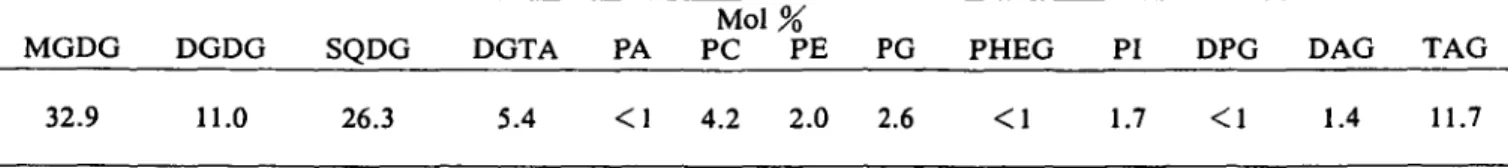
PC, PE, PG and PI were the most prominent polar lipids, while DPG, PA and PHEG were present as minor constitu-ents, as shown in Table 1. The nonpolar lipids contained TAG, DAG and the major part of pigments. A weak spot with free fatty acids was also observed.
Fatty acid composition—Major components are 16:0,
18:1, al8:3, 18:4, 20:4 and 20:5 which account for 9 1 % of the total, as shown in Table 2. The galactolipids DGDG and MGDG are rich in al8:3, 18:4 and 20:5, while the sulfolipid SQDG mainly contains 16:0, 18:1 and ctl8:3. The betaine lipid DGTA and the phospholipid PC predomi-nantly contain 16:0, 20:4 and 20:5. DPG and PE are rich in 20:4 and 20:5, while in PHEG, almost 76% of the total fat-ty acids consist of 20:4. PG mainly contains 16:0 and al8:3, but rather low amounts of rl6:l. In PI, 16:0 and 18:1 predominate and PA mainly contains 18:0, 20:0 and
Table 1 Lipid composition of E. fasciculatus MGDG 32.9 DGDG 11.0 SQDG 26.3 DGTA 5.4 PA < 1 Mol PC 4.2 % PE 2.0 PG 2.6 PHEG < 1 PI 1.7 DPG < 1 DAG 1.4 TAG 11.7
20:4. In TAG, almost all the fatty acids are present and DAG is rich in 16:0 and yl8:3.
Positional distribution of fatty acids in various lipids
—The positional distribution of fatty acids in MGDG, DGDG, PG and SQDG, as obtained by enzymatic hydroly-sis with lipase from Rhizopus arrhizus, is shown in Table 3. The sn-2 position of MGDG and DGDG is occupied by only 1 and 2%, respectively, by Ci6 fatty acids. This in-dicates that 98 to 99% of both galactolipids are of the eukaryotic type. The proportion of eukaryotic species in PG, in contrast, is only 62% and, surprisingly, in SQDG as low as 23%. This indicates that in turn, SQDG is by almost 80% of the prokaryotic type with C16 acids in the sn-2 posi-tion.
Incorporation of [l-l4C]18:l into polar and nonpolar lipids—In order to investigate the incorporation of
radiola-belled oleic acid into the polar and nonpolar lipids, whole plants of Ectocarpus fasciculatus were incubated with [1-14
C]18:1 in a pulse-chase manner. Lipids were extracted at the end of the pulse and at different times of the chase peri-od, and then separated by two-dimensional TLC. During 30 min of incubation, almost 15% of the radiolabelled sub-strate was incorporated into the polar and nonpolar lipids.
At the end of the pulse, TAG, PG, PE and PC were the most strongly labelled lipids containing 33, 16, 14 and 10%, respectively, of the total lipid label, as shown in Fig. 1. During 7 days of chase, the label mainly decreased in TAG (down to 10% of the total), but to a minor extent also in PG, PE, PC, PI and DAG. In the same time, the la-bel drastically increased in MGDG from 8% up to 4 1 % of the total. A minor increase was also observed in DGDG, SQDG, DGTA and DPG. Almost no radioactivity was found in PHEG and in PA (not shown).
Incorporation of label into prokaryotic and euka-ryotic molecular species of SQDG—It was important to
find out whether of the plastidial lipids either eukaryotic only or both eukaryotic and prokaryotic species were la-belled. Since in E. fasciculatus, SQDG is the only lipid class which is synthesized exclusively in the chloroplast and, in the same time, contains a high proportion of prokaryotic species (Table 2), the labelling of this lipid was further in-vestigated. The total labelled SQDG was isolated and the unsaturated fatty acids were converted into their saturated form by hydrogenation. Then, the saturated SQDG was chromatographed by RP-HPLC and the fractions monitor-ed. Fractions 17-24 containing most part of the
radioactiv-Table 2 Fatty acid composition of lipids from E. fasciculatus Fatty acid 14:0 16:0 rl6:l cl6:l 18:0 18:1 18:2 yl8:3 al8:3 18:4 20:0 20:4 20:5 22:0 others Total lipids 1.6 16.7 0.6 0 1.0 12.5 4.2 1.4 15.2 22.6 0.3 10.8 13.4 0 0.7 MGDG 1.1 5.2 0 0.2 1.0 3.9 2.6 1.1 14.2 48.0 0 4.6 18.0 0.1 0 DGDG 1.3 8.3 0 0 0 0 3.0 1.4 17.2 29.8 0 2.3 36.7 0 0 SQDG 3.9 42.7 0.4 0.1 0.4 25.7 8.2 0.1 16.9 0.8 0.1 0.3 0.2 0.1 0.1 DGTA 3.9 18.5 0 0 2.0 3.2 6.1 2.4 3.1 1.6 0 42.4 16.8 0 0 PA 0 5.8 0 0 35.8 1.7 0 0 1.5 0 23.1 24.9 6.2 0 1.0 M o l % PC 4.7 30.7 0 0 0.8 4.8 6.5 1.3 5.7 2.0 0 23.7 17.7 0 2.1 fatty acids PE 0 7.2 0 0 2.3 4.0 1.5 3.8 2.2 1.7 1.7 55.2 18.0 1.4 1.0 PG 0.3 30.6 4.8 1.1 0.6 11.3 9.6 0.2 35.1 1.0 0.1 4.0 1.3 0 0 PHEG 0 4.5 0 0 3.3 1.7 0 3.0 4.1 0 0 76.4 7.0 0 0 PI 0 45.0 0 0 3.1 43.0 4.8 1.5 2.6 0 0 0 0 0 0 DPG 0 4.7 2.2 0 2.4 5.2 4.7 3.0 2.9 9.4 0 24.7 37.4 0 3.4 DAG 4.6 21.9 2.8 0 3.6 13.7 4.5 29.9 5.4 2.8 2.2 3.6 5.0 0 0 TAG 5.3 17.2 2.4 1.5 3.6 19.2 6.8 0 10.0 6.7 1.6 8.0 12.8 3.2 1.7
Table 3 Positional distribution of fatty acids in MGDG, DGDG, PG and SQDG of E. fasciculatus Fatty acid 14:0 16:0 tl6:l cl6:l 18:0 18:1 18:2 yl8:3 al8:3 18:4 20:0 20:4 20:5 22:0 others
zc
l6 MGDG sn-l 2.2 11.6 0 0.1 1.7 1.5 1.7 0.7 17.1 24.3 3.5 5.7 28.8 1.1 0 sn-2 0 1.0 0 0 0.3 7.5 4.5 1.3 12.3 62.3 0 3.6 7.2 0 0 1.0 99.0 M o l % DGDG sn-l 2.3 16.6 0 1.2 2.2 0 0 1.5 6.5 0.9 0 5.0 63.8 0 0 sn-2 0 1.8 0 0.7 0.5 7.3 4.8 0.8 28.6 54.0 0 0.5 1.0 0 0 2.5 97.5 fatty acids PG sn-l 0.3 30.8 0 0.2 0.6 1.3 7.9 0.2 56.8 0 0 1.5 0.3 0.1 0 sn-2 0 27.5 9.4 0.8 1.4 26.2 12.5 0.2 15.3 1.8 0 3.4 1.4 0 0.1 37.7 62.3 sn-l 6.9 13.6 0 0.5 0.9 47.4 8.0 0 22.0 0 0.2 0.2 0.3 0 0 SQDG sn-2 1.8 76.6 0 0.3 0.3 2.9 7.3 0 9.3 1.3 0 0.2 0 0 0 76.9 23.1ity, as shown in Fig. 2, were pooled and treated with lipase from Rhizopus arrhizus. The label was measured in both the fatty acid from the sn-l position and in the lyso SQDG containing the acyl group in the sn-2 position. The results are summarized in Table 4. From the total SQDG, 74% of the label was recovered in the fractions 17-24 containing 16:0 and 18:0 in a ratio of 1 : 1 (Fig. 2, insert) and thus con-sisting of 16:0/18:0 and 18:0/16:0 combinations only. Since, by the use of 18:1 as radiolabelled substrate, 18:0
but not 16:0 was labelled, the label found in the sn-l posi-tion can exclusively be ascribed to 18:0 from prokaryotic species. SQDG is significantly labelled in the sn-l position and, hence, in its prokaryotic species, as demonstrated in Table 4.
Incorporation of label into different fatty acids—In
order to trace the path of radiolabel from [1-MC]18:1 through individual fatty acids of total and particular lipids, fatty acid phenacyl esters were prepared and separated by
50 40 -% radioactivit y
8
8
10 -n cfill
I
I
I
• i
m
i
1
Ml
LI
u
MGDG DGDG SQDG DGTA PG PC PI PE PHEG BPG DAG TAG
• 0 E36 EB24 01148 ^ 9 6 hours iS7 days chase
800 600 ' • % 4 0 0 O CD
g
2 200
0 16:0 14:0 1 0Fatty acid composition of fractions 17-24 18.0 20:0 i 10min
Q.
nnr-i 110
20 30 fractionFig. 2 RP-HPLC separation of hydrogenated SQDG from E.fasciculatus labelled with [1-14C]18:1. 40
RP-HPLC, and the label in individual fatty acids was moni-tored. At the end of the 30-min pulse, the label mainly ap-peared in 18:1 acid, as shown in Fig. 3. During the chase, most label disappeared from 18:1 and, simultaneously ap-peared in 18:2, 18:3, 18:4. After 48 h chase, a minor increase of label was also observed in 20:4 and 20:5. This in-dicates that most of the incorporated oleate was desatu-rated to give 18:2, 18:3 and 18:4, while only a minor por-tion of the Ci8 acids was elongated and desaturated to give 20:4 and 20:5. Since only insignificant labelling of C14 and
C]6 acids was observed during the experiment, degradation of the substrate and reincorporation of the label by de novo synthesis of fatty acids can almost be excluded. The radiolabelling of fatty acids in glycolipids is shown in Fig. 4. In the two galactolipids and in the sulfolipid, most part of the label appeared in 18:1 at the end of the pulse, but then decreased in this acid during the chase period. In both MGDG and DGDG, the rapid decrease of label in 18:1 was accompanied by an accumulation of radioactivity mainly in 18:3 and 18:4 acids. In all the glycolipids, only
••s
s
1
100 -f 80 60 -14:0 16:0 16:1 18:1 18:2 18:3 18:4 20:4 20:5 40-• 0 ^16 E324 11348 1196 hours 117 days chase
Table 4 Incorporation of label from [1-I4C]18:1 into prokaryotic and eukaryotic SQDG
of E. fasciculatus
Fatty acid composition
(mol %) Radioactivity
RP-HPLC fractions 17-24
sn-\ fatty acid
sn-2 fatty acid (lyso SQDG)
16:0 52 17 82 18:0 47 83 17 others 1 1 4,980 2,282 271
minor amounts of label were found in 20:4 and 20:5 at the end of the chase. The labelling kinetics in phospholipids are presented in Fig. 5. In all of them, the radiolabel first ap-peared in 18:1, but behaved differently in these three lipids during the chase period. In PC, a shift of label into 18:2 and 18:3 and a final accumulation in 20:4 and 20:5 was
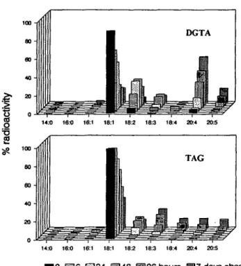
ob-served. In PE, the label was shifted more slowly but pri-marily to 20:4. In PG, little change of label in 18:1 and a si-multaneous increase in 18:2 and 18:3 but not in CM fatty acids, was observed. In DGTA, the label rapidly shifted from 18:1 temporarily to 18:2 and 18:3 but finally accumu-lated in 20:4 and, to a smaller extent, in 20:5, as shown in
M.-O 16:0 16:1 18:1 182 18:3 18:4 20:4 20:5 14.-0 16:0 16:1 18:1 18:2 183 18:4 20:4 203 14:0 16:0 16:1 18:1 18:2 183 18:4 20:4 20:5 10 S 6 E324 14:0 16:0 16:1 18:1 182 183 18:4 20:4 20:5 ioo -A 80 60 40 20 -0 -«=
•h
i
1
1
PE 14.1] 16:0 16:1 18:1 182 183 18:4 20:4 20:5 16:0 16:1 18:1 18:2 18:3 18:4 20:4 20:510 B 6 E324 96 hours ffl7 days chase
Fig. 4 Labelling of fatty acids of MGDG, DGDG and SQDG of Fig. 5 Labelling of fatty acids of PC, PE and PG of
14:0 16:0 16:1 18:1 18:2 18:3 18:4 20:4 20:5
14:0 16:0 16:1 18:1 18:2 18:3 18:4 20:4 20:5
• 0 S 6 E324 IH148 ^ 9 6 hours H 7 days chase
Fig. 6 Labelling of fatty acids of DGTA and TAG of E.
fasci-culatus after incubation with [1-14C]18:1.
Fig. 6. In TAG, a shift of label from 18:1 to almost all kinds of polyenoic fatty acids was observed.
Discussion
Ectocarpus fasciculatus shows the lipid pattern which
is typical of a photosynthetic plant with the glycolipids MGDG, DGDG and SQDG and the phospholipids PC, PE, PG, PI and DPG. In addition, it contains the common brown algal phospholipid PHEG (Eichenberger et al. 1995) and belongs to those members of the order Ectocarpales which also produce the betaine type lipid DGTA (Eichen-berger et al. 1993). Significant amounts of TAG and minor portions of DAG are also present. Major fatty acids are 16:0, 18:1, 18:2, 18:3, 18:4, 20:4 and 20:5 with C,6, C18 and C20 acyl chains accounting for 17, 57 and 25 mol%, respec-tively, of the total. Typically, the plastidial lipids including the glycolipids and PG contain large amounts of C18 fatty acids, while in PA, PC, PE, PHEG, DPG and DGTA, long-chain C20 polyenoic acids predominate. A similar tendency in fatty acid patterns has also been reported for other brown algae (Arao and Yamada 1989, Caron et al. 1985, Fleurence et al. 1994, Jones and Harwood 1992, Smith and Harwood 1984).
The positional distribution of fatty acids among the positions of the glycerol moiety, as determined by treat-ment with lipase of Rhizopus arrhizus, merits special atten-tion. In higher plants, glycerolipids containing a C,6 fatty
acid in their sn-2 position are ascribed to the plastidial or prokaryotic pathway using a DAG precursor of plastidial origin (Frentzen 1986). Correspondingly, lipids contain-ing in the same position an acyl chain with more than 16 carbons are thought to be synthesized by the eukaryotic pathway using a DAG backbone of cytoplasmic origin (Roughan and Slack 1984). In the sn-2 position of MGDG and DGDG of Ectocarpus, C!6 fatty acids acount for only 1% and 2%, respectively, while for PG and SQDG these values are 38 and 77%, respectively. Since the assembly of MGDG, DGDG and SQDG occurs in the plastid envelope (Heinz 1993, Joyard and Douce 1987, Mudd and Klepp-inger-Sparace 1987), according to the two-way concept for higher plants, the DAG used for the assembly of galac-tolipids in E. fasciculatus has to be imported by almost 100% from the cytoplasm, while for the synthesis of SQDG, DAG of predominantly plastidic origin is used. For PG, the proportions of prokaryotic (38%) and eukaryotic (62%) molecular species, reflect the amounts of PG in the chloroplasts and the extraplastidial compartments, respec-tively, since chloroplasts contain prokaryotic PG only (Roughan and Slack 1984). On labelling with [1-14C]18:1 during 30 min, 73% of the label appeared in TAG, PG, PE and PC. During the chase period, the label decreased in these lipids, but drastically increased in MGDG and, to a minor extent, also in DGDG, SQDG and DGTA. This shift of label strongly suggests an incorporation of the exoge-nous fatty acid into TAG and phospholipids and a subse-quent transfer of fatty acids to plastidial glycolipids. In this process, TAG apparently plays an important role as an in-termediate acyl carrier. During our experiment, 18:1 is con-verted mainly by desaturation to give 18:3 and 18:4 acids. The label in 20:4 and 20:5 acids, in contrast, accounts for only 16% of the total after 7 days indicating the production of C20 fatty acids to be rather slow under these conditions. The proportion of labelled Ci8 and C20 fatty acids, how-ever, is different in the various lipids. In glycolipids, an ac-cumulation of C)8 acids is observed. The rapid decrease of label in 18:1 acid and the concomitant increase in C,8 dienes, trienes and tetraenes strongly suggest a desatura-tion of lipid-bound C18 fatty acids. In both MGDG and DGDG, the end products arel8:3 and 18:4. The same pro-cess has also been reported from Isochrysis galbana (Stern and Tietz 1993) and Echium plantagineum (Williams and Khan 1996). Almost no labelled CM acids are incorporated into galactolipids during the experiment. In SQDG, the con-version of 18:1 is slower and mainly produces 18:3. In PC and DGTA where a shift of label from 18:1 to 18:2 and 18:3 temporarily also occurs, a significant accumulation of 20:4 and 20:5 is observed suggesting that these lipids act as final acceptors of long-chain fatty acids. A similar process, but to a minor extent also occurs in PE, while in PG only a slow turnover is observed. In TAG, about equal propor-tions of the different labelled fatty acids appear suggesting
that most part of the labelled 18:1 acid initially present is rapidly removed suggesting TAG to act as a donor of fatty acids for processing and/or transfer to polar lipids. A meta-bolic role of TAG has also been reported by Kim et al. (1996) who found a seasonal variation of the TAG content in Fucus serratus.
In higher plants, fatty acids used for the synthesis of eukaryotic glycolipids in the chloroplast are imported from the cytoplasm in the form of DAG. In E. fasciculatus, there is strong evidence for a translocation of single fatty acids from the cytoplasm to the chloroplast. The significant labelling of the prokaryotic molecular species of SQDG
(11% of the total) by a C18 acid in the sn-1 position is possi-ble only either by introduction of a single fatty acid in a de novo synthesized lipid molecule or by exchange of the sn-l acyl group in a preexisting molecule. Evidence that single fatty acids rather than DAG might be transferred from the cytoplasm to the chloroplasts, was also obtained from the haptophyte alga Pavlova lutheri (Eichenberger and Gribi 1997). This indicates that in chromophyte algae, the biosyn-thetic pathways for lipids may be different from those of higher plants as already suggested by Sato (1991). Further investigations should therefore be carried out also on the mechanism by which the surprisingly high proportion of eukaryotic MGDG is synthesized in these algae. The elonga-tion of C)8 to C20 fatty acids in higher plants is ascribed to an elongase system of the endoplasmic reticulum using acyl-CoA esters as substrates (Cassagne et al. 1994). Also in the eustigmatophyte alga Nannochloropsis, the elonga-tion has been attributed to the cytoplasm (Schneider and Roessler 1994). The desaturation steps leading to 20:4 and 20:5 are suggested to occur on PC-bound fatty acids in the red alga Porphyridium cruentum and the eustigmatophyte alga Monodus subterraneus (Khozin and Cohen 1996). This is in accordance with the general view that fatty acid desaturases in plants operate on lipid-bound substrates (Ohlrogge and Browse 1995). The labelling kinetics in
E. fasciculatus suggest that not only PC but also DGTA is
involved in the desaturation of C20 fatty acids indicating that the betaine lipid DGTA, as its isomer DGTS in green algae, may be involved in fatty acid desaturation.
We are indebted to Prof. D.G. Muller, University of Konstanz, for the gift of cultures of Ectocarpus fasciculatus and for fruitful discussions. The work has been supported by the Swiss National Science Foundation (Grant 31-45901.95).
References
Arao, T. and Yamada, M. (1989) Positional distribution of fatty acids in galactolipids of algae. Phytochemistry 28: 805-810.
Borch, R.F. (1975) Separation of long-chain fatty acids as phenacylesters by HPLC. Anal. Chem. 47: 2437-2439.
Browse, J. and Somerville, C. (1991) Glycerolipid synthesis: biochemistry and regulation. Annu. Rev. Plant Physiol. Plant Mol. Biol. 42: 467-506. Caron, L., Dubacq, J.P., Berkaloff, C. and Jupin, H. (1985)
Subchloro-plast fractions from the brown alga Fucus serratus: phosphatidylglycerol contents. Plant Cell Physiol. 26: 131-139.
Cassagne, C , Lessire, R., Bessoule, J.J., Moreau, P., Creach, A., Schneider, F. and Sturbois, B. (1994) Biosynthesis of very long chain fat-ty acids in higher plants. Prog. Lipid Res. 33: 55-69.
Chapman, V. J. and Chapman, D.J. (1980) Seaweeds and their Uses. Chap-man and Hall, London and New York.
Dittmer, J.C. and Lester, R.L. (1964) A simple, specific spray for the de-tection of phospholipids on thin-layer chromatograms. J. Lipid Res. 5: 126-127.
Eichenberger, W., Araki, S. and Muller, D.G. (1993) Betaine lipids and phospholipids in brown algae. Phytochemistry 34: 1323-1333. Eichenberger, W., Bigler, P., GfeUer, H., Gribi, C. and Schmid, C.E.
(1995) Phosphatidyl-O-[/V-(2-hydroxyethyl) glycine] (PHEG), a new gly-cerophospholipid from brown algae (Phaeophyceae). /. Plant Physiol. 146: 398-404.
Eichenberger, W. and Gribi, C. (1997) Lipids of Pavlova lutheri (Hap-tophyceae): cellular site and metabolic role of DGCC. Phytochemistry (in press).
Fahmy, R.A., Niederwieser, A., Pataki, G. and Brenner, M. (1961) Diinn-schicht-chromatographie von Aminosauren auf Kieselgel G. 2. Mitt., Eine Schnellmethode zur Trennung und zum qualitativen Nachweis von 22 Aminosauren. Helv. Chim. Ada 44: 2022-2026.
Fischer, W., Heinz, E. and Zeus, M. (1973) The suitability of lipase from Rhizopus arrhizus delemar for analysis of fatty acid distribution in dihex-osyl diglycerides, phospholipids and plant sulfolipids. Z. Physiol. Chem. 354: 1115-1123.
Fleurence, J., Gutbier, G., Mabeau, S. and Ceray, C. (1994) Fatty acids from 11 marine macro-algae of the French Brittany coast. J. Appl. Phycol. 6: 527-532.
Frentzen, M. (1986) Biosynthesis and desaturation of the different diacylglycerol moieties in higher plants. J. Plant Physiol. 124: 193-209. Harwood, J.L. and Jones, A.L. (1989) Lipid metabolism in algae. Adv.
Bot. Res. 16: 1-53.
Heinz, E. (1967) Ueber die enzymatische Bildung von Acylgalactosyldigly-cerid. Biochim. Biophys. Ada 144: 333-343.
Heinz, E. (1993) Recent investigations on the biosynthesis of the plant sulfolipid. In Sulfur Nutrition and Assimilation in Higher Plants. Edited by De Kok, L.J. et al. p. 163-178. Academic Publishing bv, The Hague. Jensen, A. (1979) Industrial utilization of seaweeds in the past, present and future. In Proc. Int. Seaweed Symp. 9. Edited by Jensen, A. and Stein, J.R. p. 17-34. Science Press, Princeton.
Jones, A.L. and Harwood, J.L. (1987) Comparative aspects of lipid metab-olism in marine algae. Biochem. Soc. Trans. 15: 482-483.
Jones, A.L. and Harwood, J.L. (1992) Lipid composition of the brown algae Fucus vesiculosus and Ascophyllum nodosum. Phytochemistry 31: 3397-3403.
Joyard, J. and Douce, R. (1987) Galactolipid synthesis. In The Biochemis-try of Plants. Vol. 9. Edited by Stumpf, P.K. and Conn, E.E. pp. 215-274. Academic Press, New York.
Khotimchenko, S.V. (1995) Uncommon 16:1 (n-5) acid from Dictyota dichotoma and fatty acids from some brown algae of Dictyotaceae. Phytochemistry 38: 1411-1415.
Khozin, I. and Cohen, Z. (1996) Differential response of microalgae to the substituted pyridazinone, Sandoz 9785, reveal different pathways in the biosynthesis of eicosapentaenoic acid. Phytochemistry 42: 1025-1029. Kim, M.K., Dubacq, J.-P., Thomas, J.-C. and Giraud, G. (1996) Seasonal
variations of triacylglycerols and fatty acids in Fucus serratus. Phyto-chemistry 43: 49-55.
Mudd, J.B. and Kleppinger-Sparace, K.F. (1987) Sulfolipids. In The Bio-chemistry of Plants. Vol. 9. Edited by Stumpf, P.K. and Conn, E.E. p. 275-289. Academic Press, New York.
Muller, D.G. and Eichenberger, W. (1994) Betaine lipid content and spe-cies delimitation in Ectocarpus, Feldmannia and Hincksia (Ectocarpales, Phaeophyceae). Eur. J. Phycol. 29: 219-225.
Munier, R. and Macheboeuf, M. (1951) Microchromatographie de partage sur papier des alcaloi'des et de diverses bases azotees biologiques. III. Ex-emples de separation de divers alcaioldes par la technique en phase solvente acide (families d'atropine, de la cocaine, de la nicotine, de la sparteine, de la strychnine et de la corynanthine). Bull. Soc. Chim. Biol.
33: 846-856.
Ohlrogge, J. and Browse, J. (1995) Lipid biosynthesis. Plant Cell 7: 957-970.
Roughan, P.G. and Slack, C.R. (1982) Cellular organization of glycer-olipid metabolism. Annu. Rev. Plant Physio!. 33: 97-132.
Roughan, O. and Slack, R. (1984) Glycerolipid synthesis in leaves. Trends Biochem. Sci. 9: 383-386.
Sato, N. (1991) Lipids in Cryptomonas CR-1. Biosynthesis of betaine lipids and galactolipids. Plant Cell Physiol. 32: 845-851.
Schneider, J.C. and Roessler, P. (1994) Radiolabeling studies of lipids and fatty acids in Nannochloropsis (Eustigmatophyceae), an oleaginous marine alga. J. Phycol. 30: 594-598.
Smith, K.L., Bryan, G.W. and Harwood, J.L. (1984) Changes in the lipid metabolism of Fucus serratus and Fucus vesiculosus caused by copper. Biochim. Biophys. Ada 796: 119-122.
Smith, K.L. and Harwood, J.L. (1984) Lipids and lipid metabolism in the brown alga Fucus serratus. Phytochemistry 23: 2469-2473.
South, G.R. and Whittick, A. (1987) Introduction to Phycology. p. 27.
Blackwell Scientific Publications, Oxford.
Starr, R.C. and Zeikus, J.A. (1993) UTEX: the culture collection of algae at the University of Texas in Austin. /. Phycol. 29: Suppl: 1-106. Stern, N. and Tietz, A. (1993) Octadecatetraenoate synthesis in the
unicellu-lar alga Isochrysis galbana: studies with intact and broken chloroplasts. Biochim. Biophys. Ada 1167: 248-256.
Stratmann, K., Boland, W. and Muller, D.G. (1993) Biosynthesis of pheromones in female gametes of marine brown algae (Phaeophyceae).
Tetrahedron 49: 3755-3766.
Thies, W. (1971) Schnelle und einfache Analyse der Fettsaurezusammenset-zung in einzelnen Raps-Kotyledonen. Z. Pflanzenzuchtg. 65: 181-202. Vaskovsky, V.E., Khotimchenko, S.V., Xia, B. and Hefang, L. (1996)
Polar lipids and fatty acids of some marine macrophytes from the yellow sea. Phytochemistry 42: 1347-1356.
Williams, J.P. and Khan, M.U. (1996) Lipid metabolism in leaves of an 18:4-plant, Echium plantagineum: a model of galactolipid biosynthesis in 18:3 ans 18:4 plants. Plant Physiol. Biochem. 34: 93-100.
![Fig. 1 Incorporation of [1- 14 C] 18:1 into lipids of Ectocarpus fasciculatus.](https://thumb-eu.123doks.com/thumbv2/123doknet/14892387.649853/4.893.146.749.801.1093/fig-incorporation-of-c-into-lipids-ectocarpus-fasciculatus.webp)
![Fig. 2 RP-HPLC separation of hydrogenated SQDG from E.fasciculatus labelled with [1- 14 C]18:1.](https://thumb-eu.123doks.com/thumbv2/123doknet/14892387.649853/5.893.138.796.157.506/fig-rp-hplc-separation-hydrogenated-sqdg-fasciculatus-labelled.webp)
![Table 4 Incorporation of label from [1- I4 C]18:1 into prokaryotic and eukaryotic SQDG of E](https://thumb-eu.123doks.com/thumbv2/123doknet/14892387.649853/6.893.169.728.216.352/table-incorporation-label-i-c-prokaryotic-eukaryotic-sqdg.webp)