HAL Id: hal-00470652
https://hal.archives-ouvertes.fr/hal-00470652
Submitted on 7 Apr 2010HAL is a multi-disciplinary open access archive for the deposit and dissemination of sci-entific research documents, whether they are pub-lished or not. The documents may come from teaching and research institutions in France or abroad, or from public or private research centers.
L’archive ouverte pluridisciplinaire HAL, est destinée au dépôt et à la diffusion de documents scientifiques de niveau recherche, publiés ou non, émanant des établissements d’enseignement et de recherche français ou étrangers, des laboratoires publics ou privés.
Immunological responses of the Manila clam (Ruditapes
philippinarum) with varying parasite (Perkinsus olseni)
burden, during a long-term exposure to the harmful
alga, Karenia selliformis, and possible interactions.
Patricia Mirella da Silva, Helene Hegaret, Christophe Lambert, Gary Wikfors,
Nelly Le Goïc, Sandra Shumway, Philippe Soudant
To cite this version:
Patricia Mirella da Silva, Helene Hegaret, Christophe Lambert, Gary Wikfors, Nelly Le Goïc, et al.. Immunological responses of the Manila clam (Ruditapes philippinarum) with varying parasite (Perkinsus olseni) burden, during a long-term exposure to the harmful alga, Karenia selliformis, and possible interactions.. Toxicon, Elsevier, 2008, 51, pp.563-573. �10.1016/j.toxicon.2007.11.006�. �hal-00470652�
Immunological responses of the Manila clam (Ruditapes philippinarum) with varying parasite (Perkinsus olseni) burden, during a long-term exposure to the
harmful alga, Karenia selliformis, and possible interactions.
Patricia Mirella da Silvaa, Hélène Hégaretb, Christophe Lamberta, Gary H. Wikforsc, Nelly Le Goïca, Sandra E. Shumwayb, Philippe Soudanta*.
a : Université de Bretagne Occidentale - IUEM, LEMAR CNRS UMR 6539, Place Nicolas Copernic, Technopôle Brest Iroise, 29280 Plouzané, France.
b : Department of Marine Sciences, University of Connecticut, 1080 Shennecossett Road, Groton, CT 06340, USA.
c : Milford Laboratory, Northeast Fisheries Science Center, NOAA Fisheries Service, 212 Rogers Avenue, Milford, CT 06460, USA.
*Corresponding author. Tel.: +33298498623; fax: +33298498645. E-mail address: Philippe.Soudant@univ-brest.fr (P. Soudant).
Abstract
The present study evaluated the possible effects of a toxic dinoflagellate,
Karenia selliformis, upon immunological hemocyte functions of the Manila clam Ruditapes philippinarum, and on the progression of infection by Perkinsus olseni.
Clams with variable levels of perkinsosis were exposed for 6 weeks to simulated blooms of cultured the K. selliformis (102 and 103 cell ml-1). Samples were collected after 0, 2, 3, and 6 weeks of exposure. The following hemocyte parameters were measured by flow cytometry: percentage of dead cells, cell size and complexity, apoptosis, phagocytosis, and production of reactive oxygen species. Agglutination activities of K. selliformis on horse erythrocytes, serum protein concentration, and condition index of clams were also assessed. The harmful alga K. selliformis caused a significant decrease in hemocyte size and percentage of apoptotic cells. In contrast,
P. olseni did not affect clams strongly; the only significant effect was an increase in
hemocyte size in heavily infected clams. After 2 and 3 weeks, the prevalence and burden of P. olseni decreased in clams exposed to K. selliformis, but after 6 weeks, and a diminution in K. selliformis cell density in the exposure, this effect disappeared. In vitro tests exposing P. olseni to K. selliformis showed direct algal toxicity to the parasite (increased percentage of dead cells and altered morphology). Initial exposure of P. olseni-infected clams to K. selliformis appeared to modify the host-parasite interaction by causing effects in both organisms.
Keywords: Bivalve; Ruditapes philippinarum; Hemocyte; Harmful algae bloom;
1. Introduction
Harmful-algal blooms (HABs), or ‘‘red tides,’’ are global phenomena caused by a number of microalgal species, mainly dinoflagellates and diatoms, that produce biotoxins of various kinds (Hallegraeff, 1993; Smayda, 1997a, b). Suspension-feeding bivalves naturally ingest most dinoflagellate species (Gainey and Shumway, 1988; Lesser and Shumway, 1993) and are thus exposed to a variety of toxic components. Accumulation and persistence of toxicity in bivalves is species-dependent and varies according to the concentration of the bloom and rates of feeding and toxin elimination in the shellfish (see the review by Shumway, 1990).
Some HABs may cause pathologies and mortalities in the shellfish themselves (see reviews by Shumway, 1990; Landsberg, 2002). Most previous studies have addressed general physiological responses of bivalves, such as shell-valve closure, clearance and filtration rates, oxygen consumption, scope for growth, and mortality (Gainey and Shumway, 1988; Shumway, 1990, 1995; Lesser and Shumway, 1993;
Landsberg, 2002; Leverone et al., 2007). A few studies have demonstrated deleterious effects of harmful algae upon bivalves at the cellular, tissue, and organ levels (Wikfors and Smolowitz, 1993, 1995; Smolowitz and Shumway, 1997; Pearce et al., 2005), or upon immunological function in bivalves (Hégaret and Wikfors, 2005a, b; Hégaret et al., 2007a, b; Galimany et al., 2008). Thus, the literature on interactions between filter-feeding molluscs and HABs demonstrates a wide diversity of effects, even though only a small number of potential HAB–bivalve pairs have been investigated. Moreover, the increase in HABs has been recognized as a factor that may contribute to acceleration of pathological conditions in aquatic animals (Landsberg, 1996; Harvell et al., 1999).
The dinoflagellate Karenia selliformis is thought to be responsible for mass mortalities of fish in the Gulf of Gabès, Tunisia, in 1994 (Arzul et al., 1995), and probably in Kuwait Bay, Arabian Sea (Heil et al., 2001). This dinoflagellate showed strong hemolytic activity against horse red blood cells (Jenkinson and Arzul, 2000). One toxin produced by K. selliformis, gymnodimine, has been characterized chemically (Seki et al., 1995; Miles et al., 2000, 2003). Gymnodimine was first characterized in New Zealand, where it is widely distributed but generally in low concentration (Stirling, 2001). The presence of this toxin in Tunisia, concentrated in digestive-gland tissues of clams, has been reported (Biré et al., 2002). This species is, thus, a good model for studying the effects of HABs upon bivalves because it occurs naturally in molluscs and has demonstrated a variety of toxic effects.
When a bivalve is exposed to a toxic or noxious particle, shell-valve closure and reduced filtration may constitute the first responses and may serve to minimize contact of the soft tissues (Gainey and Shumway, 1988). As bivalves need to move water through the shell for respiration, these protective responses are temporary. Moreover, shell-valve closure and reduced pumping are not always completely effective in isolating soft tissues from harmful algae; therefore, internal defense responses may be stimulated (Hégaret et al., 2007c). Cellular and humoral responses comprise the internal defense mechanisms in bivalves. The cellular immune response is attributed to the hemocytes, usually exhibiting two morphological types: granulocytes and hyalinocytes (Cheng, 1981). Hemocytes play an important role in recognition and destruction of invading organisms, mainly by phagocytosis, the most effective mechanism of defense, but also by producing immune factors such as lectins, enzymes, antimicrobial peptides, etc. The impairment of these cells could
result in higher disease susceptibility. As harmful algae cells can pass intact and viable into the bivalve digestive system (Bricelj et al., 1993; Scarratt et al., 1993;
Laabir and Gentien, 1999; Bauder and Cembella, 2000; Hégaret et al., in press), the algal cells are likely to be in contact with hemocytes in the digestive system or in other tissues. Possible impairment of bivalve hemocytes by harmful algae is poorly understood.
Parasites of the genus Perkinsus infect marine molluscs throughout the world causing, in some cases, mass mortalities (Villalba et al., 2004). The parasite
Perkinsus olseni occurs in Europe, including France (Goggin, 1992; Lassalle et al., 2007), within two species of clams, Ruditapes decussatus and Ruditapes
philippinarum. As hemocytes in molluscs can be affected by both parasitic diseases
and harmful algae, understanding the relationship between immune function in bivalves and harmful algae can contribute to more informed interpretation and management of consequences resulting from simultaneous effects of both parasites and HABs upon shellfish populations.
The present study employed a long-term (6-week) exposure of Manila clams,
R. philippinarum, to an ichthyotoxic alga, K. selliformis, to elucidate possible
relationships between three biological components: the harmful alga, the clam, and the parasite. The experiment tested whether or not infection by P. olseni, or exposure to K. selliformis, is associated with changes in immune characteristics of Manila clams, and whether or not one phenomenon can amplify the effect of the other.
2. Material and methods 2.1. Experimental animals
In October 2005, approximately 300 Manila clams, R. philippinarum (35-45mm shell length), were collected from a natural population affected by perkinsosis near Bailleron Island, Golfe of Morbihan (Brittany, NW France). The clams were conditioned for 1 week prior to the experiment in a recirculating-seawater system wherein temperature was raised progressively from 16 to 18 °C, a temperature that can sustain development of P. olseni infections.
2.2. Algal cultures
The GM94GAB strain of K. selliformis used was obtained from the Department of Dyneco, IFREMER (Brest, France). This strain was isolated from Gulf of Gabès (Tunisia), after a massive fish mortality event, by E. Erard-Le Denn, who identified it as Gymnodinium sp. (Arzul et al., 1995). The species was recently re-described by
Haywood et al. (2002) as K. selliformis, having been formerly referred to as
Gymnodinium maguelonnense or Karenia sp. (Guillou et al., 2002; Shao et al., 2004). This strain has been maintained in the IFREMER culture collection for 12 years since its isolation. This alga was chosen for its marked cytotoxicity (Hégaret et al., 2007b). This dinoflagellate was cultured in 6-l carboys with F/2-enriched (Guillard and Ryther, 1962; Guillard, 1975), filtered (1 mm), autoclaved seawater. Algal cultures were maintained in a 12 h/12 h light–dark cycle and reached a maximum cell density of 104 cells ml-1. Batch cultures of K. selliformis were harvested in late-exponential or early stationary phase after 9–11 days of growth. The non-toxic alga Chaetoceros
Batch cultures of C. neogracile were harvested after 3–6 days of growth, usually at a concentration approaching 4x10⁶ cells ml-1. Algal-cell density was quantified using a Malassez (C. neogracile) or Nageotte chamber (K. selliformis), depending upon algal species.
2.3. Dependent variables measured
2.3.1. Immunological analysis - hemocytes and plasma
Hemocytes and hemolymph analyses were done on individual clams to allow correlation of dependent variables with the P. olseni status of each clam. Hemocytes and plasma were collected and prepared as described in Hégaret et al. (2007a, b). Hemocyte parameters include number and characterization (size, complexity) of hemocytes, percentage of dead hemocytes, percentage of apoptotic hemocytes, phagocytosis activity, and reactive oxygen species production. Hemocytes were analyzed according to the procedures described in Hégaret et al. (2007a, b) using a FACSCalibur flow cytometer (Becton Dickinson, San Jose, CA, USA). Plasma parameters - agglutination titer and protein concentration - were determined according to Hégaret et al. (2007a, b).
2.3.2. Detection and quantification of P. olseni infection
P. olseni was detected and quantified according to Choi et al. (1989). The number of hypnospores per g of gill tissue obtained was log10 transformed. To facilitate statistical analysis, clams were categorized into two classes of P. olseni infection intensity: null–light (≤1000 hypnospores per g gill wet weight) and moderate–heavy (>1000 hypnospores per g gill wet weight).
2.3.3. Condition index
Condition index (CI; Mann, 1978) was calculated using the dry meat weight (DMW) in relation with dry shell weight (DSW) as follows: CI = DMW x 100/DSW).
2.4. Exposure of Manila clams R. philippinarum to K. selliformis
One-hundred and sixty clams were distributed into eight 10-l tanks (20 clams per tank) and 20 clams were used as an initial control. Experimental treatments were as follows:
(A) Unialgal C. neogracile, at 4x10⁸ cells clam-1 day-1, which corresponded to a maintenance ration (4% of clam dry weight in algal dry weight per day; n = 4)).
(B) C. neogracile at the same quantity as above plus K. selliformis at 102 cells ml-1 (Hansen et al., 2004) for the first 2 weeks and the last 3 weeks of experiment, to simulate the beginning and the end of a bloom, and 103 cells ml-1 during the third week to simulate the peak of an algal bloom (n = 4).
A peristaltic pump was used to distribute algal suspensions and to ensure a continuous seawater flow, with renewal of the total tank volume in 2 days. Sampling was carried out at different times: before the exposure started (T0), after 2 weeks of low concentration (T2), after the peak bloom (T3), and at the end of the experiment
(T6). At each time, 5 clams were collected from each replicate tank to analyze 20 clams per treatment.
2.5. In vitro challenge of P. olseni with K. selliformis
An additional experiment was carried out to test the hypothesis that K.
selliformis can affect the viability and morphology of P. olseni.
2.5.1. P. olseni culture
The P. olseni isolate used (PJg5F) was established from gills of infected R.
decussatus collected in Galicia (NW Spain). P. olseni cells were maintained in
DMEM:HAM’s F-12 culture medium (Gauthier and Vasta, 1993) at 1000 mOsm kg-1 and 18 °C. Trophozoites of P. olseni in log growth phase were used for the experiment. The culture was passed three times through a 23-G needle with a 5 ml syringe to break up cell aggregates, centrifuged (800g, 10 min) to eliminate the medium, and resuspended in filter-sterilized seawater (FSSW). To ascertain the quality of the culture before the experiment, cell viability and counts were assessed by staining with neutral red solution (50 mg l-1). The P. olseni concentration used was approximately 106 cells ml-1.
2.5.2. K. selliformis culture
The K. sellifomis culture (as specified in Section 2.2) was tested as the complete culture and as cell-free medium prepared as follows: 30 ml of culture was dispensed into each of three tubes. The tubes were centrifuged (360g, 5 min); the supernatant was transferred to other tubes, and the process was repeated twice with the last centrifugation at 800g for 15 min to ensure complete removal of algae. Uninoculated medium was used as a control. The Nageotte chamber was used for microscope counts. The age and concentration of K.
selliformis culture used in the experiments were 9 days and 4x10³ cells ml-1, respectively. The K. selliformis:P. olseni cell ratio was approximately 1:250; this corresponds to a 1:10 ratio by biovolume.
2.5.3. In vitro exposure of P. olseni to K. selliformis
The experiment was performed using five different culture flasks of P.
olseni as replicates. Treatments were as follows:
(A) P. olseni alone (PA);
(B) P. olseni plus uninoculated medium (PUM);
(C) P. olseni plus surpernatant of culture of K. selliformis (PSK); (D) P. olseni plus complete culture of K. selliformis (PCK).
After 30 min, 1 h 30 min, and 3 h 30 min of incubation in flow-cytometer tubes, viability, size, and complexity of P. olseni cells were assessed by flow cytometry as described in Soudant et al. (2005).
2.6. Statistical analysis
All clam measurements made were analyzed by a multifactor analysis of variance (multifactor ANOVA), wherein algal diet and the level of infection by P.
olseni (two classes established) were the two factors, and the time of exposure
was run as a covariable. Multifactor ANOVAs did not include the first sampling (T0). The percentages of phagocytic, dead, and apoptotic hemocytes were arcsin-transformed to meet homogeneity and normal-distribution requirements before multifactor ANOVAs. The differences in prevalence of P. olseni were analyzed using a Friedman test, in which the treatments were the algal diet and the blocks the weeks. A T-test was used to analyze the differences between means for all parameters, contrasting the two classes of P. olseni intensity at T0. To analyze differences in P. olseni infection intensity between treatments, a one-way ANOVA was used at each time of sampling (except T0). Percentage of dead cells, and size, and complexity of P. olseni were analyzed using a multifactor ANOVA (treatment and time of incubation as main factors). Statgraphics Plus statistical software was used for all analysis, except for the Friedman test for which Minitab 14 was used.
3. Results
3.1. Exposure of Manila clams R. philippinarum to K. selliformis
3.1.1. Variation in prevalence and intensity of infection with P. olseni No clam mortality was observed during the entire experiment in any of the treatments. The RFTM analysis revealed numerous positive samples for P. olseni, with different parasite burdens. Of 140 clams analyzed, 98 were infected and less than half did not show the presence of the parasite. The distribution of clams into the two classes of P. olseni infection is shown in Table 1: 92 clams were categorized as having a ‘‘Null or Light’’ infection, and 48 a ‘‘Moderate to Heavy’’ infection, which represents sufficient numbers of clams in both categories to run the following statistical analyses (multifactor ANOVAs). The prevalence of infection by
P. olseni was the highest (90%) at the beginning of the experiment, and began to
decrease after 2 weeks in both treatments (K. selliformis 65%; C. neogracile 70%); the trend was more pronounced in the clams exposed to K. selliformis, although the differences were not significant (Friedman test, P = 0.083). In the latter group, the lowest prevalence was observed after 3 weeks, i.e. after 2 weeks of exposure to 102 cells ml-1 followed by 1 week with 103 cells ml-1 K. selliformis (K.
selliformis 50%; C. neogracile 70%). After 6 weeks, parasite prevalence increased
to levels similar to values in clams fed C. neogracile (K. selliformis 70%, C.
neogracile 75%). A similar pattern in parasite burden to that of P. olseni
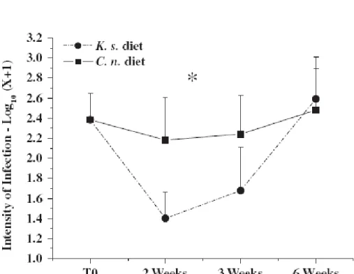
prevalence was observed, i.e. intensity was high at the beginning of the experiment, decreased afterward for both treatments, and was lower in clams exposed to K. selliformis for 2 weeks (significant difference, ANOVA, P = 0.007) and after 3 weeks (no significant difference) than clams fed C. neogracile (Fig. 1).
3.1.2. Effect of K. selliformis, infection with P. olseni, and their interaction, upon hemocyte immune parameters in Manila clams R. philippinarum
The effect of infection by the protozoan parasite P. olseni upon hemocyte parameters of clams was tested before the experiment started, i.e. before
exposing clams to any cultured microalgae. No effect of a natural infection of clams with P. olseni on any clam parameter was observed (data not shown). Likewise, during the 6 weeks of exposure to K. selliformis, the impact of the parasite was quite mild; only one parameter was affected strongly— hemocyte size, which was significantly (P = 0.016) higher in clams moderately to heavily infected with P. olseni than in lightly infected clams (Fig. 2). The effect of the long-term exposure of R. philippinarum to K. selliformis was mainly observed on two hemocyte parameters: hemocyte size (P = 0.022) and percentage of apoptotic hemocytes (P = 0.021), which were severely reduced with respect to the control during the entire exposure (Fig. 3).
Moreover, some tendencies were observed in the clams fed K. selliformis. An increase in agglutination activity was observed, starting at the beginning of the exposure and intensifying after 3 weeks of exposure; clams exposed to K.
selliformis tended to have a much higher agglutination activity than control clams
(Fig. 3). Fewer circulating hemocytes were observed in clams fed K. selliformis compared with clams fed C. neogracile, especially after the third week (Fig. 3).
No differences in condition index between treatments were observed, although values were slightly lower for clams fed K. selliformis (T2: 4.6±0.21; T3: 4.7±0.20; and T6: 4.7±0.20), in comparison to control groups fed C. neogracile (T2: 4.8±0.26; T3: 4.8±0.22; and T6: 5.0±0.30). Variation over time of exposure, for each individual parameter, was often significant, and was quite similar for both microalgal treatments (Fig. 3).
Hemocyte size, complexity, and percentages of dead and phagocytic hemocytes showed similar, strong decreases (P<0.05) after 3 weeks, but increased thereafter (except for percentage of dead hemocytes). Conversely, percentages of apoptotic hemocytes increased (P<0.001) after 3 weeks of exposure (Fig. 3). Production of ROS by clam hemocytes was highest (P = 0.034) at the beginning of the exposure and decreased over time. Total hemocyte count (THC), agglutination titer, protein concentration, and condition index were not affected by time of exposure.
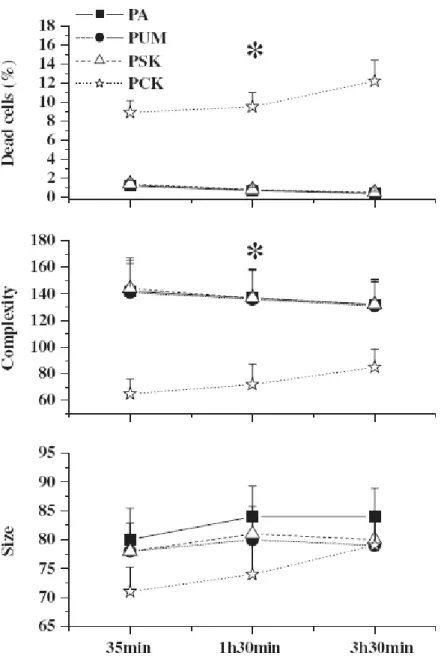
3.3. In vitro exposure of P. olseni to K. selliformis
As the severity of P. olseni infection decreased in clams fed K. selliformis, one question that arose was whether or not the harmful alga had toxic effects upon the parasite itself. Thus, in vitro studies were conducted co-incubating K.
selliformis and P. olseni. The trophozoites of P. olseni were greatly affected by the
presence of K. selliformis cells. Percentages of dead parasite cells increased approximately 10 times (P<0.001) and trophozoites of P. olseni showed significant reduction in cell complexity (P<0.001) (Fig. 4). Parasite cell size was also reduced, but differences were not significant (P = 0.320) (Fig. 4). The effect of cultured K. selliformis upon the trophozoites of P. olseni was found only with the complete culture, not with cell-free medium, indicating that the presence of the algal cells is necessary to induce toxic effects upon P. olseni.
4. Discussion
The main goal of the present study was to understand the combined effects of both parasitic infection and a simulated HAB on hemocyte parameters and condition index of Manila clam, thereby establishing a linkage between the three components of the biological-interaction model: harmful alga–clam–parasite.
As there was no interactive effect of perkinsosis and the HAB exposure on any of the hemocyte parameters or condition index, the effect of K. selliformis upon clam hemocytes will be discussed independently of the effect of infection by
P. olseni. Clam hemocyte size was diminished when K. selliformis was present.
Results from short-term, Manila clam–hemocyte exposure experiments with K.
selliformis and K. mikimotoi also showed reduction in hemocyte size and
complexity (Hégaret et al., 2007b). The effects of harmful-algal cells upon morphology of bivalve hemocytes, recorded in the literature, are very variable, probably because of the variety of mechanisms of toxicity specific to each algal species. The presence of yessotoxin in mussel hemocyte cytoplasm has also been associated with hemocyte shape changes, including reduction in size (Franchini et al., 2003; Malagoli and Ottaviani, 2004), which is attributed by the authors to an increase in intracellular Ca2+. On the contrary, significant increases in hemocyte size and complexity have been reported in Crassostrea virginica after a Prorocentrum minimum bloom (Hégaretand Wikfors, 2005a), but no effects were found on oyster hemocytes (C. virginica and C. gigas) with other toxic dinoflagellates, Alexandrium fundyense and A. catenella (Hégaretet al., 2007a). The first studies on the effects of a harmful dinoflagellate upon bivalve hemocyte characteristics were reported by Hégaret and Wikfors (2005a, b); main effects observed were on concentrations and proportions of the different hemocyte types, production of reactive oxygen species, phagocytosis and hemocyte mortality in both C. virginica and Argopecten irradians after simulated and natural blooms of P.
minimum. In the present work, K. selliformis triggered a decrease in the
percentage of apoptotic hemocytes. In contrast, phycotoxin-induced apoptosis has been reported in several human cell types exposed to algal toxins, such as okadaic acid (Cabado et al., 2003; Lago et al., 2005) or yessotoxin (Leira et al., 2002). Apoptosis eliminates over-abundant and potentially harmful cells; thus, apoptosis and cell proliferation are coupled to maintain homeostasis, as shown for human lymphocytes (Fortner and Budd, 2005). Thus far, in bivalves, apoptosis is little studied; mechanisms and possible inducers could be quite different from those in vertebrate organisms. Some contaminants, such as cadmium, can induce apoptosis in oyster hemocytes (Sokolova et al., 2004). Goedken et al. (2005)
showed that two oyster species, C. virginica and C. gigas, had repressed hemocyte apoptosis resulting from P. marinus infection in hemocytes. These authors also demonstrated, however, that C. gigas, which is resistant to P.
marinus, seemed to overcome that suppression faster than C. virginica.
The K. selliformis phycotoxin, gymnodimine (Seki et al., 1995), apparently persists over the long term (even years) in mussel and oyster tissues (Stirling, 2001; MacKenzie et al., 2002). Gymnodimine accumulates in the digestive gland as well as in adjacent tissues (MacKenzie et al., 2002). Thus, this toxin may be expected to affect all organs and tissues, including hemocytes. Franchini et al. (2003) reported that yessotoxin is found in abundance in mussel hemocyte cytoplasm, which led these authors to hypothesize that hemocytes probably are the main vectors transporting these toxins throughout shellfish tissues. It is,
however, still unknown whether or not this mechanism may be active for gymnodimine.
The effects of the parasite P. olseni upon clam–hemocyte parameters and condition index were also assessed. Categories of P. olseni infection intensity established in the present study paralleled the Mackin scale for P. marinus infection in eastern oysters, C. virginica (Ray, 1954). Our categories were shown to be very useful for experimental studies wherein the number of samples does not permit categorization of the clams into more than 2 groups (Hégaret et al., 2007b). In the present study, moderate–heavy P. olseni infection had little effect upon the hemocyte parameters and condition index of the Manila clam, as it was associated with an increase in hemocyte size only. The higher hemocyte size in clams with moderate–heavy infections of P. olseni is possibly attributable to the engulfment of tissue debris resulting from heavy infection, which provokes tissue disruption and organ dysfunction (Villalba et al., 2004; Kim et al., 2006). The engulfment of P. olseni by phagocytosis is believed to be rare in clams; Perkinsus spp. are instead encapsulated by hemocytes (Villalba et al., 2005). The low impact of P. olseni on the clam immune system was a bit surprising. Ordás et al. (2000) had reported a significant increase in lectins (agglutination titer) in carpet shell clams, R. decussatus, naturally infected with P. olseni. Lectins have been shown to be induced selectively in Manila clams upon P. olseni infection (Kim et al., 2006). In contrast, herein P. olseni did not induce lectin production.
The length of the experiment seemed to affect both groups of clams, exposed or not to the harmful algae, indicating that the clams may have been responding to external factors, such as the effect of the containers. This experiment did not highlight any specific changes in the immune parameters over time attributable to the exposure to K. selliformis. The most striking result obtained in this study suggests an immediate toxic effect of the harmful alga K.
selliformis upon P. olseni infection; indeed, the intensity and prevalence of P. olseni in the clams decreased after 2 and 3 weeks when the simulated HAB was
the highest (103 cell ml-1). It is unlikely that K. selliformis affects P. olseni development by stimulating a clam immune response against P. olseni as no major changes were observed in the functional hemocyte parameters of infected or uninfected clams. The hypothesis of a direct effect of K. selliformis upon P.
olseni within exposed clams implies some mechanism whereby K. selliformis toxin
is transported to P. olseni cells within clam tissues. As mentioned above, the toxin of K. selliformis accumulates in the digestive gland, as well as in adjacent tissues (MacKenzie et al., 2002). This tissue transfer of toxin may favor contact between parasite cells and toxin in clam tissues. It seems, however, that the effects of K.
selliformis on P. olseni also depend upon the intensity of infection. Thus, we
propose the hypothesis that heavily infected clams would have a higher probability of contact between parasites and toxins. The highest P. olseni intensity was observed at the start of experiment; subsequently, and not surprisingly, perkinsosis intensity declined quickly, leaving less-heavily infected clams present at the time when the clams were exposed to the higher concentration of K.
selliformis. Finally, intensity of P. olseni re-increased at the end of the experiment,
as the concentration of K. selliformis to which clams were exposed decreased. To test the hypothesis described above on the potential toxicity of K.
selliformis to P. olseni cells, in vitro tests were performed. The results showed that
cell mortality, and decreases in complexity of P. olseni trophozoites. Moreover, the intensity of P. olseni in the clams exposed to the HAB was strongly reduced from the first week of exposure throughout the bloom exposure, likely because of microalgal toxicity.
The exact mode of action of gymnodimine from K. selliformis has not been elucidated (Kong et al., 2005); therefore, it is not possible to attribute the increased mortality and the reduction in parasite complexity and size observed to a specific, chemical effect. Thus, other experiments using pure extracts of gymnodimine or other biotoxins from K. selliformis would provide insights into understanding the biochemical and physiological mechanism by which K.
selliformis exerts toxic effects upon P. olseni cells.
We conclude from the present study that initial impairment of P. olseni infection in clams feeding on K. selliformis is short-lived. This phenomenon may reflect an ancestral interaction between phylogenetically close, unicellular organisms—Perkinsae and dinoflagellates (Leander and Keeling, 2004). Results also highlight that P. olseni alone had minimal impacts upon the immunological and physiological functions of clams that we measured. Conversely, K. selliformis did affect hemocytes and other physiological functions within Manila clams.
Acknowledgments
The authors are grateful to Alain Marhic, Isabelle Queau, Christian Mingant, Jean René Le Coz, Marcos Paiva Scardua, Agnès Travers, Thibaud de Bettignies, Hansy Haberkorn, Philippe Miner, and Stéphane Pouvreau for technical assistance. We thank Geneviève Arzul and Marie Pierre Crassous who kindly provided K. selliformis strain. We thank Antonio Villalba who kindly provided the P.
olseni strain. The work was partially supported by funds from the French National
Program on Coastal Environment, PNEC. Hélène Hégaret was supported by the EPA/ECOHAB Grant no. 523792 to S.E. Shumway, G.H. Wikfors, and J.M. Burkholder, supplemental funding was provided by the Lerner Gray Fund for Marine Research from the American Museum of Natural History, the Grants in- Aid of Research from Sigma Xi, the National Shellfisheries Association, and the Feng Fund from the University of Connecticut. Patricia Mirella da Silva was supported by a scholarship from Conseil Général du Finistère.
References
Arzul, G., Turki, S., Namza, A., Daniel, P., Merceron, M., 1995. Fish kills induced by phycotoxins. Toxicon 33, 1119.
Bauder, A.G., Cembella, A.D., 2000. Viability of the toxic dinoflagellate Prorocentrum lima following ingestion and gut passage in the bay scallop Argopecten irradians. J. Shellfish Res. 19, 321– 324.
Biré , R., Krys, S., Fremy, J.M., Dragacci, S., Stirling, D., Kharrat, R., 2002. First evidence on occurrence of gymnodimine in clams from Tunisia. J. Nat. Toxins 11, 269–275.
Bricelj, V.M., Greene, M., Cembella, A.D., 1993. Growth of the blue mussel Mytilus edulis on toxic Alexandrium fundyense and effects of gut passage on dinoflagellate cells. In: Smayda, T.J., Shimizu, Y. (Eds.), Toxic Phytoplankton Blooms in the Sea, pp. 371–376.
Cabado, A.G., Leira, F., Vieites, J.M., Vieytes, M., Botana, L.M., 2003. Caspase-8 activation initiates okadaic acid induced apoptosis in neuroblastoma. In: Villalba, A., Reguera, B., Romalde, J.L., Beiras, R. (Eds.), Consellerıá de Pesca e Asuntos Maritimos da Xunta de Galicia and Intergovernmental Oceanographic Commission of UNESCO, Santiago de Compostela, pp. 107–117.
Cheng, T.C., 1981. Bivalves. In: Ratcliffe, N.A., Rowley, A.F. (Eds.), Invertebrate Blood Cells. Academic Press, London, pp. 233–300.
Choi, K.S., Wilson, E.A., Lewis, D.H., Powell, E.N., Ray, S.M., 1989. The energetic costs of Perkinsus marinus parasitism in oysters: quantification of the thioglycollate method. J. Shellfish Res. 8, 125–131.
Fortner, K.A., Budd, R.C., 2005. The death receptor Fas (CD95/ APO-1) mediates the deletion of T lymphocytes undergoing homeostatic proliferation. J. Immunol. 175, 4374–4382.
Franchini, A., Milandri, A., Poletti, R., Ottaviani, E., 2003. Immunolocalization of yessotoxin in the mussel Mytilus galloprovincialis. Toxicon 41, 967–970.
Gainey, L., Shumway, S.E., 1988. A compendium of the responses of bivalve molluscs to toxic dinoflagellates. J. Shellfish Res. 7, 623–628.
Galimany, E., Place, A.R., Ramon, M., Jutson, M., Pipe, R.K., 2008. The effects of feeding
Karlodinium veneficum (PLY #103; Gymnodinium veneficum Ballantine) to the blue mussel Mytilus edulis. Harmful Algae 7(1), 91–98.
Gauthier, J.D., Vasta, G.R., 1993. Continuous in vitro culture of eastern oyster parasite Perkinsus
marinus. J. Invert. Pathol. 62, 321–323.
Goedken, M., Morsey, B., Sunila, I., Dungan, C., De Guise, S., 2005. The effects of temperature and salinity on apoptosis of Crassostrea virginica hemocytes and Perkinsus marinus. J. Shellfish Res. 24, 177–183.
Goggin, C.L., 1992. Occurrence of parasites of the genus Perkinsus in France. Bull. Eur. Assoc. Fish Pathol. 12, 174–176.
Guillard, R.R.L., 1975. Culture of Phytoplankton for Feeding Marine Invertebrates Animals. Plenum, New York, NY.
Guillard, R.R.L., Ryther, J.H., 1962. Studies of marine planktonic diatoms. I. Cyclotella nana Hustedt and Denotula confervacea (Cleve) Gran. Can. J. Microbiol. 8, 229–239.
Guillou, L., Nézan, E., Cueff, V., Erard-Le Denn, E., Cambon- Bonavita, M.A., Gentien, P., Barbier, G., 2002. Genetic diversity and molecular detection of three toxic dinoflagellate genera (Alexandrium, Dinophysis, and Karenia) from French coasts. Protist 153, 223–238.
Hallegraeff, G.M., 1993. A review of harmful algal blooms and their apparent global increase. Phycologia 32, 79–99.
Hansen, G., Erard-Le Denn, E., Daugbjerg, N., Rodriguez, F., 2004. Karenia selliformis responsible for the fish-kills in the Gulf of Gabes, Tunisia 1994. Communication Ifremer.
Harvell, C.C., Kim, K., Burkholder, J.M., Colwell, R.R., Epstein, P.R., Grimes, D.J., Hofmann, E.E., Lippe, E.K., Osterhaus, A.D.M.E., Ovestreet, O.M., Porter, J.W., Smith, G.W., Vasta, G.R., 1999. Emerging marine diseases— climate links and anthropogenic factors. Science 285, 1505–1510.
Haywood, A.J., Steidinger, K.A., Truby, E.W., MacKenzie, L., 2002. Comparative morphology and molecular phylogenetic analysis of the three new species of the genus Karenia (Dinophyceae) from New Zealand. J. Phycol. 40, 165–179.
Hégaret, H., Wikfors, G.H., 2005a. Effects of natural and fieldsimulated blooms of the dinoflagellate
Prorocentrum minimum upon hemocytes of eastern oysters, Crassostrea virginica, from two
different populations. Harmful Algae 4, 201–209.
Hégaret, H., Wikfors, G.H., 2005b. Time-dependent changes in hemocytes of eastern oysters,
Crassostrea virginica, and northern bay scallops, Argopecten irradians irradians, exposed to a
cultured strain of Prorocentrum minimum. Harmful Algae 4, 187–199.
Hégaret, H., Wikfors, G.H., Soudant, P., Lambert, C., Shumway, S.E., Bérard, J.B., Lassus, P., 2007a. Toxic dinoflagellates (Alexandrium fundyense and A. catenella) have minimal apparent effect on oyster hemocytes. Mar. Biol. 152, 441–447.
Hégaret, H., da Silva, P., Wikfors, G.H., Lambert, C., De Bettignies, T., Shumway, S.E., Soudant, P., 2007b. Hemocyte responses of Manila clams, Ruditapes philippinarum, with varying parasite,
Perkinsus olseni, severity to toxic-algal exposures. Aquat. Toxicol. 84, 469–479.
Hégaret, H., Wikfors, G.H., Shumway, S.E., 2007c. Diverse feeding responses of five species of bivalve mollusc when exposed to three species of harmful algae. J. Shellfish Res. 26, 549– 559.
Hégaret, H., Shumway, S.E., Wikfors G.H., Pate, S., Burkholder, J.M., in press. Potential transport of harmful algae through relocation of bivalve molluscs. Mar. Ecol. Prog. Ser.
Heil, C.A., Glibert, P.M., Al-Sarawi, M.A., Faraj, M., Behbehani, M., Husain, M., 2001. First record of a fish-killing Gymnodinium sp. bloom in Kuwait Bay, Arabian Sea: chronology and potential causes. Mar. Ecol. Prog. Ser. 214, 15–23.
Jenkinson, I., Arzul, G., 2000. Mitigation by cysteine compounds of rheotoxicity, cytotoxicity and fish mortality caused by the dinoflagellates, Gymnodinium mikimotoi and G. maguelonnense. In: Hallegraeff, G., et al. (Eds.), Harmful Algal Blooms 2000. Intergovernmental Oceanographic Commission of UNESCO 2001, pp. 461–464.
Kim, Y.M., Park, K.-I., Choi, K-S., Alvarez, R.A., Cummings, R.D., Cho, M., 2006. Lectin (MCL) from the Manila clam Ruditapes philippinarum is induced upon infection with the protozoan parasite
Perkinsus olseni. J. Biol. Chem. 281, 26854–26864.
Kong, K., Moussa, Z., Romo, D., 2005. Studies toward a marine toxin immunogen: enantioselective synthesis of the spirocyclic imine of (_)-gymnodimine. Org. Lett. 7, 5127–5130.
Laabir, M., Gentien, P., 1999. Survival of toxic dinoflagellates after gut passage in the Pacific oyster
Crassostrea gigas Thunburg. J. Shellfish Res. 18, 217–222.
Lago, J., Santaclara, F., Vieites, J.M., Cabado, A.G., 2005. Collapse of mitochondrial membrane potential and caspases activation are early events in okadaic acid-treated Caco-2 cells. Toxicon 46, 579–586.
Landsberg, J.H., 1996. Neoplasia and biotoxins in bivalves: is there a connection? J. Shellfish Res. 15, 203–230.
Landsberg, J.H., 2002. The effects of harmful algal blooms on aquatic organisms. Rev. Fish. Sci. 10, 113–390.
Lassalle, G., de Montaudouin, X., Soudant, P., Paillard, C., 2007. Parasite co-infection of two sympatric bivalves, the Manila clam (Ruditapes philippinarum) and the cockle (Cerastoderma
Leander, B.S., Keeling, P.J., 2004. Early evolutionary history of dinoflagellates and apicomplexans (Alveolata) as inferred from hsp90 and actin phylogenies. J. Phycol. 40, 341–350.
Leira, F., Alvarez, C., Vieites, J.M., Vieytes, M.R., Botana, L.M., 2002. Characterization of distinct apoptotic changes induced by okadaic acid and yessotoxin in the BE(2)-M17 cell line. Toxicol. Vitro 16, 23–31.
Lesser, M.P., Shumway, S.E., 1993. Effects of toxic dinoflagellates on clearance rates and survival in juvenile bivalve mollusks. J. Shellfish Res. 12, 377–381.
Leverone, J.R., Shumway, S.E., Blake, N.J., 2007. Comparative effects of the toxic dinoflagellate
Karenia brevis on clearance rate in juveniles of four bivalve molluscs from Florida, USA.
Toxicon 49, 634–645.
MacKenzie, L., Holland, P., McNab, P., Beuzenberg, V., Selwood, A., Suzuki, T., 2002. Complex toxin profiles in phytoplankton and greenshell mussels (Perna canaliculus), revealed by LC–MS/MS analysis. Toxicon 40, 1321–1330.
Malagoli, D., Ottaviani, E., 2004. Yessotoxin affects fMLPinduced cell shape changes in Mytilus
galloprovincialis immunocytes. Cell Biol. Int. 28, 57–61.
Mann, R., 1978. A comparison of morphometric biochemical and physiological index of condition inmarine bivalves molluscs. In: Thorpand, J.H., Gibbons, I.W. (Eds.), Energy and Environmental Stress in Aquatic Systems.
Miles, C.O., Wilkins, A.L., Stirling, D.J., MacKenzie, A.L., 2000. New analogue of gymnodimine from a
Gymnodinium species. J. Agric. Food Chem. 48, 1373–1376.
Miles, C.O., Wilkins, A.L., Stirling, D.J., MacKenzie, A.L., 2003. Gymnodimine C, an isomer of gymnodimine B, from Karenia selliformis. J. Agric. Food Chem. 51, 4838–4840.
Ordás, M.C., Ordás, A., Beloso, C., Figueras, A., 2000. Immune parameters in carpet shell clams naturally infected with Perkinsus atlanticus. Fish Shellfish Immunol. 10, 597–609.
Pearce, I., Handlinger, J.H., Hallegraeff, G.M., 2005. Histopathology in Pacific oyster (Crassostrea
gigas) spat caused by the dinoflagellate Prorocentrum rhathymum. Harmful Algae 4, 61–74.
Ray, S.M., 1954. Biological studies of Dermocystidium marinum. Rice Inst. Pamph. 41, 1–114. Scarratt, A.M., Scarratt, D.J., Scarratt, M.G., 1993. Survival of live Alexandrium tamarense cells in
mussel and scallop spat under simulated transfer conditions. J. Shellfish Res. 12, 383–388. Seki, T., Satake, M., MacKenzie, L., Kaspar, H.F., Yasumoto, T., 1995. Gymnodimine, a new marine
toxin of unprecedented structure isolated from New Zealand oysters and the dinoflagellate,
Gymnodinium sp. Tetrahedron Lett. 36, 7093–7096.
Shao, P., Chen, Y.-Q., Zhou, H., Yuan, J., Qu, L.-H., Zhao, D., Lin, Y.-S., 2004. Genetic variability in Gymnodiniaceae ITS regions: implications for species identification and phylogenetic analysis. Mar. Biol. 144, 215–224.
Shumway, S.E., 1990. A review of the effects of algal blooms on shellfish and aquaculture. J. World Aquacult. Soc. 21, 65–104.
Shumway, S.E., 1995. Phycotoxin-related shellfish poisoning: bivalve molluscs are not the only vectors. Rev. Fish. Sci. 3, 1–31. Smayda, T.J., 1997a. What is a bloom? A commentary. Limnol. Oceanogr. 42, 1132–1136.
Smayda, T.J., 1997b. Harmful algal blooms: their ecophysiology and general relevance to phytoplankton blooms in the sea. Limnol. Oceanogr. 42, 1137–1153.
Smolowitz, R., Shumway, S.E., 1997. Possible cytotoxic effects of the dinoflagellate, Gyrodinium
aureolum, on juvenile bivalve molluscs. Aquacult. Int. 5, 291–300.
Sokolova, I.M., Evans, S., Hughes, F.M., 2004. Cadmium induced apoptosis in oyster hemocytes involves disturbance of cellular energy balance but no mitochondrial permeability transition. J. Exp. Biol. 207, 3369–3380.
Soudant, P., Chu, F.-L., Lund, E.D., 2005. Assessment of the cell viability of cultured Perkinsus
SYBRgreen–propidium iodide double staining and flow cytometry. J. Eukaryot. Microbiol. 52, 492–499.
Stirling, D., 2001. Survey of historical New Zealand shellfish samples for accumulation of gymnodimine. NZJ. Mar. Freshwater Res. 35, 851–857.
Villalba, A., Reece, K.S., Ordás, M.C., Casas, S.M., Figueras, A., 2004. Perkinsosis in molluscs: a review. Aquat. Living Resour. 17, 411–432.
Villalba, A., Casas, S.M., Lopez, C., Carballal,M.J., 2005. Study of perkinsosis in the carpet shell clam
Tapes decussatus in Galicia (NW Spain). II. Temporal pattern of disease dynamics and
association with clam mortality. Dis. Aquat. Org. 65, 257–267.
Wikfors, G.H., Smolowitz, R.M., 1993. Detrimental effects of a Prorocentrum isolate upon hard clams and bay scallops in laboratory feeding studies. In: Smayda, T.J., Shimizu, Y. (Eds.), Toxic Phytoplankton Blooms in the Sea, pp. 447–452.
Wikfors, G.H., Smolowitz, R.M., 1995. Experimental and histological studies of four life-history stages of the eastern oyster, Crassostrea virginica, exposed to a cultured strain of the dinoflagellate
Table 1 : Categorization of P. olseni infection intensity according to the number of P. olseni cells in
clam gills (X). Two categories are defined as null–light and moderate–heavy, respectively, with less or more that 1000 parasites per g of gill wet weight. Log10(X+1) is indicated between parentheses. The results include clams before the exposure and after 2, 3 and 6 weeks of exposure. n = number of clams in each class
Fig. 1. Infection intensity (mean±SE) of P. olseni in Manila clams, R. philippinarum, during the 6 weeks
of exposure to two different algal treatments; K. selliformis plus C. neogracile (K. s., n = 60) or C.
neogracile alone (C. n., n = 60) (T0, n = 20). Asterisk indicates where algal treatment affected
significantly the P. olseni burden (ANOVA, P<0.05). Results are expressed as log10(X+1) where X is the number of hypnospores of P. olseni per g of gill wet weight.
Fig. 2. Effect of P. olseni on Manila clam R. philippinarum hemocyte size (in arbitrary units, mean±SE).
Asterisks indicate significant differences in hemocyte size between classes of infection by P. olseni (null–light, n = 92; moderate–heavy, n = 48) (ANOVA, P<0.05).
Fig. 3. Effect of K. selliformis on Manila clam, R. philippinarum, hemocyte parameters (means±SE)
during the 6 weeks of exposure to two different algal treatments; K. selliformis plus C. neogracile (K. s.) or C. neogracile solely (C. n.). Hemocyte size is in arbitrary units. Apoptosis values are missing for 6 weeks. Asterisks indicate significant differences between algal treatments (ANOVA, P<0.05). Apoptosis: K. s., n = 20, C. n., n = 24; hemocyte size, agglutination and THC: K. s., n = 60; C. n., n = 60; T0: n = 20.
Fig. 4. Effects of K. selliformis on P. olseni cell parameters (mean±SE, n = 5 per treatment at each
time): the percentage of dead cells, size and complexity (in arbitrary units), after different times of in vitro incubation with cultured algal samples. PA: Perkinsus olseni alone; PUM: Perkinsus olseni plus uninoculated medium; PSK: Perkinsus olseni plus supernatant of culture of Karenia selliformis; PCK:
Perkinsus olseni plus complete culture of Karenia selliformis. Asterisks indicate significant differences
between P. olseni incubated with complete culture of K. selliformis and other treatments (ANOVA, P<0.05).