HAL Id: hal-00271618
https://hal.archives-ouvertes.fr/hal-00271618
Submitted on 9 Apr 2008HAL is a multi-disciplinary open access
archive for the deposit and dissemination of sci-entific research documents, whether they are pub-lished or not. The documents may come from teaching and research institutions in France or
L’archive ouverte pluridisciplinaire HAL, est destinée au dépôt et à la diffusion de documents scientifiques de niveau recherche, publiés ou non, émanant des établissements d’enseignement et de recherche français ou étrangers, des laboratoires
Distinct effects of DNA-PKcs and Artemis inactivation
on signal joint formation in vivo.
Cédric Touvrey, Chrystelle Couedel, Pauline Soulas-Sprauel, Rachel Couderc,
Maria Jasin, Jean-Pierre de Villartay, Patrice Marche, Evelyne
Jouvin-Marche, Serge Candéias
To cite this version:
Cédric Touvrey, Chrystelle Couedel, Pauline Soulas-Sprauel, Rachel Couderc, Maria Jasin, et al.. Distinct effects of DNA-PKcs and Artemis inactivation on signal joint formation in vivo.. Molecular Immunology, Elsevier, 2008, 45 (12), pp.3383-91. �10.1016/j.molimm.2008.04.004�. �hal-00271618�
Distinct effects of DNA-PKcs and Artemis inactivation on signal joint formation
in vivo.
Cédric Touvrey1,4, Chrystelle Couedel2, Pauline Soulas3, Rachel Couderc1,5, Maria Jasin2,
Jean-Pierre de Villartay3, Patrice N. Marche1,5, Evelyne Jouvin-Marche1,5
and Serge M. Candéias1,6*
1: CEA, DSV, DRDC, Laboratoire d’Immunochimie ; INSERM U548 ; Université Joseph Fourier ;
Grenoble, F-38054 France.
2: Molecular Biology Program, Memorial Sloan-Kettering Cancer Center, 1275 York Avenue, New
York, New York 10021
3: INSERM U768 Développement Normal et Pathologique du Système Immunitaire ; Hôpital
Necker-Enfants Malades ; Paris, F-75015 France; Université Paris Descartes, Faculté de Médecine René Descartes ; Paris, F-75005 France.
4: present address : Ludwig Institute for Cancer Research / CHUV, University Hospital Lausanne
HO 05/1552, CH-1005 Lausanne Switzerland
5: present address : INSERM U823; Université Joseph Fourier; La Tronche, France.
6: present address : CEA, DSV, iRTSV, LBBSI; CNRS UMR 5092; Université Joseph Fourier;
F38054 Grenoble, France.
* Corresponding author : Laboratoire Lésions des Acides Nucléiques, INAC/SCIB,
CEA-Grenoble,17 rue des martyrs, 38054 Grenoble, France. Tel : +33 4 38 78 92 49 ; Fax : + 33 4 38 78 50 90; E-mail: serge.candeias@cea.fr
Abbreviations: TR: T cell receptor; Ig : immunoglobulin RSS: recombination signal sequence; SE:
signal end; CE: coding end; SJ: signal joint; CJ: coding joint; NHEJ: non homologous end joining; DSB: double strand break; TdT: terminal nucleotidyl transferase; DNA-PK: DNA dependent protein kinase; DNA-PKcs: DNA-PK catalytic subunit ; DN: double negative
Abstract
The assembly of functional immune receptor genes via V(D)J recombination in developing lymphocytes generates DNA double stranded breaks intermediates that are repaired by non homologous end joining (NHEJ). This repair pathway requires the sequential recruitment and activation onto coding and signal DNA ends of several proteins, including the DNA-dependent protein kinase and the nuclease Artemis. Artemis activity, triggered by the DNA-dependent protein kinase, is necessary to process the genes hairpin-sealed coding ends but appears dispensable for the ligation of the reciprocal phosphorylated, blunt-ended signal ends into a signal joint. The DNA-dependent protein kinase is however present on signal ends and could potentially recruit and activate Artemis during signal joint formation. To determine whether Artemis plays a role during the resolution of signal ends during V(D)J recombination, we analyzed the structure of signal joints generated in developing thymocytes during the rearrangement of T cell receptor genes in wild type mice and mice mutated for NHEJ factors. These joints exhibit junctional diversity resulting from N nucleotide polymerization by the terminal nucleotidyl transferase and nucleotide loss from one or both of the signal ends before they are ligated. Our results show that Artemis participates in the repair of signal ends in vivo. Furthermore, our results also show that while the DNA-dependent protein kinase complex protects signal ends from processing, including deletions, Artemis seems on the opposite to promote their accessibility to modifying enzymes. In addition, these data suggest that Artemis might be the nuclease responsible for nucleotide loss from signal ends during the repair process.
Keywords : V(D)J recombination ; T cell receptor genes ; signal joints ; Artemis ; DNA-PKcs ;
Introduction
Functional genes coding for the antigenic T cell receptor (TR) and immunoglobulin (Ig) subunits are assembled from discrete variable (V), diversity (D) and joining (J) genes by a site specific rearrangement mechanism in developing T and B lymphocytes, respectively (Jung and Alt, 2004). In germ line configuration, the individual V, D and J components are flanked by specific recombination signal sequences (RSS) that are the target of the recombination machinery. After synapsis of the two genes to be rearranged, the lymphoid specific RAG-1 and RAG-2 proteins introduce a single strand nick exactly at the border of the genes and their respective RSS, which is converted in a double strand break after attack of the other strand. This process generates hairpin-sealed coding ends (CE) and blunt, phosphorylated signal ends (SE). These DNA extremities are ligated pairwise to create the coding and signal joints (CJ and SJ), respectively. The joining step of V(D)J recombination is carried out by the non-homologous end joining (NHEJ) double strand break (DSB) repair pathway (Rooney et al., 2004). Although several repair pathways are possible (Couedel et al., 2004; Lee et al., 2004; Weinstock and Jasin, 2006), the association of the RAG proteins with CEs and SEs forces those physiological DSBs to be repaired by Ku-dependent mechanisms (Corneo et al., 2007; Cui and Meek, 2007).However, because of their structural differences, CEs and SEs are handled differentially.
The hairpin structure sealing the CEs must be opened to make these ends ligatable. Hairpin opening and CJ formation require both the DNA dependant protein kinase complex (DNA-PK), composed of Ku70, Ku80 and the catalytic subunit DNA-PKcs, and the nucleaseArtemis (Li et al., 2005; Moshous et al., 2001; Rooney et al., 2004). In vitro, free Artemis is an exonuclease; bound to and phosphorylated by DNA-PK, it becomes an endonuclease able to open hairpins and cleave DNA at the transition between double stranded and single stranded regions (Ma et al., 2002; Povirk et al.,
2007). In DNA-PKcs-mutated SCID mice (Roth et al., 1992), DNA-PKcs-/- mice (Gao et al., 1998;
Taccioli et al., 1998) and Artemis-/- mice (Li et al., 2005; Rooney et al., 2002), the hairpin structure
is not resolved and CJ formation is severely inhibited. The hairpin can be opened at its apex or asymmetrically. In the latter case, the single stranded extension can be filled in by DNA polymerases, resulting in addition of P nucleotides, or cleaved/digested. A yet unknown exonuclease can eventually remove a limited number of bases. Random nucleotides can be polymerized at DNA ends by the terminal nucleotidyl transferase (TdT), recruited to the repair complex by Ku80 (Purugganan et al., 2001). A CJ is created after ligation of processed CEs by the DNA LigIV complexed to XRCC4 and Cernunnos/XLF (Ahnesorg et al., 2006; Buck et al., 2006; Tsai et al., 2007; Wu et al., 2007). Because the combination of the different types of modification (hairpin opening, nucleotide removal, nucleotide addition) is essentially random, a large array of different CJs can be generated from a single pair of rearranging genes (Davis and Bjorkman, 1988). This high level of junctional diversity in CJs is found in all the rearranged immune receptor genes is one of the hallmarks of V(D)J recombination.
In contrast, SJ junctional diversity is less extensive and more restricted. Indeed, since SEs are blunt and phosphorylated (Schlissel et al., 1993), they can be ligated directly by the DNA LigIV-XRCC4-Cernunnos/XLF complex without prior processing/remodelling (Lewis et al., 1985). However, SEs are retained within a post cleavage complex by the RAG proteins with the Ku heterodimer, DNA-PKcs (Agrawal and Schatz, 1997) and DNA-damage sensors like ATM and p53 (Perkins et al., 2002). In this complex, they are therefore exposed to the same enzymatic activities as CEs: Ku86 can recruit and activate both the TdT polymerase (Purugganan et al., 2001) and the Artemis nuclease (Ma et al., 2002; Povirk et al., 2007). The SJ repertoire found in human and mice therefore includes both SJs formed by the precise juxtaposition of the rearranged genes RSSs and
modified SJs in which SEs have been processed before ligation. The sequence at the gene/RSS border is known to influence CE processing and CJ sequence (Ezekiel et al., 1997; Gauss and Lieber, 1996; Nadel and Feeney, 1995), and this phenomenon probably operates also on SEs, as the frequency of SJs displaying modifications and the nature of the modifications are variable depending on the rearranged genes For example, in vivo, in developing thymocytes, the frequency of SJ with N nucleotide insertions varies according to the V gene used during TRB gene rearrangement (Candéias et al., 1996; Kanari et al., 1998; Talukder et al., 2004), and base removal from SEs before ligation is found for TRA and TRD, but not TRB and TRG genes (Candéias et al., 1996; Iwasato and Yamagishi, 1992; Kanari et al., 2003; Kanari et al., 1998; Touvrey et al., 2006a). In contrast, ex vivo, the recombination of artificial recombination substrates in non lymphoid cells (fibroblasts or embryonic stem cells) following introduction of the genes coding for the RAG-1 and RAG-2 proteins generates only precise joints in cells with a wild type NHEJ activity (Dai et al., 2003; Gao et al., 1998; Lieber et al., 1988a). Co-transfection of the TdT gene eventually results in the formation of N-containing SJs (Purugganan et al., 2001). Similarly, in lymphoid cell lines, the extent of N diversity in SJs following recombination of artificial substrates correlates with the endogenous level of TdT expression (Lieber et al., 1988b). In these experimental systems, base loss from SEs is almost never observed in the context of a proficient NHEJ activity (Dai et al., 2003; Gao et al., 1998; Lieber et al., 1988a; Lieber et al., 1988b). The discrepancy between SJ repertoires generated in vivo and ex
vivo probably reflects both the importance of the nuclear environment in the regulation of V(D)J
recombination (Stanhope-Baker et al., 1996; West et al., 2005) and the tissue specificity and developmental regulation of the TdT (Bentolila et al., 1997; Li et al., 1993).
SEs are blunt ended. The DNA-PKcs-Artemis complex, responsible for CE hairpin opening, is therefore dispensable for SJ formation both in vivo and ex vivo. Consequently SJs but not CJs
resulting from TCR gene rearrangement are found in developing thymocytes in the absence of DNA-PKcs (Carroll et al., 1993; Gao et al., 1998; Taccioli et al., 1998) or Artemis (Li et al., 2005; Rooney et al., 2002). SJ abundance and/or fidelity was found to be reduced in DNA-PKcs-deficient mice and cell lines (Binnie et al., 1999; Bogue et al., 1998; Lieber et al., 1988a) whereas SJ fidelity was reported to be unaffected following rearrangement of artificial extra-chromosomal substrates in Artemis-deficient MEFs or ES cells (Rooney et al., 2003; Rooney et al., 2002). However, because of the biases inherent to extra-chromosomal recombination assays (see above), we found important to analyze the eventual involvement of Artemis, DNA-PKcs effector in CE processing, in the repair of SEs and SJ formation in vivo, in a physiological nuclear environment.
To determine whether Artemis plays a role in SJ formation, we compared the structure of SJs created following recombination of endogenous TCR genes in Artemis-/- mice with those generated
in Ku80-/- and SCID mice in which the DNA-PK/Artemis pathway is disrupted at the level of its
recruitment to the DNA ends or at the level of Artemis activation, respectively. As expected, very few SJs were recovered from Ku80-/- thymocytes, and most are severely deleted. In SCID mice, most
of the SJs recovered are modified by N nucleotide additions and/or base loss. Surprisingly, in Artemis-deficient thymocytes, the frequency of modified SJs is lower than in wt and SCID mice, and none of the modified SJs exhibits base loss. Our results show that Artemis participates in the repair of blunt DNA ends in vivo, that DNA-PKcs and Artemis inactivation have different effects on SJ diversity developing thymocytes, and strongly suggest that Artemis is the nuclease responsible for base loss from SEs during V(D)J recombination.
Materials and Methods
Mice
C57BL/6 mice were bred in the animal facility of the “Commissariat à l’Energie Atomique-Grenoble”. SCID mice were purchased from Charles River Laboratories, L’Arbresle, France. Artemis–deficient mice will be described elsewhere (PS and JPdeV, unpublished results). Ku80
-/-mice (Nussenzweig et al., 1996) were bred in the animal facility of the Memorial Sloan-Kettering Cancer Center, New-York, USA. Mice were sacrificed between 4 and 10 wks of age.
DNA preparation-Signal joint analysis
The thymi were removed and crushed. Thymocyte DNA was prepared from single cell suspensions either with the Nucleospin Tissue Kit (Macherey-Nagel, Hoerdt, France) according to the manufacturer’s instructions, for C57BL/6 and Artemis-deficient mice, or by SDS/proteinase K digestion, extraction and ethanol precipitation for Ku80-/- mice. SJ were amplified from C57BL/6,
SCID and Artemis-/- thymocytes as described (Candéias et al., 1996; Touvrey et al., 2006a; Touvrey
et al., 2006b). The relative location of the genes analyzed in this study and the strategy used for SJ amplification are depicted in Fig. 1. For SJ amplification from Ku80-/- thymocytes, a first PCR
reaction was performed in the same conditions using the primers 5’DD2ext (5’-CCAGCAGGGCGGCAAGCATTAGACA-3’) and 3’DD1. One µl of this product was then used as a template to amplify SJ as for the other samples. For each SJ type, amplifications were performed from 2 to 3 different DNA samples. Cloning and sequencing of the PCR products was performed as described (Candéias et al., 1996; Touvrey et al., 2006a; Touvrey et al., 2006b), except for the Ku80
-/-PCR products that were cloned and sequenced without prior discrimination of R and ApaL1-S ApaL1-SJs. ApaL1-SJ sequences were aligned with the RApaL1-SApaL1-S sequences of the respective V and D genes as
published in IMGT, the international ImMunoGeneTics information system® http://imgt.cines.fr (founder and director: Marie-Paule Lefranc, Montpellier, France), except for the 3’TRDD1 RSS, that was re-assigned as described in (Touvrey et al., 2006b). Non-templated bases were scored as N nucleotides. S and R represent the total numbers of ApaL1-S and ApaL1-R SJ, respectively. Identical ApaL1-R sequences identified from the same DNA sample were counted as a single occurrence to determine R*. The R*/R ratio was considered to reflect the level of redundancy of the modified SJ repertoire. It was used to calculate S* (S x R*/R), the number of ApaL1-S SJ corrected for this redundancy. The frequency of SJ modification was calculated as (R*/R* + S*) x 100 (Tables 1-3).
Results
Processing of SEs in wild type mice
The perfect juxtaposition of canonical RSSs creates an ApaL1 restriction site (CACGTG). If SEs are ligated in absence of any processing, the resulting SJ is sensitive to ApaL1 digestion (ApaL1-S) whereas it is resistant to digestion (ApaL1-R) if nucleotides are removed from and/or added to at least one SE. ApaL1 digestion therefore allows an easy discrimination between perfect and modified SJs, and sequencing of the ApaL1-R SJs allows to determine the nature of SE processing. We focused our attention on SJs created during recombination of TRD genes because a low level of V(D)J recombination at the TRD locus have been described in SCID (Bogue et al., 1997) and Artemis-/- mice (Li et al., 2005; Rooney et al., 2002). Thymocyte development is arrested
at the immature CD4-CD8- double negative (DN) stage in these mice (data not shown, PS and
JPdeV, unpublished results). We recently showed that N nucleotides are present in about 74% of the SJs resulting from the recombination of TRDD1 and TRDD2 genes in CD3-deficient mice, in which thymocyte development is also arrested at the DN stage (Touvrey et al., 2006b). To verify that our observation was not biased by this maturation block, we first established the pattern of TR DD1/DD2 SJ diversity in un-manipulated wt thymocytes.
TR DD1/DD2 SJs were amplified from total DNA extracted from C57BL/6 thymocytes as described (Touvrey et al., 2006b) (see Fig 1 for a schematic representation). The resulting PCR products were cloned, and DNA was prepared from individual SJ-containing colonies and digested to completion with ApaL-1. Out of a total of 50 cloned SJs produced from 3 mice, 38 were ApaL1-R. To determine the nature of SE processing, these ApaL1-R SJs were sequenced (Fig 2 and Table 1). Only 3 junctions were found more than once, indicating that the ApaL1-R TRDD1/DD2 SJ repertoire is diverse. All the ApaL1-R sequences were found to contain non-templated N
nucleotides. In two cases, nucleotides were deleted from the TRDD2 SE. Finally, in one case, a TRDD1/DD2 CJ was inserted in the SJ. This event results from the recombination of the 5’ TRDD1 and 3’ TRDD2 RSSs as described (Kanari et al., 2003; Touvrey et al., 2006b). Altogether, our results indicate that the repertoire of TR DD1/DD2 SJs is similar in wt and CD3-deficient mice. The profile of SE modifications is not affected by the developmental block at the DN stage in mice with functional V(D)J recombination mechanisms. Finally, it is important to note that among the nucleotide inserted between the TR DD1 and TR DD2 RSS, very few are attributable to coding flank retention. We never observe more than one putative coding flank base adjacent to each heptamer, a distribution perfectly compatible with the random insertion of non-templated nucleotide by the TdT. Therefore, and for all this study, we will consider that the SJ diversity results from the processing of blunt-ended SEs generated when the RAG proteins introduce a DSB at the border of the RSSs.
Effects of DNA-PK inactivation on SE processing
In order to specifically identify the effects of Artemis deficiency, we first characterized the repertoire of TR DD1/DD2 SJ modifications in mice mutated for Ku80 or DNA-PKcs, two other components of the DNA-PK/Artemis pathway.
In Ku80-/- mice, SJs level was very low and two successive nested PCR reactions were
needed to obtain enough material for cloning (data not shown). Upon cloning and sequencing (Fig 3), all the SJs resulting from the first animal and most of those from the second animal were modified. The junctions amplified from the first animal carry deletions ranging from 16 to 100 nucleotides on both SEs. In the second, only two different modified junctions were identified in which deletions from either only one or both of the RSSs range from 6 to 10 bases. Thus, in absence of Ku80, the 3’ TRDD1 and 5’ TRDD2 SEs are not protected from deletion. The rare signal joints
that are nonetheless created, presumably by alternative, Ku-independent NHEJ mechanisms (Corneo et al., 2007; Cui and Meek, 2007), do not contain non-templated nucleotides, in accordance with the role of Ku80 in the recruitment of the TdT in the recombinase complex (Purugganan et al., 2001), and a few are even generated without end processing, at least in the Ku2 animal (Fig 3). These results, including the slight inter-individual variations that probably result from the amplification procedure, are perfectly consistent with the documented effects of Ku80 inactivation on signal joint creation in vivo (Bogue et al., 1997).
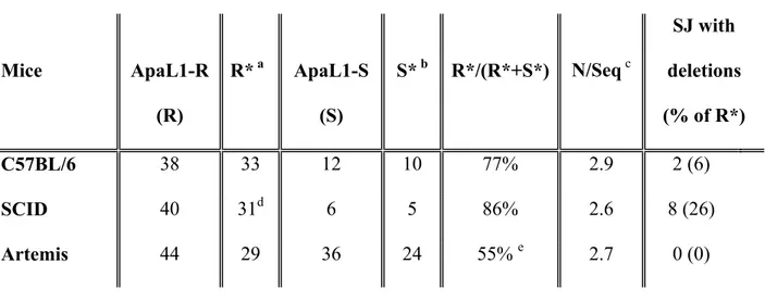
In DNA-PKcs mutated SCID mice, TR DD1/DD2 SJs were readily amplified from total thymocyte genomic DNA, in one round of amplification. After ApaL-1 digestion of individual cloned SJs, the vast majority of these junctions (40 out of 46) was found to be ApaL1-R. They were sequenced to determine the nature of the modifications (Fig 4 and Table 1). Few sequences (5) were found several times, indicating that the TR DD1/DD2 SJ repertoire is diverse in SCID thymocytes. In contrast to the situation found in Ku80-/- mice, almost all of these SJs (90%) have N nucleotide
insertions. Nucleotides were deleted from SEs in 8 of them. These deletions are limited (1 to 7 nucleotides), except in one junction where 12 and 27 nucleotides have been lost from the DD2 and DD1 RSS, respectively. In 5 cases, SJ modification consists of both deletion and N addition; in 3 sequences, SJ modification results exclusively from SEs deletion in absence of N nucleotide additions. A TRDD1/DD2 CJ was identified in the SJ in 2 cases. When compared to wt, DNA-PKcs deficiency induces only a small increase (+12%) in the frequency of modified TR DD1/DD2 SJs (Table 1). The modifications include N nucleotide additions, and, in about 25 % of the events, controlled deletion of SEs. The average number of N nucleotides added per junction is similar to that of wt mice.
Overall, these results are in agreement with the documented roles of Ku80 and DNA-PKcs in V(D)J recombination (Rooney et al., 2004) and validate our experimental procedure to analyze the eventual function of Artemis in SJ formation.
Lower level of SE processing in Artemis-deficient thymocytes
We then analyzed the repertoire of TR DD1/DD2 SJs produced in Artemis-/- thymocytes.
These joints were readily amplifiable from total thymocyte DNA, in one round of PCR (not shown), indicating, as already shown (Li et al., 2005), that Artemis deficiency does not impair SJ formation as severely as Ku80 inactivation. After cloning and ApaL1 digestion of 80 individual SJs obtained from 3 mice, only a short majority (44) was found to be ApaL1-R. Following sequencing, 11 were found several times (2 or 3) and only 29 unique sequences were identified (Fig 5). In each of these junctions, SE processing consisted exclusively in N nucleotide additions, ranging from 1 to 6 bases per sequence. None of the SJs exhibits base loss. The SEs are full length in all the junctions. Thus, the TR DD1/DD2 SJ repertoire created in Artemis-/- mice have a lower frequency of modification
than the repertoire created in wt and SCID mice (Table1), and SJ modifications are exclusively N nucleotide additions. These results show that, event though they cooperate in hairpin opening at CEs (Ma et al., 2002; Moshous et al., 2001), DNA-PKcs and Artemis inactivation impact on SE processing are different. In SCID mice, SJs appear to be more prone to processing, indicating that SEs are protected from modifications (N nucleotide polymerization and deletion) by DNA-PKcs. On the opposite, Artemis inactivation results in a lower frequency of modified SJs. Thus, either directly of indirectly, Artemis seems to promote the availability of SEs for processing, including nucleotide removal.
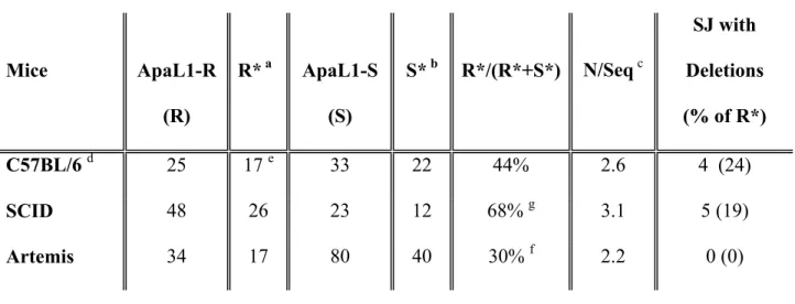
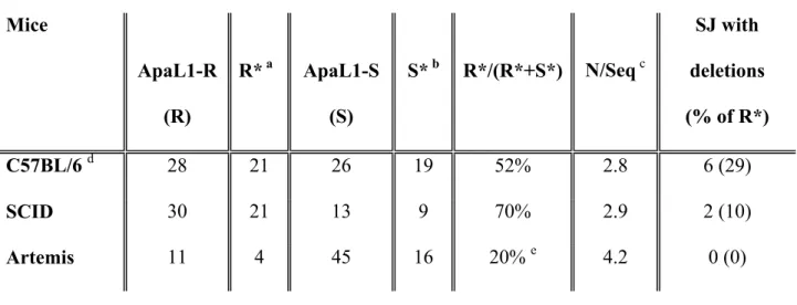
To confirm these findings, we extended our analysis to other TRD SJ repertoires for which the profile of modification has already been established in wt mice, namely SJs resulting from TRDV1/DD2 and TRAV14/DD2 rearrangement (Touvrey et al., 2006a) (see Fig 1 for a schematic representation of the TRAD locus). Although classified as TRA genes, the genes of the TRAV14 family, previously known as the ADV2 gene family, can be rearranged with TRD genes in DN thymocytes (Gallagher et al., 1998; Jouvin-Marche et al., 1998). These rearrangements generate a diverse SJ repertoire in wt thymocytes (Touvrey et al., 2006a). In both cases, the frequency of modified SJs was significantly decreased in Artemis deficient mice when compared to SCID animals (Tables 2 and 3, sequences available upon request). As in wt mice, SE processing consists of N nucleotide additions and/or deletions in wt and SCID mice. Only one of the TR DV1/DD2 SJ is modified only by SE deletion in absence of N nucleotides. In contrast, in absence of Artemis, SJs are modified exclusively through N nucleotide addition. Base loss from SEs is never observed in Artemis deficient thymocytes.
These results largely confirm our initial observation and extend our findings to rearrangement events involving V genes. It should be noted that the average length of N nucleotide additions is similar in both types of mice (wt, SCID, Artemis-/-; Tables 1-3). The TdT works with the same
efficiency on SEs whether DNA-PKcs and Artemis are present or not in the post-synaptic complex. It seems that the proportion of SEs available for modification is higher in SCID mice than in Artemis deficient thymocytes. Thus, although DNA-PKcs and Artemis cooperate in hairpin opening, their inactivation has clearly different effects on the resolution of SEs in vivo and results in distinct SJ repertoires.
Discussion
Opposite influence of DNA-PKcs and Artemis inactivation on SE availability for processing
NHEJ plays an essential role in developing lymphocytes for the repair of RAG-induced DNA breaks during V(D)J recombination. This process creates blunt ended, phosphorylated SE intermediates (Schlissel et al., 1993) which represent a category of “simple” DNA lesions that can be repaired by Ku-dependent NHEJ without processing (Lobrich and Jeggo, 2005). However, numerous studies showed that, in vivo, SEs can be modified by the TdT and a yet unknown nuclease activity before they are ligated, generating a diverse SJ repertoire. Here, we analyzed quantitatively and qualitatively the junctional diversity in three different SJ generated during the recombination of TRD genes in vivo in the thymus of wt, SCID and Artemis-deficient mice. Surprisingly, our results show a significant decrease in the frequency of imprecise junctions and a total absence of SE deletions in Artemis-deficient mice. Therefore, this genetic approach shows that, when present, Artemis participates in the re-joining of blunt DNA ends in vivo, at least in the context of SJ formation. Importantly, the impact of Artemis absence on SJ diversity is very different from the effects observed when DNA-PKcs is mutated.
In absence of DNA-PKcs activity, the frequency of imprecise SJ increases by 12% for DD1/DD2, 61% for DV1/DD2 and 33% for AV14/DD2 when compared to wt mice and reaches values ranging from 68% to 86%. More than two thirds of the SJs are resistant to ApaL1 digestion in SCID mice. The overall frequency and extent of SE deletions are similar in wt and SCID mice, even if variations are observed when the different junctions are considered individually. Consequently, this increase in ApaL1-R SJs reflects an increased frequency of joints containing N nucleotides. The activity of the TdT is however not affected, as the average number of N nucleotides added per joint is similar in both types of mice. These results suggest that in SCID mice, RAG-generated SEs are
more accessible to the TdT. It should be pointed out that the pattern of SJ modification (N nucleotide additions, limited occurrence of short deletions) clearly differs from that observed in Ku80-deficient thymocytes (no N nucleotides, large deletions). The increased frequency of SJ modification in SCID thymocytes is therefore due specifically to the inactivation of DNA-PKcs, and does not reflect a more general failure of assembling a functional DNA-PK complex on SEs.
Until now, Artemis has been thought to be functional only after association with and activation by DNA-PK on hairpin-sealed (Ma et al., 2002; Moshous et al., 2003; Niewolik et al., 2006) or “complex” (Lobrich and Jeggo, 2005; Povirk et al., 2007; Riballo et al., 2004) DNA ends. Our results now show that, in the absence of Artemis, the pattern of SJ modification is strikingly different from the one observed in SCID mice. We find for each of the SJs examined a decrease in the frequency of modified SJs, to levels even lower than in wt mice for TR DD1/DD2 and TR AV14/DD2 SJ. It can be that SEs with N nucleotides are less efficiently ligated; it can also be that SJs are formed more rapidly in Artemis deficient cells, and that the TdT does not have enough time to polymerize N nucleotide on SEs. However, the average number of N nucleotides per sequence in Artemis-deficient mice is similar to that observed in wt and SCID mice. This observation suggests that there is no counter selection for sequences with shorter stretches of untemplated bases and that there is no competition for ligation vs nucleotide polymerization. In addition, none of the ApaL1-R SJ sequenced from Artemis-deficient thymocytes shows any sign of SE deletion. Thus, it appears that SEs are less accessible to end modifying activities, both the TdT and a nuclease, when DNA-PKcs is present and Artemis absent than when DNA-DNA-PKcs is inactive and Artemis present.
An eventual role for Artemis in SJ formation has previously been sought but not found, in non-lymphoid cell lines by analyzing the rearrangement of artificial extra-chromosomal recombination substrate in SV 40-transformed fibroblasts from Artemis-mutated patients (Nicolas et
al., 1998) or in Artemis-deficient MEFs (Rooney et al., 2002) or ES cells (Rooney et al., 2003). As mentioned earlier (see introduction), this assay produces only precise SJs in NHEJ-proficient cells. It was therefore not possible in this experimental system to detect an increase in the overall SJ fidelityin absence of Artemis. In contrast, the variations in the pattern of junctional diversity in SJs generated in vivo during the recombination of endogenous TR genes in their normal, chromatinized environment allow us to propose that Artemis, although not required, participates in SJ formation when it is expressed.
Interplay of DNA-PKcs and Artemis in vivo
Several recent articles addressed the interplay of Ku, DNA-PKcs and Artemis at DNA ends. The emerging picture is that DNA-PK activity and DNA-PKcs autophosphorylation are required for Artemis recruitment and activation (Goodarzi et al., 2006; Li et al., 2005; Soubeyrand et al., 2006). PK activity seems to be triggered by the synapsis of DNA ends (DeFazio et al., 2002). DNA-PKcs phosphorylation induces conformational changes that facilitate DNA threading to make ends more accessible or expose them in a better configuration for processing and ligation (Block et al., 2004; DeFazio et al., 2002; Goodarzi et al., 2006; Niewolik et al., 2006; Uematsu et al., 2007). DNA-PKcs-dependent phosphorylation triggers Artemis endonucleolytic activity, possibly by exposing its active site (Niewolik et al., 2006). Phosphorylated DNA-PKcs dissociates from the complex after Artemis action, to allow ligation of the DNA ends (Block et al., 2004; Drouet et al., 2006; Goodarzi et al., 2006). Artemis seems to be critically required for the resection of long overhangs (Goodarzi et al., 2006), maybe because DNA-PKcs can fold then in hairpin-like structure (Niewolik et al., 2006). This sequence of events results from several studies conducted with cell lines over-expressing -or mutated for- one of the NHEJ components, or from in vitro reactions with
nuclear extracts or purified proteins. In most of them artificial DNA substrates with 5’ or 3’ extensions were used to promote Artemis activation. This proposed model was largely confirmed by a recent analysis of DNA end processing during NHEJ repair of plasmids by cell-free extracts (Budman and Chu, 2005).
Our results now provide evidence for a similar scenario for the interplay of DNA-PKcs and Artemis in vivo, during the repair of RAG-generated blunt DNA ends. The level of SJ modification is lower in Artemis-deficient animals than in SCID mice. It is possible that in SCID thymocytes, SEs become more susceptible to modifications because no phosphorylation-competent DNA-PK complex is assembled to thread/protect them correctly before ligation, whereas in the absence of Artemis, DNA-PKcs dissociation does not occur, and the DNA ends are maintained in a tight complex with DNA-PK, where they are less susceptible to be modified. Although they do not play an obligatory role in SJ formation, when present, DNA-PKcs and Artemis actively participate in the regulation of this reaction as revealed by the impact of their inactivation on the profile of SJ modification. Even if they are a priori ligatable without requiring further processing, SEs are associated with several proteins including RAG-1, RAG-2, the TdT, the Ku heterodimer, DNA-PKcs, ATM and p53 (Agrawal and Schatz, 1997; Perkins et al., 2002; Purugganan et al., 2001). Our results suggest that, in vivo, Artemis can also participate in these complexes, where it could regulate the stability of the DNA-PK complex assembled on “simple” DNA end. A definitive proof will await the availability of an efficient anti-Artemis antibody, so that its presence in a physiological context can be analyzed. It will also be interesting to determine whether DNA-PKcs activation of Artemis endonucleolytic activity is required for this new property, or if its recruitment to the DNA repair complex (Soubeyrand et al., 2006) is sufficient to trigger DNA-PKcs dissociation.
Artemis as an exonuclease during the repair of blunt DNA ends
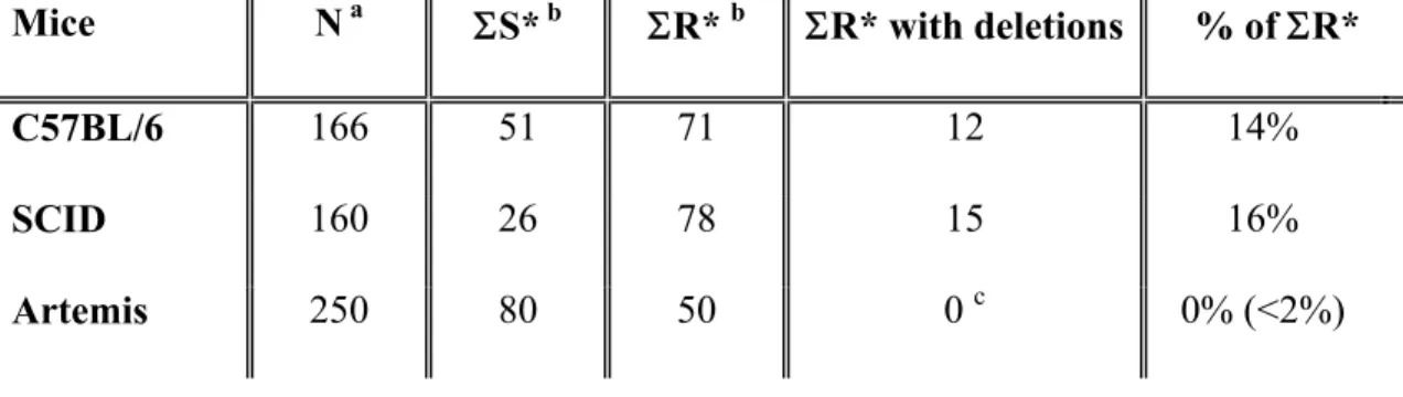
Finally, it is interesting to note that for each of the three different SJ analyzed in this study, deletions are found only in wt and SCID mice but not in Artemis-deficient animals. Deletions were found in 2 to 8 sequences depending on the SJ considered (Tables 1-3). They contribute to junctional diversity in 6% to 29% of the joints in wt and SCID thymocytes. When SJs are considered individually, the frequency of deletions is significantly lower (p<0.05, Student’s test) in joints created in absence of Artemis only for DV1/DD2 SJs when compared to wt and DD1/DD2 SJs when compared to SCID. However, when SJ sequences are pooled and analyzed globally (Table 4), we found that the occurrence of deletions in SJs created in absence of Artemis (0 out of 50 ApaL1-R unique sequences, <2%) is significantly decreased when compared to wt (12 out of 71, 14%, p<0.002) and SCID (15 out of 78, 16%, p<0.001) mice. Thus, deletions are observed only when Artemis is present.
The simplest explanation for this correlation is that Artemis itself is responsible for these deletions. Indeed, it is the only identified nuclease that participates in V(D)J recombination. In vitro, the purified free protein possesses an exonuclease activity which does not require triggering through association with DNA-PKcs (Niewolik et al., 2006). SE deletions could also be due to Artemis endo-rather than exo-nuclease activity. In any case, it was recently shown that a mutated Artemis protein that can no longer interact with DNA-PKcs is nonetheless functional and recruited at the site of V(D)J recombination (Soubeyrand et al., 2006). It is not known yet whether it is still endowed with exonuclease activity, but this result suggests that wt Artemis can be recruited at the site of DNA damage independently of DNA-PKcs. This feature could explain that SE deletions are observed in SCID mice. Whatever the exact underlying mechanism, our results show for each of the SJ analyzed a correlation between Artemis expression and SE deletions. Although we cannot exclude that
Artemis is only indirectly responsible for SE deletions by recruiting another enzyme, this correlation and its known activities strongly argue that Artemis is the nuclease responsible for the deletions observed in SJ generated in vivo, in wt and SCID mice, by Ku-dependent NHEJ mechanisms. Indeed, we do not known whether Artemis can be recruited onto DNA ends and act as a nuclease in the absence of Ku. It is therefore not possible at that stage to speculate about its possible involvement in the formation of the large deletions found in Ku-/- thymocytes by alternative NHEJ
mechanisms.
Concluding remarks
The resolution of DSBs by NHEJ is of the utmost importance for cell survival following exposure to genotoxic stress and for V(D)J recombination during lymphocyte development. DNA-PKcs and Artemis are both required for the repair of a subset of “complex” DSBs (Povirk et al., 2007; Riballo et al., 2004) and for CJ formation. In contrast, it was proposed that DNA-PKcs plays at most only a “facilitating” role during the Artemis-independent repair of “simple” ligatable DNA ends such as SEs (Lobrich and Jeggo, 2005). SEs therefore represent bona fide DNA lesions that are repaired by the Ku-dependant NHEJ pathway and SJ structure reflects the mechanisms operating during the repair of physiological substrates in their natural environment, i.e. complexes where free DNA ends are associated with endogenous proteins stoichiometrically expressed at physiological level. Our findings show that Artemis shapes the outcome of the repair process involving blunt and phosphorylated DNA ends, possibly by promoting DNA-PKcs dissociation from the complex assembled on DNA ends. This in turn suggests that, in non-lymphoid cells, Artemis and DNA-PKcs can be recruited to the site of the DNA lesions together with Ku70, Ku80, the DNA Lig IV, XRCC4, Cernunnos, even when these lesions do not require the resection of protruding ends or the removal of
complex damage. How the different scaffolding and repair factors are recruited to DNA ends and how all of these proteins interact with one another in vivo is still to be uncovered to fully understand DNA DSB repair and V(D)J recombination.
Acknowledgements
The authors want to thank Drs C. Aude-Garcia, P. Calsou,C. Lemercier and S. Marcand for critical reading of the manuscript and S. Fremy, N. Chaumontel and F. Sergent for animal care. CT was supported in part by a grant from “l’Association pour la Recherche contre le Cancer”.
References
Agrawal A. and Schatz D. G. (1997) RAG1 and RAG2 form a stable postcleavage synaptic complex with DNA containing signal ends in V(D)J recombination. Cell 89, 43-53.
Ahnesorg P., Smith P. and Jackson S. P. (2006) XLF interacts with the XRCC4-DNA ligase IV complex to promote DNA nonhomologous end-joining. Cell 124, 301-13.
Bentolila L., Wu G., Nourrit F., Fanton d'Andon M., Rougeon F. and Doyen N. (1997) Constitutive expression of terminal deoxynucleotidyl transferase in transgenic mice is sufficient for N region diversity to occur at any Ig locus throughout B cell differentiation. J Immunol 158, 715-723.
Binnie A., Olson S., Wu G. E. and Lewis S. M. (1999) Gamma-irradiation directly affects the formation of coding joints in SCID cell lines. J.Immunol. 163, 5418-5426.
Block W. D., Yu Y., Merkle D., Gifford J. L., Ding Q., Meek K. and Lees-Miller S. P. (2004) Autophosphorylation-dependent remodeling of the DNA-dependent protein kinase catalytic subunit regulates ligation of DNA ends. Nucleic Acids Res 32, 4351-7.
Bogue M. A., Jhappan C. and Roth D. B. (1998) Analysis of variable (diversity) joining recombination in DNAdependent protein kinase (PK)-deficient mice reveals DNA-PK-independent pathways for both signal and coding joint formation.
Proc.Natl.Acad.Sci.U.S.A. 95, 15559-15564.
Bogue M. A., Wang C., Zhu C. and Roth D. B. (1997) V(D)J recombination in Ku86-deficient mice: distinct effects on coding, signal, and hybrid joint formation. Immunity. 7, 37-47.
Buck D., Malivert L., de Chasseval R., Barraud A., Fondaneche M. C., Sanal O., Plebani A., Stephan J. L., Hufnagel M., le Deist F., Fischer A., Durandy A., de Villartay J. P. and Revy
P. (2006) Cernunnos, a novel nonhomologous end-joining factor, is mutated in human immunodeficiency with microcephaly. Cell 124, 287-99.
Budman J. and Chu G. (2005) Processing of DNA for nonhomologous end-joining by cell-free extract. Embo J 24, 849-60.
Candéias S., Muegge K. and Durum S. K. (1996) Junctional diversity in signal joints from T cell receptor beta and delta loci via terminal deoxynucleotidyl transferase and exonucleolytic activity. J.Exp.Med. 184, 1919-1926.
Carroll A. M., Slack J. K. and Chang W. T. (1993) Biased T-cell receptor delta element recombination in scid thymocytes. Mol Cell Biol 13, 3632-40.
Corneo B., Wendland R. L., Deriano L., Cui X., Klein I. A., Wong S. Y., Arnal S., Holub A. J., Weller G. R., Pancake B. A., Shah S., Brandt V. L., Meek K. and Roth D. B. (2007) Rag mutations reveal robust alternative end joining. Nature 449, 483-6.
Couedel C., Mills K. D., Barchi M., Shen L., Olshen A., Johnson R. D., Nussenzweig A., Essers J., Kanaar R., Li G. C., Alt F. W. and Jasin M. (2004) Collaboration of homologous recombination and nonhomologous end-joining factors for the survival and integrity of mice and cells. Genes Dev 18, 1293-304.
Cui X. and Meek K. (2007) Linking double-stranded DNA breaks to the recombination activating gene complex directs repair to the nonhomologous end-joining pathway. Proc Natl Acad Sci
U S A 104, 17046-51.
Dai Y., Kysela B., Hanakahi L. A., Manolis K., Riballo E., Stumm M., Harville T. O., West S. C., Oettinger M. A. and Jeggo P. A. (2003) Nonhomologous end joining and V(D)J recombination require an additional factor. Proc Natl Acad Sci U S A 100, 2462-7.
Davis M. M. and Bjorkman P. J. (1988) T-cell antigen receptor genes and T-cell recognition. Nature 334, 395-402.
DeFazio L. G., Stansel R. M., Griffith J. D. and Chu G. (2002) Synapsis of DNA ends by DNA-dependent protein kinase. Embo J 21, 3192-200.
Drouet J., Frit P., Delteil C., de Villartay J. P., Salles B. and Calsou P. (2006) Interplay between Ku, Artemis, and the DNA-dependent protein kinase catalytic subunit at DNA ends. J Biol Chem 281, 27784-93.
Ezekiel U. R., Sun T., Bozek G. and Storb U. (1997) The composition of coding joints formed in V(D)J recombination is strongly affected by the nucleotide sequence of the coding ends and their relationship to the recombination signal sequences. Mol Cell Biol 17, 4191-7.
Gallagher M., Candéias S., Martinon C., Borel E., Malissen M., Marche P. N. and Jouvin-Marche E. (1998) Use of TCR ADV gene segments by the delta chain is independent of their position and of CD3 expression. Eur.J.Immunol. 28, 3878-3885.
Gao Y., Chaudhuri J., Zhu C., Davidson L., Weaver D. T. and Alt F. W. (1998) A targeted DNA-PKcs-null mutation reveals DNA-PK-independent functions for KU in V(D)J recombination.
Immunity 9, 367-76.
Gauss G. H. and Lieber M. R. (1996) Mechanistic constraints on diversity in human V(D)J recombination. Mol Cell Biol 16, 258-69.
Goodarzi A. A., Yu Y., Riballo E., Douglas P., Walker S. A., Ye R., Harer C., Marchetti C., Morrice N., Jeggo P. A. and Lees-Miller S. P. (2006) DNA-PK autophosphorylation facilitates Artemis endonuclease activity. Embo J 25, 3880-9.
Iwasato T. and Yamagishi H. (1992) Novel excision products of T cell receptor gamma gene rearrangements and developmental stage specificity implied by the frequency of nucleotide insertions at signal joints. Eur J Immunol 22, 101-6.
Jouvin-Marche E., Aude-Garcia C., Candéias S., Borel E., Hachemi-Rachedi S., Gahery-Segard H., Cazenave P. A. and Marche P. N. (1998) Differential chronology of TCRADV2 gene use by alpha and delta chains of the mouse TCR. Eur.J.Immunol. 28, 818-827.
Jung D. and Alt F. W. (2004) Unraveling V(D)J recombination; insights into gene regulation. Cell 116, 299-311.
Kanari Y., Muto M. and Yamagishi H. (2003) TCR delta gene rearrangements revealed by fine structure of the recombination junction in mice. Microbiol Immunol 47, 883-94.
Kanari Y., Nakagawa R., Arakawa H. and Yamagishi H. (1998) Variable gene segment-specific N-insertions at the signal joint of T-cell receptor Vbeta-Dbeta recombinations. Immunol Lett 61, 151-5.
Lee G. S., Neiditch M. B., Salus S. S. and Roth D. B. (2004) RAG proteins shepherd double-strand breaks to a specific pathway, suppressing error-prone repair, but RAG nicking initiates homologous recombination. Cell 117, 171-84.
Lewis S., Gifford A. and Baltimore D. (1985) DNA elements are asymmetrically joined during the site-specific recombination of kappa immunoglobulin genes. Science 228, 677-85.
Li L., Salido E., Zhou Y., Bhattacharyya S., Yannone S. M., Dunn E., Meneses J., Feeney A. J. and Cowan M. J. (2005) Targeted disruption of the Artemis murine counterpart results in SCID and defective V(D)J recombination that is partially corrected with bone marrow transplantation. J Immunol 174, 2420-8.
Li Y., Hayakawa K. and Hardy R. (1993) The regulated expression of B lineage associated genes during B cell differentiation in bone marrow and fetal liver. J. Exp. Med. 178, 951-960. Lieber M. R., Hesse J. E., Lewis S., Bosma G. C., Rosenberg N., Mizuuchi K., Bosma M. J. and
Gellert M. (1988a) The defect in murine severe combined immune deficiency: joining of signal sequences but not coding segments in V(D)J recombination. Cell 55, 7-16.
Lieber M. R., Hesse J. E., Mizuuchi K. and Gellert M. (1988b) Lymphoid V(D)J recombination: nucleotide insertion at signal joints as well as coding joints. Proc Natl Acad Sci U S A 85, 8588-92.
Lobrich M. and Jeggo P. A. (2005) Harmonising the response to DSBs: a new string in the ATM bow. DNA Repair (Amst) 4, 749-59.
Ma Y., Pannicke U., Schwarz K. and Lieber M. R. (2002) Hairpin opening and overhang processing by an Artemis/DNA-dependent protein kinase complex in nonhomologous end joining and V(D)J recombination. Cell 108, 781-94.
Moshous D., Callebaut I., de Chasseval R., Corneo B., Cavazzana-Calvo M., Le Deist F., Tezcan I., Sanal O., Bertrand Y., Philippe N., Fischer A. and de Villartay J. P. (2001) Artemis, a novel DNA double-strand break repair/V(D)J recombination protein, is mutated in human severe combined immune deficiency. Cell 105, 177-86.
Moshous D., Callebaut I., de Chasseval R., Poinsignon C., Villey I., Fischer A. and de Villartay J. P. (2003) The V(D)J recombination/DNA repair factor artemis belongs to the metallo-beta-lactamase family and constitutes a critical developmental checkpoint of the lymphoid system.
Ann N Y Acad Sci 987, 150-7.
Nadel B. and Feeney A. J. (1995) Influence of coding-end sequence on coding-end processing in V(D)J recombination. J Immunol 155, 4322-9.
Nicolas N., Moshous D., Cavazzana-Calvo M., Papadopoulo D., de Chasseval R., Le Deist F., Fischer A. and de Villartay J. P. (1998) A human severe combined immunodeficiency (SCID) condition with increased sensitivity to ionizing radiations and impaired V(D)J rearrangements defines a new DNA recombination/repair deficiency. J Exp Med 188, 627-34. Niewolik D., Pannicke U., Lu H., Ma Y., Wang L. C., Kulesza P., Zandi E., Lieber M. R. and
Schwarz K. (2006) DNA-PKcs Dependence of Artemis Endonucleolytic Activity, Differences between Hairpins and 5' or 3' Overhangs. J Biol Chem 281, 33900-9.
Nussenzweig A., Chen C., da Costa Soares V., Sanchez M., Sokol K., Nussenzweig M. C. and Li G. C. (1996) Requirement for Ku80 in growth and immunoglobulin V(D)J recombination.
Nature 382, 551-5.
Perkins E. J., Nair A., Cowley D. O., Van Dyke T., Chang Y. and Ramsden D. A. (2002) Sensing of intermediates in V(D)J recombination by ATM. Genes Dev 16, 159-64.
Povirk L. F., Zhou T., Zhou R., Cowan M. J. and Yannone S. M. (2007) Processing of 3'-phosphoglycolate-terminated DNA double strand breaks by Artemis nuclease. J Biol Chem 282, 3547-58.
Purugganan M. M., Shah S., Kearney J. F. and Roth D. B. (2001) Ku80 is required for addition of N nucleotides to V(D)J recombination junctions by terminal deoxynucleotidyl transferase.
Nucleic Acids Res 29, 1638-46.
Riballo E., Kuhne M., Rief N., Doherty A., Smith G. C., Recio M. J., Reis C., Dahm K., Fricke A., Krempler A., Parker A. R., Jackson S. P., Gennery A., Jeggo P. A. and Lobrich M. (2004) A pathway of double-strand break rejoining dependent upon ATM, Artemis, and proteins locating to gamma-H2AX foci. Mol Cell 16, 715-24.
Rooney S., Alt F. W., Lombard D., Whitlow S., Eckersdorff M., Fleming J., Fugmann S., Ferguson D. O., Schatz D. G. and Sekiguchi J. (2003) Defective DNA repair and increased genomic instability in Artemis-deficient murine cells. J Exp Med 197, 553-65.
Rooney S., Chaudhuri J. and Alt F. W. (2004) The role of the non-homologous end-joining pathway in lymphocyte development. Immunol Rev 200, 115-31.
Rooney S., Sekiguchi J., Zhu C., Cheng H. L., Manis J., Whitlow S., DeVido J., Foy D., Chaudhuri J., Lombard D. and Alt F. W. (2002) Leaky Scid phenotype associated with defective V(D)J coding end processing in Artemis-deficient mice. Mol Cell 10, 1379-90.
Roth D. B., Menetski J. P., Nakajima P. B., Bosma M. J. and Gellert M. (1992) V(D)J recombination: broken DNA molecules with covalently sealed (hairpin) coding ends in scid mouse thymocytes. Cell 70, 983-91.
Schlissel M., Constantinescu A., Morrow T., Baxter M. and Peng A. (1993) Double-strand signal sequence breaks in V(D)J recombination are blunt, 5'-phosphorylated, RAG-dependent, and cell cycle regulated. Genes Dev 7, 2520-32.
Soubeyrand S., Pope L., De Chasseval R., Gosselin D., Dong F., de Villartay J. P. and Hache R. J. (2006) Artemis phosphorylated by DNA-dependent protein kinase associates preferentially with discrete regions of chromatin. J Mol Biol 358, 1200-11.
Stanhope-Baker P., Hudson K. M., Shaffer A. L., Constantinescu A. and Schlissel M. S. (1996) Cell type-specific chromatin structure determines the targeting of V(D)J recombinase activity in vitro. Cell 85, 887-97.
Taccioli G. E., Amatucci A. G., Beamish H. J., Gell D., Xiang X. H., Torres Arzayus M. I., Priestley A., Jackson S. P., Marshak Rothstein A., Jeggo P. A. and Herrera V. L. (1998) Targeted
disruption of the catalytic subunit of the DNA-PK gene in mice confers severe combined immunodeficiency and radiosensitivity. Immunity 9, 355-66.
Talukder S. R., Dudley D. D., Alt F. W., Takahama Y. and Akamatsu Y. (2004) Increased frequency of aberrant V(D)J recombination products in core RAG-expressing mice. Nucleic Acids Res 32, 4539-49.
Touvrey C., Borel E., Marche P. N., Jouvin-Marche E. and Candeias S. M. (2006a) Gene-specific signal joint modifications during V(D)J recombination of TCRAD locus genes in murine and human thymocytes. Immunobiology 211, 741-51.
Touvrey C., Cowell L. G., Lieberman A. E., Marche P. N., Jouvin-Marche E. and Candeias S. M. (2006b) Reassignment of the murine 3'TRDD1 recombination signal sequence.
Immunogenetics 58, 895-903.
Tsai C. J., Kim S. A. and Chu G. (2007) Cernunnos/XLF promotes the ligation of mismatched and noncohesive DNA ends. Proc Natl Acad Sci U S A 104, 7851-6.
Uematsu N., Weterings E., Yano K., Morotomi-Yano K., Jakob B., Taucher-Scholz G., Mari P. O., van Gent D. C., Chen B. P. and Chen D. J. (2007) Autophosphorylation of DNA-PKCS regulates its dynamics at DNA double-strand breaks. J Cell Biol 177, 219-29.
Weinstock D. M. and Jasin M. (2006) Alternative pathways for the repair of RAG-induced DNA breaks. Mol Cell Biol 26, 131-9.
West K. L., Singha N. C., De Ioannes P., Lacomis L., Erdjument-Bromage H., Tempst P. and Cortes P. (2005) A direct interaction between the RAG2 C terminus and the core histones is required for efficient V(D)J recombination. Immunity 23, 203-12.
Wu P. Y., Frit P., Malivert L., Revy P., Biard D., Salles B. and Calsou P. (2007) Interplay between Cernunnos-XLF and nonhomologous end-joining proteins at DNA ends in the cell. J Biol
Figure legends
Fig 1: Schematic representation of the genes and their RSS
The relative location of the TRAV14, TRDV1, TRDD1 and TRDD2 genes on the murine TRAD locus is represented. The genes are presented as grey squares, and their RSS as triangles. Open and black triangles represent RSS with spacers of 23 and 12 bases, respectively. Following V(D)J recombination, the recombination signals created by the juxtaposition of the two RSS is located on a T cell receptor excision circle. The recombination of TRDD1 and TRDD2 is shown as an example. The arrows represent the forward and reverse primers used for amplification. Given their orientation, amplification of a signal joint is possible only after V(D)J recombination did occur. The drawing is not to scale. Only one member of the TRAV14 family is shown.
Fig 2: Sequence of the ApaL1-R TR DD1/DD2 SJs amplified from C57BL/6 mice.
The sequences of TRDD1 and TRDD2 RSS heptamers (capital letters) and of the first flanking TRDD1 and TRDD2 bases (lowercase italics) are indicated at the top. The nucleotides listed under the “N” column represent putative non-templated nucleotides. Bold nucleotides in the “N” column could putatively represent coding flank retention resulting from mis-cutting by the recombinase complex. Dashes indicate deletions. The number in the last column indicates the number of times the sequence on that line was found in a sample from one mouse. Identical sequences found in different samples were considered as distinct events. The last two sequences represent non-standard rearrangement products generated through the recombination of the 5’TR DD1 and 3’TR DD2 RS (Kanari et al., 2003). In these junctions, the sequence of the TRDD1 and TRDD2 genes are underlined. Sequences were obtained from 3 independent animals.
Fig 3: Sequence of the TR DD1/DD2 SJs amplified from Ku80-/- mice.
See legend to figure 1. The sequences obtained from two individual animals are identified as Ku1 and Ku2, respectively. The products obtained after the nested PCR amplification were directly cloned and sequenced without prior ApaL1 discrimination between perfect and modified SJs. No putative non-templated nucleotides were identified. Dashes indicate deletions. When more than 6 bases where deleted, the number of nucleotides deleted from the TR DD1 or TR DD2 heptamer is indicated in the relevant column.
Fig 4: Sequence of the ApaL1-R TR DD1/DD2 SJs amplified from SCID mice.
See legend to figures 1 and 2 for details. Sequences were obtained from 2 individual mice.
Fig 5:Sequence of the ApaL1-R TR DD1/DD2 SJs amplified from Artemis-deficient-mice.
Table 1: Analysis of TR DD1/DD2 signal joint repertoire Mice ApaL1-R (R) R* a ApaL1-S (S) S* b R*/(R*+S*) N/Seq c SJ with deletions (% of R*) C57BL/6 38 33 12 10 77% 2.9 2 (6) SCID 40 31d 6 5 86% 2.6 8 (26) Artemis 44 29 36 24 55% e 2.7 0 (0)
a: multiple occurrences of a sequence from one DNA sample were counted only once and signal
joints containing a DD1/DD2 coding joint were not considered to determine R*.
b: S* is the value of S corrected by R*/R to consider the “redundancy” of the SJ repertoire.
c:average number of un-templated nucleotides per unique sequence (R*).
d: three of the R* sequences have only deletions and no N insertions
e: p<0.025 and p<0.002 when compared to C57BL/6 and SCID mice by Student test, respectively.
Table 2: Analysis of TR DV1/DD2 signal joint repertoire Mice ApaL1-R (R) R* a ApaL1-S (S) S*b R*/(R*+S*) N/Seq c SJ with Deletions (% of R*) C57BL/6 d 25 17e 33 22 44% 2.6 4 (24) SCID 48 26 23 12 68% g 3.1 5 (19) Artemis 34 17 80 40 30% f 2.2 0 (0)
a: multiple occurrences of a sequence from one DNA sample were counted only once.
b, c: see footnotes to Table I.
d: data from Touvrey et al, 2006, corrected for redundancy as explained in Materials and Methods
e: one of the R* sequences has only deletions and no N insertions.
f: p<0.0002 when compared to SCID mice by Student test
Table 3: Analysis of TR AV14/DD2 signal joint repertoire Mice ApaL1-R (R) R* a ApaL1-S (S) S*b R*/(R*+S*) N/Seq c SJ with deletions (% of R*) C57BL/6 d 28 21 26 19 52% 2.8 6 (29) SCID 30 21 13 9 70% 2.9 2 (10) Artemis 11 4 45 16 20% e 4.2 0 (0)
a: multiple occurrences of a sequence from one DNA sample were counted only once.
b, c, d: see footnotes to Table I and II.
Table 4: Absence of SJ with deletions in Artemis-deficient mice
Mice Na S*b R*b R* with deletions % of R*
C57BL/6 166 51 71 12 14%
SCID 160 26 78 15 16%
Artemis 250 80 50 0c 0% (<2%)
a: N is the total number of SJ analyzed in each type of mice.
bS* and R* represent the total numbers S* and R* SJs found in each type of mice,
respectively.
c: p<0.002 and p<0.001 when compared to C57BL/6 and SCID mice by a Student’s test, respectively
TRDD1 TRDD2 TRDV1 TRAV14
+
V(D)J recombination
Fig 1
TableFigure 2:
ApaL1-R TR DD1/DD2 signal joints in C57BL/6 thymocytes
DD2 N DD1
CACCGTG atcg atat CACACAG
CACCGTG GGGGCCT CACACAG CACCGTG GGGGAG CACACAG CACCGTG ATCTCT CACACAG CACCGTG CGCC CACACAG CACCGTG AGAC CACACAG CACCGTG AACT CACACAG CACCGTG GGG CACACAG 2x CACCGTG GGC CACACAG CACCGTG GGA CACACAG CACCGTG GCA CACACAG CACCGTG GCG CACACAG CACCGTG CCG CACACAG CACCGTG CCT CACACAG CACCGTG TCC CACACAG CACCGTG AGC CACACAG CACCGTG AAA CACACAG CACCGTG AGA CACACAG CACCGTG ACA CACACAG CACCGTG AAC CACACAG CACCGTG GG CACACAG 2x CACCGTG GG CACACAG CACCGTG CC CACACAG CACCGTG GA CACACAG CACCGTG CG CACACAG 3x CACCGTG CC CACACAG CACCGTG CC CACACAG CACCGTG CT CACACAG CACCGTG TC CACACAG CACCGTG AG CACACAG CACCGTG C CACACAG CACCGTG G CACACAG CACC--- CC CACACAG CACCG-- G CACACAG CACCGTG ATCGGAGGGATACGACCCAATGGCATAT CACACAG
Figure 3
TR DD1/DD2 signal joints in Ku80-/- mice
DD2 N DD1
CACCGTG atcg atat CACACAG
Ku1 -37 -52 Ku1 -41 -60 Ku1 -47 -60 Ku1 -83 -96 Ku1 -70 -90 7X Ku1 -79 -100 2X Ku1 -16 -43 3X Ku1 -82 -36 Ku1 -67 -86 Ku2 CACCGTG CACACAG 9x Ku2 C--- CACACAG 2x Ku2 -10 ---AG 31x
Figure 4
ApaL1-R TR DD1/DD2 signal joints in SCID mice
DD2 N DD1
CACCGTG atcg atat CACACAG
CACCGTG GGGGCCT CACACAG CACCGTG AACCC CACACAG CACCGTG CCCCC CACACAG CACCGTG GGGG CACACAG CACCGTG AGA CACACAG CACCGTG GAC CACACAG CACCGTG GGG CACACAG CACCGTG GCT CACACAG CACCGTG TCT CACACAG CACCGTG CCT CACACAG CACCGTG CAA CACACAG CACCGTG GGG CACACAG 2x CACCGTG CCC CACACAG CACCGTG GTA CACACAG CACCGTG GGC CACACAG 2x CACCGTG GTC CACACAG CACCGTG CTC CACACAG CACCGTG TCC CACACAG CACCGTG GG CACACAG 4x CACCGTG CT CACACAG CACCGTG GA CACACAG CACCGTG G CACACAG 2x CACCGTG G CACACAG CACCGT- CC CACACAG CACCGTG CG -ACACAG CACCGTG GG ---ACAG CACCGT- AG CACACAG (-7)---- G ---G CACCGTG --CACAG CACCGTG --CACAG 2x (-12)--- ---(-27) CACCGTG ATCGGAGGGAGGGGCATAT CACACAG CACCGTG ATCGGGGGCACGTGGCATAT CACACAG
Figure 5
ApaL1-R TR DD1/DD2 signal joints in Artemis-deficient mice
DD2 N DD1
CACCGTG atcg atat CACACAG
CACCGTG GGGACA CACACAG CACCGTG AGACAC CACACAG CACCGTG GGGGG CACACAG CACCGTG GCGG CACACAG CACCGTG GGGA CACACAG CACCGTG ACGA CACACAG
CACCGTG CCC CACACAG 2x CACCGTG GGG CACACAG CACCGTG CCT CACACAG CACCGTG CCC CACACAG CACCGTG GGG CACACAG CACCGTG TGC CACACAG CACCGTG GGG CACACAG CACCGTG AC CACACAG CACCGTG CG CACACAG 2x CACCGTG AG CACACAG 2x CACCGTG CC CACACAG 2x CACCGTG GG CACACAG 3x CACCGTG GG CACACAG 2x CACCGTG CC CACACAG 2x CACCGTG CT CACACAG 2x CACCGTG CC CACACAG CACCGTG CG CACACAG CACCGTG CG CACACAG CACCGTG AG CACACAG 2x CACCGTG G CACACAG CACCGTG A CACACAG CACCGTG G CACACAG 2x CACCGTG G CACACAG
Supplementary Material