Beta Oscillations in Frontal Cortex and Striatum Represent
Post-Processing of Successful Behavior
by
Joseph Feingold
AB Philosophy and Physics
Harvard University, 1999
Master of Advanced Study in Mathematics
Cambridge University, 2000
Submitted to the Division of Health Sciences and Technology
in Partial Fulfillment of the Requirements for the Degree of
Doctor of Philosophy in Health Sciences and Technology
at the
Massachusetts Institute of Technology
MASSACHUSETTS INSTIUTE OF TEcHNOLOGY
SEP
2 1
2011
LIBRARIES
ARCHIVES
September 2011
© 2011 Joseph Feingold. All rights reserved
The author hereby grants to the Massachusetts
Institute
of Technology permission to
reproduce and to distribute publicly paper and electronic copies of this thesis document
in whole or in part in any medium now known or hereafter created
/7'(7
Signature of Author:
Certified by:
Speech and Hearing Bic
sienc
a'nd T'chnology Prorgram
September 1, 2011
'
7
Ann M.
Graybiel, PhD
Institute Professor and Professor in Brain and Cognitive Sciences
Thesis Supervisor
Accepted by:
Ram Sasisekharan, PhD
Director, Harvard-MIT Division of Health Sciences and Technology
Edward Hood Taplin Professor of Health Sciences & Technology and Biological Engineering
ACKNOWLEDGEMENTS
TABLE OF CONTENTS
ACKNOWLEDGEMENTS 2
TABLE OF CONTENTS 3
ABSTRACT 4
CHAPTER I: BACKGROUND AND SIGNIFICANCE 5
REFERENCES 11
CHAPTER II: A SYSTEM FOR RECORDING NEURAL ACTIVITY CHRONICALLY
AND SIMULTANEOUSLY FROM MULTIPLE CORTICAL AND SUBCORTICAL
REGIONS IN NON-HUMAN PRIMATES 13
ABSTRACT 14
INTRODUCTION 15
MATERIALS AND METHODS 18
RESULTS 30
RECONFIGURABLE CHRONIC ELECTRODE IMPLANT SYSTEM 30
SIMULTANEOUSLY RECORDED NEURAL ACTIVITY FROM MULTIPLE CORTICAL AND
SUBCORTICAL SITES 31
SESSION-TO-SESSION ADJUSTMENT OF INDIVIDUAL ELECTRODES' DEPTHS 32
SESSION-TO-SESSION STABILITY OF NEURAL ACTIVITY 34
MICROSTIMULATION AND INJECTION USING THE CHIME SYSTEM 34
DISCUSSION 36 ACKNOWLEDGEMENTS 42 GRANTS 42 REFERENCES 43 TABLES 47 FIGURE CAPTIONS 48
CHAPTER III: BETA OSCILLATIONS IN FRONTAL CORTEX AND STRIATUM REPRESENT POST-PROCESSING OF SUCCESSFUL BEHAVIOR 64
ABSTRACT 65
PROMINENT POST-PERFORMANCE BETA OSCILLATIONS 68
BURSTS OF SPATIALLY LOCALIZED BETA ACTIVITY 70
TEMPORAL RELATIONSHIPS BETWEEN BETA BURSTS 75
DISCUSSION 78
METHODS 84
REFERENCES 93
FIGURE LEGENDS 96
ABSTRACT
Beta band (13-30 Hz) oscillations in sensorimotor cortex are associated with motor performance, but the nature of this relationship is not clear. Recently, excessive beta activity in cortico-basal ganglia circuits has been recognized as a hallmark of Parkinson's disease. Renewed interest in beta oscillations has since led to the suggestion that they might reflect the preservation of the current output or state of a given brain region. To investigate the potential role of beta activity in the brain, we recorded local field potentials in the frontal cortex and striatum of monkeys as they performed single and sequential arm movement tasks. To facilitate these experiments, we developed novel methods for recording simultaneously from independently moveable electrodes implanted chronically at over 100 sites in cortical and subcortical areas of the monkey brain. We found that, across tasks, beta oscillations occurred in brief, spatially localized bursts that were most prominent following task performance. Across brain regions, post-performance bursts were differentially modulated by the preceding task. In motor cortex they tracked the number of movements just performed. In contrast, striatal and prefrontal burst rates were proportional to the number of visual cues, or to a combination of the cues and movements, respectively, and were higher following correct, rewarded, trials than unrewarded errors. Pairs of striatal-prefrontal sites exhibited increased cross-covariance and coherence during post-trial beta bursts, suggesting that these bursts might be involved in communication or coordination across brain regions. Based on our results, we propose that beta oscillations may represent post-performance reinforcement of the network dynamics that led to the desired behavioral outcome obtained immediately prior.
CHAPTER I
Neural activity is characterized by oscillations at multiple time-scales. A growing body of evidence implicates oscillatory neural activity in an array of behavioral and cognitive functions, as well as in clinical manifestations of neurological disorders. Recent work has focused on the roles of hippocampal theta (4-7 Hz) and cortical gamma (> 35 Hz) activity in navigation and attention, respectively. The function of activity in the beta band, in between these well-characterized frequency ranges, remains less clear (Engel and Fries, 2010). Beta activity has been studied primarily in motor areas of the cortex and basal ganglia (subthalamic nucleus, STN, and globus pallidus, GP), and only recently in the striatum (Courtemanche et al, 2003). The mechanistic origins of beta oscillations in frontal cortex and basal ganglia are unknown, though they are thought to arise from the interplay of excitatory and inhibitory feedback (Tsai et al., 2008). In the basal ganglia, computational work has identified the GPe-STN network as a likely candidate generator of beta oscillations, which can arise from the inhibition of STN by GPe, coupled with the excitation of GPe by the STN (Nevado Holgado et al., 2010). In vitro slice work suggests that beta oscillations in sensorimotor cortex may reflect gap-junction-dependent firing of pyramidal cell layer neurons (Roopun et al., 2006), a view that is supported by the coherence observed between LFPs and hand muscle activity in monkeys during maintenance of a precision grip (Baker et al., 1997).
Two main beta-range phenomena have been described in the literature. First, studies have shown that LFPs in cortical sensorimotor areas of humans (Neuper et al., 2006) and monkeys (Baker et al., 1997; Sanes & Donoghue, 1993; Rubino et al., 2006) follow a pattern of peri-movement ERD and ERS, as described in Specific Aims. Following an instructional cue, there is a consistent decrease in beta power that reaches a
minimum (ERD) around the movement and then rebounds (ERS) before returning to a "resting" baseline (Fig. 1). Second, recent studies of the basal ganglia in human Parkinson's disease (PD) patients and animal models have reported increased synchrony in the beta range (Hammond et al., 2007). While the clinical consequences of high beta synchrony in the cortex and basal ganglia are not clear, evidence has been mounting that it can be reduced by the leading treatments for PD, whose pathology involves a loss of the major source of dopanimergic input to the striatum. Remarkably, recent studies have shown that decreases in beta activity in the subthalamic nucleus can correlate with decreases in motor symptoms of PD, specifically bradykinesia and rigidity (Kuhn et al., 2008).
Three main ideas have emerged regarding the functional role of beta activity. First, the classical idea of beta oscillations as the hallmark of an "idling" motor cortex arose from the observation of ongoing beta oscillations during rest, which are suppressed around voluntary movement (Jasper and Penfield, 1949; Pfurtscheller et al., 1996). Second, beta oscillations have been interpreted as being directly "anti-kinetic" (Brown and Williams, 2005), in view of the ERD phenomenon, as well as the elevated levels of beta synchrony in PD and the evidence of a specific link between high cortical beta power and bradykinesia (Pogosyan et al., 2009). Recently, a third hypothesis posited a role for beta oscillations in promoting the status quo (Gilbertson et al, 2005; Engel & Fries, 2010) when the brain does not expect a forthcoming change in sensorimotor set.
These ideas are largely based on recordings of beta from motor areas of the cortex during the performance of simple motor tasks. It is difficult to reconcile these ideas with the few observations of beta phenomena that have been reported outside the traditional
motor areas during performance of more sophisticated behaviors, including the findings of increased beta-range coherence between cortical areas in free vs. instructed search (Pesaran et al., 2008) and of beta as a clocking mechanism in prefrontal cortex (Buschman & Miller, 2009). It is likely that the differences in functional roles ascribed to beta oscillations are due at least in part to differences in brain areas or behavioral tasks. In this thesis we will search for a single interpretation of beta activity that accounts for the patterns of modulation observed across motor and non-motor brain regions in a set of behavioral tasks involving single and sequential arm movements. We will use our results to discover whether the function of beta activity differs across brain regions.
To date, the patterns of beta activity in many of these brain regions have not been reported. This lack of data on beta activity in non-motor brain regions is striking given that several of the published reports on beta activity during movement behavior include speculation about the effects of attention or planning on the observed activity (e.g., Sanes & Donoghue, 1993; Donoghue et al., 1998). This is supported by studies of human cortical sensory areas that reported influences of attention on the power in the alpha (8-14 Hz, Kelly et al., 2006), and to a lesser extent, the beta range (Jones et al., in press). In particular, power in these areas appears to be inversely related to the degree of spatial or somatic attention. In primary motor cortex (Ml), the ERD has been reported to follow a brief peri-cue increase in beta power (Rubino et al., 2006), thought to be related to planning or preparation. Recently, 10-45 Hz oscillations in monkey M1 have been shown to phase-lock to the onset of a visual target for arm movement (Reimer & Hatsopoulos, 2010). The degree to which the spiking of individual neurons became phase-locked to the "event-locked" oscillations was correlated with the amount of stimulus-related
information that could be extracted from the spikes, suggesting a role for beta oscillations in the transfer of sensory information to motor cortex.
Taken together, these observations emphasize the need to study beta activity in non-motor areas during performance of movement tasks specifically designed to isolate cognitive functions from motor execution. The purpose of this thesis is to begin to address this need. Some of the main questions we will attempt to answer are: What is the time-course of beta activity across multiple brain regions involved in voluntary motor behavior? To what extent is beta activity spatially localized across and within brain regions? Are there temporal relationships between beta activity patterns across distributed sites? These questions can be addressed experimentally by using behavioral tasks where movement execution can be separated from cognitive aspects of behavior, such as planning and engagement (concentration), and by recording from brain regions that are not predominantly motor-related, such as the dorsolateral prefrontal cortex (dlPFC) and caudate nucleus (CN). In order to discover the functional roles of beta oscillations in frontal cortex and striatum, we recorded LFP activity simultaneously from multiple sites across these structures as macaques performed a set of joystick tasks requiring them to execute single or sequential movements or to withhold movement in response to visual cues. We will focus on characterizing beta activity in primary motor and dorsal premotor cortex (MlPMC), dlPFC and striatum (CN and putamen), a major input stage of the basal ganglia. These structures are thought to be involved in higher-level cognitive processing and specifically in sequencing. Additionally, the involvement of the basal ganglia in PD, along with the observation of excessive beta activity in PD, makes the striatum and the frontal areas anatomically connected to it natural targets for studying beta in the normal
brain. We expect that by characterizing beta activity in these structures in relation to a variety of sequential and single movement tasks, we will be able to assess the functional role of beta activity in frontal cortex and striatum.
REFERENCES
Baker, S.N., Olivier E. & Lemon R.N. Coherent oscillations in monkey motor cortex and hand muscle EMG show task-dependent modulation. JPhysiol 501, 225-41 (1997). Brown, P. & Williams, D. Basal ganglia local field potential activity: Character and functional significane in the human. Clin Neurophysiol 116, 2510-2519 (2005).
Buschman, T.J. & Miller, E.K. Serial, covert shifts of attention during visual search are reflected by the frontal eye fields and correlated with population oscillations. Neuron 63,
386-396 (2009).
Courtemanche, R., Fujii, N. & Graybiel, A.M. Synchronous, focally modulated beta-band oscillations characterize local field potential activity in the striatum of awake behaving monkeys. JNeurosci 23, 11741-11752 (2003).
Donoghue, J.P., et al. Neural discharge and local field potential oscillations in primate motor cortex during voluntary movements. JNeurophysiol 79, 159-173 (1998).
Gilbertson, T., et al. Existing motor state is favored at the expense of new movement during 13-35 Hz oscillatory synchrony in the human corticospinal system. JNeurosci 25, 7771-7779 (2005).
Hammond, C., Bergman H. & Brown, P. Pathological synchronization in Parkinson's disease: networks, models and treatments. Trends Neurosci 30, 357-364 (2007).
Huang, N.E., et al. The empirical mode decomposition and the Hilbert spectrum for nonlinear and non-stationary time series analysis. Proc R Soc Lond A 454, 903-995 (1998).
Huang, N.E., et al. A confidence limit for the empirical mode decomposition and Hilbert spectral analysis. Proc R Soc LondA 459, 2317-2345 (2003).
Jasper, H.H. & Penfield, W. Electrocorticograms in man: effect of the voluntary movement upon the electrical activity of the precentral gyrus. Arch Psychiat Z Neurol 183, 163-174 (1949).
Jones S.R., et al. Cued spatial attention drives functionally-relevant modulation of the mu rhythm in primary somatosensory cortex. JNeurosci (in press).
Kelly, S.P., et al. Increases in alpha oscillatory power reflect an active retinotopic mechanism for distracter suppression during sustained visuospatial attention. J Neurophysiol 95, 3844-3851 (2006).
Kopell, N., et al. Gamma rhythms and beta rhythms have different synchronization properties. PNAS 97, 1867-1872 (2000).
Kuhn, A.A., et al. High-frequency stimulation of the subthalamic nucleus suppresses oscillatory beta activity in patients with Parkinson's disease in parallel with improvement in motor performance. JNeurosci 24, 6165-6173 (2008).
Neuper, C., Wortz, M. & Pfurtscheller, G. ERD/ERS patterns reflecting sensorimotor activation and deactivation. Prog Brain Res 159, 211-222 (2006).
Nevado Holgado, A.J., Terry, J.R. & Bogacz, R. Conditions for the generation of beta oscillations in the subthalamic nucleus-globus pallidus network. J Neurosci 30,
12340-12352 (2010).
Pesaran, B., Nelson, M.J. & Andersen, R.A. Free choice activates a decision circuit between frontal and parietal cortex. Nature 453, 406-410 (2008).
Pogosyan, A., et al. Boosting cortical activity at beta-band frequencies slows movement in humans. Curr Biol 19, 1637-1641 (2009).
Pfurtscheller, G., Stancak, Jr., A. & Neuper C. Post-movement beta synchronization. A correlate of an idling motor area? Electroencephal Clin Neurophysiol 98, 281-293 (1996).
Reimer, J. & Hatsopoulos, N.G. Periodicity and evoked responses in motor cortex. J
Neurosci 30, 11506-11515 (2010).
Roopun, A.K., et al. A beta2-frequency (20-30 Hz) oscillations in nonsynaptic networks of somatosensory cortex. PNAS 103, 15646-15650 (2006).
Rubino, D., Robbins, K.A. & Hatsopoulos, N.G. Propagating waves mediate information transfer in the motor cortex. Nat Neurosci 9, 1549-1557 (2006).
Sanes, J.N. & Donoghue, J.P. Oscillations in local field potentials of the primate motor cortex during voluntary movement. PNAS 90, 4470-4474 (1993).
Tsai, T.Y-C., et al. Robust, tunable biological oscillations from interlinked positive and negative feedback loops. Science 321, 126 -129 (2008).
CHAPTER II
A system for recording neural activity chronically and simultaneously
from multiple cortical and subcortical regions in non-human primates
Joseph Feingold1,2*, Theresa M. Desrochers2,3*, Naotaka Fujii4*, Ray Harlan5, Patrick L. Tiemey' , Hideki Shimazu 2, Ken-ichi Amemorir and Ann M. Graybiel2'3
* These authors contributed equally to this work IHST, MIT, Cambridge, MA, USA
2
MIBR, MIT, Cambridge, MA, USA
3BCS, MIT, Cambridge, MA, USA
4
BSI, RIKEN, Saitama, Japan
ABSTRACT
A major goal of neuroscience is to understand the functions of networks of neurons in cognition and behavior. Recent work has focused on implanting fixed arrays of ~100 electrodes or smaller numbers of individually adjustable electrodes, designed to target a few cortical areas. We have developed a recording system that allows the independent movement of hundreds of electrodes chronically implanted in several cortical and subcortical structures. We have tested this system in macaque monkeys, recording simultaneously from up to 127 electrodes in 14 brain areas for up to one year at a time. A key advantage of the system is that it can be used to sample different combinations of sites over prolonged periods, generating multiple snapshots of network activity from a single implant. Used in conjunction with microstimulation and injection methods, this versatile system represents a powerful tool for studying neural network activity in the primate brain.
INTRODUCTION
Microelectrode-based recording techniques provide the most direct measure of electrical activity in the brains of behaving animals. Extracellular recordings of neural signals in the awake, behaving monkey, pioneered by Evarts (Evarts 1968), have shaped our understanding of how the primate brain operates as animals perceive the world, make decisions, and select appropriate actions to reach goals. Despite their remarkable contributions to neuroscience, these classic single-electrode recording methods fall short of the capability of measuring the activity of many neurons simultaneously. Solving this problem is critical, given the widely recognized need to analyze neural computations across distributed networks (Alexander et al. 1986; Cohen and Maunsell 2009; Pesaran et al. 2008), including both cortical and subcortical nodes, the interactions among which are crucial to the function of the entire network (Contreras et al. 1996; Pennartz et al. 2009; Siapas et al. 2005; Sommer and Wurtz 2008) and to the pathophysiology of major neurological diseases (Graybiel and Rauch 2000; Hammond et al. 2007; Rivlin-Etzion et al. 2006).
In order to increase the number of simultaneously recorded sites in the monkey brain, three approaches have been pursued. First, neuronal activity has been recorded simultaneously from multiple brain structures (Hernandez et al. 2008; Pasupathy and Miller 2005) or from multiple sites within a single structure (Baker et al. 1999; Courtemanche et al. 2003; Gray et al. 2007) in acute preparations. Second, chronically
implanted arrays of immovable electrodes have increased rates of data acquisition by at least one order of magnitude over acute single electrode techniques (Nicolelis et al. 2003; Nordhausen et al. 1996; Suner et al. 2005; Vetter et al. 2004; Ward et al. 2009). The
pioneering use of these arrays has led to new insights into the correlated activity of multiple neurons (Cohen and Maunsell 2009), as well as to novel therapies centered on brain-machine interfaces for human patients (Carmena et al. 2003; Hochberg et al. 2006; Velliste et al. 2008). Third, small numbers of chronically implanted adjustable-depth electrodes have been used to afford some control over the placement of electrodes post-implantation (Jackson and Fetz 2007; Sun et al. 2006; Swadlow et al. 2005). Other techniques (deCharms et al. 1999; Ecker et al. 2010; Lei et al. 2004), including those adapted from methods used in rodents (Jog et al. 2002; Johnson and Welsh 2003; Yamamoto and Wilson 2008), have begun to be scaled up to record simultaneously from more than one structure in the monkey.
There remains, however, a key need to permit the independent movement of large numbers of electrodes implanted for prolonged periods of time in multiple brain structures. To address this need, we developed a system for recording simultaneously from hundreds of independently movable electrodes implanted in cortical and subcortical sites in the monkey. Using this Chronic Independently Movable Electrode (ChIME) system, we recorded from up to 127 electrodes simultaneously in 14 brain regions with implants lasting up to a year. Like chronically implanted arrays, the ChIME system allows repeated sampling of the same sites from session to session, and like acute methods, it enables the sampling of multiple sites along individual recording tracks, providing an opportunity to improve the unit isolation at each site by adjusting the depth of the electrode. The implant procedure is reversible, and individual monkeys can receive multiple implants in succession, with different configurations of microdrives, targeting the same or different brain regions. The ChIME system is straightforward to use and
highly flexible - the number and locations of the microdrives can be completely reconfigured implant-to-implant, and the microdrives can be used with many different types of electrodes and in combination with electrical stimulation, pharmacological injection and optogenetic techniques. These are essential tools for determining the function of interconnected networks in the brain. The ChIME system thus represents a novel synthesis of numerous critical capabilities into a single, integrated platform suitable for studying the electrophysiology of the non-human primate brain - the definitive animal model for the human brain.
MATERIALS and METHODS
Surgical procedures. Procedures were performed under sterile conditions on anesthetized monkeys placed in a standard stereotaxic apparatus, in accordance with National Institutes of Health guidelines and as approved by the Massachusetts Institute of Technology's Committee on Animal Care. Prior to the chamber implant procedure, in a separate procedure, a mold of the skull was made (VP Mix, Henry Schein) and subsequently used to create a plastic chamber (Delrin, DuPont, DE; Specialty Machining, Wayland, MA). The curved bottom surface of the chamber was machined to fit the contours of the skull precisely. Based on pre-operative MRI of the monkey (TI and T2 weighted structural images, 1.5-3 Tesla, Siemens, Germany) and in accordance with Rhesus monkey atlases, the chamber was positioned under stereotaxic guidance to facilitate recordings from target brain areas. The chamber could be placed to allow recording either from a single hemisphere or from both hemispheres. During the initial procedure, a portion (10 mm x 20 mm) of the skull was removed over the intended recording area in each hemisphere. The chamber was then secured to the skull with radiopaque bone cement (Palacos, Zimmer, OH), anchored by ceramic screws (Thomas Recording, Germany). Divots around the outside of the lower portion of the chamber facilitated the adhesion of the bone cement. The chamber was designed so that the bottom of the removable grid into which the microdrives can be inserted would be -10 mm above the highest point on the skull, leaving room for fluid to escape through the side ports, rather than rise above the grid. The placement of the chamber was confirmed post-operatively with structural MRI.
At least one month after the chamber implant, the remaining bone covered by the chamber was removed in one or more procedures. Ultimately, sufficient bone was removed to allow access to the entire volume of brain beneath the chamber, resulting in a single opening in the skull of up to 1600 mm 2. Following recovery, additional physiological mapping was performed to reconfirm the 3D coordinates of the brain in relation to the grid. Additional procedures were performed periodically in order to remove growing bone and to thin the dura mater and overlying granulation tissue.
Electrophysiological mapping. Initially, under the guidance of structural MRIs, the cortex and striatum of each monkey was mapped to determine the locations relative to the grid of known brain landmarks. In each mapping session, a 7 mm thick rectangular plastic grid (area: 30 x 30 mm2, 30 x 40 mm2, 30 x 50 mm2 or 40 x 40 mm 2, Specialty
Machining, Wayland, MA, Fig. IA) was inserted into the recording chamber and secured in place with a screw in each corner. Depending on the size of the grid, its holes (0.025 in diam., spaced 1 mm center-to-center) could provide access to the brain across an area of up to 1600 mm2. The holes in some grids were offset such that a 900 rotation would
provide access to tracks that are shifted by 0.5 mm from those accessible in the original orientation. This design enabled sampling from non-overlapping recording tracks when accessing the underlying brain through the same grid holes in successive chronic implants.
In each mapping session, up to 12 epoxy-insulated tungsten microelectrodes (Frederick Haer, Inc., ME, 1-2 MOhm at 1 kHz, 110 to 130 mm long, 125 pim shank, -3 ptm diam. tip) glued to screw microdrives (Fig. 1A) were acutely implanted in the brain,
using sharp stainless steel guide-tubes to penetrate the dura mater while protecting the tips of the electrodes. Neuronal responses were characterized by standard somatosensory, visual and auditory tests and by manipulation of the limbs and electrical microstimulation (Master-8, A.M.P.I., Israel, and Bak Electronics, MD, trains of 24-64 250 pts wide biphasic pulses, 333 Hz, 10-150 pA). Several sessions were performed to map the somatotopic organization of the motor and oculomotor cortical areas, including Ml (Strick and Preston 1982), FEF (Funahashi et al. 1989; Sommer and Wurtz 2000), SMA and pSMA (Luppino et al. 1991; Matsuzaka et al. 1992; Mitz and Wise 1987), and to confirm the depths of the CN, Put and other subcortical targets, as needed. The dIPFC was defined as the area rostral to the FEF, surrounding the principal sulcus and corresponding to Brodmann Area 9/46.
Chronic implant preparation. Depending on the experiment and based on the MRIs and the results of electrophysiological mapping, the desired number and locations of electrodes were selected. The electrodes were loaded onto custom-made screw-based microdrives (Specialty Machining, Wayland, MA). Each microdrive consisted of 1-3 groups of three adjacent screws each (length, 0.825 in; 160 threads per inch, so that six 360' turns = 0.9525 mm of vertical travel distance), spaced 1 mm apart, and supported by a plastic frame. A pair of stainless steel pins on the bottom of the microdrive frame fit into grid holes and was used to secure the microdrive to the grid inside the chamber rigidly. The microdrive was designed so that the heads of the screws were flush with the top of the microdrive, and could be turned with a flat-head screwdriver (Fig. IB-E). Each screw was threaded through a 5 mm-long plastic sled with a slot to which one or more
electrodes could be glued. The screws rested on the bottom of the microdrive frame, so that turning a screw would cause the attached sled to move along the screw's shaft. The
microdrive's plastic frame provided friction to prevent the sleds on the outer screws of each group of three from twisting around the screw threads. The sleds on neighboring screws provided additional stabilizing friction, so long as the center-points of the sleds were less than a sled's length apart in height. The sleds guided the electrodes through the grid holes in a row immediately adjacent to the grid holes occupied by the microdrive itself.
Prior to an implant procedure, the grid was prepared in the following manner. First, the entire top and under side of the grid was covered in a general purpose silicone sealant to prevent fluid from below the grid from contaminating the space above the grid. After curing, the silicone was cleared with a 23-gauge needle from the grid holes needed for the implant. Bare copper wire for carrying the ground was looped below the grid and the free ends were fed through the cleared grid holes and capped with pins to interface with the connectors. In some implants, the copper wire was soldered to a piece of flattened copper mesh, designed to rest over as large a portion of the granulation tissue as possible (without obstructing the path of the electrodes). The outer guide-tubes (23-gauge, extra thin wall) of a set of two telescoping guide-tubes designed to protect each electrode were then inserted into the holes cleared for electrode tracks. These tubes were short enough so that they would not touch the surface of the granulation tissue above the dura. To carry reference signals, a number of electrodes or 23-gauge stainless steel tubes that could reach the tissue under the grid were inserted through cleared grid holes. The entire grid construct was bathed in 70% ethyl alcohol prior to the implant procedure.
The microdrives and electrodes were prepared in parallel with the grid. First, connector pins were crimped onto the ends of the electrodes (same as mapping electrodes or Parylene-coated tungsten or platinum/iridium (Pt/Ir), 125 Pm shank diameter, impedance < 1.5 MQ, WeSense, Israel) and then glued (2-hour epoxy, extra slow cure). Second, the impedance of each electrode was tested to confirm that it fell within the acceptable range (0.5-1.5 MOhms). Third, each electrode was glued to a slotted sled on one microdrive screw, so that, upon implanting, the recording tip of the electrode would be at the desired depth relative to the grid. After the glue cured, securing the electrodes to the microdrive, a sharp-edged stainless-steel guide-tube (27-gauge, regular wall hypodermic disposable needle or custom cut and beveled tubing) was slipped over the shank of each electrode and left covering the tip to protect it during the implant procedure. The guide-tubes were filled with mineral oil to keep out blood and other fluids. The electrode leads of each loaded microdrive were grouped together by microdrive using labeled, folded paper slips, intended to prevent the leads from tangling during the implant procedure.
Chronic implant procedure. The implant procedure was performed under aseptic conditions, with the monkey under light general anesthesia (ketamine, xylazine and atropine) and the head fixed within a stereotaxic apparatus. After thoroughly cleaning and drying the chamber and the grid, silicone sealant was applied around the outer edge of the grid. The grid was inserted into the chamber and secured to it with four screws and washers in the corners. Then, one by one working from the center of the implant outward, each microdrive was carefully positioned above the target grid holes. The 27-gauge
guide-tubes covering the electrode tips were lowered into the 23-gauge guide-tubes that had already been inserted into the grid. Once all the sharp guide-tubes for a given microdrive were properly situated, they were used to punch small holes through the dura mater, using forceps or fine needle holders. The microdrive was then slowly lowered into position and its bottom pins were fit into the grid holes, so that the bottom of the microdrive frame was flush with the surface of the grid. Openings in the sides of the chamber were used to monitor the tissue underlying the grid and remove any excess fluid during this process.
Upon completion of the implantation of all the electrodes, a bead of silicon sealant was applied along the upper junction of the chamber and grid and the grid anchor screws covered. Any cleared holes in the grid not filled by implant components were also filled in. Then the pins from the ends of the electrode leads, references, and ground wires were plugged into the appropriate spots along the connector strips. Efforts were made to minimize crisscrossing of the leads above the microdrives. Finally, all the wires were carefully bent with forceps in order to accommodate the connectors fastened to the edges of the chamber, and a thick coat of varnish was applied to protect and insulate them (completed implant, Fig. 1C). Plastic spacers (Fig. 1A,C,D) were used in between the connectors to adjust their heights as necessary to accommodate the preamplifiers. The location of holes used to screw the connectors to the chamber and the dimensions of the plastic spacers could easily be adjusted to accommodate different sizes or configurations of preamplifiers.
Recording sessions and implant maintenance. During the first few weeks of a chronic implant, electrodes were slowly lowered to their initial recording positions in the brain, the earliest points at which unit activity could be detected in the target structures. Once a sufficiently large fraction of electrodes had reached their targets, recordings commenced in daily sessions. Either at the start of recording sessions or in between them (on non-recording days), a subset of electrodes was advanced carefully in ~20 pim steps to isolate units. Typically, on any given day no more than approximately 1/3 of the total electrodes in the implant were moved, and an effort was made not to move adjacent electrodes in order to promote the stability of recordings.
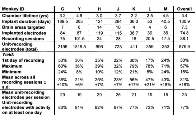
Large slots in the lower portion of the chamber (Fig. ]A) provided access to the underside of the grid for daily cleaning and observation of the surface of the granulation tissue, which had grown to cover the dura mater. This granulation tissue effectively sealed the brain from the underside of the grid and chamber. At the start of each session, the chamber beneath the grid was flushed thoroughly with sterile saline via these side ports. Depending on the monkey and the chronic implant, this was followed by diluted (20:1) Novalsan Solution (chlorhexidine diacetate 2%) or Betadine Solution (povidone-iodine 10%) 2-5 times per week. Possible infections, as indicated by the type and amount of discharge, were treated with dilute antibiotic applied inside the chamber. Systemic administration of antibiotics was rarely used. Topical antibiotic ointment was occasionally applied along the margins of the chamber. With these precautions, the chambers could be maintained for up to five years (Chamber Lifetime, Table 1).
The chamber beneath the grid was filled with saline for the duration of the session. Prior to some sessions, the saline was mixed with viscous methyl cellulose, in
order to reduce noise resulting from the motion of the saline. When necessary, application of petroleum jelly or silicon grease to the electrode leads between the tops of the microdrives and the connectors effectively dampened mechanical vibrations of the electrodes. Additionally, if fluid were suspected of traveling up the guide tubes to the surface of the grid, silicon grease was used to fill the space between the electrodes and guide tubes, sealing the tops of the tubes, while permitting the free movement of the electrodes.
Over the course of the implant, the space above the grid was kept as dry and clean as possible, in order to preserve the quality of recorded signals and the ability to manipulate the depths of electrodes with the microdrives. The grid was sealed with silicone and the side slots in the chamber beneath the grid were left partially open, as needed, to minimize fluid build-up. Further protection from fluid was provided by a raised ridge around the top of the inner surface of the chamber (surrounding the grid) that served as a dam.
In between experimental sessions, the chamber was covered with either a raised cap, while electrodes were implanted in the brain, or a flat cap, otherwise (Fig. 1A top). The side panels on the raised cap could be removed to connect pre-amplifiers to the connector strips without having to remove the main portion of the cap. Ventilation slots along the non-removable ends of the raised cap helped to keep the space above the grid dry.
Neuronal signals were amplified and filtered (600-6000 Hz for spikes, and 1-475 Hz for LFPs) by the Cheetah system (Neuralynx Inc., Bozeman, MT). Spike waveforms (32 kHz sampling rate) and LFP signals (2 kHz) were continuously collected during daily
recording sessions. The Cheetah system was configured to accommodate up to 128 single electrodes along with 8 analog input channels (for behavioral data). At the start of each recording session, custom software was used to configure each electrode channel to record either spike or LFP data. Spike and LFP signals could be recorded simultaneously from up to 32 electrodes. Each neural (spike or LFP) data channel was stored to a separate file for offline analysis.
In some implants, the impedance of the electrodes was measured periodically and those with high impedance (> 2 MOhm) were stimulated (Master-8, A.M.P.I., Israel, and Bak Electronics, MD, 100-200 ms trains of biphasic pulses at 1 kHz, < 20 [LA). Stimulation was repeated up to five times or until the impedance fell below 2 MOhm. Post-stimulation impedance values were measured and recorded.
Electrical stimulation experiments. The ACC stimulation experiments were conducted with a chronic implant of 48 independently movable electrodes targeting the ACC, CMA, CN and dIPFC. In each trial a single bipolar, biphasic pulse (current amplitude: 200 pIA, cathodal-anodal pulse duration: 400 pis) was delivered between two of the Pt/Ir electrodes chronically implanted in the ACC (4 mm apart). The responses of single units in the ACC and CMA were analyzed by binning the spike counts of each unit in a 20 ms window surrounding the stimulus delivery. The analysis was repeated for two bin sizes (0.1 ms and I ms). Bins with spike counts beyond the 99% confidence limits were defined as statistically significant. Those units exhibiting significant responses irrespective of bin size were considered to be modulated significantly by the electrical stimulation.
Stimulation experiments in the dlPFC were performed with a chronic implant of 24 independently movable tungsten electrodes (12 each in dlPFC and CN). Four of the dIPFC electrodes (2 each in areas 9L and 46) were used for delivering electrical stimuli. In each of 40 trials, spaced 5 s apart, a single monopolar, biphasic pulse (current amplitude: 20 pA, cathodal-anodal pulse duration: 300 Rs) was delivered. LFPs were recorded from eight electrodes in the CN (2 mm apart in a 4x4 configuration). LFPs were low-pass filtered at 20 Hz and stimulation-triggered waveform averages were computed on 400 ms of LFP data centered on the onset of stimulation. Significant modulation of waveform averages was detected by comparing the post-stimulation activity to the 95%
confidence limits estimated from the pre-stimulation activity.
Pharmacological microinjection experiments. A microelectrode nested within a steel canula was used to record electrophysiological activity during local drug infusion. The canula was connected via polyethylene tubing to a tabletop pump (Harvard Apparatus, Cambridge, MA). Drugs were delivered at a rate of 100 nL/min for 2 minutes. Periods of significant changes in firing rate were detected using a sliding bin average, as follows. Spike counts were aggregated into 10 s bins and the bin values underwent 3-point smoothing. Baseline activity was defined as the mean bin value over the 5 min immediately preceding drug infusion. The onset of a significant change in firing rate was defined as the 1st of 10 consecutive bins with values at least 2 s.d. from the baseline.
Response offset was defined as the 1st of 10 consecutive bins with values within 2 s.d. of the baseline.
Implant removal. At the end of a chronic implant, the electrodes were slowly raised to their initial depths, over a few sessions. With the alert monkey head-fixed, the implant was then removed in a single procedure. First, the electrode leads above the microdrives and reference and ground wires were cut, and the connector strips unscrewed from the chamber. Then, each microdrive was removed by first ensuring the electrodes are retracted as far as possible, if possible using forceps to raise guide-tubes and then gently using force perpendicular to the grid to extract the manipulator. Once all the manipulators have been removed in this manner, the silicon sealing the grid was stripped away and the grid itself removed by unscrewing the corner screws. The chamber was then thoroughly rinsed with sterile saline. The monkey was allowed to recover for at least a few weeks before the next implant. The grid, microdrives and most 23-gauge guide-tubes that could be recovered for use in subsequent implants were cleaned with ethanol, bleach and acetone. If needed, prior to the next implant, a small drop of oil was added to each of the plastic sleds that travel along the microdrive screws, in order to ensure free movement of the sled and minimal wear and tear on the plastic.
Histology. After experiments were completed, the monkey was perfused intracardially with fixative (0.9% NaCl followed by 4% paraformaldehyde in 0.1 M Na2 /K- PO
4
buffer, pH 7.4). Whenever possible, this was done before removing the final chronic implant. In some cases, electrolytic lesions (10 pA DC for 10 s, < 15 sites) were made to mark locations in the brain relative to the grid. Conventional Nissl staining (60 pm thick slices) was performed to visualize electrode tracks. The slices were analyzed to reconstruct the location of each electrode in each recording session. Tracks from earlier
chronic implants were reconstructed on the assumption that the distance from the grid to the surface of the brain was constant across implants. In some monkeys, anatomical tracing software and 3D reconstruction (Neurolucida, MicroBrightField, Inc., Williston, VT) was used.
RESULTS
Reconfigurable chronic electrode implant system
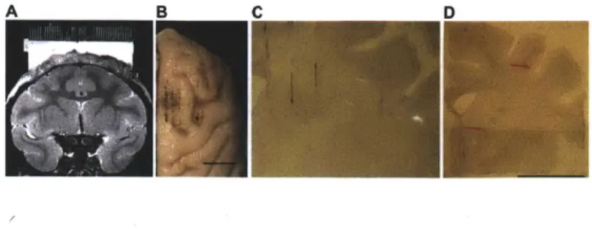
We designed and implemented the ChIME system to obtain simultaneous recordings of neural activity from multiple, individually movable microelectrodes chronically implanted in cortical and subcortical structures of the primate brain. The key components of the ChIME system are compact screw-based microdrives that can be placed in nearly any configuration on a grid (Fig. 1). Improving upon the concept of the widely used (Pasupathy and Miller) method of Wurtz and colleagues (Nichols et al. 1998) for acute, single-electrode recordings, the ChiME microdrives employ a novel mechanism whereby each recording track is targeted by a single screw occupying a single grid hole, thus maximizing the density of independently movable electrodes. The grid can be inserted into a plastic chamber fixed to the skull, in order to deliver electrodes to the underlying brain. Structural magnetic resonance images (MRIs) are used (Fig. 2A) to confirm the location of the grid holes relative to the target regions in the brain. We describe here the use of the system based on results from 16 implants of electrodes placed in cortical and subcortical sites in seven monkeys (Table 1). Preliminary experimental results based on the use of pilot versions of the ChIME system have been published elsewhere (Fujii and Graybiel 2005; Fujii et al. 2007).
Microdrives loaded with 3, 6 or 9 individually movable electrodes were placed on grid within the chamber, and the electrodes were lowered into the brain in a single implant procedure (see Methods). Subsequently, across multiple daily sessions, the electrodes were advanced to their intended target sites. The depth of each electrode was controlled by turning the microdrive screw (158.75 pm/turn) to which it was attached.
Implants were left in place for periods of weeks to months, during which the depths of individual electrodes were continually adjusted (maximum travel distance: 13 mm from initial implant depth). Following completion of the recordings, histological analysis was performed to reconstruct the locations of the electrodes (Fig. 2B-D).
Simultaneously recorded neural activity from multiple cortical and subcortical sites To test the ChIME system, we have used implants ranging in size from 27 to 127 electrodes and lasting from 22 to 365 days. In each implant, single-unit spike activity and local field potentials (LFPs) were recorded simultaneously from electrodes implanted in 6-14 brain structures bilaterally, including medial and dorsolateral prefrontal cortex (mPFC and dlPFC), frontal eye fields (FEF), supplementary eye fields (SEF), primary motor cortex (M1), premotor cortex (PM), supplementary and pre-supplementary motor areas (SMA and pSMA), anterior cingulate cortex (ACC), cingulate motor area (CMA), orbitofrontal cortex, parietal cortex, caudate nucleus (CN), putamen (Put), globus pallidus, thalamus and amygdala. Recording sessions occurred on up to 59% of the days an implant was in place. In a single session, up to 57 electrodes recorded unit activity simultaneously along with 84 LFP signals. Under the constraints of the data acquisition system that we used, when fewer units were recorded, up to 127 LFPs were recorded simultaneously.
We used the ChIME system to record neural signals simultaneously from several neocortical and subcortical regions participating in widely distributed brain networks. The left half of Fig. 3 gives examples of LFPs recorded simultaneously from the left hemisphere of a bilateral implant in monkey H, showing different activity patterns across
seven regions, during performance of a joystick movement task. The right half of Fig. 3 shows examples of unit activity recorded simultaneously from the right hemisphere of a separate implant in monkey G, during performance of an oculomotor scan task. For each implant, all of the recording sites were accessed through a single craniotomy and grid.
Session-to-session adjustment of individual electrodes' depths
We have found that electrode depths can be adjusted either immediately prior to a recording session or on a previous day. Manipulating the depths of individual electrodes at different times enabled us to monitor activity from different combinations of sites within multiple regions across the recording sessions performed during a single implant (Fig. 4A). The vertical travel of the microdrives (up to 13 mm) enabled individual electrodes to progress through multiple brain structures over the course of a single chronic implant (Fig. 4B). Within a given brain structure, it was possible to record from sites at which neurons exhibited different activity profiles along a single track (Fig. 4C).
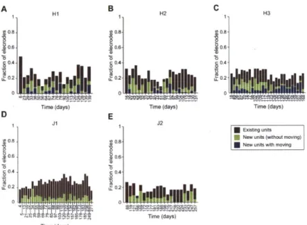
We defined the yield in each recording session as the fraction of electrodes that recorded unit activity. The average yield over all sessions was 31% (Mean Yield, Table 1), but the yield varied from implant to implant, ranging from 18% to 65%. The yield averaged across all implants decreased over months (Fig. 5), though the time-course of the yield differed across implants (Fig. 6). Implants with fewer electrodes and of shorter durations were associated with higher yields (Fig. 6, right vs. left columns; note difference in vertical scale). In most implants the yield converged within a few weeks to the overall mean value (31%).
To investigate the time-course of the yield in individual implants, in each recording session of two monkeys (H and J, 5 implants total), we classified the electrodes that recorded unit activity into two groups: electrodes that had recorded units in the preceding session and electrodes that had not (Fig. 7). These two groups both contributed persistently to the yield over the course of each implant. Of the electrodes that recorded at least one unit in a given session, 35% ± 18% (mean ± s.d. unless otherwise noted) recorded new unit activity, that is, they had not recorded any units in the previous session (means for each implant in Monkey H: 43% ± 16%, 35% ± 21% and 44% ± 12%; in Monkey J: 25% + 13% and 32% ± 20%). The emergence of new activity on many electrodes represents a key advantage of the ChIME system over fixed multi-electrode arrays, and new unit activity can continue to arise for several months after the start of an implant.
To assess the effect of manipulating the depths of the implanted electrodes on the yield, we present an analysis of data from monkey H, because session yields did not differ significantly across all three implants in this monkey (chi-square test, P > 0.5). Of the electrodes that recorded new unit activity in a given session, on average 37% ± 32% had been moved since the preceding session (Fig. 7A-C). As this percentage varied with the rate at which electrodes were moved, it does not imply a causal relationship between moving electrodes and recording new unit activity. Focusing on the electrodes that had not recorded units in a given session, we asked whether moving them increased the likelihood of recording new units in the following session. In each of the three implants, we found that it did. Electrodes that had been moved were significantly more likely to record new units in the following session than electrodes that had not been moved
(chi-square test, P < 10-15). This comparison likely underestimates the effect of moving electrodes on the yield, as we examined the probability of obtaining unit activity only in the recording session immediately following a session in which an electrode was moved, and we did not take into account the cumulative effects of moving electrodes multiple times before new units were recorded.
Session-to-session stability of neural activity
We tested the ability of the ChIME system to record stable neural activity during consecutive sessions from multiple electrodes that were not moved in the interim (Fig. 8). We analyzed the durations of such stretches of stable activity in six implants across three monkeys. On average, an electrode recorded unit activity in 2.1 ± 2.8 consecutive sessions (3.9 ± 8.5 days) without being moved. Stationary electrodes that recorded unit activity in at least two consecutive sessions recorded unit activity in a total of 4.2 ± 4.1 consecutive sessions (9.7 ± 12.8 days), on average. Such activity was not only stable, but typically exhibited striking similarities in wave-shape, inter-spike interval distribution and responses to task events across sessions (Fig. 8A, Days 106-115). LFPs recorded from stationary electrodes also showed extraordinary stability over periods of several weeks to months (Fig. 8B). When the electrodes were moved, the recorded signals changed (Fig. 4B,C; Fig. 8A first and last day; Fig. 8B, last day), suggesting that the preceding recordings were likely obtained from small volumes of brain with similar
functional properties.
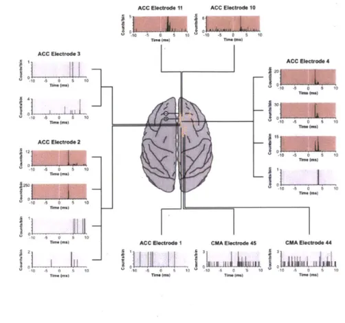
We have exploited the versatility of the ChIME system to incorporate electrical stimulation and pharmacological injection methods with chronic recordings of neural activity. In a subset of experiments, we tested for within-area modulation of spike activity using single-pulse electrical stimuli in the ACC. We found fixed, short-latency responses from multiple simultaneously recorded units in the ACC, indicating synaptic connectivity between sites targeted by the chronically implanted electrodes (Fig. 9). The stimulation-evoked responses were sufficiently localized, so that units recorded in the CMA did not respond to the ACC stimulation. Moreover, on electrodes that recorded multiple units in the ACC, not all single units exhibited a significant response. In separate experiments, we used electrical stimulation to detect functional connectivity between brain areas. By stimulating at four sites in the dlPFC, we found differential modulation of the LFP activity recorded simultaneously at multiple sites in the CN (Fig. 10A).
We have also begun to develop injection methods for use with the ChIME system. In pilot experiments, we recorded units in dlPFC and CN simultaneously and examined their responses to injections made in the CN. In the example shown in Fig. 1OB, all four of the striatal units exhibited changes in firing rate, whereas none of the eight prefrontal units showed changes in firing rate for up to 10 minutes following the injection. This example demonstrates the possibility of using the ChIME system to perform pharmacological manipulations of subsets of recording sites in the context of an extended-duration implant. Stimulation and injection techniques can be used to search for and identify desired recording sites, as well as to record the neural and behavioral effects of localized electrical or pharmacological manipulations, making the ChIME system a powerful tool for studying the functions of brain circuits.
DISCUSSION
Large-scale simultaneous recordings from multiple superficial and deep brain structures test the limits of existing methods for extracellular recordings in alert non-human primates. We developed the ChIME system to meet this need. The most important advantage of our system is the opportunity it provides to sample neural activity at multiple depths chronically and flexibly. The ability to move individual electrodes can dramatically improve the yield of single units, as demonstrated by the comparison of the unit activity between electrodes that had or had not been moved across pairs of successive sessions. The single-unit yields in the initial recording sessions of each implant were similar to those reported for experiments using fixed-electrode arrays, chronically implanted in multiple cortical areas (Chhatbar et al. 2010; Nicolelis et al.). However, the ability to move electrodes at will over the lifetime of the implant led to sustainably high yields over the long-term, in marked contrast to the typical time-dependent deterioration of yields seen with fixed-electrode arrays. In addition to its critical effect on the yield, the independent manipulation of electrodes enabled recordings from different combinations of depths across the recording tracks of a single implant (Fig. 4). This cannot be done using silicon probes with multiple contacts, the distances between which are fixed (Kipke et al. 2003). Furthermore, fixed arrays have not been used to study subcortical structures of the primate brain because of the substantial damage to the overlying cortex and white matter, as has been observed in the brains of non-primates (McCreery et al. 2006). Finally, whereas there is little control over the types of cells recorded with fixed arrays, by adjusting the depths of electrodes independently
with the ChIME system, cells that are anatomically connected (as confirmed by microstimulation) or that belong to specific classes (based on physiological characteristics and responses) can be targeted and studied selectively. Taken together, the numerous advantages of the movable electrodes implanted with the ChIME system thus provide a unique opportunity to collect neural data for analyzing circuits spanning cortical and deep structures.
The ChIME system is the first to enable simultaneous recordings from chronically implanted electrodes in cortical and subcortical sites in the non-human primate brain, controlled by as many as hundreds of independently movable drives. Other recording methods share some, but not all, of the features of the ChIME system. Jackson and Fetz (Nichols et al. 1998), and separately Wilson and colleagues (Sun et al. 2006), used chronically implanted, independently movable electrodes in the non-human primate. However, these methods were designed for use only with a small number of electrodes, and primarily in the freely moving monkey. A method by Merzenich and colleagues (deCharms et al. 1999) used 49 electrodes, but to record from.a single cortical area of non-human primates with brains considerably smaller than those of macaques. It is not clear whether or how any of these or similar methods (Galashan et al.) could be used to record simultaneously from multiple structures in the macaque brain, as we have done using the ChIME system. Other chronic recording methods using movable microwires have been developed for non-primate species, including rabbits (Swadlow et al. 2005) and rodents (Jog et al. 2002; Johnson and Welsh 2003; Yamamoto and Wilson 2008). Adapting these methods to record from multiple cortical and deep structures in the macaque brain would require reducing the physical footprint of the microdrives
substantially, as well as overcoming the challenge of traversing sulci with microwires, as opposed to the sharp microelectrodes used with the ChIME system.
Others have recorded neuronal activity in the monkey brain simultaneously from multiple structures (Buschman and Miller 2007; Hernandez et al. ; Pasupathy and Miller 2005) or from multiple sites within a single structure (Baker et al. 1999; Courtemanche et al. 2003; Gray et al. 2007) using acute methods, in which the electrodes were implanted daily. Recordings commenced within a few hours following electrode implantation, and the electrodes were extracted from the brain at the end of each recording session. These acute multi-electrode methods require considerable preparation time prior to each recording session, constraining the duration and frequency of experimental sessions. These methods have additional drawbacks, including the difficulty of maintaining stable signals following the acute implantation of multiple electrodes in a confined region; an increased potential for tissue damage from repeated daily penetrations of the dura and brain; and the inability to track learning-related changes in the activity of a localized neural population over daily recording sessions. Most importantly, the maximum number of independently movable drives that can be implanted acutely is likely to be at least one order of magnitude smaller than what can be achieved with the ChIME system.
In our experiments using the ChIME system, the neural signals on many electrodes were stable across numerous sessions (Fig. 7,8). The durations of periods of stable activity recorded by individual electrodes were likely limited by the fact that most of the electrodes in an implant were moved periodically, in order to sample new sites along each track. Moving electrodes could have reduced the stability of signals recorded on stationary neighboring electrodes. We suspect that the long-term stability of signals
recorded with the ChIME system is due to the combination of continued growth of granulation tissue overlying the exposed dura mater within the chamber and to the multiple guide-tubes stabilizing the dural surface. It is possible for the dura mater and overlying calvarium to re-grow over the course of several weeks, providing even further stabilization of the electrodes. Indeed, we found evidence for dural adhesions to the guide-tubes in at least one monkey that was perfused with the implanted electrodes left in place (see Methods).
As signal quality was often maintained day-to-day, minimal pre-recording preparation time was required, with the bulk of it spent adjusting electrode depths in
order to improve single-unit isolation. On any given day, we only adjusted the depths of a fraction of the electrodes. With implants of greater numbers of electrodes, we moved electrodes only on non-recording days. This made the system essentially "plug-and-play" on recording days, maximizing the length of recording sessions.
The number of electrodes used in our experiments to date has been limited only by the capacity of available data acquisition systems. We have demonstrated the capabilities of the ChIME system using commercially available, epoxy-coated tungsten or parylene-coated platinum/iridium microelectrodes, but we have successfully tested the use of microwire bundles to increase the density of recording sites by increasing the number of recording channels per track. These could be substituted by tetrodes, stereotrodes, or multi-contact probes, all of which can be attached to a microdrive screw. The number of simultaneously recorded channels could also be increased by adding more microdrives, which can drive ~9 tracks per 30 grid holes, allowing up to ~450 tracks in a single implant (using a 40 mm x 40 mm grid).
The flexibility of the ChIME system permits the incorporation of other methods in conjunction with electrode-based recordings. Electrical stimulation can be used to map the functional connectivity within and across brain areas over the course of a chronic implant (Figs. 9,10A). Microinjections can be made through the grid to perform pharmacological manipulations and anatomical labeling (Fig. 10B). Fiber optics can be passed through the grid for use with optogenetic methods, which have recently been extended to the monkey brain (Diester et al. 2011; Han et al. 2009). With minor changes (e.g., replacing the microdrive screws with plastic ones), the ChIME system could also be used in paired recording and functional MR-imaging experiments. Using these and other innovative techniques with the ChIME system should make it possible to manipulate and record chemical, electrical and optical signals in nearby tracks simultaneously over long periods of time, giving unprecedented insight into the circuit-level functions of cortical and subcortical networks.
The gradual deterioration of signal quality is a challenge facing all chronic recording methods. The fall-off in yield and accompanying increase in the impedance of many electrodes over the course of an implant has been attributed to the process of gliosis (Stice and Muthuswamy 2009). The ChIME system can be used to reduce the effects of gliosis by moving the electrodes. Nevertheless, in our experience, the signal-to-noise ratio tended to degrade over the duration of the implant along with the proportion of well-isolated single units. Using platinum/iridum electrodes, we have been able to reduce electrode impedances by stimulating frequently to "clean" the electrode tips (Otto et al. 2006). The stimulation paradigm effectively reduced even high impedances (> 2 MOhm)