by
Peter Joseph Schatz
Sc.B., Sc.M., Brown University (1982)
Submitted in partial fulfillment of the requirements
for the degree of Doctor of Philosophy
at the
Massachusetts Institute of Technology
December 1987
© Peter Joseph Schatz 1987
The Author hereby grants to MIT permission to reproduce and to distribute copies of this thesis document in whole or in part.
Signature of Author Department of Biology November 1987 Certified by Certified by Accepted by David Botstein Thesis Supervisor Frank Solomon Thesis Supervisor I .
MASSACHUSETS INSTIUTEOF TECHNOLOGY
DEC 1 ' /
LIBRAR!ES
Robert T. Sauer Chairperson, Departmental Committee on Graduate Studies
ARCHIVES 7 l 1
Submitted to the Department of Biology in November 1987 in partial fulfillment of the
requirements for the Degree of Doctor of Philosophy
in Biology
ABSTRACT
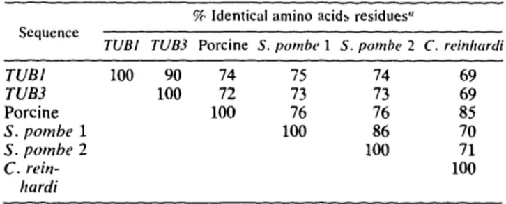
Two a-tubulin genes from the budding yeast Saccharomyces cerevisiae were identified and cloned by cross-species DNA homology. Nucleotide sequencing studies revealed that the two genes, named TUBI and TUB3, encoded gene products of 447 and 445 amino acids,
respectively, which were highly homologous to a-tubulins from other species. Comparison
of the sequences of the two genes revealed a 19% divergence between the nucleotide
sequences and a 10% divergence between the amino acid sequences. Each gene had a single intervening sequence, located at an identical position in codon 9. Cell fractionation studies
showed that both gene products were present in yeast microtubules. These two genes, along with the TUB2 Vtubulin gene, probably encode the complete complement of tubulin in
budding yeast.
Microtubules in yeast are functional components of the mitotic and meiotic spindles and are essential for nuclear movement during cell division and mating. The relative importance in
these processes of TUBI and TUB3 was examined through the construction of null mutations and also by increasing their copy number on chromosomes and on plasmids. Experiments with null alleles of TUB3 showed that TUB3 was not essential for mitosis, meiosis or mating. Null alleles of TUB3, however, did cause several phenotypes including hypersensitivity to the antimicrotubule drug benomyl and poor spore viability. On the other hand, the TUB1 gene was essential for growth of normal haploid cells. Even in diploids heterozygous for a TUB] null allele, several dominant phenotypes were evident including slow growth and poor
sporulation. This functional difference between the two genes was apparently due to different
levels of expression because extra copies of either gene could suppress the defects caused by a null mutation in the other. We concluded that in spite of the 10%1 divergence between the products of the two genes, there was no essential functional difference between them.
Comparisons of sequences from several species revealed the presence of a variable region
near the amino terminus of a-tubulin proteins. We have perturbed the structure of this region in TUB3 by inserting into it 3, 9, or 17 amino acids and have tested the ability of these altered
proteins to function as the only a-tubulin protein in yeast cells. We found that each of these altered proteins was sufficient on its own for mitotic growth, mating, and meiosis of yeast.
We concluded that this region could tolerate considerable variation without losing any of the
highly conserved functions of a-tubulin. Our results suggested that variabiltiy in this region
occurred because it could be tolerated, not because it specified an important function for the
protein.
We have isolated 70 conditional-lethal mutations in TUB1 using a plasmid replacement technique commonly called the plasmid shuffle. Of the 70 mutations isolated, 69 resulted in cold-sensitivity. We have mapped 31 of the mutations to deletion intervals within the TUB1
role of microtubules in yeast mitosis. Several of the mutants arrested growth with a
sufficiently uniform morphology to indicate that TUB1 has a specific role in the progression of the yeast cell cycle. A number of the mutants had gross defects in microtubule assembly at the restrictive temperature, some with no microtubules and some with excess microtubules. Other mutants contained disorganized microtubules and nuclei. Greater than 90% of the mutants examined were hypersensitive to the antimicrotubule drug benomyl. None of the
mutants were resistant to benomyl.
Thesis Supervisors: Dr. David Botstein, Professor of Biology Dr. Frank Solomon, Professor of Biology
Acknowledgments:
During the course of this work, I have had the fortune of being associated with the people in the labs of David Botstein and Frank Solomon. I would like to thank all of them for their gifts of advice, of time, and, most importantly, of good humor even in the face of my sometimes surly moods.
Three people did some of the experiments that are described in this thesis. Lorraine Pillus led me through the fractionation of the yeast cytoskeleton to allow the identification of the
proteins which were the focus of this study. Paula Grisafi sequenced substantial portions of the genes. George Georges initiated the experiments that led to Chapter 4.
The first course that I attended at MIT was called Method and Logic in Molecular Biology and was taught by David Botstein and Frank Solomon. In that course, they communicated to me their enthusiasm for the process of scientific discovery. Unable to get enough in that brief first semester, I signed on for a 5 year extension of the course as a joint student in their labs. My work was enhanced at every step by their complementary (and rarely conflicting)
viewpoints. My interest in the cytoskeleton dates to several lectures given in the Cell Biology
course by Frank, in which he was so excited that he breathed only three times during six
hours of very rapid talking.
My brother David and Leilani Miller deserve special honors for having suffered through the first drafts of most of the writing that follows. I would also like to thank them for helping to make my graduate student years one of the most enjoyable periods of my life.
Pag
Title Page 1
Abstract 2
Acknowledgments 4
Table of Contents 5
Chapter One - Microtubules, Mitosis and Yeast 6
Chapter Two - Two Functional a-Tubulin Genes of the Yeast
Saccharomyces cerevisiae Encode Divergent Proteins 54 Chapter Three - Genetically Essential and Nonessential a-Tubulin
Genes Specify Functionally Interchangeable Proteins 66 Chapter Four - Insertions of up to 17 Amino Acids into a Region
of a-Tubulin Do Not Disrupt Function In Vivo 79 Chapter Five - Isolation and Characterization of Mutations in the TUB 1
a-Tubulin Gene of the Yeast Saccharomyces cerevisiae 112 Appendix 1 - Plasmid Construction by Recombination in Yeast 166
Chapter 1:
Microtubules are polymers of globular protein subunits which are present in most
eukaryotic cells and function in a wide variety of roles in cellular motility and structure. The
study of microtubules has been concerned with both the tubulin subunits that are their main structural element and on other components in cells that interact with them. In the first part of this introduction, I will discuss microtubule structure, assembly, and function. I will
emphasize the surprising extent to which the properties of tubulin and microtubules in vivo can be explained by their behavior in vitro. The properties of tubulin alone, however, are not sufficient to explain the diverse functions of microtubules. Many other cellular constituents must be involved. The challenge of current microtubule research is to identify these other components and to determine their roles and the roles of tubulin in the mechanism of
microtubule action.
This thesis is concerned with the study of microtubular function in the yeast
Saccharomyces cerevisiae. The study of microtubules in yeast has been motivated by a desire
to correlate biochemical and structural data with information about in vivo functions, obtained
through the use of the sophisticated classical and molecular genetic system of yeast. Because the major role of microtubules in the yeast cell cycle is in mitosis, the second part of this
introduction will consist of a brief discussion of mitosis in organisms other than yeast. Finally, I will discuss the function of microtubules in yeast mitosis, meiosis and mating.
Part 1: Microtubules:
Structure:
Microtubules are found in a very wide variety of cellular structures and, in the electron
microscope, appear as hollow tubes about 24 nm in diameter (reviewed in Kirschner, 1978 and Dustin, 1984). Transverse sections usually reveal a tube consisting of 13 smaller
protein subunits are stacked in a regular helical array. The regular stacking of these subunits
on top of one another leads to the parallel appearance of the protofilaments. Although 13 is
the most common number of protofilaments observed, other numbers of protofilaments are found in cylindrical microtubules. In addition, irregular arrangements of protofilaments occur in specialized structures such as cilia and basal bodies.
Microtubules are most commonly found in structures that play a role in motility. These
include mitotic and meiotic spindles, flagella, cilia and nerve cell processes. Other
microtubules have a more obvious structural role, such as those in the marginal band of red blood cells in birds and cold-blooded vertebrates. Furthermore, most actively dividing eukaryotic cells display arrays of microtubules during interphase that originate from a single
area (called the microtubule organizing center or MTOC). In higher cells, these microtubules
form a lacy array which weaves throughout the cytoplasm and play a role in cell shape and
motility.
Tubulin and Microtubule assembly in vitro:
Tubulin was originally identified as a soluble protein from eukaryotic cells that bound to the antimitotic drug colchicine (Borisy and Taylor, 1967). In its soluble form, tubulin
probably exists as a 110 kd heterodimer of a- and V- subunits (Luduena et al., 1975). Under conditions of neutral pH, warm temperature, low Ca++, sufficient concentrations of GTP, Mg+ +, and tubulin dimers, the dimers will assemble into microtubules (Weisenberg, 1972).
There are two binding sites for GTP on a dimer. One is referred to as the E-site (exchangeable with the external medium) and the the other is the N-site (for
nonexchangeable). GTP binding to these two sites is thought to have distinct functions. Since the N-site GTP has a half life comparable to that of the dimer, it probably functions as a stable structural part of the dimer (Spiegelman et al., 1977). In contrast, the E-site GTP is hydrolyzed to GDP during the microtubule assembly/disassembly process.
The fact that microtubule assembly/disassembly is coupled with the hydrolysis of GTP gives microtubules several interesting properties. If the assembly/disassembly process did not use energy, then microtubules would be equilibrium polymers, with their growth (in the
absence of other factors) determined by the concentration of dimer at free ends and by the
dissociation rate of dimers from the ends. The two ends of the polymer (commonly called "plus" and "minus" ends) could have different assembly rates, but the equilibrium constant for
growth at each end would have to be identical (Asakura, 1968). Such a situation would place several constraints on the assembly process including: [1] relative insensitivity of the rate of
assembly to monomer concentration, [2] slow rates of disassembly, and [3] static
microtubule arrays (reviewed in Kirschner and Mitchison, 1986). As suggested by Wegner
(1976) for the case of actin filaments and ATP, energy consumption allows differentiation
between the ends of a linear polymer. At a steady state, net assembly could occur at one end accompanied by net disassembly at the other end, a process called "treadmilling". The plus end of a microtubule is defined as the favored end for assembly. Thus, treadmilling would involve addition of subunits at the plus end and loss of subunits at the minus end. Evidence
for treadmilling has been reported by Margolis and Wilson (1978). It has been the basis of
several models of microtubular organization and motility (Kirschner, 1980; Margolis and Wilson, 1981). Two crucial aspects of these models are: [1] selective stabilization of certain microtubules by capping of their minus ends by MTOC's and [2] production of force by treadmilling microtubules. Observations of microtubules in vivo and in vitro, however, led to
a reexamination of the treadmilling models (see below).
More recent models of microtubule assembly also rely on the hydrolysis of GTP during the assembly/disassembly process, but they incorporate the possibility that the kinetics of
assembly may not be the same as the kinetics of GTP hydrolysis. Two observations concerning in vitro assembly of nficrotubules are relevant to newer models. First,
nonhydrolyzable analogues of GTP will support assembly of microtubules (Aria and Kaziro, 1976; Penningroth et al., 1976). Thus, the energy of GTP hydrolysis is not essential for
tubulin concentrations (Carlier and Pantaloni, 1981), although the extent and even the
existence of this lag is disputed (O'Brien et al., 1987). Thus, a microtubule may be a mixed polymer of GDP-tubulin and GTP-tubulin. Theoretically, this situation gives the polymer several useful properties (Kirschner and Mitchison, 1986). In a pure tubulin polymer, there are three possible reactions: [1] the addition and loss of GTP-tubulin, [2] the addition and loss of GDP-tubulin, and [3] irreversible hydrolysis of GTP-tubulin to GDP-tubulin. If assembly of GTP-tubulin and disassembly of GDP-tubulin are strongly favored, then cells can regulate microtubule assembly by regulating GTP hydrolysis in microtubules.
Alternatively, some other property of microtubule ends might distinguish the growing phase from the shrinking phase. Experimentally determined assembly and disassembly rates are consistent with such a phase transition and inconsistent with simple equilibrium models (Kirschner and Mitchison, 1986).
The existence of subpopulations of microtubules that could be distinguished on the basis of intrinsic assembly competence would predict that a population of microtubules would contain both growing and shrinking microtubules. This two-phase mode of microtubule
polymerization has been called "dynamic instability" (Mitchison and Kirschner, 1984). In the
most widely publicized version of the model, the distinguishing characteristic between
growing and shrinking phases is the presence of GTP-tubulin at the ends of the microtubules. In the model, GTP is hydrolyzed to GDP with a constant probability per unit time. Therefore, faster growth leads to a larger GTP "cap" on the tubulin. Slow or stopped growth increases the probability that the GTP will be hydrolyzed to GDP, thus exposing GDP-tubulin ends and leading to rapid depolymerization. Support for the GTP cap model comes from theoretical
studies (eg. Hill and Chen, 1984) and from the observation that nonhydrolyzable analogues of
GTP stabilize the ends of microtubules (Weisenberg and Deery, 1976; Penningroth and
Kirschner, 1977).
proteins or some conformational change in the microtubule after GTP hydrolysis. Without energy consumption, however, it is impossible for steadily growing and shrinking
microtubules to coexist in the same population (Kirschner and Mitchison, 1986). The simplest version of the GTP cap model would predict that once a microtubule started
shrinking, it would be very likely to disappear because of the low amount of GTP tubulin in the middle (the oldest part) of the polymer.
The behavior of microtubules in vitro is consistent with the dynamic instability model, although other explanations have been proposed (Farrell et al., 1987; Rothwell et al., 1987).
Using light and electron microscopy, Mitchison and Kirschner (1984a,b) examined the
growth of individual free microtubules and microtubules nucleated from purified centrosomes. At high concentrations of tubulin, microtubules grew steadily from centrosomes. When the tubulin concentration was lowered by dilution, however, some microtubules continued to grow while others disappeared quite rapidly. Clearly, there were two populations of microtubules distinguished by their assembly competence. Free microtubules were used as
seeds for assembly to a steady level of polymer mass. Once the steady state of bulk assembly
was reached, however, the number of microtubules declined while their average length
increased. A more recent study using biotinylated tubulin as a marker has shown that both
ends of the polymer grow steadily in most microtubules at steady state while a minority of microtubules are shrinking rapidly (Kristofferson et al., 1986). No evidence of treadmilling was observed. The length of microtubules in solution did not reach a stable steady state
because: [1] growing microtubules were more likely to continue growing than shrinking ones
and [2] shrinking microtubules were more likely to continue shrinking than growing ones. The coexistence of growing and shrinking phases has reportedly been visualized in vitro by observation of individual microtubules using dark-field microscopy (Horio and Hotani, 1986). In these experiments, the ends of the microtubules apparently changed randomly between growing and shrinking phases, and the two ends behaved independently. In addition, the presumptive plus end grew and shrunk faster than the minus end and
phase interconversions were quite frequent and disappearance was rare. These frequent phase conversions are inconsistent with the simple version of the GTP cap model, which would
predict that most microtubules that started to shrink would disappear. There are several
possible explanations for this inconsistency. [1] The observed variations in length may have been due in part to movement of the microtubules into and out of the microscope's plane of focus. [2] The tubulin may have been contaminated with sufficient microtubule associated proteins to affect the phase transition frequency; this possibility is supported by the
observation that added microtubule associated proteins suppressed the phase conversion and stabilized the growing phase. [3] The simple GTP cap model may be incorrect.
Observations in vivo have cast doubts on the relevance of these frequent phase conversions (see below).
It is clear that dynamic instability of microtubule growth occurs in vitro, but the factor that determines the difference between growing and shrinking microtubules is as yet unknown.
Evidence for Dynamic Instability in vivo:
Microtubule dynamics have also been studied in vivo, using microinjection of labeled tubulin into cells. Bulk measurements of the rate of incorporation of tubulin into mitotic spindles and interphase cytoskeletons and rates of recovery of photobleached areas within cells demonstrated that microtubule arrays are extremely dynamic in vivo (Salmon et al.,
1984, Saxton et al., 1984). Rates of incorporation were extremely high, particularly in mitotic spindles, which is consistent with dynamic instability and inconsistent with any simple
equilibrium model. Observation at the level of individual microtubules indicated that most
microtubules were continuously growing at their ends at a fairly steady rate (Soltys and Borisy, 1985; Schulze and Kirschner, 1986, Mitchison et al., 1986). These studies also showed that the unlabeled microtubules dissappeared quite rapidly, leading to rapid turnover of most microtubules in the cell. A few microtubules, however, were considerably more
stable than the average (Schulze and Kirschner, 1986). The rapid and complete turnover of
most microtubules in the cells casts doubt on the relevance of the frequent in vitro growth phase transitions reported by Horio and Hotani (1986), although some in vivo phase transitions have been reported (Sammak 2mnd Borisy, 1987).
Theoretical Consequences of Dynamic Instability:
Dynamic instability is the basis for models of cellular regulation of microtubule organization and function (Kirschner and Mitchison, 1986): Due to the phase transition between growth states, microtubules can both grow and shrink quite rapidly from either end. Different populations of microtubules in a single cell can be growing and shrinking, as is often required in complicated functions such as mitosis (see below). Growth can occur below
the critical concentration as long as there are nucleation sites available (eg. centrosomes). These nucleation sites will be greatly favored as sites of initiation because they give a kinetic
advantage to assembly at low tubulin concentrations. Free microtubules that disappear during random fluctuations predicted by the dynamic instability model will be unable to regrow
because of the lack of the ability to nucleate. Consistent with this, microtubules in most cell
types appear to arise from organizing centers called MTOC's. Finally, the rapid turnover of
microtubules gives the cell the opportunity to reorganize its cytoskeleton quickly by selectively stabilizing a subset of them in a region where they may be needed. In summary, it is clear that a number of the in vivo properties of microtubule assemblies can be explained as a
consequence of fairly simple interactions between tubulin, GTP, and nucleation sites.
The current emphasis on dynamic instability is certainly appropriate when examining cells
in mitosis or cells with dynamic cytoskeletons. Many cells, however, have very stable
microtubular structures. Examples include neuronal cells and blood cells with marginal
bands. In these cases, dynamic instability might explain very little about the in vivo behavior of microtubules.
So far, this discussion has focused on the properties of tubulin and microtubules without
consideration of many mechanisms that might regulate their function and organization in living
cells. The need for such regulation is demonstrated most simply by considering the wide variety of structures of which microtubules are an integral part (Dustin, 1984). At the same time, the ultrastructure of microtubules is extremely conserved. Three possible sources for
this functional diversity in the midst of structural conservation are: [1] variations in the
primary structure of tubulins which form the microtubules in diverse structures, [2] specific modifications of tubulins to effect functional diversity, and [3] accessory proteins that regulate microtubular structures. The evidence for these three mechanisms will be discussed in the next three sections.
The Role of Tubulin Sequences:
While it is clear that tubulin sequences on the whole are highly conserved, there is
abundant information about the existence of a variety of subtle structural variants of tubulins within species and within single cells (for reviews see Raff, 1984; Cleveland and Sullivan, 1985). Fulton and Simpson (1976) first proposed the hypothesis that different a- and P-tubulin molecules might form the basis for functionally distinct microtubules. This idea, called the "multitubulin hypothesis", gained momentum through the years from many observations of multiple tubulin proteins, some of which appear to segregate into different structures (Stephens, 1978). That some of this variation arises from the expression of different genes has been documented for a number of species (for reviews see Raff, 1984; Cleveland and Sullivan, 1985). Some of these genes encode proteins which can be classified into groups based on sequence similarity. These groups, called "isotypes", are often more
conserved across species barriers than they are conserved compared to other isotypes in the
same species. The isotypes also often share patterns of expression in the tissues of higher eukaryotes. This conservation of sequence and expression patterns has been interpreted to
indicate that each isotype familiy shares some functional specialization (see Cleveland and Sullivan, 1985).
The most extreme version of the multitubulin hypothesis would say that tubulins are segregated into different microtubular structures based on their primary sequence and that this segregation has some essential role in microtubule function. Such a model would predict that specific tubulins might have a restricted ability to function in a variety of contexts. Several observations, however, argue against the possibility that different tubulin gene products have restricted functions. In Drosophila melanogaster spermatogenesis, a sperm-specific p-tubulin gene has a role in the meiotic spindle, in nuclear shaping by cytoplasmic microtubules, and in the sperm flagella (Kemphues et al., 1982). In Aspergillus nidulans, the -tubulin gene normally used for conidiation can be replaced by the divergent vegetative gene (May et al.,
1985; Weatherbee et al., 1985). The two divergent -tubulin proteins of each of the very distantly related yeasts Schizosaccharomyces pombe and Saccharomyces cerevisiae show a similar lack of functional specialization (Adachi et al., 1986; Chapter 3). In both species, either of the a-tubulin proteins is sufficient, on its own, for all known microtubule dependent processes (Chapter 3, M. Yanagida, personal communication).
Two studies of the function of divergent -tubulins in animal cells have shown that their primary sequences placed no restrictions on their ability to assemble into all microtubules of the cells. Bond et al. (1986) showed that a chicken-yeast chimeric V-tubulin assembled efficiently into all microtubules of mouse fibroblasts and had no apparent effect on either growth rate or morphology. Lewis et al. (1987) showed that a variety of naturally occurring
-tubulin isotypes demonstrate neither complete nor partial segregation into different structures. Even a divergent isotype normally expressed only in hematopoietic cells was assembled into all microtubules in its normal context and in transfected human fibroblasts.
The above results suggest that any functional significance of divergent tubulin isotypes is likely to be subtle. The observation of the conservation of isotype sequences and expression
tubulin. The presence of multiple genes could have arisen as an adaptation to this need for stage-specific regulation of tubulin synthesis during differentiation (Raff, 1984). Instead of evolving complicated promoters for a few genes, these organisms could have evolved simple promoters for many genes. Assuming that this duplication of genes occurred before the radiation of various vertebrate species, the expected result is a closer relationship between isotypes in a variety of species than between isotypes in the same species. The observed conservation of these isotypes over long periods of evolutionary time could be explained by tubulin's known resistance to divergence. Most random changes would be recessive lethals.
Only genes expressed in specialized tissues would be free to diverge, as has been observed in a number of species (Villasante et al., 1986; Theurkauf et al., 1986; Pratt et al., 1987). In the
absence of evidence that tubulin isotypes show structural or biochemical specialization, tests of subtle functions of these isotypes will necessarily involve gene replacement experiments. The most complex organism in which such studies are currently feasible is Drosophila.
The Role of Covalent Modifications of Tubulin:
A variety of covalent modifications of tubulin have been observed and have been suggested to play a role in microtubule function (for review see Cleveland and Sullivan, 1985). Gard and Kirschner (1985) observed phosphorylation of -tubulin in mouse neuroblastoma cells and showed that levels of phosphorylation were correlated with levels of microtubule assembly. Two covalent modifications of a-tubulin have been observed. L'Hernault and Rosenbaum (1983) showed that the majority of a-tubulin in Chlamydomonas flagella was
acetylated on the epsilon-amino group of a lysine residue. Although originally thought to be
specific to cilia and flagella, acetylated a-tubulin was found in subsets of cytoplasmic microtubules in Chlamydomonas and Physarum (LeDizet and Piperno, 1986; Diggins and Dove, 1987 ). These studies used a monoclonal antibody that is probably specific for the acetylated form of a-tubulin (Piperno and Fuller, 1985). This antibody also recognized
microtubules in some but not all mammalian cells, including primary cilia, centrioles, mitotic spindles, and subsets of cytoplasmic microtubules (Piperno et al., 1987). The epitope also
was detected in rat nerve cell processes (Cambray-Deakin and Burgoyne, 1987). One common feature of the microtubules containing acetylated a-tubulin is that they are generally more stable than other microtubules (eg. Piperno et al, 1987). Whether this relationship is cause or effect remains to be determined (see below).
The second well-studied posttranslational modification of a-tubulin is the removal and replacement of the carboxy-terminal tyrosine residue (Barra et al., 1973a, 1973b, 1974). Most a-tubulin genes encode tyrosine as the last residue (Cleveland and Sullivan, 1985). This residue can be removed by a specific carboxypeptidase (Argarana et al., 1978; Kumar and Flavin, 1981) and replaced by the enzyme tubulin tyrosine ligase (Raybin and Flavin, 1977a; Murofushi, 1980). Although the absence of the terminal tyrosine did not seem to alter the in vitro assembly properties of tubulin (Raybin and Flavin, 1977b; Arce et al., 1978; Kumar and Flavin, 1982), the fraction of tyrosinated tubulin was different in the assembled and unassembled pools (Rodriguez and Borisy, 1979). Individual microtubules in
mammalian cells could be distinguished on the basis of the relative presence or absence of this tyrosine (Gundersen et al., 1984).
The currently accepted model for tyrosination/detyrosination involves four steps: [1]
assembly of Tyr-tubulin into microtubules, [2] detyrosination of Tyr-microtubules, [3]
eventual disassembly of Glu-microtubules (detyrosinated), and [4] tyrosination of Glu-tubulin (Gunderson et al., 1987, Webster et al., 1987). The fact that Tyr-tubulin is the primary species which forms new microtubules is supported by the known preference of the ligase for unassembled tubulin in vitro (Arce et al., 1978) and the observation of very low amounts of
Glu-tubulin in the unassembled pool (Gunderson et al., 1987). Step [2] is carried out by the
carboxypeptidase, that has a preference for polymerized tubulin as a substrate (Kumar and Flavin, 1981; Arce and Barra, 1985). Support for this step also comes from experiments with agents that prevent tubulin depolymerization (Gunderson et al., 1987), that lead to an
by the drug-induced depolymerization of microtubules and the demonstration that >98% of the tubulin was present in the tyrosinated form (Gunderson et al., 1987).
The morphology of Glu-microtubules in interphase animal cells is substantially different from the majority Tyr-microtubules (Gunderson et al., 1984). Tyr-microtubules originate
from a central area (MTOC), fill the whole cell, and tend to be fairly straight. In contrast, the
Glu-microtubules are sinuous and restricted to the central part of the cell. This arrangement of Glu-microtubules is similar to that observed for acetylated microtubules (Piperno et al., 1987) and for a subset of stable microtubules, identified because of slow exchange rates with
injected tubulin (Schulze and Kirschner, 1987). In spite of the similarity of appearance, the extent of overlap between these three sets of microtubules is unclear. Even if the three subsets
are the same, it is not clear whether increased stability causes detyrosination and acetylation or vice versa. It is clear that these microtubules are biochemically and morphologically different and may be used for some specialized function, but the identity of this function remains obscure.
The Role of Other Proteins:
Due to the limited amount of variation in tubulins and the wide variety of structures of which microtubules are a part, investigators have focused much attention on other proteins in the cell that may have a role in regulating microtubule structure and function. Even "simple" microtubule structures, such as cilia and flagella, contain more than a hundred non-tubulin proteins (Lefebvre and Rosenbaum, 1986). The most notable protein identified by early experiments on cilia and flagella is dynein, an ATPase which is responsible for force
generation during the beating of flagella and cilia.
One of the major criteria used in attempts to identify microtubule associated proteins (MAPs) has been the ability to bind to microtubules in vitro (for review see Olmsted, 1986).
specific activity through multiple cycles of polymerization and depolymerization. Although this assay is prone to artifactual binding by nonspecific "sticky" proteins, it has been
successfully used to identify several proteins that probably have some role in microtubule function. A number of these proteins promote microtubule assembly in vitro and are associated with microtubular structures in vivo (Olmsted, 1986). Although many of these proteins have suggestive patterns of expression in the tissues of higher organisms and in differentiating cells, information about their in vivo function is scarce. One exception is a set of proteins called tau, which stabilized microtubules when injected into cells (Drubin and
Kirschner, 1986).
A more rigorous criterion has been used to identify MAPs from a number of cell types (Solomon et al., 1979; Duerr et al., 1981; Pallas and Solomon, 1982) and specific cell stages (Zieve and Solomon, 1982). This technique utilizes extraction of cells in buffers that stabilize
microtubules followed by depolymerization of the stabilized tubules in order to solubilize
associated proteins. A parallel extraction with cells that have previously been treated with drugs or cold to depolymerize microtubules, defines the background proteins that are not
specific to the microtubular structures. A set of proteins identified by this technique, called
chartins, colocalized with microtubules in vivo (Magendantz and Solomon, 1985). Pillus and
Solomon (1986) used a combination of this technique and copolymerization to identify MAPs
from yeast.
MAPs have also been identified by functional criteria. Recently, a protein named kinesin
has been identified by its ability to bind to microtubules in the presence of a nonhydrolyzable
ATP analogue and to generate microtubule based motile events. Kinesin has been purified from a variety of species and tissues. In vitro, it can move microtubules along glass slides and synthetic beads along microtubules. It appears to have a role in fast axonal transport of vesicles towards the plus end of microtubules (for review see Vale et al., 1986). Kinesin has also been observed in mitotic spindles of dividing sea urchin eggs (Scholey et al., 1985), but its role in mitosis is unclear.
Genetic Analysis of Microtubular Systems:
The frustrations inherent in correlating biochemical and structural information with
function have led a number of investigators to perform genetic analysis of microtubules.
These studies represent an effort to generate more conclusive information about the functions
of tubulins and MAPs in vivo (for review see Raff, 1984; Cleveland and Sullivan, 1985). In
Aspergillus nidulans, mutations resulting in resistance to the anti-mitotic drug benomyl
mapped to a -tubulin gene (Sheir-Neiss et al., 1978). Reversion analysis of
temperature-sensitive drug resistant mutations in this gene allowed identification of a gene for
a-tubulin (Morris et al., 1979). Morris' group has shown that Aspergillus has two a- and
two V-tubulin genes and has begun to analyze the relative functions of these genes
(Weatherbee et al., 1985; May et al., 1985, see above). They have identified mutants defective in microtubule assembly that are defective in mitosis and nuclear migration. In
addition, they have isolated mutants defective in microtubule disassembly which are defective in these processes (Oakley and Morris, 1981; Gambino et al., 1984). Mutations resulting in
resistance to drugs related to benomyl (called benzimidazoles) have also been isolated in several tubulin genes of the slime mold Physarwn polycephalwn (Burland et al., 1984; Schedl
et al., 1984).
The type of reversion analysis used by Morris et al. (1979) to identify a gene for a-tubulin by starting with a V-tubulin mutant had been previously used to study structural proteins important for bacteriophage morphogenesis (Jarvik and Botstein, 1975). It relies on
suppression of mutations in genes encoding structural proteins by mutations in genes coding for proteins that physically interact with the original defective protein. It is one of the major genetic techniques used for identifying unknown components of complex structures such as microtubular and actin microfilament arrays (Huffaker et al., 1988a).
Drosophila melanogaster is the most complex organism whose tubulins have been analyzed genetically. These studies have been informative concerning the function of tubulin genes
expressed in specialized tissues and in introducing novel methods for identifying genes whose products interact with tubulin in vivo (for review see Raff, 1984). The 2-tubulin gene was originally identified by the isolation of dominant male sterile mutants (Kemphues et al.,
1979). Since this gene is only expressed in testes, the one tissue in Drosophila where
flagellar axonemes are formed, these investigators speculated that the V-2 gene product might be specialized to construct flagella. Subsequent study of recessive alleles of this gene, however, have shown that it plays a role in a variety of microtubular processes during spermatogenesis. These include meiosis, shaping of the nucleus by cytoplasmic
microtubules, and the construction of sperm flagellar axonemes (Kemphues et al., 1982).
Mutations in more widely expressed tubulin genes in Drosophila have a variety of defects
(Matthews and Kaufman, 1987). One of the most interesting results to arise from the
Drosophila work was the isolation of recessive mutations that fail to complement recessive -2
mutations, but map to distinct, nontubulin genes (Raff and Fuller, 1984; Fuller, 1986). This method is likely to lead to the identification of genes that code for proteins involved in the regulation of microtubular structure and function.
Another genetic approach to microtubule function in Drosophila has been the direct
isolation of genes that code for microtubule associated proteins. Goldstein et al. (1986)
isolated a gene for a 205 kD Drosophila MAP from an expression library and mapped the
single copy gene to a region of a chromosome.
A second major genetic approach to the study of microtubules is the direct isolation of
mutants defective in the function of specific microtubular structures. Studies of
Chlamydomonas mutants defective in flagellar structure and/or motility have identified
proteins that form various substructures and perform specific functions (for review see Luck,
1984). These experiments have also revealed various regulatory interactions that control
flagellar function.
In the nematode Caenorhabditis elegans, a distinctive microtubular structure in a set of
and are packed into rigid hexagonal arrays (Chalfie and Thomson, 1982). Mutations in the mec-7 gene result in loss of touch sensitivity and the replacement of this set of 15
protofilament microtubules with 11 protofilament microtubules (Chalfie and Sulston, 1981;
Chalfie and Thomson, 1982). Thus, the mec-7 gene product is involved in organizing a
microtubular array whose function may rely on a specific microtubule structure.
Finally, extensive genetic studies of microtubule function have been carried out in the two distantly related yeasts Saccharomyces cerevisiae and Schizosaccharomyces pombe. These
studies will be described below in the section on yeast microtubules.
Part2: Mitosis:
Mitosis is the process by which dividing eukaryotic cells separate their genetic material. Although the morphology of the mitotic apparatus varies widely in divergent eukaryotes, microtubules are always a major structural component (for reviews see Dustin, 1984; Inoue,
1981; Pickett-Heaps et al., 1982). Mitosis is generally characterized by two poles. In animal cells, these poles are defined by centrosomes; in fungi and many other lower eukaryotes the
poles are specific structures embedded in the nuclear envelope. Animal cells have what is called an "open" mitosis because the nuclear membrane breaks down and then reforms after
chromosome separation. The "closed" mitosis of fungi like yeast is characterized by the
persistence of the nuclear membrane throughout the cell cycle.
Description of Mitosis:
Despite wide variation in the morphology of mitosis in eukaryotes, several structural features are widely conserved. Although the existence of all of the features described below has not been confirmed in all organisms, I will assume that they are universal for the purposes of this discussion. In most cells, microtubules of the mitotic spindle form three distinct
structures: [1] Some microtubules connect the poles with specific structures, called
kinetochores, located at the centromeres of the chromosomes. [2] Pole-to-pole microtubules
originate at each pole and form a region of overlap in the center of the mitotic spindle. [3] The third set of microtubules emerges from the pole and projects away from the center of the cell towards the periphery. In cells with closed mitoses, the third set of microtubules are the only
ones in the cytoplasm. All of the microtubules in the spindle appear to have the same polarity,
with their plus ends distal from the pole (Haimo et al., 1979; Tippit et al., 1980; Euteneuer and McIntosh, 1981; Euteneuer et al., 1982). Once the spindle has formed, two kinds of motion occur, called anaphase A and anaphase B. Anaphase A consists of motion of chromosomes toward the poles along shortening kinetochore microtubules. Anaphase B consists of the motion of the poles away from each other while the pole-to-pole microtubules
lengthen.
Mechanism of Mitosis:
In spite of an enormous amount of work on mitosis, the molecular basis of anaphase A and B movements remains obscure. Many models have been proposed, but strong evidence for any of them is lacking (for reviews see Dustin, 184; Inoue, 1981; Pickett-Heaps et al., 1982). I will briefly discuss a few relevant experiments, making no effort at a complete review.
Anaphase B:
One question that has been addressed by several recent experiments concerns the sites of microtubule assembly and disassembly during anaphase movements. Using an in vitro system from diatoms, Masuda and Cande (1987) showed that tubulin could add to the plus ends of the pole-to-pole microtubules in the overlap zone. In the absence of this additional
polymerization, ATP-dependent spindle elongation was limited to the length of the overlap
regions flanking this zone; as the spindle elongated (anaphase B), these two labeled zones
moved together. These results suggest that anaphase B movements involve antiparallel sliding of the interdigitated midzone microtubules which grow at their plus ends distal to the poles. The results from this in vitro system are supported by in vivo experiments using
microinjection of labeled tubulin and photobleaching techniques (Saxton and McIntosh, 1987). It is unclear whether the force for anaphase B is generated in this midzone region because the destruction of spindle microtubules with a UV microbeam has been observed to increase the rate of anaphase B (Aist and Berns, 1981).
Kinetochores:
Mitchison and Kirschner (1985a, 1985b) have studied the properties of kinetochores in
vitro to determine how kinetochore microtubules assemble and act. They found that
kinetochores can nucleate microtubules, but with mixed polarity. Previous results with cells recovering from treatment with depolymerizing drugs also showed that kinetochores can
nucleate microtubule assembly in vitro, but in these experiments the microtubules were of the
wrong polarity (that is plus end distal to the kinetochore, Bergen et al., 1980). When
Mitchison and Kirschner (1985b) mixed kinetochores with microtubule assemblies previously nucleated by isolated centrosomes, they found that the kinetochores could capture the
mAicrotubule plus ends and stabilize them to depolymerization. They suggested that this
observation, in conjunction with dynamic instability of microtubule growth, could explain formation of kinetochore microtubules in vivo: During the initial stages of mitosis,
microtubules are continuously polymerizing from the centrosomes and then depolymerizing. Microtubules whose plus ends happen to bind to a kinetochore are selectively stabilized, leading to the correct polarity observed in vivo. The ability of kinetochores to nucleate microtubules under special conditions was explained as an artifact of their observed affinity
for tubulin.
Along with the ability to capture microtubule ends, the kinetochores studied by Mitchison and Kirschner (1985b) also had the ability to translocate along microtubules in an
ATP-dependent fashion. When tubulin and ATP were added to kinetochore-microtubule
complexes, the microtubules grew at the plus end attached to the kinetochore and the
kinetochore moved with the growing plus end. If tubulin was added first and then ATP, the kinetochores could move along the preexisting microtubule lattice. Of course, the direction of this movement is opposite to that of kinetochore movement during anaphase A. During construction of the spindle, however, the chromosomes of animal cells move to the center of the spindle (the metaphase plate). Mitchison and Kirschner thus suggested that the ATPase may play a role in establishment of the metaphase plate through movement of chromosomes
away from the pole.
Anaphase A:
The sites of microtubule disassembly during kinetochore fiber shortening in anaphase A
have been investigated by two different methods. Mitchison et al. (1986) microinjected
labeled tubulin into fibroblasts during metaphase and then investigated sites of incorporation at various times thereafter by electron microscopy. When the labeled tubulin was visualized at progressively later stages of metaphase, they observed increasing lengths of incorporation into
individual kinetochore microtubules at the kinetochore ends. When labeled microtubules were examined in anaphase, 60% of the individual kinetochore fibers were unlabeled. Mitchison et
al. interpreted these results as evidence of a slow poleward flux of tubulin subunits in kinetochore microtubules at metaphase, though it is also possible that this is a result of random chromosome oscillation around the metaphase plate. The incorporation at metaphase
is followed by depolymerization at kinetochore ends during anaphase, presumably as the
kinetochore lives up to its name and walks down the shrinking microtubules.
before metaphase to allow uniform labeling of the spindle. When the cells entered anaphase, a
band of labeled tubulin was bleached at varying distances from the chromosomes. Later in
anaphase, the cells were fixed and examined with an antibody specific for unbleached
fluorescein. In all cases, they observed that the chromosomes invaded the bleached region as
they moved towards the pole. The problem with this experiment, however, is that it is difficult to distinguish kinetochore microtubules from pole-to-pole microtubules using light microscopy. Further studies will be required to prove that kinetochore microtubules depolymerize only at their plus (kinetochore) ends and not at the poles.
Part 3: Microtubule Function in Yeast:
This thesis is concerned with microtubule function in the budding yeast Saccharomyces
cerevisiae. The study of cytoskeletal elements in yeast has been a growing field in recent
years for a number of reasons. Yeast are simple, single-celled eukaryotes which contain a number of the proteins, like actin and tubulin, that have been studied for many years in other eukaryotes (see Huffaker et al., 1988a). Their cell cycle has been well-studied
morphologically and genetically (see Byers, 1981; Pringle and Hartwell, 1981). Most
importantly, classical and molecular genetic techniques have been developed that allow the
detailed study of protein function unsurpassed in any other eukaryotic organism (Botstein and Davis, 1982). It is the power of the combined approaches of morphological analysis,
biochemistry, and genetics which holds great promise for giving us a detailed understanding of the function of structural elements of the eukaryotic cell.
Microtubule Structures in Yeast:
Microtubules are one of the best understood structures with known, specific functions in the progression of the yeast cell cycle. Early microscopic studies revealed their presence in a
relatively simple, "closed" intranuclear mitotic spindle (Matile et al., 1969). They are also
integral parts of the meiotic spindle during sporulation of diploid yeast v(Moens and Rapport, 1971). These spindles radiate from special structures, called spindle pole bodies (SPB's), embedded in the nuclear envelope (Byers and Goetsch, 1975; Moens and Rapport, 1971). Microtubules also radiate into the cytoplasm from the SPB. These cytoplasmic microtubules stretch toward the developing bud during mitotic growth and stretch between nuclei in the process of fusion during mating of haploid yeast ( Byers and Goetsch, 1975; Kilmartin and Adams, 1984; Adams and Pringle, 1984). The central role of the SPB in organizing
microtubules in yeast is generally accepted because all microtubules in wild-type yeast are
thought to have one end associated with it (Byers and Goetsch, 1975; Peterson and Ris, 1976). SPB's can also nucleate microtubule assembly in vitro (Byers et al., 1978, Hyams and Borisy, 1978).
A Description of the Yeast Cell Cycle:
Microtubules persist throughout the mitotic cycle of yeast and undergo a characteristic series of morphological changes which have led to hypotheses (both correct and incorrect) about their function in growth. These observations have been made by electron microscopy (Byers and Goetsch, 1975; Peterson and Ris, 1976; King et al., 1982) and by indirect immunofluorescence light microscopy (Kilmartin and Adams, 1984; Adams and Pringle,
1984).
The start of the yeast cell cycle is defined as the stage with an unbudded cell with a single nucleus containing unreplicated DNA (for review see Pringle and Hartwell, 1981). At this
stage, the SPB is a single densely stained disc embedded in the nuclear membrane with an adjacent densely stained portion called the half-bridge. Microtubules radiate from the SPB
amorphous "satellite" of dense material on the other side of the half-bridge. At the time of bud emergence, a double SPB forms, which consists of two single SPB's that share a bridge. Byers and Goetsch (1975) found that this double SPB was absent from unbudded cells and present in all budded cells examined. The cytoplasmic microtubules that radiate from the double SPB are preferentially oriented towards the emerging bud. The double SPB persists for about 30% of the budded portion of the cycle, coincident with the period of DNA replication (Byers and Goetsch, 1975).
At about the time of the completion of DNA replication, the double SPB splits into two single ones which move to opposite sides of the nucleus as the spindle forms between them. No intermediates in this process have been observed, so it must occur very rapidly. By t:s time, the nucleus has moved into the bud neck. The spindle at this stage is about 1 gm lo. ,
and contains microtubules analogous to the three types typical of eukaryotic spindles (sex
above). The pole-to-pole or "continuous" microtubules emerge from either SPB and run straight towards the other pole. The "lateral" microtubules are shorter and splay out from the axis of the spindle. Since chromosomes do not condense in Saccharomyces, it is not clear if these microtubules correspond to the kinetochore microtubules in higher eukaryotes, though they sometimes appear to associate with chromatin fibers (Peterson and Ris, 1976). The number of intranuclear microtubules that issue from each SPB is slightly more than the number of chromosomes in yeast, which implies that there is only one per
centromere/kinetochore plus only a few continuous pole-to-pole ones (Peterson and Ris, 1976). The cytoplasmic microtubules at this stage run from both SPB's and stretch towards the far ends of the mother and bud.
The spindle elongates with movements corresponding to anaphase B and A; the
continuous microtubules grow until the spindle reaches a maximum length of 6 to 8 gm and the lateral microtubules shorten as the replicated chromosomes are separated into two distinct
spindle breaks down from the middle and cytokinesis occurs. Thus, the movements of yeast mitosis are analogous to those in other eukaryotes. It is unclear if the sites of microtubule
depolymerization in the yeast spindle correspond to those described earlier for higher cells. King et al. (1982) have even reported that only a single microtubule exists in the midzone of the yeast spindle during the later stages of elongation. Although this result could be due to
microtubule instability during sample preparation, it would, if correct, challenge simple models of antipolar sliding of microtubules during anaphase B.
Functional Analysis of Microtubules in Yeast:
The role of microtubules in separation of chromosomes on mitotic and meiotic spindles and in nuclear movement during mitosis and mating has been confirmed using specific drugs and
conditional lethal tubulin mutants. The drugs that have been used are of the benzimidazole
class of compounds and include benomyl, methyl-benzimidazol-2-yl carbamate (MBC) and nocodazole (for structures see Davidse and Flach, 1977). Several lines of evidence suggest
that these drugs specifically inhibit tubulin function. In Aspergillus nidulans, MBC has been
shown to bind to tubulin purified from drug sensitive strains and not to that of drug resistant strains (Davidse and Flach, 1977). In addition, the in vitro assembly of purified yeast tubulin is inhibited by MBC (Kilmartin, 1981). The most convincing evidence, however, comes from mutations that result in resistance to very high levels of benomyl. These mutations are in the single -tubulin gene of yeast (Thomas et al., 1985) and in one of the p-tubulin genes of
Aspergillus (Sheir-Neiss et al., 1978).
The drugs described above inhibit the mitotic cell cycle of yeast subsequent to DNA replication and before nuclear division; the cells arrest with single large buds, though metabolism and cell growth continue (Quinlan et al., 1980; Wood and Hartwell, 1982). Treatment with MBC causes a high frequency of chromosome loss events in diploid cells (Wood, 1982). MBC also causes a high rate of failure of nuclear fusion, but not cell fusion, during mating of haploid yeast (Delgado and Conde, 1984). Nocodazole causes the
MBC (Pringle et al., 1986). In addition, migration of the nucleus of growing yeast to the bud neck is inhibited by nocodazole (Pringle et al., 1986), consistent with previous results in
Aspergillus (Oakley and Morris, 1980).
Experiments with conditional lethal tubulin mutants have confirmed the role of
microtubules in nuclear migration and chromosome separation. The yeast Saccharomyces cerevisiae has a single P-tubulin gene, named TUB2, which is essential for growth (Neff et al., 1983). As mentioned above, mutations that result in resistance to high levels of benomyl
occur exclusively in this gene (Thomas et al., 1985). Among a large set of benomyl resistant
mutants, Thomas et al. (1985) found several that were also temperature-sensitive or
cold-sensitive for growth. At the nonpermissive temperature, these mutants have a terminal arrest phenotype characteristic of a failure in mitosis, are defective in nuclear fusion during
mating and are defective for meiosis during sporulation (Thomas, 1984). Additional alleles of TUB2 have been isolated by in vitro mutagenesis of the cloned gene (Huffaker et al., 1988b).
Experiments with these mutants have shown a correlation of the loss of cytoplasmic
microtubules at the nonpermissive temperature with a loss of nuclear migration to the bud
neck (Huffaker et al., 1988b).
Functions Independent of Tubulin:
One of the more interesting aspects of the study of microtubules in yeast is the discovery of a number of processes in which they apparently have no role. Early electron microscopic observations that cytoplasmic microtubules extend towards or into the bud caused speculation
that they were responsible for bud site selection and transport of vesicles into the bud (Byers
and Goetsch, 1975). This hypothesis is consistent with the inhibition of secretion by antimicrotubule drugs which has been observed in many higher cells (Dustin, 1984). Two kinds of experimental data, however, argue against this hypothesis. First, nocodazole treated
unbudded cells (isolated by differential centrifugation) produce large buds in the absence of
detectable microtubules (Pringle et al., 1986). Second, conditional lethal tubulin mutants that
rapidly lose all microtubules at the nonpermissive temperature arrest with large buds
(Huffaker et al., 1988b; Chapter 5). Thus, intact microtubules are not essential for bud
emergence and growth. Microtubules are also not required for secretion of the external
enzyme invertase (Huffaker et al., 1988b).
Analysis of Microtubules in Fission Yeast:
The yeast Schizosaccharomyces pombe is related only very distantly to Saccharomyces
cerevisiae, but the parallel study of tubulin in both has revealed many similarities. S. pombe has three large chromosomes which can be visualized in light microscopy under conditions of
mitotic arrest produced by benzimidazole compounds or tubulin mutants (Umesono et al., 1983a; Hiraoka et al., 1984). This ability to examine chromosomes has allowed the production of many beautiful images of mitosis under various normal and disruptive
conditions (Toda et al., 1981; Hiraoka et al., 1984; Uemura et al., 1987). Tubulin mutants of
S. pombe have been isolated by direct screening for cold-sensitive nuclear division arrest (Toda et al., 1983) or by altered sensitivity to benzimidazole compounds (Yamamoto et al.,
1980; Roy and Fantes, 1983; Umesono et al., 1983b). Two a-tubulin genes and one
V-tubulin gene have been isolated and sequenced (Toda et al., 1984; Hiraoka et al., 1984). As
is the case in Saccharomyces cerevisiae, these tubulin mutants show characteristic defects in
mitotic and meiotic spindles and in nuclear migration (Toda et al., 1983; Umesono et al., 1983a,b; Toda et al., 1984; Hiraoka et al., 1984). The relative functions of the two a-tubulin
genes have been explored genetically (Adachi et al., 1986; for a more complete discussion see Chapter 3).
Genes that code for proteins likely to be components of the Saccharomyces cerevisiae spindle have been isolated by a number of methods (for review see Huffaker et al., 1988a). Since tubulin is the only well-characterized component of spindles, most of these methods
have relied on finding mutants with phenotypes expected for defects in spindle function. The
NDC1 gene was originally identified using a cold-sensitive allele, ndcl-1, which causes a
weak cell cycle arrest at the large budded stage (Thomas and Botstein, 1986). At
nonpermissive temperature, ndcl-1 causes a failure of chromosome separation such that one
of the progeny receives a diploid chromosome complement and the other receives no nuclear
DNA. Despite the lack of chromosome separation, the SPB's are segregated to the two
progeny. Thus NDC1 may encode a component of the yeast mitotic apparatus necessary for attachment of chromosomes to the SPB (Thomas and Botstein, 1986).
Several genes have been identified that may be functional components of the SPB (see
Baum et al., 1986b). The KAR1 gene was originally identified by mutations that affected only the efficiency of nuclear fusion (karyogamy) during yeast mating (Conde and Fink,
1976). Subsequent construction of temperature-sensitive and null alleles has shown that
KAR1 is essential for mitotic growth (Rose and Fink, 1987). Both the temperature-sensitive
mutants and mutants conditional for overproduction of the KARl protein cause a conditional
cell cycle arrest as large budded cells with a single nucleus in the neck and an unduplicated
SPB. Aberrantly long extranuclear microtubules are produced by the temperature-sensitive mutants at the nonpermissive temperature and by the original allele during mating. The
observation that KAR1-lacZ protein fusions localize to the SPB suggests that the KAR1 gene product may be a part of the SPB (M. Rose, personal communication).
The cdc31-1 mutation causes temperature-sensitive cell cycle arrest as large budded cells
with a single nucleus (Pringle and Hartwell, 1981). The SPB fails to duplicate, but doubles
in size, leading to a doubling of the chromosome complement of transiently arrested cells
calcium binding proteins, suggesting that the CDC31 protein may regulate SPB duplication in response to calcium fluxes (Baum et al., 1986a). The SPA1 gene was identified because it
encoded an antigen recognized by a human serum that reacts with mammalian spindle poles
(Snyder and Davis, 1986). The 59 kD protein product of SPA1 copurifies with nuclei and is overproduced by a mutant that overproduces spindle pole bodies (Baum et al., 1986b). SPAl
is not essential for growth, but null alleles are slightly temperature-sensitive. At permissive
temperature, null alleles result in a higher frequency of chromosome loss, a karyogamy defect, and abnormal numbers of nuclei in many cells, consistent with a role in SPB function (Snyder and Davis, 1986).
Genes that affect spindle function have also been identified by directly screening for
mutants that lose chromosomes. The assays used have depended on colored sectoring of colonies that lose marked chromosomes at an elevated rate (Heiter et al., 1985; Koshland et
al., 1985; Meeks-Wagner and Hartwell, 1986; Meeks-Wagner et al., 1986; M.A. Hoyt,
personal communication). Many of these mutants are also hypersensitive to benomyl. One example is the CIN1 gene, originally identified both by chromosome loss screening and by direct screening for benomyl hyper-sensitivity (M.A. Hoyt, T. Steams, and D. Botstein, personal communication). CIN1 null strains are viable, but benomyl hyper-sensitive and slightly cold-sensitive. They have reduced amounts of microtubules in the cold, have a weak karyogamy defect, and show lethality when they are crossed to make double mutants with
certain tubulin alleles. Thus, the CIN1 gene product may function to stabilize microtubules by
some mechanism.
As was mentioned above, Pillus and Solomon (1986) have identified several
microtubule-associated proteins from yeast using a fractionation procedure followed by
copolymerization with mammalian microtubules. Analysis of these proteins will have to await
purification of reasonable amounts of proteins or the identification of the genes that encode them.
mechanism of the yeast spindle. In addition to genetic analysis, the problem will have to be
examined through improved techniques for localization of gene products and improved biochemical analysis of the functions of interesting proteins.
This thesis describes the characterization and mutational analysis of two a-tubulin genes in yeast. Chapters 2 and 3 describe the isolation of these genes, the demonstration of their presence in , t microtubules, and analysis of null mutations. Chapter 4 describes a brief foray into functional analysis of one region of one of the molecules. Finally, Chapter 5 presents the isolation and characterization of a large set of conditional lethal a-tubulin mutations, that I hope will be used in the future to help answer more of the mechanistic questions about microtubules in yeast.
References:
Adachi, Y., T. Toda, O. Niwa, and M. Yanagida. 1986. Differential expression of essential and nonessential a-tubulin genes in Schizosaccharomyces pombe. Mol. Cell. Biol. 6:
2168-2178.
Adams, A.E.M., and J.R. Pringle. 1984. Localization of actin and tubulin in wild-type and morphogenetic mutants of Saccharomyces cerevisiae. J. Cell Biol. 98: 934-945.
Aist, J.R., and M.W. Berns. 1981. Mechanics of chromosome separation during mitosis in
Fusariwn (Fungi imperfecti): New evidence from ultrastructural and laser microbeam
experiments. J. Cell Biol. 91: 446-458.
Arce, C.A., and H.S. Barra. 1985. Release of C-terminal tyrosine from tubulin and
microtubules at steady state. Biochem. J. 226: 311-317.
Arce, C.A., M.E. Hallak, J.A. Rodriguez, H.S. Barra, and R. Caputto. 1978. Capability of tubulin and microtubules to incorporate and to release tyrosine and phenylalanine and the effect of the incorporation of these amino acids on tubulin assembly. J. Neurochem. 31: 205-210.
Argarana, C.E., H.S. Barra, and R. Caputto. 1978. Release of [14C] tyrosine from tubulinyl-[14C] tyrosine by brain extract. Separation of a carboxy peptidase from tubulin tyrosine ligase. Mol. Cell Biochem. 19: 17-22.
Aria, T., and Y. Kaziro. 1976. Effect of guanine nucleotides on the assembly of brain microtubules: ability of 5'-guanlyl imidodiphosphate to replace GTP in promoting the
Asakura, S. 1968. A kinetic study of the in vitro polymerization of flagellin. J. Mol. Biol. 35: 237-239.
Barra, H.S., C.A. Arce, J.A. Rodriguez, and R. Caputto. 1973. Incorporation of
phenylalanine as a single unit into rat brain protein: reciprocal inhibition by phenylalanine and
tyrosine of their respective incorporations. J. Neurochem. 21: 1241-1251.
Barra, H.S., C.A. Arce, J.A. Rodriguez, and R. Caputto. 1974. Some common properties
of the protein that incorporates tyrosine as a single unit and the microtubule proteins.
Biochem. Biophys. Res. Commun. 60: 1384-1390.
Barra, H.S., J.A. Rodriguez, C.A. Arce, and R. Caputto. 1973. A soluble preparation from rat brain that incorporates into its own proteins [14C] arginine by a ribonuclease-sensitive system and [1 4C] tyrosine by a ribonuclease-insensitive system. J. Neurochem. 20: 97-108.
Baum, P., C. Furlong, and B. Byers. 1986a. Yeast gene required for spindle pole body duplication: homology of its product with Ca2+-binding proteins. Proc. Natl. Acad. Sci.
USA 83: 5512-5516.
Baum, P., L. Goetsch, and B. Byers. 1986b. Genetics of spindle pole body regulation. p. 155-158., in Yeast Cell Biology, J. Hicks, ed., Alan R. Liss, Inc., NY.
Bergen, L.G., R. Kuriyama, and G.G. Borisy. 1980. Polarity of microtubules nucleated by centrosomes and chromosomes of Chinese hamster ovary cells in vitro. J. Cell Biol. 84: 151-159.
Bond, J.F., J.L. Fridovich-Keil, L. Pillus, R.C. Mulligan, and F. Solomon. 1986. A chicken-yeast chimeric -tubulin protein is incorporated into mouse microtubules in vivo. Cell 44: 461-468.
Borisy, G.G., and E.W. Taylor. 1967. The mechanism of action of colchicine. Binding of
colchicine- 3H to cellular protein. J. Cell Biol. 34: 525-534.
Botstein, D., and R.W. Davis. 1982. Principles and practice of recombinant DNA research
with yeast. In: The Molecular Biology of the Yeast Saccharomyces, vol. 1, edited by
Strathern, J.N., E.W. Jones, and J.R. Broach, Cold Spring Harbor Monograph Series, Cold Spring Harbor, NY, pp. 637-638.
Burland, T.G., T. Schedl, K. Gull, and W.F. Dove. 1984. Genetic analysis of resistance to benzimidazoles in Physarum: differential expression of Vtubulin genes. Genetics 108:
123-141.
Byers, B. 1981. Cytology of the yeast life cycle. In: The Molecular Biology of the Yeast Saccharomyces, vol. 1, edited by Strathern, J.N., E.W. Jones, and J.R. Broach, Cold Spring Harbor Monograph Series, Cold Spring Harbor, NY, pp. 59-96.
Byers, B., and L. Goetsch. 1975. Behavior of spindles and spindle plaques in the cell cycle and conjugation of Saccharomyces cerevisiae. J. Bacteriol. 124: 511-523.
Byers, B., K. Shriver, and L. Goetsch. 1978. The role of spindle pole bodies and modified microtubule ends in the initiation of microtubule assembly in Saccharomyces cerevisiae. J. Cell Sci. 30: 331-352.
Cambray-Deakin, M.A., and R.D. Burgoyne. 1987. Posttranslational modifications of a-tubulin: acetylated and detyrosinated forms in axons of rat cerebellum. J. Cell Biol. 104: 1569-1574.
Carlier, M.F., and D. Pantaloni. 1981. Kinetic analysis of guanosine 5'-triphosphate hydrolysis associated with tubulin polymerization. Biochemistry 20: 1918-1924.
Chalfie, M., and J. Sulston. 1981. Developmental genetics of the mechanosensory neurons
of Caenorhabditis elegans. Dev. Biol. 82: 358-370.
Chalfie, M., and J.N. Thomson. 1982. Structural and functional diversity in the neuronal microtubules of Caenorhabditis elegans. J. Cell Biol. 93: 15-23.
Cleveland, D.W., and K.F. Sullivan. 1985. Molecular biology and genetics of tubulin. Ann. Rev. Biochem. 54: 331-365.
Conde, J., and G.R. Fink. 1976. A mutant of Saccharomyces cerevisiae defective for nuclear fusion. Proc. Natl. Acad. Sci. USA 73: 3651-3655.
Davidse, L., and W. Flach. 1977. Differential binding of
methyl-benzimidazol-2-yl-carbamate to fungal tubulin as a mechanism of resistance to this
antimitotic agent in strains of Aspergillus nidulans. J. Cell. Biol. 72: 174-193.
Delgado, M.A., and J. Conde. 1984. Benomyl prevents nuclear fusion in Saccharomyces
cerevisiae. Mol. Gen. Genet. 193: 188-89.
![FIG. 6. Two-dimensional gel analysis of yeast tubulin. Yeast cells were labeled with [ 35 S]methionine, and the tubulin was fractionated into unassembled and assembled pools (44)](https://thumb-eu.123doks.com/thumbv2/123doknet/13954645.452511/62.864.167.689.680.979/dimensional-analysis-tubulin-labeled-methionine-fractionated-unassembled-assembled.webp)