Chitin-Binding Domains of Escherichia coli chiA Mediate
Interactions With Intestinal Epithelial Cells in Mice with Colitis
Daren Low1,*, Hoa T. Tran1,*, In-Ah Lee1,*, Nicolas Dreux2,3, Alan Kamba1, Hans-Christian
Reinecker1,4, Arlette Darfeuille-Michaud2,3,5, Nicolas Barnich2, and Emiko Mizoguchi1,4
1Gastrointestinal Unit, Department of Medicine, Massachusetts General Hospital and Harvard
Medical School, Boston, MA, USA
2M2iSH, UMR1071 Inserm/Université d’Auvergne, 63000 Clermont-Ferrand, France
3USC-INRA 2018, 63000 Clermont-Ferrand, France
4Center for the Study of Inflammatory Bowel Disease, Massachusetts General Hospital, Boston,
MA, USA
5Centre Hospitalier Universitaire, 63000 Clermont-Ferrand, France
Abstract
Background & Aims—Inducible chitinase 3-like-1 (CHI3L1) is expressed by intestinal epithelial cells (IECs) and adheres to bacteria under conditions of inflammation. We performed a structure–function analysis of the chitin-binding domains (CBDs) encoded by the chiA gene, which mediates the pathogenic effects of adherent invasive Escherichia coli (AIEC). Methods—We created AIEC (strain LF82) with deletion of chiA (LF82-ΔchiA) or that expressed chiA with specific mutations. We investigated the effects of infecting different IEC lines with these bacteria, compared with non-pathogenic E coli; chitinase activities were measured using the colloidal chitin-azure method. Colitis was induced in C57/Bl6 mice by administration of dextran sodium sulfate (DSS), and mice were given 108 bacteria for 15 consecutive days by gavage. Stool/tissue samples were collected and analyzed.
Results—LF82-ΔchiA had significantly less adhesion to IEC lines than LF82. Complementation of LF82-ΔchiA with the LF82 chiA gene, but not chiA from non-pathogenic (K12) E coli, increased adhesion. We identified 5 specific polymorphisms in the CBD of LF82 ChiA (at amino acids 362, 370, 378, 388, and 548) that differ from chiA of K12 and were required for LF82 to interact directly with IECs. This interaction was mediated by an N-glycosylated asparagine in CHI3L1 (amino acid 68) on IECs. Mice infected with LF82, or LF82-ΔchiA complemented with LF82 chiA, developed more severe colitis following administration of DSS than mice infected with LF82-ΔchiA or LF82 that expressed mutant forms of chiA.
© 2013 The American Gastroenterological Association. Published by Elsevier Inc. All rights reserved.
Correspondence: Emiko Mizoguchi, M.D., Ph.D., Massachusetts General Hospital, GRJ 825D, Boston, MA02114, USA, Tel: (617) 726-7892, Fax: (617) 726-3673, emizoguchi@partners.org.
*The authors contributed equally to this study.
Financial Disclosure: All authors have nothing to disclose any potential conflicts that are relevant to this manuscript.
Author Contributions: DL, HTT, IAL performed the major experiments; DL, IAL, NB, EM prepared the manuscript, DL, IAL, AK, NB, EM; provided some technical support; NB, ND; critically proofread the manuscript; ND, HCR, ADM, NB; EM is the major organizer of this work and designed the experiments with DL, HTT, IAL, NB, ND; All authors declared no conflict of interest. Publisher's Disclaimer: This is a PDF file of an unedited manuscript that has been accepted for publication. As a service to our customers we are providing this early version of the manuscript. The manuscript will undergo copyediting, typesetting, and review of
NIH Public Access
Author Manuscript
Gastroenterology
. Author manuscript; available in PMC 2014 September 01.Published in final edited form as:
Gastroenterology. 2013 September ; 145(3): 602–612.e9. doi:10.1053/j.gastro.2013.05.017.
NIH-PA Author Manuscript
NIH-PA Author Manuscript
Conclusion—AIEC adhere to an N-glycosylated CHI3L1 on IEC via the CBD of chiA. This mechanism of promotes pathogenic effects of AIEC in mice with colitis.
Keywords
mouse model; ulcerative colitis; bacterial infection; AIEC
Introduction
Inflammatory bowel disease (IBD), including Crohn’s disease (CD) and ulcerative colitis (UC), are chronic relapsing inflammatory disorders. The pathogenesis of IBD has been attributed to exaggerated host immune responses to enteric microbial dysbiosis and host genetic susceptibility.
Host factors expressed specifically during intestinal inflammation, such as chitinase 3-like 1 (CHI3L1), have been shown to play pivotal roles in facilitating enteric bacterial infection [1]. CHI3L1 belongs to the glycohydrolase 18 family of chitinases and contains chitin-binding domain (CBD) at the C-terminus but is enzymatically inactive. Colonic CHI3L1 expression is undetectable in healthy individuals, but was reported to be up-regulated during intestinal inflammation, predominately on IECs and lamina propria (LP) macrophage [1, 2]. Our group previously demonstrated that acute colitis can be exacerbated by CHI3L1 through facilitating bacterial adhesion and internalization into IECs [1]. However, the molecular mechanism underlying the interaction between CHI3L1 and intestinal microbiota under inflammatory conditions remains poorly understood.
The bacterial community found in patients with IBD consists of a diminished number of protective bacteria with an increased number of harmful bacteria including adherent invasive Escherichia coli (AIEC) [3]. AIEC has been isolated from patients with active IBD, CD in particular, and also from healthy individuals to a lesser extent [4, 5]. AIEC LF82 strain, isolated from a CD ileal lesion, utilizes its type 1 pili and flagella as virulence factors to adhere to and invade into IECs [6, 7]. During disease onset, AIEC first colonizes the intestinal epithelium and forms a biofilm followed by adherence and invasion into the epithelium thus crossing the mucosal barrier and increasing intestinal permeability by inducing claudin-2 expression [8–10]. After internalization, it resides in LP macrophages [11, 12].
Recent data demonstrated that luminal bacteria adhere to host IECs through interactions with endogenous CHI3L1 via bacterial proteins that contain CBDs [13]. For example, Serratia marcescens and Vibrio cholerae secrete chitin-binding proteins called CBP21 and GbpA, respectively, which are required for the adhesion to host IECs [13, 14]. Therefore, better identification and characterization of these bacterial CBDs, especially in potentially pathogenic strains present in normal microflora, are important to determine the degree of virulence of these particular strains in disease conditions.
Here, we demonstrate that the AIEC LF82 chitinase (chiA; LF82_0302) utilizes specific pathogenic CBDs to interact with CHI3L1 expressed on host cells, which mediates a close interaction between host cells and bacteria. In addition, we demonstrate that N-glycosylation of the 68th asparagine residue on mouse CHI3L1 is a critical factor that mediates adherence to host cells.
NIH-PA Author Manuscript
NIH-PA Author Manuscript
Materials & Methods
Ethics statement and mouse strains
C57Bl/6 mice were purchased from the Jackson Laboratory (Bar Harbor, ME) and housed in the Massachusetts General Hospital specific pathogen free facility under an Institutional Animal Care and Use Committee approved protocol and compliance.
Cell culture and transient transfection
SW480, Caco-2, HEK293, HT29 and T84 cell lines were purchased from the American Type Culture Collection (Manassas, VA). All cell lines, except T84 cells, were cultured in Dulbecco’s modified Eagle medium with L-glutamine (Cellgro, Lawrence, KS)
supplemented with 10% fetal calf serum and antibiotics cocktail. T84 cells were cultured in complete DMEM-Ham’s F12 medium on transwell filter with 0.4 μm pore size (Coster, Cambridge, MA) as previously described [15]. Transfection was performed using Lipofectamine 2000 (Invitrogen, Carlsbad, CA) according to manufacturer’s instructions. Bacterial strains and plasmids constructions
The plasmids and bacterial strains used in this study are listed in Supplementary Table 1. AIEC LF82 strain, isolated from an ileal lesion of a CD patient, was used as the reference strain for AIEC [9]. AIEC LF82-ΔchiA isogenic mutants were generated using the method described earlier [6]. Briefly, competent cells of LF82/pKOBEG were electroporated with 50–200 ng of PCR products, which were amplified with the following primers (F: 5 ′-CCTGCGTAGGACTTTTGTTTTGCAGTTTTTACGTTACAAGGGATTATAATGGTGT
AGGCT GGAGCTGCTTC-3′, R:
5′-CGATACCGGAAGGTATCGCCAACACATTTATTGCTTAGTA AA
CGGCGCCATATGAATATCCTCCTTAG-3′). To construct plasmids pHGS575/chiALF82 and pHGS575/chiAK12, coding sequence of chiA were amplified with a specific primer set
(F: 5′-GGTCGGATCCTTCATATTGAAGGGTTCTCG, R:
5′-CCTGCAAGCTTTCGCCAACACATTTATTGC), and ligated with pHGS575. Chitinase activity assay
Chitinase activities of the respective AIEC LF82 strains were determined using colloidal chitin-azure method as previously described [16, 17].
In vivo AIEC infection
Eight- to ten-week-old C57BL/6 mice weighing 20–25 grams were subjected to 1.5% dextran sulfate sodium (DSS) (MP Biomedicals, Solon, OH) treatment in the drinking water for 15 days and were orally gavaged daily with 108 of the respective bacteria suspended in 0.5% carboxylmethylcellulose (CMC) (Sigma-Aldrich, St. Louis, MO). Fresh mouse stools collected at day 7 and 14 were suspended in 20 μl PBS/mg of stool, plated on LB agar plates. Serum, liver, spleen and mesenteric lymph nodes (MLNs) were extracted and sonicated in PBS on day 15. Serial dilutions were made and spread on LB agar plates followed by the determination of CFU per gram of tissue. Clinical and histological scores were determined based on parameters as previously described [1].
Glycosylation inhibition assay
SW480 cells were treated with 10, 25, 50 or 100 μg/mL of Tunicamycin (Sigma), or 1, 3 or 4 mM of Benzyl-GalNac (Sigma) for 24 hours prior to LF82 inoculation followed by the adhesion assay as described in Supplemental Materials and Methods.
NIH-PA Author Manuscript
NIH-PA Author Manuscript
Statistics
Statistical significance was determined by Student’s t-test or one-way analysis of variance (ANOVA) for multiple comparisons. Post-hoc Tukey’s honestly significant difference (HSD) test was performed, where applicable, to analyze significance differences between groups.
Results
Functional ChiA is required for the adhesion of pathogenic AIEC LF82 strain on IECs To determine the prevalence of CBDs in bacterial proteins, chitin-binding domain type 3 (CBD3) was used in a query search in the Simple Molecular Architectural Research Tool (SMART) online platform. This revealed approximately 65% (450/700) of known bacterial genomes encoding at least one protein that contains CBD (data not shown), including 13 different strains of both non-pathogenic and pathogenic E. coli such as the AIEC LF82 chitinase protein, ChiA [18].
To investigate whether ChiA plays an essential role in mediating AIEC adhesion to IECs, we first generated a chiA isogenic mutant (LF82-ΔchiA) in AIEC LF82 strain by replacing it with a kanamycin cassette and using this to subsequently infect Caco-2 and SW480 cells at multiplicity of infection (MOI) of 10 at 37°C for 1 hour [Supplementary Figures 1A and 1B]. As a negative control, AIEC LF82 type 1 pili negative mutant (52D11), previously shown to have impairment in adhesive/invasive capability, was also tested in parallel [6]. Bacterial adhesion was seen to be reduced with LF82-ΔchiA as compared to LF82-WT in both Caco-2 and SW480 cells [Figure 1A]. Electron microscopic analysis revealed that LF82-ΔchiA morphologically appears indistinguishable from LF82-WT, with intact type 1 pili and flagella, suggesting that the bacterial macro-structure and morphology are preserved in LF82-ΔchiA [Figure 1B].
To confirm a lack of functionality in LF82-ΔchiA, both LF82-WT and LF82-ΔchiA strains were tested for their respective chitinase enzymatic activity towards chitin-azure. We found that LF82-ΔchiA mutant is completely abolished of all chitinase enzymatic activity and confirmed this dramatic impairment in chitin association using immunofluorescence [Figure 1C; Supplementary Figure 1C]. Complementing the LF82-ΔchiA isogenic mutant with functional WT AIEC LF82 chiA gene (shown as ΔchiA/chiALF82) regained both full chitinase enzymatic potential and the ability to adhere on SW480 cells to a similar extent as the LF82-WT strain [Figures 1C and 1D]. These results indicated that ChiA is crucial for bacterial adherence to IECs independent of the bacterial macrostructure.
Polymorphisms on five specific amino acids in ChiA domains 4 and 7 regulate the adhesiveness of E. coli strains
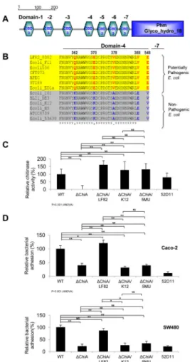
AIEC LF82 ChiA contains seven CBD3 domains upstream of the glycohydrolase catalytic domain at the C-terminus which are highly conserved among 13 other different E. coli genomes that contain CBD3 [Figure 2A]. CBD3 domain 4 showed four amino acid variations (at the 362nd, 370th, 378th and 388th positions) and domain 7 showed one amino acid variation (at the 548th position) among the different E. coli strains. Interestingly, multiple alignments of E. coli CBD3 showed that potentially pathogenic E. coli strains clustered perfectly corresponding to their respective specific polymorphisms, whereas non-pathogenic strains formed another separate group, indicating that this exclusive five amino acid variation seemed to be associated with pathogenicity of E. coli [Figure 2B].
To address the functional relevance of these 5 polymorphic residues, we created an AIEC LF82 mutant strain (LF82-ΔchiA/chiALF82-5MU), in which LF82-ΔchiA strain is
NIH-PA Author Manuscript
NIH-PA Author Manuscript
complemented with the mutated chiA gene from LF82-WT containing mutations on the 5 amino acids (Q362K, E370K, V378A, V388E and E548V) [Supplementary Table 2]. We also created an additional mutant strain (LF82-ΔchiA/chiAK12) that has been complemented with an orthologous chiA gene from the non-pathogenic E. coli strain K12. We found that other than LF82-ΔchiA mutant, the remaining five E. coli strains retained their chitinase enzymatic activities due to the intact glycohydrolase domain present at the C-terminus [Figure 2C]. However, only LF82-ΔchiA, LF82-ΔchiA/chiAK12 and LF82-ΔchiA/ chiALF82-5MU E. coli mutants had markedly reduced adhesion to Caco2 and SW480 IECs, as compared to LF82-WT and -ΔchiA/chiALF82 strains [Figure 2D]. Similar pattern of adhesion with the different AIEC strains was also observed in polarized T84 IECs
[Supplementary Figure 2A]. In addition, CHI3L1 expression was detected on the apical side of polarized T84 IECs, hence correlating localization with functionality [Supplementary Figure 2B]. These observations suggest that the specific genotype of ChiA CBDs may have an influence on the bacterial adhesiveness and hence pathogenicity of E. coli on host cells. AIEC LF82 adhesion increases IFNβ and IL-8 production but not TNFα and CHI3L1 expression
Since previous report showed that CHI3L1 facilitates the ability of bacteria to adhere and invade on/into IECs and that LF82-infected macrophages secreted large amounts of TNFα, we measured the amount of secreted CHI3L1 and TNFα in the culture supernatant of SW480 IECs infected with LF82-WT or LF82 mutant strains by ELISA [1, 12]. We found no apparent differences in both CHI3L1 and TNFα levels in SW480 IECs infected with LF82-WT or LF82 mutant strains, suggesting that AIEC LF82 binding itself does not affect CHI3L1 or TNFα production in IECs [Supplementary Figure 3A].
In line with previous reports showing elevated IL-8 production upon adhesion of mucosa-associated E. coli to IECs in UC and CD patients, cells infected by LF82-WT and -ΔchiA/ chiALF82 strains up-regulated IL-8 production, whereas LF82-ΔchiA-, -ΔchiA/chiAK12,
-ΔchiA/chiALF82-5MU- and 52D11-infected cells produced IL-8 levels that were lower than
LF82-WT- and -ΔchiA/chiALF82-infected cells [Supplementary Figure 3B] [19, 20]. To confirm this observation, we co-transfected IL-8 promoter-luciferase reporter vector and CMV promoter-renilla reporter vector into SW480 cells and infected the cells with WT or its 5 mutant strains, and found significantly higher luciferase activity levels in LF82-WT- and -ΔchiA/chiALF82-infected SW480 cells, when compared to cells infected with any of the other 4 mutant strains [Supplementary Figure 3C].
Increasing reports demonstrate that the level of cytokine IFNβ rapidly increases during bacterial infection [21]. Therefore, IFNβ production in the culture supernatant of HEK293 cells was analyzed after infection with LF82-WT or any of the 5 mutants. An approximately 8- to 10-fold increase in IFNβ production was observed in the supernatant of LF82WT or
-ΔchiA/chiALF82 infected HEK293 cells 24 hours post infection, as compared to that from
uninfected cells [Figure 3A]. In contrast, the remaining mutant strains (LF82-ΔchiA,
-ΔchiA/chiAK12, -ΔchiA/chiALF82-5MU or 52D11) showed only an approximately 2- to
5-fold increase in IFNβ levels [Figure 3A]. A subsequent renilla-normalized IFNβ-promoter luciferase reporter assay also revealed that luciferase activity is significantly upregulated (30-fold) in cells infected with the LF82-WT and -ΔchiA/chiALF82 strains whereas the activity levels of the other 4 mutants showed about 5- to 10-fold higher activity than basal level [Figure 3B].
These results indicate that the ChiA-CBDs in LF82 affect production of IL-8 and IFNβ, but not TNFα or CHI3L1 levels.
NIH-PA Author Manuscript
NIH-PA Author Manuscript
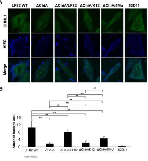
AIEC LF82 cell adhesion requires a functional specific pathogenic form of ChiA-CBMs To visualize the extent of adhesion of LF82-WT and its five mutants, we performed confocal microscopic analysis on infected SW480 cells. CHI3L1 expression was mainly observed in the peri-nucleic and cytoplasmic compartments with epithelial surface
association. High numbers of bacteria adhering to SW480 cells were observed with infection with LF82-WT and -ΔchiA/chiALF82 strains, as revealed by antibody labeling against E. coli-derived LPS, [Figure 4A, 4B]. Conversely, 52D11 strain negative control (no type-1 pili), LF82-ΔchiA, -ΔchiA/chiAK12, and -ΔchiA/chiALF82-5MU strains-infected cells showed significantly less bacterial adhesion. These results further support the fact that LF82 E. coli specifically adheres to host cells via pathogenic ChiA-containing a motif consisting of 5 crucial amino acids within the CBDs.
N-glycosylated, but not O-glycosylated, CHI3L1 is critical for ChiA-mediated AIEC adhesion to IECs
Since previous reports show that human CHI3L1 is post-transcriptionally glycosylated, we tested whether this glycosylation is involved in host-bacterial ChiA interactions by treating SW480 cells with either N-glycosylation inhibitor tunicamycin or O-glycosylation inhibitor benzyl-GalNac for 24 hours and then infecting the cells with LF82-WT [22]. We found that cells devoid of N-glycosylation by tunicamycin had significantly lower associated bacteria in a concentration-dependent manner. Conversely, O-glycosylation-inhibitor treated cells did not demonstrate any apparent changes in bacterial association rate [Figure 5A].
Treatment with the two inhibitors did not affect cell viability since total cellular protein was not altered following treatment [Supplementary Figure 4]. This indicates that
N-glycosylation, but not O-N-glycosylation, is critical in mediating bacterial adhesion on IECs. Using the NetNGly 1.0 online server (http://www.cbs.dtu.dk/services/NetNGlyc), we identified a single glycosylation site on the 68th asparagine residue of mouse CHI3L1 corresponding to the previously reported glycosylated 60th asparagine on human. To confirm this prediction, we constructed three mouse CHI3L1-expressing mutant plasmids containing a mutation in the asparagine residue changing it to proline at the 68th (N68P), 73rd (N73P) or 211th (N211P) residue [Supplementary Table 3]. SW480 or COS7 cells transfected with any of the CHI3L1 mutant plasmids showed a similar pattern of protein expression and localization compared to CHI3L1 WT [Supplementary Figure 5A]. Western blot analysis confirmed that only N68P affects proper CHI3L1 glycosylation [Figure 5B]. Overexpression of CHI3L1-mutant plasmid N68P, which lacks N-glycosylation, in SW480 cells with subsequent infection with AIEC LF82-WT strain resulted in less bacterial association, as compared to cells overexpressing WT or CHI3L1 mutant N211P, which have conserved N-glycosylation [Figure 5C].
We further investigated how CHI3L1 N68P mutant-overexpressing cells responded to different chiA mutants by overexpressing N68P- or N211P-mutant CHI3L1 or WT CHI3L1 in IECs and then infecting the cells with LF82-WT or the 4 LF82 mutants. There was significantly increased bacterial adhesion with LF82-WT and -ΔchiA/chiALF82 in CHI3L1-WT-overexpressing cells, as well as the N211P mutant CHI3L1-overexpressing cells [Figure 5D, Supplementary Figure 5B]. Bacterial counts in the groups infected with the other mutant LF82 strains (LF82-ΔchiA, -ΔchiA/chiAK12 and -ΔchiA/chiALF82-5MU) remained
significantly lower. However, there was no apparent difference in bacterial association across all groups of infected cells that overexpressed CHI3L1 mutant N68P. This indicates that N-glycosylation at the single 68th asparagine residue in mouse CHI3L1, which corresponds to human CHI3L1 60th asparagine residue, is critical for ChiA-mediated host/ microbial interactions.
NIH-PA Author Manuscript
NIH-PA Author Manuscript
LF82 ChiA plays a key role in efficient infection of the host and in exacerbating infectious colitis in vivo
To further confirm our in vitro findings and investigate the in vivo relevance of the observed virulence of LF82-WT and its four chiA mutants, 8–10-week-old C57Bl/6 mice were given 1.5% DSS in their drinking water to induce mild intestinal epithelial damage, and orally gavaged with 108 LF82-WT or its four chiA mutants for 15 consecutive days. The body weight of each mouse was monitored daily. Mice infected with LF82-WT or -ΔchiA/ chiALF82 strains did not show any signs of weight recovery until the endpoint and had higher clinical scores [Figure 6A]. Conversely, LF82-ΔchiA, -ΔchiA/chiAk12- or -ΔchiA/ chiALF82-5MU-infected mice as well as uninfected mice showed recovery after DSS day 10, with milder clinical scores [Figure 6A]. On treatment day 7, LF82-WT-infected mouse stools contained the highest number of bacteria as compared to all the other groups of mice [Figure 6B]. On day 14, the stool bacterial count was highest in mice infected with either LF82-WT- or -ΔchiA/chiALF82.
Bacteria translocation assays revealed that only LF82-WT- and -ΔchiA/chiALF82-infected mice showed appreciable bacterial counts in the liver, spleen, mesenteric lymph nodes (MLNs) and colon [Figure 6C], in association with significantly reduced colonic length as compared to the other groups [Supplementary Figure 6A]. Colonic production of CHI3L1 was up-regulated after DSS treatment with or without AIEC infection [Supplementary Figure 6B]. In addition, colonic histological sections clearly showed severe colitis development in LF82-WT and -ΔchiA/chiALF82-infected mice, with large number of infiltrating inflammatory cells in colonic LP. Conversely, mice infected with the remaining LF82-ΔchiA mutants had milder colitis, as determined by histologic scores, and less LP cellular infiltration [Figures 6D and 6E; Supplementary Figures 7A and 7B]. Up-regulation of IL-6, TNFα and IL-1β in LF82-WT and -ΔchiA/chiALF82-infected mice further supports the colitis severity and pro-inflammatory environment, as compared to -ΔchiA, -ΔchiA/ chiAK12 and -ΔchiA/chiALF82-5MU-infected mice [Figure 6F].
To visualize the extent of bacterial adhesion and invasion in in vivo infection, colonic sections from each infected mouse group were co-stained with antibodies against E. coli-LPS and CHI3L1 [Figure 7]. In uninfected mice, basal levels of endogenous E. coli can be detected, with relatively low CHI3L1 expression levels around the IECs. In contrast, in mice infected with LF82-WT, high bacterial counts were observed in both IEC as well as LP compartments. CHI3L1 expression was also significantly up-regulated in this group of mice and was no longer restricted to the IECs, but extended to the LP. An increased frequency in co-localization between CHI3L1 and LF82-WT and -ΔchiA/chiALF82 was observed in IECs as compared to LF82-ΔchiA or -ΔchiA/chiAK12 strain. Of note, mice infected with
LF82-ΔchiA/chiALF82-5MU strain showed detectable bacterial loads around colonic crypts,
indicating that this AIEC-mutant managed to translocate and invade into the colon to a lesser extent than LF82-WT or -ΔchiA/chiALF82 strain. This result suggests that
polymorphisms within the five amino acids in ChiA-CBDs can delay the invasion process, most likely through the impairment of adhesion. In LF82-ΔchiA/chiALF82-5MU-infected mice, CHI3L1 expression was strong in the IECs compartment and moderate in LP, presumably based on a progressive invasion of this strain in the colon.
Discussion
Bacterial adhesion and colonization on IECs are considered as two of the crucial initializing steps in IBD pathogenesis, before bacteria translocate and enter the submucosal
compartment. In this report, we have demonstrated for the first time that N-glycosylated CHI3L1 facilitates CD-associated AIEC LF82 adhesion to IECs by interacting with bacterial ChiA via the specific CBD that is responsible for the pathogenic genotype. The requirement
NIH-PA Author Manuscript
NIH-PA Author Manuscript
for a specific sugar component to mediate host-microbial interactions was also reported previously in Serratia marcescens and Vibrio cholera-infected IECs [13, 14]. In the ileum of CD patients, highly mannosylated epithelial glycoreceptors carcinoembryonic antigen-related cell-adhesion molecules 6 (CEACAM6) on the apical side of the ileal enterocytes is up-regulated during ileal inflammation in CD patients, which is responsible for AIEC colonization [23]. Although CEACAM6 is not up-regulated in the colonic mucosa of IBD patients, an increased number of AIEC can be detected in both ileum and colon with equal binding affinity in the intestine of these patients [23, 24]. This suggests that AIEC exploits specific glycosylated host factors in a site-specific manner (e.g. CEACAM6 in the ileum and N-glycosylated CHI3L1 in the colon). After AIEC adheres and crosses the colonic mucosal barrier, it internalizes into LP macrophages, where it resides and replicates in association with high levels of TNFα production [11, 12]. Interestingly, TNFα has been previously shown to induce and up-regulates CHI3L1 expression on IECs under inflammatory
conditions [1]. Therefore, it is conceivable that one of the effects of TNFα secretion induced by AIEC LF82 infection is an increase in CHI3L1 expression on IECs, with the probable purpose of facilitating higher affinity to IECs and subsequent entry into the mucosa. Our in vivo AIEC infection studies in mice demonstrate for the first time an essential requirement of chiA, including 5 particular crucial amino acid residues within the ChiA-CBDs in the adhesion of AIEC to IECs. We generated a LF82-ΔchiA/chiALF82-5MU mutant that was still able to cross the mucosa for a relatively short distance with an apparently retarded rate of invasion [Figure 7]. In vivo bacterial loads observed in LF82-ΔchiA/ chiALF82-5MU-infected mice may be a result of a small amount of bacteria that somehow manages to cross the mucosal barrier and then exponentially replicates within the invaded macrophages. This suggests that the 5 polymorphic amino acids are critical for the CHI3L1-dependent attachment onto mucosal epithelial cells, but likely not for invasion and
replication within the macrophages.
Susceptibility and severity in IBD also highly depends on individual genetic variation. Recently, a number of studies reported that single nucleotide polymorphisms (SNPs) in the CHI3L1 locus, especially along the promoter region, have strong associations with different immune-mediated disorders including rheumatoid arthritis and asthma [25, 26]. Although there are no reports of an association between CHI3L1 SNPs and IBD, it is likely that the SNPs may affect proper CHI3L1 gene expression and/or post-translational modification, thus affecting microbial interaction and the susceptibility and severity of IBD in certain individuals.
Given our data demonstrating that bacterial infection of IECs is highly dependent on a carbohydrate intermediate, a novel therapeutic option would be to prevent bacterial attachment by using appropriate carbohydrate components that can modify the interactions between bacteria and host cells. For instance, it was previously shown that
chitin-microparticle treatment can ameliorate intestinal inflammation in two murine models of colitis, and pre-treatment of S. marcescens with chitin can block the bacterial adhesion to IECs [13, 27].
In conclusion, we here demonstrate that ChiA-CBDs in E. coli strains are essential for the bacterial association with IECs in vitro and in vivo. Five amino acids in CBD-4 and -7 specific to pathogenic E. coli, in this case AIEC LF82, are required for high affinity to host IECs, achieved though interactions between bacterial ChiA and host N-glycosylated-CHI3L1. Mice infected with AIEC LF82 devoid of ChiA or harboring mutations in the 5 critical amino acids, experienced less colonic inflammation. Finally, these results present new insights towards therapeutic approaches for the control of potentially pathogenic E. coli infections by providing the molecular mechanistic details underlying bacterial pathogenesis.
NIH-PA Author Manuscript
NIH-PA Author Manuscript
Supplementary Material
Refer to Web version on PubMed Central for supplementary material.
Acknowledgments
Grant Supports: This work has been supported by National Institute of Health (DK80070, DK74454, DK64289 and DK43351, DK068181, DK033506, AI093588), and grants from the Broad Medical Foundation and American Gastroenterological Association Foundation to EM. DL has been awarded the fellowship grant supported by A*STAR Graduate Academy (Singapore) and IAL was supported by the National Research Foundation of Korea. This study was also supported by INSERM (UMR1071), INRA (USC-2018) and by grants from the Association F. Aupetit (AFA) and Région Auvergne (Nouveau Chercheur).
The authors are grateful to Drs. Daniel Podolsky, Ramnik Xavier, Haining Shi, Deanna Nguyen, and Hao-Sen Chiang for their helpful discussions and assistances. We would like to thank Terry Danford Lott for his secretarial assistance, and Manasa Kanneganti for her technical assistances in performing some experiments in this study.
Abbreviations
AIEC adherent-invasive Escherichia coli
CBD chitin-binding domain
CECAM6 carcinoembryonic antigen-related cell-adhesion molecules 6
CHI3L1 chitinase 3-like-1
CMC carboxymethyl cellulose
DSS dextran sulphate sodium
IEC intestinal epithelial cells
LP lamina propria
MOI multiplicity of infection
SNPs single molecule polymorphisms
WT wild type
References
1. Mizoguchi E. Chitinase 3-like-1 exacerbates intestinal inflammation by enhancing bacterial adhesion and invasion in colonic epithelial cells. Gastroenterol. 2006; 130:398–411.
2. Chen CC, Pekow J, Llado V, et al. Chitinase 3-like-1 expression in colonic epithelial cells as a potentially novel marker for colitis-associated neoplasia. Am J Pathol. 2011; 179:1494–1503. [PubMed: 21763261]
3. Chassaing B, Darfeuille-Michaud A. The commensal microbiota and enteropathogens in the pathogenesis of inflammatory bowel diseases. Gastroenterol. 2011; 140:1720–1728.
4. Darfeuille-Michaud A, Boudeau J, Bulois P, et al. High prevalence of adherent-invasive Escherichia coli associated with ileal mucosa in Crohn’s disease. Gastroenterol. 2004; 127:412–421.
5. Martin HM, Campbell BJ, Hart CA, et al. Enhanced Escherichia coli adherence and invasion in Crohn’s disease and colon cancer. Gastroenterol. 2004; 127:80–93.
6. Boudeau J, Barnich N, Darfeuille-Michaud A. Type 1 pili-mediated adherence of Escherichia coli strain LF82 isolated from Crohn’s disease is involved in bacterial invasion of intestinal epithelial cells. Mol Microbiol. 2001; 39:1272–1284. [PubMed: 11251843]
7. Barnich N, Boudeau J, Claret L, et al. Regulatory and functional co-operation of flagella and type 1 pili in adhesive and invasive abilities of AIEC strain LF82 isolated from a patient with Crohn’s disease. Mol Microbiol. 2003; 48:781–794. [PubMed: 12694621]
NIH-PA Author Manuscript
NIH-PA Author Manuscript
8. Darfeuille-Michaud A, Neut C, Barnich N, et al. Presence of adherent Escherichia coli strains in ileal mucosa of patients with Crohn’s disease. Gastroenterol. 1998; 115:1405–1413.
9. Boudeau J, Glasser AL, Masseret E, et al. Invasive ability of an Escherichia coli strain isolated from the ileal mucosa of a patient with Crohn’s disease. Infect Immun. 1999; 67:4499–4509. [PubMed: 10456892]
10. Denizot J, Sivignon A, Barreau F, et al. Adherent-invasive Escherichia coli induce claudin-2 expression and barrier defect in CEABAC10 mice and Crohn’s disease patients. Inflamm Bowel Dis. 2012; 18:294–304. [PubMed: 21688348]
11. Glasser AL, Boudeau J, Barnich N, et al. Adherent invasive Escherichia coli strains from patients with Crohn’s disease survive and replicate within macrophages without inducing host cell death. Infect Immun. 2001; 69:5529–5537. [PubMed: 11500426]
12. Bringer MA, Billard E, Glasser AL, et al. Replication of Crohn’s disease-associated AIEC within macrophages is dependent on TNF-α secretion. Lab Invest. 2012; 92:411–419. [PubMed: 22042084]
13. Kawada M, Chen CC, Arihiro A, et al. Chitinase 3-like-1 enhances bacterial adhesion to colonic epithelial cells through the interaction with bacterial chitin-binding protein. Lab Invest. 2008; 88:883–895. [PubMed: 18490894]
14. Kirn TJ, Jude BA, Taylor RK. A colonization factor links Vibrio cholerae environmental survival and human infection. Nature. 2005; 438:863–866. [PubMed: 16341015]
15. Mizoguchi E, Mizoguchi A, Takedatsu H, et al. Role of tumor necrosis factor receptor 2 (TNFR2) in colonic epithelial hyperplasia and chronic intestinal inflammation in mice. Gastroenterol. 1992; 122:134–144.
16. Gomez Ramirez M, Rojas Avelizapa LI, Rojas Avelizapa NG, et al. Colloidal chitin stained with Remazol Brilliant Blue R, a useful substrate to select chitinolytic microorganisms and to evaluate chitinases. J Microbiol Meth. 2004; 56:213–219.
17. Shen CR, Chen YS, Yang CJ, et al. Colloid chitin azure is a dispersible, low-cost substrate for chitinase measurements in a sensitive, fast, reproducible assay. J Biomol Screen. 2010; 15:213– 217. [PubMed: 20042532]
18. Schultz J, Milpetz F, Bork P, et al. SMART, a simple modular architecture research tool: identification of signaling domains. Proc Nat Acad Sci USA. 1998; 95:5857–5864. [PubMed: 9600884]
19. Steiner TS, Nataro JP, Poteet-Smith CE, et al. Enteroaggregative Escherichia coli expresses a novel flagellin that causes IL-8 release from intestinal epithelial cells. J Clin Invest. 2000; 105:1769–1777. [PubMed: 10862792]
20. Harrington SM, Strauman MC, Abe CM, et al. Aggregative adherence fimbriae contribute to the inflammatory response of epithelial cells infected with enteroaggregative Escherichia coli. Cell Microbiol. 2005; 7:1565–1578. [PubMed: 16207244]
21. Sotolongo J, Espana C, Echeverry A, et al. Host innate recognition of an intestinal bacterial pathogen induces TRIF-dependent protective immunity. J Exp Med. 2011; 208:2705–2716. [PubMed: 22124111]
22. De Ceuninck F, Pastoureau P, Bouet F, et al. Purification of guinea pig YKL40 and modulation of its secretion by cultured articular chondrocytes. J Cell Biochem. 1998; 69:414–424. [PubMed: 9620168]
23. Barnich N, Carvalho FA, Glasser AL, et al. CEACAM6 acts as a receptor for adherent-invasive E. coli, supporting ileal mucosa colonization in Crohn disease. J Clin Invest. 2007; 117:1566–1574. [PubMed: 17525800]
24. Jensen SR, Fink LN, Nielsen OH, et al. Ex vivo intestinal adhesion of Escherichia coli LF82 in Crohn’s disease. Microb Pathogen. 2011; 51:426–431. [PubMed: 21911052]
25. Nielsen KR, Steffensen R, Boegsted M, et al. Promoter polymorphisms in the chitinase 3-like 1 gene influence the serum concentration of YKL-40 in Danish patients with rheumatoid arthritis and in healthy subjects. Arthr Res Ther. 2011; 13:R109. [PubMed: 21714862]
26. Verlaan DJ, Ouimet M, Adoue V, et al. Promoter polymorphisms in CHI3L1 are associated with asthma. J Aller Clin Immunol. 2012; 130:533–535.
NIH-PA Author Manuscript
NIH-PA Author Manuscript
27. Nagatani K, Wang S, Llado V, et al. Chitin microparticles for the control of intestinal inflammation. Inflammatory bowel diseases. 2012; 18:1698–1710. [PubMed: 22241684]
NIH-PA Author Manuscript
NIH-PA Author Manuscript
Figure 1. Bacterial ChiA is required for AIEC LF82 attachment onto intestinal epithelial cell (A) Cell-associated bacteria (relative to the number of originally inoculated bacteria) were quantified after infection with AIEC LF82-WT, -chiA deletion mutant (ΔchiA) or -type 1 pili deletion mutant (52D11) for 1 hour at 37°C (MOI=10). Each value is the average of 6 separate experiments. (B) Representative images of LF82-WT (left) and ΔchiA (right) performed by electron microscopy are shown. (C and D) Bacterial chitinase activity (C) and percentage of cell-associated bacteria (D) were analyzed among the LF82-WT, -ΔchiA and
-ΔchiA/chiALF82 (ΔchiA/LF82) strains, of which the chiA gene was replaced with the chiA
gene derived of LF82-WT. Each value is the average of 4 separate experiments. *P<0.05, **P<0.01, non-significant (NS) as compared to LF82-WT strain.
NIH-PA Author Manuscript
NIH-PA Author Manuscript
Figure 2. Domain architecture of ChiA reveals a requirement of critical amino acid residues for adhesion of LF82 to IECs
(A) Bioinformatic analyses using the chitin-binding domain type 3 (CBD3) as a search query in the SMART online platform detects 7 domains of CBD3 in ChiA, in additional to a glycohydrolase domain in AIEC LF82-WT strain. (B) Multiple amino acid sequence alignments of 13 distinct E. coli strains show unique genotypic conservation at CBD3 domains 4 and 7 that is distinct between pathogenic and non-pathogenic E. coli strains. (C
and D) Relative chitinase activities in SW480 cells (C) and % of cell-associated bacteria in
Caco-2 (upper) and SW480 cells (lower) (D) infected with LF82-WT, ΔchiA, or ΔchiA-mutants that have been complemented with either non-pathogenic K12 strain chiA gene
(ΔchiA/K12), LF82 chiA gene (ΔchiA/LF82), or LF82 chiA gene containing mutations on
the 5 polymorphic amino acids (ΔchiA/5MU) were shown. Statistical analyses for multiple comparisons using ANOVA are indicated below the graph. *P<0.05, **P<0.01, and Non-significant (NS) by post-hoc Tukey HSD test between the two groups as indicated. Each value is the average of 4 separate experiments.
NIH-PA Author Manuscript
NIH-PA Author Manuscript
Figure 3. LF82-WT and -ΔchiA/LF82 strains, but not -ΔchiA, -ΔchiA/K12 and -ΔchiA/5MU strains, up-regulate IFNβ production in culture supernatant of HEK293 cells
(A) IFNβ levels were determined in cells inoculated with the different AIEC LF82 strains at 12 and 24 hours post-infection by ELISA. Each value is the average of 4 separate
experiments. (B) HEK293 cells were transiently transfected with IFNβ promoter-luciferase-and renilla-luciferase-plasmids promoter-luciferase-and subsequently inoculated with the respective AIEC LF82 bacteria (MOI=10). Relative IFNβ promoter-luciferase activities were determined after normalizing with renilla-luciferase activities after 24 h of transfection. Statistical analyses for multiple comparisons using ANOVA are indicated below the graph. *P<0.05, **P<0.01, ***P<0.001 and Non-significant (NS) by post-hoc Tukey HSD test between the two groups as indicated.
NIH-PA Author Manuscript
NIH-PA Author Manuscript
Figure 4. Five amino acid residues within chiA chitin binding domains (CBDs) in pathogenic E. coli, including LF82, are critical in host-microbial interactions
(A) SW480 cells were transfected with a CHI3L1-expression vector for 48 hour and inoculated with the respective LF82-WT or -mutants for 1 hour (MOI=10).
Immunohistochemical staining was performed with antibodies against CHI3L1 (green) and E. coli-LPS (blue) and analyzed under confocal microscopy. (B) Attached bacterial number in twenty different cells was counted under confocal microscope. Each value is the average of 3 separate experiments. Statistical analysis for multiple comparisons using ANOVA is indicated below the graph. **P<0.01 and Non-significant (NS) by post-hoc Tukey HSD test between the two groups as indicated.
NIH-PA Author Manuscript
NIH-PA Author Manuscript
Figure 5. N-glycosylated CHI3L1 is critical for the attachment of AIEC LF82 onto IECs (A) SW480 cells were treated with either N-glycosylation inhibitor tunicamycin or O-glycosylation inhibitor benzyl-GalNac 24 hours prior to infection with LF82-WT strain. (B) Mouse CHI3L1-WT and -point-mutants in N-glycosylated residue (N68P) or
non-glycosylated residues (N73P and N211P) expression vector were individually transfected into cells and detected using immunobloting with anti-X-press antibody. (C) SW480 cells were transiently transfected with mouse CHI3L1-WT or its mutant forms expression vector or LacZ-expressing vector negative control, and infected with AIEC LF82-WT strain for 1 hour (MOI=10). Each value is the average of at least 6 separate experiments. (D) Mouse CHI3L1-WT (closed bars), -N68P (hatched bars) and -N203P (open bars) -expressing vectors-transfected HT29 cells (which do not express endogenous CHI3L1) were infected with LF82-WT or -mutants to determine relative bacterial cell association. Each value is the average of at least 3 separate experiments. Statistical analyses for multiple comparisons using ANOVA are indicated below the graph. *P<0.05, **P<0.01 and Non-significant (NS) by post-hoc Tukey HSD test between the two groups as indicated.
NIH-PA Author Manuscript
NIH-PA Author Manuscript
Figure 6. AIEC LF82-derived ChiA plays a crucial role in the pathogenesis of AIEC-induced infectious colitis
C57Bl/6 mice (n=5–6 in each group) were orally administered with 1.5 % DSS for 15 days and orally infected with 1×10⁸ LF82-WT or -mutants every day. (A) Body weight (left) and clinical scores (right) are shown during the course of colitis. (B, C) Fresh stool was collected on days 7 and 14 (B) and lymphoid organs including liver, spleen, MLNs and colon were analyzed on day 15 (C). Each value represents the average of five mice. (D, E)
Representative histological images of each group (D) or histological score in each group (E) are shown. Scale bars, 100μm. Each value is the average of five or six mice in each group. (F) Colonic levels of cytokines including IL-6, TNFα, IL-1β were determined by ELISA on day 15. Each value is the average of four mice in each group. Statistical analysis for multiple comparisons using ANOVA as indicated. *P<0.05, **P<0.01 and Non-significant (NS) by post-hoc Tukey HSD test compared to LF82WT or between the two groups as indicated.
NIH-PA Author Manuscript
NIH-PA Author Manuscript
Figure 7. LF82-WT and -ΔchiA/LF82-infected mice showed increased levels of adhesion and invasiveness to the colonic mucosa as compared to the other chiA-mutant-infected mice Representative confocal microscopic images of colonic sections from the LF82WT or -mutant strains infected mice using antibodies against CHI3L1 (shown as blue-fluorescence) and E. coli-LPS (shown as green-fluorescence), which were detected by Alexa Fluor 647-and FITC-conjugated secondary antibodies, respectively. Arrow heads show strongly merged areas in the two-fluorescence.