HAL Id: hal-02093098
https://hal.archives-ouvertes.fr/hal-02093098
Submitted on 22 Oct 2020HAL is a multi-disciplinary open access archive for the deposit and dissemination of sci-entific research documents, whether they are pub-lished or not. The documents may come from teaching and research institutions in France or abroad, or from public or private research centers.
L’archive ouverte pluridisciplinaire HAL, est destinée au dépôt et à la diffusion de documents scientifiques de niveau recherche, publiés ou non, émanant des établissements d’enseignement et de recherche français ou étrangers, des laboratoires publics ou privés.
Tyrosine metabolism: identification of a key residue in
the acquisition of prephenate aminotransferase activity
by 1β aspartate aminotransferase
Cécile Giustini, Matthieu Graindorge, David Cobessi, Serge Crouzy, Adeline
Robin, Gilles Curien, Michel Matringe
To cite this version:
Cécile Giustini, Matthieu Graindorge, David Cobessi, Serge Crouzy, Adeline Robin, et al.. Tyro-sine metabolism: identification of a key residue in the acquisition of prephenate aminotransferase activity by 1β aspartate aminotransferase. FEBS Journal, Wiley, 2019, 286 (11), pp.2118-2134. �10.1111/febs.14789�. �hal-02093098�
Signature of prephenate competent 1AAT
1
Tyrosine metabolism: identification of a key residue in the acquisition of prephenate
aminotransferase activity by 1 aspartate aminotransferase.
Cecile Giustini1, Matthieu Graindorge1, David Cobessi2, Serge Crouzy3, Adeline Robin1, Gilles Curien1, Michel Matringe1*
1Univ. Grenoble Alpes, INRA, CNRS, CEA, BIG-LPCV, 38000 Grenoble France. 2Univ. Grenoble Alpes, CNRS, CEA, IBS, 38000 Grenoble France.
3Univ. Grenoble Alpes, CEA, CNRS, BIG-LCBM, 38000 Grenoble France
Running title: Signature of prephenate competent 1AAT
To whom correspondence should be addressed: Michel Matringe, Laboratoire de Physiologie Cellulaire et Végétale, CEA, BIG, 17 rue des Martyrs, Grenoble 38054, France. Tel.: 33 4 38 784 184; Fax: 33 4 38 785 091; E-mail: michel.matringe@cea.fr
Keywords: tyrosine metabolism, prephenate, arogenate route, 1 aspartate aminotransferase, structure
function, molecular modeling
Abreviation: AAT Aspartate aminotransferase, PAT Prephenate aminotransferase
ABSTRACT
Alternative routes for the post-chorismate branch of the biosynthetic pathway leading to tyrosine exist, the 4-hydroxyphenylpyruvate or the arogenate route. The arogenate route involves the transamination of prephenate into arogenate. In a previous study, we found that, depending on the microorganisms possessing the arogenate route, three different aminotransferases evolved to perform prephenate transamination i.e. 1 aspartate aminotransferase (1 AAT), N-succinyl-L,L-diaminopimelate aminotransferase, and branched-chain aminotransferase. The present work aimed at identifying molecular determinant(s) of 1 AAT prephenate aminotransferase (PAT) activity. To that purpose, we conducted X-ray crystal structure analysis of two PAT competent 1 AAT from Arabidopsis thaliana and Rhizobium meliloti, and one PAT incompetent 1 AAT from Rhizobium meliloti. This structural analysis supported by site directed mutagenesis, modelling and molecular dynamics simulations allowed us to identify a molecular determinant of PAT activity in the flexible N-terminal loop of 1 AAT. Our data reveal that a Lys/Arg/Gln residue in position 12 in the sequence (numbering according to Thermus thermophilus 1 AAT), present only in PAT competent enzymes, could interact with the 4-hydroxyl group of the prephenate, substrate and thus may have a central role in the acquisition of PAT activity by 1 AAT.
The aromatic amino acid biosynthesis pathway occurs in microorganisms, fungi and plants, and is not found in animals. This pathway links the metabolism of carbohydrates to the biosynthesis of aromatic amino acids involved in protein synthesis, and leads to the synthesis of a large diversity of aromatic secondary metabolites [1-3]. The aromatic amino acid biosynthesis pathway plays a central role for the growth of these organisms and their interaction with environment. Phenylalanine and tyrosine also represent an important source of high-value aromatic natural compounds for industry, and human health.
Two alternative routes leading from chorismate to tyrosine and phenylalanine exist: the 4-hydroxyphenylpyruvate route, and the arogenate route (Figure 1). All photosynthetic organisms and many microorganisms synthesize L-tyrosine via the arogenate route [4-7]. In addition, most viridiplantae and some microorganism also synthesize L-phenylalanine via the arogenate route [8]. In this case, a prephenate aminotransferase transforms prephenate into arogenate, and an arogenate dehydrogenase (EC 1.3.1.43) or an arogenate dehydratase (EC 4.2.1.91) decarboxylates arogenate into L-tyrosine or L-phenylalanine, respectively (Figure 1) [7,8]. Alternatively, some microorganisms and fungi such as Escherichia coli, or yeast, first transform prephenate into 4-hydroxyphenylpyruvate by a prephenate dehydrogenase (EC 1.3.1.13) or into penylpuruvate by a prephenate dehydratase (EC 4.2.1.51) [9], and then both are transaminate into L-tyrosine or L-phenylalanine, respectively.
Signature of prephenate competent 1AAT
2
Prephenate aminotransferase (PAT) activity was first identified in plants [10,11]. In Arabidopsis thaliana, a specific chloroplastic aspartate aminotransferase is in charge of PAT activity (At2g22250 gene, EC 2.6.1.1, EC 2.6.1.78, and EC 2.6.1.79) [10,11]. This prephenate aminotransferase belongs to aspartate aminotransferase (AAT) subgroup 1. In this subgroup, the dicarboxylic substrate interacts with a lysine and an arginine (K101 and R361, numbering according to Thermus thermophilus 1AAT enzyme: Q56232 abbreviated as Tth-PAT) instead of two arginines in the 1 AAT subgroup. In a previous study, we showed that the PAT competent 1 AAT from A. thaliana (Ath-PAT) catalyzes the transamination of oxaloacetate into aspartate as efficiently as prephenate into arogenate [10].
Historically in microorganisms, arogenate route has been detected in cyanobacteria, actinobacteria, some - and -proteobacteria by the group of Jensen [4-6,13]. In a previous study [14] we reported that, in these arogenate competent microorganisms, PAT activity is carried out by three different aminotransferases, which present two different fold-types. Furthermore, we showed that the capacity to transaminate prephenate is not a general catalytic property of the three classes of aminotransferases, since other members were devoid of detectable PAT activity [14]. These results revealed that the arogenate route emerged independently several times during evolution. To go further, structure-function studies are needed to identify molecular signature(s) of the competent prephenate/arogenate aminotransferases, in order to investigate their presence in genomic databases.
In the present study, by combining X-ray crystal structure analysis of two PAT competent 1 AAT from Arabidopsis thaliana and Rhizobium meliloti, and one PAT incompetent 1 AAT from Rhizobium meliloti, site directed mutagenesis, modelling and molecular dynamics simulations, we show that K/R/Q12 residue (numbering according to T. thermophilus 1AAT) is a signature of the PAT function of 1AAT. It is present in the N-terminal flexible loop only in PAT competent 1-AAT and our data strongly support its possible role in stabilizing prephenate by interacting with its 4-hydroxy group.
Results and discussion
Resolution of 3D structures of PAT competent and PAT incompetent 1 AAT.
In our previous study [14], of the two 1 AATs from Rizobium meliloti tested, one (Q02635) showed a good prephenate aminotransferase activity while the other (P58350) had no detectable prephenate aminotransferase activity under our test conditions.
We thus decided to identify molecular determinant(s) involved in the acquisition of prephenate aminotransferase function by the 1 AAT through a comparative X ray structural study of PAT competent and PAT incompetent 1 AAT. To do this, we solved the three-dimensional structures of PAT competent from Arabidopsis thaliana (Ath_Q9SIE1, Ath-PAT) and from Rizobium meliloti (Rme_Q02635, Rme-PAT), and the PAT incompetent 1 AAT from Rizobium meliloti (Rme_P58350, Rme-AAT).
Overall structures
The 3D structures of PAT competent 1 AAT Ath-PAT, and Rme-PAT and PAT incompetent 1 AAT Rme-AAT show homodimeric proteins, covalently bound to pyridoxal phosphate (PLP) and sharing a common fold (Figure 2). The monomers are related by a non-crystallographic 2-fold symmetry axis. Residues in or preceding the first -helix are observed in different positions when monomers of Ath-PAT are compared, suggesting flexibility. A citrate molecule is bound to the catalytic site of 3 monomers of Ath-PAT (two dimers are in the asymmetric unit). It is stabilized by interacting with Lys122, Trp146, Asn196, Arg398 side chains, the Gly60 NH group and water molecules. In Rme-PAT, pyridoxal phosphate cofactor is not covalently bound to the protein probably as a consequence of radiation damages.
Structure comparisons
The Cα coordinates rms deviations calculated between all the monomers are low indicating that the structures are globally very similar. The largest difference is observed when Rme-AAT is compared with Ath-PAT (1.56 Å) or Tth-PAT (PDB code 1BKG) (1.63 Å). Dimer comparison after superimposing monomers shows moderate differences in the positioning of monomers. The catalytic site is large and solvent accessible and residues from the two monomers participate to the catalytic pockets. Most of the residues observed in the Ath-PAT catalytic site are conserved in Rme-PAT, Rme-AAT and Tth-PAT. From structure comparison with Tth-PAT, residues from the first -helix (Lys33(A), Pro34(A), Ser35(A), Lys36(A), Thr37(A), Met38(A) in Ath-PAT [15,16]) are also involved in the closing of the catalytic site
Signature of prephenate competent 1AAT
3
upon ligand binding. Lys259 of Ath-PAT is covalently bound to PLP through an aldimine link. The interactions observed between maleate and residues of the catalytic site in Tth-PAT could also take place in PAT. Maleate observed in the Tth-PAT catalytic site is superimposable to citrate observed in Ath-PAT, but the position of the first -helix is different since the Ath-PAT catalytic site is in open conformation.
3D X ray structures of PAT competent 1 AAT Ath-PAT and Rme-PAT, and PAT incompetent 1 AAT Rme-AAT, present a very similar organization. Comparison of these three 3D structures did not reveal a clear molecular determinant of PAT activity, most certainly because they are in an open conformation. Indeed, during the catalytic reaction, the keto acid substrate (oxaloacetate or prephenate) binds to a PMP-enzyme complex and a short N-terminal flexible loop move to close the active site [15,16]. Only a closed conformation of prephenate-PMP-enzyme complex is relevant for structural comparison of PAT competent and incompetent 1 AAT. Unfortunately, all our attempts to obtain either a prephenate- or PMP-maleate-enzyme complex of Rme-PAT or Ath-PAT failed.
However, the observation that the 1 AATfrom Thermus thermophilus is PAT competent ([17], this study) allowed us to use its 3D structure in a closed conformation, in complex with PMP and maleate (PDB code 1BKG), resolved by Nakai et al. [15], to model the binding of prephenate in the active site.
Identification of a potential molecular determinant of PAT capacity of 1 AAT
Taking advantage of molecular elucidation of enzyme/ligand interactions in PAT competent 1
AAT from T. thermophilus by Nakai et al [15], we manually replaced the substrate analogue maleate with prephenate in the active site of the 3D structure 1BKG. Prephenate differs from oxaloacetate and ketoglutarate, the two ketoacid substrates of aspartate aminotransferase, by the presence of its 2,5-cyclohexadiene-4-hydroxy group (Figure 3A). The idea is therefore to identify potential residue(s) present only in the PAT competent 1
AAT that could interact with the 2,5-cyclohexadiene-4-hydroxy group of prephenate. We placed the two prephenate carboxylate groups in interaction with Lys101 and Arg361 while its keto group points to the PMP NH2 group and the catalytic lysine, Lys234 (Figure 3B). In this position, the 2,5- cyclohexadiene-4-hydroxy group of the prephenate substrate points toward the entrance of the active site, its 4-hydroxyl facing the flexible N-terminal loop 10AMKPSA15, which is known to move and close the active site while the substrate binds to the active site [15,16]. In that context, it is interesting to note that the alignment of the N-terminal sequences of already characterized PAT competent1
AAT (Arabidopsis thaliana [10], Sorghum bicolor [18], R. meliloti, Rhodobacter sphaeroides [14], T. thermophilus [17] (this study) and Chlorobium tepidum [17]) with those of the already characterized PAT incompetent1
AAT (R. meliloti, Streptomyces avermitilis and Synechocystis [14]) (Figure 4), reveals that a lysine residue belonging to this flexible loop, Lys12 (numbering according to T. thermophilus1
AAT), is present only in the PAT competent1
AAT or is replaced by an arginine in all Poaceae like S. bicolor or Zea mays or a glutamine in some Proteobacteria like Rhodobacter sphaeroides and in Chlorobi like Chlorobium tepidum. Each of these three amino acids has the ability to stabilize prephenate in the active site by interacting with its 4-hydroxyl group. In contrast, in PAT incompetent1
AAT, Lys12 is replaced by a glycine, a valine or a serine. None of the later three can play the same role.However, in the ternary maleate-PMP-enzyme complex (1BKG), the Lys12 side chain points outside the active site [15] (Figure 3). We hypothesize that in the presence of prephenate, the Lys12 side chain rotates to interact with the prephenate cyclohexadiene-4-hydroxy group during closure of the active site, thereby stabilizing the prephenate within the active site.
Lys12 plays a specific role in prephenate binding by PAT competent 1 AAT from R. meliloti and T.
thermophilus
The potential interaction of Lys12 with the prephenate 4-hydroxyl group was first addressed by using a site directed mutagenesis approach performed on PAT competent 1AAT from R. meliloti and T. thermophilus. We replaced the lysine of the 10AMKPSA15 motif by a glycine. Steady‐state kinetic properties of wild‐type and K12G mutants were determined with a fixed concentration (25mM) of Glutamate as amino donor and variable concentrations of either prephenate or oxaloacetate. Modification of the apparent Michaelis-Menten constant (KM) will reflect a modification in the affinity of the 1 AAT K12G mutants for the substrate whereas modification of the apparent catalytic constant (kcat) will reflect an impact of the
Signature of prephenate competent 1AAT
4
mutation on the reaction rate. If our assumption was correct we anticipate that the mutation will have a significant impact on the affinity for prephenate, much less on the affinity for oxaloacetate, and will have minor impact on the reaction rate of both oxaloacetate and prephenate aminotransferase activity. Apparent KM for prephenate was found 7 time higher in the K12G mutant of PAT competent 1AAT from R. meliloti (Q02635) (K12G: 692±143 µM versus WT: 106±12µM). In the other hand, apparent KM for oxaloacetate was only weakly affected in K12G mutant (Table 3). Furthermore, the apparent catalytic constants of the two aminotransferase reactions (kcat) were not affected.
The effect of the K12G mutation was also tested on the PAT activity of the T. thermophilus 1AAT (Q56232). The K12G mutation gave similar results with a 10 time increase of the apparent KM for prephenate (K12G: 2332±204 µM versus WT: 150± 8.7 µM) and no effect on the apparent KM for oxaloacetate (WT: 25.3±1.8 µM versus K12G: 26.4±2.3 µM). It should be noted that in our conditions, the T. thermophilus 1AAT apparent catalytic activity is slightly affected by the K12G mutation (kcat oxaloacetate WT: 32.2±4.6 s-1 versus K12G: 12.3±3.7 s-1). Prephenate aminotransferase apparent catalytic activity seems less affected by the mutation (kcat for prephenate WT: 7.7±1.6 s-1 versus K12G: 4.8 ± 2.1 s -1), although the strong degradation of affinity for prephenate of the K12G mutant makes the determination of its kinetic parameters less accurate. Steady state kinetics analysis confirms thus that in both enzymes K12G mutation induces a specific decrease in affinity for prephenate with limited effect on their prephenate aminotransferase reaction rate.
In our previous study [14], the two incompetent PATs of R. meliloti (P58350), or Synechocystis (Q55128) which respectively have a G or a V instead of K12, were found to have no PAT activity at prephenate concentrations up to 2.5 mM, by our coupled test. However, in the present study, the two K12G mutants maintain prephenate aminotransferase activity at high prephenate concentrations since the K12G mutation does not affect their reaction rate.
In their study, Dornfield et al [17] propose that two highly conserved residues among 1AAT, T84 and K169, (numbering according to A. thaliana 1AAT, T16 and L101 according to T. thermophilus 1AAT) (Figure 3 and 4), are important residues for substrate recognition in PAT competent 1 AAT. Furthermore, during the final stage of writing our manuscript, a new structural study on Arabidopsis PAT competent 1 AAT (At2g22250) from the same group was published in Plant Journal [16]. In this study, the authors proposed that, in addition to T84 and K169, E108 (numbering according to A. thaliana 1AAT, E40 according to T. thermophilus 1AAT) (Figure 3 and 4) plays an important role in the acquisition of PAT competencies by 1AAT. However, unlike K12, these residues are conserved among all (K169, and E108) or almost all (T84) 1AAT, regardless their ability to catalyze the transamination of prephenate, or not [14,17]. Therefore, although these residues may have contributed to the evolution of prephenate aminotransferase activity within class 1 AAT and not class 1AAT, they can hardly be considered to have a specific role in the prephenate aminotransferase activity of 1AAT. On the contrary, the presence of K/R/Q12 only in PAT competent 1AAT, as well as the strong and selective impact of the K12G mutation on the binding affinity for prephenate, strongly support the specific role played by Lys12 in the binding of prephenate, most likely by its stabilization at the active site through interaction with its 4-hydroxyl group.
A clear support to the specific role played by Lys12 in the binding affinity for prephenate was afforded by Nature through the case of Synechocystis Q55128 and Synechococcus Q3AK33 1 AAT. As already mentioned Synechocystis Q55128 1 AAT have a V in place of a K, and was found inactive in our coupled assay [14]. However, in their study Dornfield et al [17] were able to detect traces of arogenate by HPLC in their end point test [17]. They reported a weak activity with an apparent KM for prephenate in the range of 2600 µM (in the range of the apparent KM for prephenate of the T. thermophilus 1 AAT K12G mutant). Blast search revealed that the Cyanobacteria Synechococcus Q3AK33 1 AAT, which is a close relative to Synechocystis Q55128 1 AAT, have a K in place of a V (Figure 5A). In order to compare the catalytic properties of these two 1 AAT, we have cloned, expressed in E. coli and partially purified the recombinant Q3AK33 1 AAT (Figure 5B). In our conditions, in contrast with Synechocystis Q55128 [14], a weak prephenate activity could be detected with the recombinant Synechococcus Q3AK33 1 AAT by our coupled assay. Kinetic characterization of prephenate aminotransferase activity of recombinant Q3AK33 (Figure 5C) reveals that its apparent KM for prephenate was found in the range of 207±23 µM µM versus 2600 µM for Synechocystis Q55128 1 AAT [17], nicely confirming that the presence of K12 strongly
Signature of prephenate competent 1AAT
5
improves binding affinity for prephenate. Our data also indicate that other residues that remains to identify are responsible for the high reaction rate exhibited by all tested 1 AAT-PAT from higher plants [10 11] and by some 1 AAT from bacteria like Rhizobium meliloti (Q02635) [14] or Chlorobium tepidum (Q8KDS8) [17].
Structural evidence for Lys12 involvement in prephenate stabilization within the active site
We wanted to confirm, at the structural level, the specific role of Lys12 pointed out by the structure comparisons of PAT competent and incompetent 1AAT, and by kinetics analysis of K12G mutants. Unfortunately, as already mentioned, all our attempts to obtain either a PMP-prephenate-enzyme complex of PAT competent 1AAT from A. thaliana, R. meliloti or T. thermophilus failed, and at that time the only available 3D structure of a PAT competent 1AAT in a closed conformation was the PMP-maleate-enzyme complex of 1AAT from T. thermophilus. (PDB code 1BKG), previously solved by Nakai et al. [15]. We thus used this 3D structure as a starting model to perform structural analyses via modeling and molecular dynamics simulations of the PMP-prephenate-enzyme complex to support the interaction between Lys12 and prephenate suggested by our kinetic experiments.
After model building, introduction of sodium counter-ions and solvation in a periodic box of TIP3P water molecules (structure 1BKG_Preph_Mod, see methods), a short equilibration MD simulation of the PAT dimer in the presence of PMP and prephenate was run at 300 K for 20 ns leading to structure 1BKG_Preph_MD (See methods). Starting from a structure with low energy and low Lys12 NZ / prephenate O1 oxygen distance (dK-Prf) of 3.2Å from this equilibration, we have run a QM/MM optimization followed by a calculation of the charges adequate to fit the calculated electrostatic potential around the ligands (PMP and prephenate). In addition to prephenate, PMP and Lys12, the QM part consisted of side chains of Lys101, Trp125, Ser127, Lys234, Arg242, Tyr322, Arg321 from chain A and Tyr64 from chain B. 14 water molecules were also included in this quantum part for a total of 191 QM atoms. All MM atoms belonging to residues within a distance of 3 Å of the QM part were allowed to relax during geometry optimizations, and the remaining structure was kept frozen.
The optimized structure of the catalytic site (see materials and methods for details) is shown in Figure 6 and 7. Stabilizing interactions through hydrogen bonds exist between Lys12, Lys101, Trp125, Arg361, Tyr322 and prephenate, between prephenate and PMP and between PMP and Tyr64, Arg242, Tyr322 and Lys234.
The atomic partial charges calculated using the Merz-Kollman routine [20] in Gaussian were directly transferred to the protein structure file describing the MM system for prephenate, PMP and surrounding residues.
Molecular dynamics simulations
Using this modified structure file, 24 ns constant pressure and temperature CPT (Constant Pressure and Temperature) dynamics were run restarting from structure 1BKG_Preph_MD. The nuclear Overhauser enhancement (NOE) type restraints used to obtain 1BKG_Preph_MD (see materials and methods for details) were kept for 4 ns and then removed for the remaining 20 ns. This distance dK-Prf oscillates between 2.5 and 4.5 Å during the first 20 ns and increases to up to 14 Å when the restraints are released (See Figure 8). Interestingly, this distance drops back down to around 2.8 Å after 42 ns, meaning that without any restraint on the protein, the distance between Lys12 and prephenate is occasionally compatible with the reaction proposed in the experimental section.
Improving conformational sampling
The previous MD simulation confirms that the N-terminal part of the protein could undergo conformational transitions during substrate binding as shown by the structural study of Nakai et al [15], enabling Lys12 to be stabilized in the vicinity of the prephenate. Still, a method to explore more conformational space is to use Replica eXchanging Self-Guided Langevin Dynamics [21]. 16 replicas were created starting from structure 1BKG_Preph_Mod and vacuum RXGLD simulations were run for 10 ns in the presence of restraints (see materials and methods). Coordinates from Met1 to Gln29 (chain A), PMP and prephenate were extracted from the MD trajectory every ps (20000 structures) and grouped into 16 clusters corresponding to coordinate root mean square deviation (rmsd) larger than 3.5Å. From the
Signature of prephenate competent 1AAT
6
evolution of cluster population versus time (shown in Figure 9), 6 most populated clusters, visited many times all along the simulation, seem particularly interesting and have been studied further.
Final production dynamics from selected clusters
The coordinates of the lowest energy structure in the 6 selected clusters were computed and one molecular dynamics simulation was started from each resulting minimized structure (see materials and methods). The evolution of dK-Prf is plotted in Figure 10 for the six selected clusters. One particular cluster (number 6) simulated during 85 ns, shows very large fluctuations of dK-Prf from 2.6 to more than 21.6 Å, demonstrating the high mobility of the N-terminal part of the protein. The corresponding two structures are shown in Figure 11 after superimposition of backbone atoms of residues 33 to 382. Backbone atom coordinate rmsd is 4.18 Å for residues 13 to 32 and 1.09 Å for residues 33 to 382. Between the two structures, conformational changes around Lys12 appear with transitions of the backbone dihedral angle Φ=(Met11)CNCαCβ from 180° to 120° and the side chain dihedral angle χ1=NCαCβCγ from 60° to -60°, for “far” and “close” conformation, respectively.
The presence of Lys/Arg/Gln12 could be considered as a molecular signature of the PAT competent 1 AAT
Our experimental kinetics data, revealed that Lys12 plays a specific role in the acquisition of PAT activity by R. meliloti and T. thermophilus 1 AAT most certainly by stabilizing prephenate though an interaction with its 4-hydroxyl group. Accordingly, we were able to optimize at the QM level a structure of the active site respecting this restraint. Releasing this restraint during MD simulations results in a rapid drift of dK-Prf followed by frequent returns to dK-Prf lower than 3 Å. This means that, although no crystal structure of PAT competent 1 AAT from T. thermophilus, or homologues, has been obtained showing proximity between Lys12 and the catalytic site, interactions are possible and even frequent between Lys12 and prephenate, on the short time scale accessible to MD simulations.
These simulations strongly support, at the structural level, our kinetics data. All together our data point lysine12 and the corresponding arginine or glutamine, present in other PAT competent 1 AAT, as key residues in the acquisition of a physiologically relevant prephenate aminotransferase activity by 1 AAT during evolution. As already mentioned, our data also revealed that other residues that remain to be identified are responsible for the good reaction rate exhibited by all tested plant and some bacteria 1 AAT PAT.
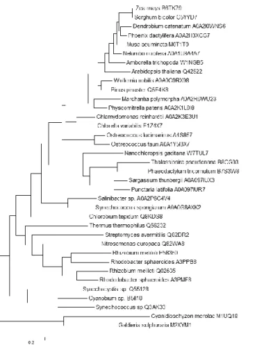
Phylogenetic distribution of 1 AAT among different classes of organisms (Figure 12) confirms the presence of a 1 AAT with a Lys/Arg/Gln in position 12 in all photosynthetic organisms at the exception of most Cyanobacteria, which are known to synthesize arogenate via a BCAT-PAT [14]. The relative distribution of K/R/Q12 reveals the presence of a Lysine residue (corresponding to K12) in the majority of land plants except in the Poaceae (like Zea maïs or Sorghum bicolor) which possess an arginine. Most green algae also have a Lysine residue, but in the 1 AAT from Ostreococcus lucimarinus (A4S857) the lysine is replaced by an arginine, like in Poaceae. It is difficult to have a clear idea of the situation for the brown and the red algae due to the small number of sequenced genomes. Some brown algae like Sargassum thunbergii have a 1 AAT with a serine residue (AOA097IUX3) and presumably synthesize arogenate with a BCAT-PAT like Cyanobacteria or a N-succinyl-l,l-diaminopimelate aminotransferase like the two tested actinobacteria, Streptomyces avermitilis and Mycobacterium tuberculosis [14], or do not synthesize tyrosine and phenylalanine via the arogenate route. In the other hand, some brown algae like Punctaria latifolia possess a 1 AAT (AOA097IUR7) with an arginine like Poaceae. The few sequenced genomes of red algae seem indicate that they are devoid of 1 AAT, the first hit concern aspartate aminotransferases with a highly divergent N-terminal sequence but with a conserved K101 and a more conserved C-terminal sequence. In contrast, Diatoms which result from secondary endosymbiosis events between an algae and a eukaryote, have either a Lysine like green algae, or a Q like some photosynthetic bacteria, confirming the possible dual origin (green or red algae) of their chloroplasts [19]. Intriguingly, Nanochloropsis have a 1 AAT with a Glu in the corresponding 12 position. Chlorobi and Bacteroidetes have a Q, and interestingly the 1 AAT of the symbiotic cyanobacteria Synechococcus spongiarum also have à Q and branch with Chlorobi and Bacteroidetes rather than with other Cyanobacteria. Lys12 residues are found in 1 AAT from many non-photosythetic bacteria (most -proteobacteria and hadobacteria and some -proteobacteria) known to be arogenate competent organisms. In the other hand, most Actinobacteria like Streptomyces
Signature of prephenate competent 1AAT
7
avermitilis have 1 AAT with a Serine12, which is in agreement with their capacity to synthesize arogenate through an S-DAP-PAT [14].
Experimental procedures
Bacterials strains- Two strains of E. coli were used in this work: the strain DH5Invitrogen
strain; F-80lacZM15 (lacZYA-argF) U169 deoR recA1 endA1 hsdR17 (rk-, mk +) phoA supE44 thi-1 gyrA96 relA1 -) was used for plasmid DNA amplification and subcloning. The strain BL21 Rosetta 2 (DE3) (Merck; F-ompThsdSB (rB-mB-) gal dcm (DE3) pRARE2 (CamR)) was used for overproduction of recombinant proteins. These two strains were grown at 37°C in Luria-Bertani medium supplemented with 1% agar (w/v) for the agar media, and supplemented with Carbenicillin 1°/°° (DH5or Kanamycine 1°/°° (BL21 The 2011 RCR strain of Rhizobium meliloti was provided by G. Alloing (UMR6192, Sophia Antipolis). Thermus thermophilus (strain HB8) Genomic DNA was provided by A. Atteia (UMR 7281, Marseille). Synechococcus CC9605 genomic DNA was provided by F. Partensky (UMR7144, Roscoff)
Chemicals- Prephenate, oxaloacetate, and glutamate were purchased from Sigma.
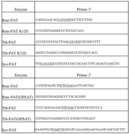
Primers- Primers used for amplification of the 1 aspartate aminotransferase genes from the corresponding genomic DNA are presented in Table 4. Restriction sites introduced by PCR are underlined. These cDNAs were cloned into pET30 b(+) vector, and the recombinant proteins were produced without any tag.
Overproduction and partial purification of recombinant proteins- Fresh colonies of transformed
BL21 (DE3) Rosetta2 bacteria (Novagen, Darmstadt, Germany) containing pET30 b(+) vector expressing the desired recombinant protein were transferred into 15 ml of LB medium supplemented with Kanamycine 1°/°°, and grown at 37°C. Saturated culture was transferred into 800 ml LB medium supplemented with Kanamycine 1°/°°,, and growth was continued until DO600 nm=0.6. IPTG (0.4 mM) was added to the medium, and the bacteria were grown at 20°C for 16 h. Cells were harvested by centrifugation (4000 g, 45 min), and pellets were re-suspended in 30 ml of 25 mM HEPES pH 8.0, 1 mM EDTA, 1 mM DTT, 10% glycerol (v/v), 50 µM PLP, 5 mM -aminocaproic acid, and 1 mM benzamidine (Buffer A), and sonicated for 10 min at 4°C on a Branson sonicator. Streptomycin sulphate (0.1% (w/v)) was added to precipitate DNA and the extract was centrifuged for 45 min at 40 000 g (Sorvall SS-34) at 4°C. The resulting supernatant of soluble proteins were stored at -80°C until used for purification.
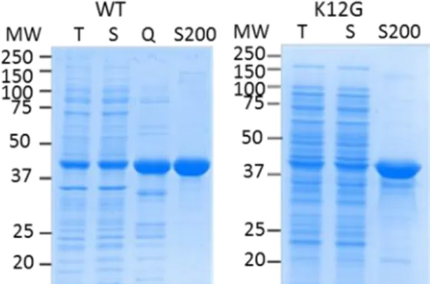
Partial purification of recombinant proteins from R. meliloti Q02635 WT and K12G mutant were performed as follows: crude extract of soluble proteins (20-50 mg) were loaded on a 30 ml of Q-sepharose resin (GE Healthcare) in an XK-16 column (Amersham Pharmacia) equilibrated with Buffer A, proteins were eluted by a linear gradient (0-0.5 M) of NaCl in buffer A. Active fractions were pooled and loaded on HiPrep 16/60 Sephacryl S-200 column (Amersham Pharmacia) equilibrated with Buffer A supplemented with 100 mM NaCl (Figure 13). The most active fractions were pooled, desalted in buffer A supplemented with 10% glycerol, frozen in liquid nitrogen and stored at -80°C.
Partial purification of recombinant proteins from Thermus thermophilus Q56232 WT and K12G mutant were performed as follows: crude extract of soluble proteins (20-50 mg) were loaded on 100 ml of EMD DEAE 650(M) resin Merck) in an XK 26 column (Amersham Pharmacia) equilibrated with Buffer A, proteins were eluted by a linear gradient (0-0.5 M) of NaCl in buffer A (Figure 14). The most active fractions were pooled, desalted in buffer A supplemented with 10% glycerol, frozen in liquid nitrogen and stored at -80°C.
Partial purification of recombinant proteins from Synechoccocus Q3AK33 were performed as follows: crude extract of soluble proteins (20-50 mg) were loaded on a 30 ml of Q-sepharose resin (GE Healthcare) in an XK-16 column (Amersham Pharmacia) equilibrated with Buffer A, proteins were eluted by a linear gradient (0-0.5 M) of NaCl in buffer A (Figure 5). The most active fractions were pooled, desalted in buffer A supplemented with 10% glycerol, frozen in liquid nitrogen and stored at -80°C
Signature of prephenate competent 1AAT
8
Determination of enzyme activities- Glutamate-oxaloacetate aminotransferase activities were
assayed as described in [8].
Prephenate aminotransferase activities were assayed by coupling the reaction with purified Tyr-insensitive arogenate-specific dehydrogenase from Synechocystis [22] and following the reduction of NADP at 340 nm. The reaction was carried out at 30°C in 50 mM HEPES buffer (pH 8.0) in the presence of 40 nM coupling enzyme, 100 µM NADP and variable amounts of prephenate, and fixed concentration of glutamate (25mM). Activities were calculated using an epsilon for NADPH of 6250 M-1.cm-1 at 340 nm. Arogenate formation was confirmed by HPLC analyses as described in [10].
Kinetic data analysis- Initial velocity data were fitted to the Michaelis–Menten equation using the
Kaleidagraph program (Synergy Software, Reading, PA, USA).
Crystallization- Extensive searches for crystallization conditions were performed in 96 well plates at 293 K using the high throughput crystallization facilities of EMBL (Grenoble). Crystallization hits were refined in 24 well Linbro plates. All the crystals were grown at 293 K. Ath-PAT crystals grew in 0.1 M Na citrate pH 4.0, 11 % PEG 4K. Rme-AAT/PAT crystals were obtained in 0.2 M ammonium formate, 19 % PEG 3350 and Rme-AAT grew in 0.1 M Na acetate pH 4.5, 5% PEG 4K. The protein concentration was 10 mg/ml.
Data collection- Diffraction data (Table 1) were collected at 100 K at the European Synchrotron
Facilities either on FIP-BM30A [23] (Rme-AAT) or ID23-1 (Ath-PAT and Rme-AAT/PAT) using an ADSC 315r detector. Prior freezing, the crystals were soaked in mother liquor with glycerol in a concentration range from 20 % (Ath-PAT and Rme-AAT/PAT) to 25 % (Rme-AAT). All the data were processed and scaled using XDS [24].
Phasing and Refinement- All the phase calculations were performed by molecular replacement after
modifying the models using CHAINSAW [25] from CCP4 [26]. Phases for Ath-PAT were calculated using Molrep [27] and the atomic coordinates of aspartate aminotransferase from Phormidium lapideum (PDB code 1J32) as a model. Phaser [28] was used to calculate phases for Rme-AAT/PAT and Rme-AAT using the atomic coordinates of Ath-PAT and those of the aspartate aminotransferase from Phormidium lapideum (PDB code 1J32), respectively. All the refinements and rebuilding were carried out using Phenix [29] and COOT [30]. Non-crystallographic symmetry was applied in all cases during refinement. Rmsd calculations were done using PDBefold [31].
Atomic coordinates and X-ray data were deposited in the PDB with the accession numbers: At-PAT (6F5V), Rme-PAT (6F77) and Rme-AspT (6F35).
Structural modeling
I- Modelling of Lys12 conformational transitions using MD simulations
Initial model- It was built from the structure of aspartate aminotransferase from Thermus
thermophilus (Tth-PAT) complexed with maleate (PDB id 1BKG). 1BKG gives the structure from Met1 to Leu382 in both A and B chains. It was obtained in the presence of 4'-deoxy-4'-aminopyridoxal-5'-phosphate (PMP) and maleate.
Experimentalists have modified the structure "manually" changing the position of Lys12 "by eye" to fit experimental observations and introducing global rotations and translations with respect to 1BKG to obtain a structure called Sexp. They also manually inserted an ideal structure of prephenate in place of maleate. The global atomic coordinate rms deviation between Sexp and 1BKG is 0.85 Å (considering the 5888 atoms of the dimer without water and ligands). To start from the experimental structure 1BKG without losing the information coming from experiments, we started by calculating the reverse transformation to be applied to Sexp to bring it back close to 1BKG while maintaining the rearrangement of Lys12. Then, this transformation was applied to prephenate and crystal waters, as well.
Structure preparation- Finally the structure of the dimer of Tth-PAT from residue 1 to 382, PMP,
prephenate and 141 crystal waters was completed and refined with the molecular dynamics program CHARMM [32]. The all-atom force field all27 for proteins [33,34] was used. An initial structure of prephenate was obtained from the Zinc database (zinc1532584) while the initial structure of pyridoxamine
Signature of prephenate competent 1AAT
9
phosphate (PMP) was loaded into chimera with its PubChem ID (cid1053). Both structures were saved to mol2 format and submitted to the SwissParam server which provided initial topology and parameter files for CHARMM. All histidines were protonated on their Nδ atom, whereas aspartate and glutamate residues were kept unprotonated (negative charge). All arginine and lysine residues were positively charged. Missing hydrogen atom coordinates were built with CHARMM and the structure was energy minimized down to a gradient of 0.1 kcal/mol/Å with the ABNR algorithm and subject to harmonic restraints on heavy atoms with force constant of 5 kcal/mol/Å2. This led to an initial refined structure with a dK-Prf of 2.9 Å. To complete the initial unrestrained model, a 1 ns Langevin Molecular Dynamics simulation at a temperature of 300 K was run on this system containing the 1BKG dimer, the X-ray waters, prephenate and PMP. (The frame giving the highest CHARMM interaction energy between prephenate or PMP and the protein is shown in Figure 3).
Solvation and restrained optimization- Twelve sodium ions were placed around the protein at
initial locations minimizing their interaction energy with the protein to neutralize the system. Then the system was solvated inside a 109*66*66 Å3 water box initially containing 15886 waters molecules. Periodic boundary conditions were applied in the orthorhombic symmetry. The protein atoms were initially fixed, the crystal water oxygen atoms were harmonically restrained with a force constant of 2.0 kcal/mol/Å2 and the solvent around the protein was equilibrated with 5000 steps (10 ps) Langevin Molecular Dynamics simulation at a temperature of 300 K yielding structure 1BKG_Preph_Mod (after introduction of prephenate into 1BKG, solvation and energy minimization).
Then all protein atoms were set free to move, and harmonic restraints were introduced on protein backbone atoms, crystal water oxygens and heavy atoms of PMP with a force constant of 1.0 kcal/mol/Å2. Additionally, Nuclear Overhauser Enhancement (NOE)-type distance restraints using a bi-harmonic potential with a force constant of 40 kcal/mol/Å2 were applied between:
- Lys12 NZ atom and prephenate O1 oxygen, dK-Prf (above 2.0 and below 3.5 Å) - PMP H1 or H2 atoms and prephenate O2 oxygen (above 1.0 and below 3.5 Å) - Lys101 NZ atom and prephenate O6 oxygen (above 2.0 and below 3.5 Å)
- Arg361 HH11 and HH21 atoms and prephenate O3 or O5 oxygen (above 1.0 and below 3.0 Å) The system was energy minimized and subject to 10 more ps Langevin MD.
Equilibration- Starting with the equilibration phase, electrostatics was calculated using the Particle
Mesh Ewald summation and the protein center of mass was restrained to the center of the coordinate system. The NOE-type distance restraints previously described were applied together with harmonic restraints on protein backbone atoms and heavy atoms of PMP with a force constant of 1.0 kcal/mol/Å2. Two rounds of steepest descent followed by ABNR minimization were run, the latter acting on the box dimensions as well as the atom coordinates. The system was then equilibrated for 100 ps using Langevin dynamics at 300 K. Harmonic restraints on the protein and PMP were removed leaving NOE-restraints only. Then, the system was subject to a constant pressure of 1 atm and temperature of 300 K and a constant pressure and temperature CPT (Constant Pressure and Temperature) dynamics based on Berendsen's weak coupling method [35] was run for 20 ns leading to Structure 1BKG_Preph_MD (after introduction of prephenate into 1BKG, solvation, equilibration and 20 ns production Molecular Dynamics simulation).
II-Parameter optimization
Partial charge optimization through QM/MM study- A refined parameterization of the two
substrates: prephenate and PMP and surrounding residues was done. Indeed, the atomic charges given by SwissParam are probably deduced from small compounds and certainly do not take into account the specific environment of the molecules. Starting from a structure with low energy and low dK-Prf (3.2Å) from previous equilibration, we have run a QM/MM optimization followed by a calculation of the charges adequate to fit the calculated electrostatic potential around the ligands.
We have used the Gaussian03 package [36] for the quantal parts (QM parts hereafter) and Tinker 4.2 [37] for the part described by molecular mechanics (MM part). In addition to prephenate PMP and residue Lys12, the QM part consisted of side chains of Lys101, Trp125, Ser127, Lys234, Arg242, Tyr322, Arg361 from chain A and Tyr64 from chain B. 14 water molecules were also included in this quantum part for a total of 191 QM atoms. All MM atoms belonging to residues within a distance of 3 Å of the QM part were allowed to relax during geometry optimizations, and the remaining structure was kept frozen. Dangling
Signature of prephenate competent 1AAT
10
bonds between the QM and MM subsystems were treated using the Link-Atom scheme [38] as implemented in Ref. [39].
For the QM part, the B3LYP density functional method [40,41,42] with a 6-31G* basis set for all QM atoms was used, which represents 1484 basis functions. The MM part was treated with the ChARMM27 force field. Missing residues and hydrogens were added to complete the structure. From this energy-minimized structure, charges on atoms in the active region were calculated using the Merz-Kollman routine [20] in Gaussian to fit the ab initio electrostatic potential. After checking that the calculated charges for water atoms were very close to those used in the CHARMM MM force field, these charges were directly transferred to the protein structure file (psf) describing the MM system for prephenate, PMP and surrounding residues.
New molecular dynamics simulations- Using this modified structure file, 24 ns Constant Pressure
and Temperature dynamics were run restarting from structure 1BKG_Preph_MD. The NOE (Nuclear Overhauser Enhancement) type restraints used to obtain 1BKG_Preph_MD were kept for four ns and then removed for the remaining 20 ns.
Improving conformational sampling- A method to explore more conformational space is to use
Replica eXchanging Self-Guided Langevin Dynamics as implemented in CHARMM in the RXSGLD module [21]. SGLD simulations enhance conformational search efficiency through acceleration of low frequency motions in a molecular system. The low frequency motion represents the slow conformational change that contributes the most in conformational search. A guiding force resonant with the low frequency motion, which is calculated based on a local average of momentums, is introduced into the equation of motion to enhance the low frequency motion. Here, temperatures of replicas and their self-guiding temperatures were exponentially spaced from 300 to 400 K and from 300 to 600 K, respectively. The guiding factor increment was set to 0.2 and exchange was attempted every 50 steps [43]. 16 replicas were created starting from structure 1BKG_Preph_Mod and vacuum RXGLD simulations were run for 10 ns with a time step of 1 fs on 16 processors. To explore conformational transitions compatible with a proximity between Lys12 and prephenate, NOE-type distance restraints on Lys12, prephenate and PMP were turned on and protein backbone atom coordinates were harmonically restrained with force constant of 2.0 kcal/mol/Å2 except for the 29 N-Terminal amino acids of monomer A which were left completely free to move. Coordinates of Met1 to Gln29, chain A and PMP + prephenate were extracted from the MD trajectory every ps (20000 structures) and grouped into 16 clusters corresponding to coordinate rmsd larger than 3.5 Å.
Final production dynamics from selected clusters- The coordinates of the lowest energy structure
in the 6 selected clusters were computed and one molecular dynamics simulation was started from each resulting minimized structure. Each system was neutralized, solvated and equilibrated for 100 ps with 4 NOE-type distance restraints as explained above. Harmonic restraints on the protein and PMP were removed leaving NOE-restraints only. Then, for the production phase, the system was subject to a constant pressure of 1 atm and temperature of 300 K and a CPT dynamics based on Berendsen's weak coupling method [35] was run for 5 ns. Then the NOE-restraint on Lys12 was removed and the dynamics was continued.
Acknowledgments
The diffraction experiments were conducted on beamline FIP-BM30A and ID23-1 at the ESRF (Grenoble, France). We thank the beamline staff for technical help. This work used the platforms of the Grenoble Instruct centre (ISBG; UMS 3518 CNRS-CEA-UJF-EMBL) with support from FRISBI (ANR-10-INSB-05-02) and GRAL (ANR-10-LABX-49-01) within the Grenoble Partnership for Structural Biology (PSB).
This work was supported by the French National Institute for Agricultural Research (INRA) (post doc fellowship to AR), the Centre National de la Recherche Scientifique (CNRS), the Commissariat à l’Energie Atomique et aux Energies Alternatives (CEA), and the University of Grenoble Alpes (PhD fellowship to MG, and a grant from the Grenoble Alliance for Integrated Structural Cell Biology: ANR-10-LABEX-04).
Signature of prephenate competent 1AAT
11
Footnote
*During the final stage of writing our Manuscript, a new structural study on Arabidopsis prephenate amino transferase appeared online in plant journal (14). This study confirms that Ath-PAT behaves very similarly to Tth-PAT during substrate binding.
References
1- Haslam, E (1993) Shikimic acid, metabolism and metabolites, Chichester, UK
2- Razal, RA, Ellis, S, Singh, S, Lewis, N, & Towers, GHN (1996) Nitrogen recycling in phenylpropanoid metabolism. Phytochemistry 41, 31-35
3- Dewick, PM (1998) The biosynthesis of shikimate metabolites. Nat Prod Rep 15, 17-58
4- Stenmark, SL, Pierson, DL, & Jensen, RA (1974) Blue-green bacteria synthesize L-tyrosine by pretyrosine pathway. Nature 247, 290-292
5- Fazel, AM, & Jensen, RA (1979) Obligatory biosynthesis of L-tyrosine via the pretyrosine branchlet in coryneform bacteria. J Bacteriol 138, 805-815
6- Jung E, Zamir LO, & Jensen RA (1986) Chloroplasts of higher plants synthesize L-phenylalanine via arogenate. Proc Natl Acad Sci USA 83: 7231-7235.
7- Rippert, P., & Matringe, M. (2002) Purification and kinetic analysis of the two recombinant arogenate dehydrogenase isoforms of Arabidopsis thaliana. Eur. J. Biochem. 269, 4753–4761
8- Cho M-H, Corea ORA, Yang H, Bedgar DL, Laskar DD, Anterola AM, Moog-Anterola FA, Hood RL, Kohalmi SE, Bernards MA, Kang CH, Davin LB, & Lewis NG (2007) Phenylalanine Biosynthesis in Arabidopsis thaliana Identification and characterization of arogenate dehydratases. J. Biol. Chem., 282, 30827-30835
9- Christendat, D, & Turnbull, JL (1999) Identifying groups involved in the binding of prephenate to prephenate dehydrogenase from Escherichia coli. Biochemistry 38, 4782-4793
10- Graindorge, M, Giustini, C, Jacomin, AC, Kraut, A, Curien, G, & Matringe, M (2010) Identification of a plant gene encoding glutamate/aspartate-prephenate aminotransferase: The last homeless enzyme of aromatic amino acids biosynthesis. FEBS Lett 584, 4357-4360
11- Maeda, H, Yoo, HJ, & Dudareva, N (2011) Prephenate aminotransferase directs plant phenylalanine biosynthesis via arogenate. Nat ChemBiol 7, 19-21
12- Jensen, RA, & Gu, W (1996) Evolutionary recruitment of biochemically specialized subdivisions of family I within the protein superfamily of aminotransferases. J Bacteriol 178, 2161-2171
13- Abouzeid, A, Euverink, GJW, Hessels, GI, Jensen, RA, & Dijkhuizen, L (1995) Biosynthesis of L-phenylalanine and L-tyrosine in the actinomycete Amycolatopsis methanolica. Applied Env Microbiol 61, 1298-1302
14- Graindorge, M, Giustini, C, Kraut, A, Moyet, L, Curien, G, & Matringe, M (2014) Three different classes of aminotransferases evolvedprephenate aminotransferase functionality in arogenate-competent microorganisms J Biol Chem., 289, 3198-31208
15- Nakai, T, Okada, K, Akutsu, S, Miyahara, I, Kawaguchi, S, Kato, R, Kuramitsu, S, and Hirotsu, K (1999) Structure of Thermus thermophilus HB8 aspartate aminotransferase and its complex with maleate. Biochemistry, 38, 2413-2424
16- Holland, CK, Berkovich DA, Kohn, ML, Maeda, H, & Jez, JM (2018) Structural basis for substrate recognition and inhibition of prephenate aminotransferase from Arabidopsis. Plant Journal Apr;94(2):304-314
17- Dornfeld, C, Weisberg, AJ, Ritesh, KC., Dudareva, N, Jelesko, JG., & Maeda, HA. (2014). Phylobiochemical characterization of class-I aspartate/prephenate aminotransferases reveals evolution of the plant arogenate phenylalanine pathway. Plant Cell, 3101-3114
18- Siehl, DL., Connelly, JA. & Conn, EE (1986). Tyrosine biosynthesis in Sorghum bicolor: characteristics of prephenate aminotransferase. Z Naturforsch C. ;418(1):79–86.
19- Moustafa A, Beszteri B, Maier UG, Bowler C, Valentin K, & Bhattacharya D. (2009) Genomic footprints of a cryptic plastid endosymbiosis in diatoms. Science
Signature of prephenate competent 1AAT
12
20- Besler, BH., Merz, KM Jr & Kollman, PA (1990) Atomic charges derived from semi-empirical methods. J. Comput. Chem., 11, 431-439
21- Wu, X, Hodoscek, M, & Brooks, BR (2012) Replica exchanging self-guided Langevin dynamics for efficient and accurate conformational sampling. J. Chem. Phys. 137044106
22- Legrand, P, Dumas, R, Seux, M, Rippert, P, Ravelli, R, Ferrer, J L, & Matringe, M (2006) Biochemical characterization and crystal structure of Synechocystis arogenate dehydrogenase provide insights into catalytic reaction. Structure, 14, 767-776
23- Roth, M, Carpentier, P, Kaïkati, O, Joly, J, Charrault, P, Pirocchi, M, Kahn, R, Fanchon, E, Jacquamet, L, Borel, F, Bertoni, A, Israel-Gouy, P & Ferrer, JL (2002). FIP: a highly automated beamline for multiwavelength anomalous diffraction experiments. Acta Cryst., D 58, 805-814
24- Kabsch, W (2010). XDS. Acta Cryst., D 66, 125-132
25- Stein, N, (2008). CHAINSAW: a program for mutating pdb files used as templates in molecular replacement. J. Appl. Cryst., 41, 641-643
26- CCP4. Collaborative computational project, Number 4. The CCP4 suite: programs for protein crystallography. ActaCryst., D 50, 760-763
27- Vagin, A, & Teplyakov A (1997). MOLREP: an automated program for molecular replacement. J. Appl. Cryst., 30, 1022-1025
28- McCoy, AJ, Grosse-Kunstleve, RW, Adams, PD, Winn, MD, Storoni, LC & Read, RJ (2007). J. Appl. Cryst., 40, 658-674
29- Adams, PD, Afonine, PV, Bunkóczi, G, Chen, VB, Davis, IW, Echols, N, Headd, JJ, Hung, LW, Kapral, GJ, Grosse-Kunstleve, RW, McCoy, AJ, Moriarty, NW, Oeffner, R, Read, RJ, Richardson, DC, Richardson, JS, Terwilliger, TC. & Zwart, PH (2010). PHENIX: a comprehensive Python-based system for macromolecular structure solution. ActaCryst., D 66, 213-221
30- Emsley, P, Lohkamp, B, Scott WG. & Cowtan, K (2010). Features and development of Coot. ActaCryst., D 66, 486-501
31- Krissinel, E & Henrick, K (2004). Secondary-structure matching (SSM), a new tool for fast protein structure alignment in three dimensions. ActaCryst., D 60, 2256-2268
32- Brooks, BR, Bruccoleri, RE, Olafson, BD, States, DJ, Swaminathan, S, & Karplus, M (1983). CHARMM: a program for macromolecular energy, minimization, and dynamics calculations.J. Comput. Chem., 4, 187-217
33- MacKerell, AD, Bashford, D, Bellot, M, Dunbrack, R, Evanseck, J, Field, M, Fischer, S, Gao, J, Guo, H, Ha, S, Joseph-McCarthy, D, Kuchnir, L, Kuczera, K, Lau, F, Mattos, C, Michnick, S, Ngo, T, Nguyen, D, Prodhom, B, Reiher III, W, Roux, B, Schlenkrich, M, Smith, J, Stote, R, Straub, J, Watanabe, M, Wiorkiewicz-Kuczera, J, & Karplus, M (1998) All-atom empirical potential for molecular modeling and dynamics studies of proteins. J. Phys. Chems. B 102, 3586-3616
34- Mackerell, AD, Feig, M, and Brooks, CL (2004) Extending the treatment of backbone energetics in protein force fields: limitations of gas-phase quantum mechanics in reproducing protein conformational distributions in molecular dynamics simulations. J. Comput. Chem. 25, 1400-1415
35- Berendsen, H, Postma, J, van Gunsteren, W, DiNola, A, & Haak, J (1984) Molecular dynamics with coupling to an external bath. J. Chem. Phys. 81, 3684-3690
36- Frisch, MJ, Trucks, GW, Schlegel, HB, Scuseria, GE, Robb, MA, Cheeseman, JR, Zakrzewski, VG, Jr., JAM, Stratmann, RE., Burant, JC., Dapprich, S, Millam, JM, Daniels, AD, Kudin, KN, Strain, MC, Farkas, O, Tomasi, J, Barone, V, Cossi, M, Cammi, R, Mennucci, B, Pomelli, C, Adamo, C, Clifford, S, Ochterski, J, Petersson, GA, Ayala, P., Cui, Q, Morokuma, K, Rega, N, Salvador, P, Dannenberg, JJ, Malick, DK, Rabuck, AD, Raghavachari, K, Foresman, JB, Cioslowski, J, Ortiz, JV, Baboul, AG, Stefanov, BB, Liu, G, Liashenko, A, Piskorz, P, Komaromi, I, Gomperts, R, Martin, RL, Fox, DJ, Keith, T, Al-Laham, MA, Peng, CY, Nanayakkara, A, Challacombe, M, Gill, PMW, Johnson, B, Chen, W, Wong, MW, Andres, JL, Gonzalez, C, Head-Gordon, M, Replogle, ES, & Pople, JA Gaussian 98, Revision A.11.3. Gaussian, Inc., Pittsburgh PA, 2002
37- Ponder, JW (2001). TINKER: Software Tools for Molecular Design, Version 4.2. 38- Field, M, Bash, P, & Karplus, MA (1990). Combined quantum mechanical and molecular mechanical potential for molecular dynamics simulations. J. Comput. Chem. 11, 700-733
39- Ferre, N & Olivucci, M (2003) Probing the rhodopsin cavity with reduced retinal models at the CASPT2//CASSCF/AMBER level of theory. J. Am. Chem. Soc. 125, 6868-6869
Signature of prephenate competent 1AAT
13
40- Becke, AD (1988) Density-functional exchange-energy approximation with correct asymptotic behavior. Phys. Rev. A 38, 3098-3100
41- Lee, C, Yang, W, & Parr, RG (1988) Development of the Colle-Salvetti correlation-energy formula into a functional of the electron density. Phys. Rev. B Condens. Matter, 37,785- 789 42- Becke, AD (1993) Density-Functional Thermochemistry. III. The Role of Exact Exchange. J.Chem. Phys. 98, 5648-5652
43- Jiang, W, Hodoscek, M, & Roux, B (2009) Computation of Absolute Hydration and Binding Free Energy with Free Energy Perturbation Distributed Replica-Exchange Molecular Dynamics (FEP/REMD). J. Chem. Theory. Comput. 5, 2583-2588
Author contribution
All the authors have contributed to the writing of the manuscript. CG and MG performed biochemical and crystallization experiments, DC and AR solved the 3D structures, SG performed molecular modeling, DC, SC, GC and MM designed the research
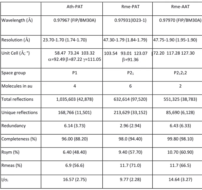
Table 1: data collection statistics
Ath-PAT Rme-PAT Rme-AAT
Wavelength (Å) 0.97967 (FIP/BM30A) 0.97931(ID23-1) 0.97970 (FIP/BM30A)
Resolution (Å) 23.70-1.70 (1.74-1.70) 47.30-1.79 (1.84-1.79) 47.75-1.90 (1.95-1.90) Unit Cell (Å; °) 58.47 73.24 103.32 =92.49 =87.22 =111.05 103.54 93.01 123.07 = =91.36 72.20 117.28 127.30 Space group P1 P21 P21212 Molecules in au 4 6 2 Total reflections 1,035,603 (42,878) 632,614 (97,520) 551,325 (38,783) Unique reflections 168,766 (11,501) 213,629 (33,152) 85,690 (6,128) Redundancy 6.14 (3.73) 2.96 (2.94) 6.43 (6.33) Completeness (%) 96.00 (88.20) 98.0 (94.40) 99.80 (98.10) Rsym (%) 6.40 (48.40) 9.40 (57.70) 10.70 (60.90) Rmeas (%) 6.9 (56.6) 11.7 (71.0) 11.7 (66.5) I/I 16.57 (2.75) 9.77 (2.28) 14.64 (3.27)
Signature of prephenate competent 1AAT
14
CC1/2 99.9 (82.9) 99.5 (72.9) 99.8 (86.9)
Rsym=∑ ∑|𝐼∑ ∑ 𝐼i−𝐼m|
i , where Ii is the intensity of the measured reflection and Im is the mean intensity of this
reflection. Values indicated in parentheses correspond to the statistics in the highest resolution shell.
Table 2: Refinement statistics
Ath-PAT Rme-PAT Rme-AAT
Resolution (Å) 23.70-1.70 (1.72-1.70) 47.30- 1.79 (1.81-1.79) 47.75-1.90 (1.92-1.90) Rcryst (F=0) (%) 17.23 (26.58) 17.44 (29.39) 15.25 (21.96) Rfree (F=0) (%) 20.12 (25.87) 20.87 (33.67) 18.07 (25.83) Number of atoms 13,825 19,872 7,127 Water molecules 1,232 1,616 851 B average (Å2) 31.0 29.0 21.0 Rmsd bonds (Å) 0.006 0.007 0.006 Rmsd angle (°) 1.075 0.876 0.859 Ramachandran statistics
Residues in allowed zones (%) 99.8 99.8 100.0
Residues in disallowed zones (%) 0.0 0.0 0.0
Values indicated in parentheses correspond to the statistics in the highest resolution shell.
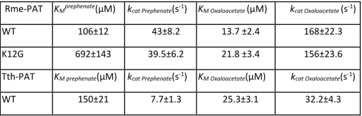
Table 3: kinetics analyses of 1b AAT Wild Type and K12G mutants from R. meliloti (Q02635) and
T. thermophilus (Q56232).
Rme-PAT KMprephenate(µM) kcat Prephenate(s-1) KM Oxaloacetate (µM) kcat Oxaloacetate (s-1)
WT 106±12 43±8.2 13.7 ±2.4 168±22.3
K12G 692±143 39.5±6.2 21.8 ±3.4 156±23.6
Tth-PAT KM prephenate(µM) kcat Prephenate(s-1) KM Oxaloacetate(µM) kcat Oxaloacetate(s-1)
Signature of prephenate competent 1AAT
15
K12G 2323±250* 5.2±2.5* 26.4±2.8 12.3±1.7
Values given are the average of at least three independent determinations from a single enzyme
preparation ± SD. *Note that K12G mutant kinetic apparent parameters KM and kcat for prephenate were difficult to determine accurately due to the important deterioration of the apparent KM.
Table 4: primers used for amplification of Rhizobium meliloti (Q02635), Thermus thermophilus (Q56232) and Synechochoccus Q3AK33 1AAT, and primers used for directed mutagenesis of K12G mutants. Enzyme Primer 5’ Rme-PAT CAGGAAACACCATATGGCCTTCCTTGC Rme-PAT K12G CCCGTGTAGGGCCCTCCGCCACC Tth-PAT CCCGCGTATACTTAGCATATGCGCGGCCTTT Tth-PAT K12G GGTCCTAGGCCATGGGGCCCTCGGCCACG Sco-PAT TTGCATATGCCGCGCCCGCCAGAACTTTCAGACCGAGCTG Enzyme Primer 3’ Rme-PAT CATGTCGGTCTGCTCGAGAATTATCTGC Rme-PAT(GPSAT) GGTGGCGGAGGGCCCTACACGGG Tth-PAT CCCCAGGAAAACGTCGACTAGGCGCGCCCCA Tth-PAT(GPSAT) CGTGGCCGAGGGCCCCATGGCCTGGACC Sco-PAT GAAGTGATGAGCTCGGATCAAAAGGAACGAAGCAGCCGCTTC Figures Legends
Figure 1. Tyrosine biosynthesis pathway. The alternative routes for the synthesis of tyrosine are
represented. Synthesis of phenylalanine from arogenate occurring in plants is also represented. Multiple enzymatic steps involved are shown by dotted lines. ADH: arogenate dehydrogenase; ADT: arogenate dehydratase; E4P: erythrose 4-phosphate; PAT: prephenate aminotransferase, PDH: prephenate dehydrogenase; PEP: phosphoenolpyruvate; TYR-AT: tyrosine aminotransferase
Signature of prephenate competent 1AAT
16
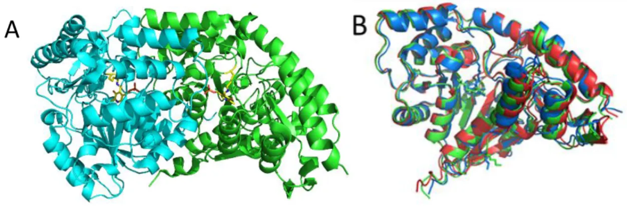
Figure 2: Three dimensional structure comparison of PAT competent and incompetent 1AAT.
A- 3D structure of A. thaliana PAT competent 1AAT dimer with PLP in yellow sticks. B- Superimposition of 3D structures of a monomer of A. thaliana PAT competent 1AAT (green), and R. meliloti PAT competent 1AAT (blue) and R. meliloti PAT incompetent 1AAT (red).
Figure 3: Docking of prephenate in the active site of Thermus thermophilus PAT competent 1AAT
(PDB id 1BKG)
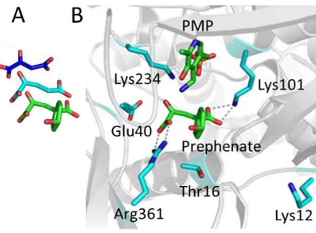
A Structure of the three ketoacid substrates of PAT competent 1 AAT, oxaloacetate (PDB ID 3PDB) (blue), ketoglutarate (build from prephenate) (cyan), and prephenate (green).
B Initial docking of prephenate in the active site of Tth-PAT (PDB id 1BKG). Prephenate was inserted manually in the active site of 1BKG in place of maleate and the resulting structure energy minimized. Then a short 1 ns unrestrained MD simulation was run. The structure giving the highest interaction energy between prephenate or PMP and the protein is displayed. The two carboxylate groups of prephenate interact with Lys101 and Arg361, and its keto group points in direction of the NH2 of PMP and the catalytic Lys234. In this position, the hydroxyl group of prephenate faces a 10AMKPSA15 amino-acid motif of the N-terminal flexible loop. In this motif Lys12 is only present in PAT competent enzymes. In the maleate-PMP-Enzyme ternary complex (1BKG), the side chain of Lys12 points outside the active site (10.5 Å from the prephenate OH group). In this simulation E40 and T16 were close to the active site but not in interaction with either PMP or prephenate.
Figure 4: N-terminal sequence comparison of PAT competent (PAT) and PAT incompetent (AAT) 1β AAT. Arabidopsis thaliana (Q42522): A. thal PAT, Sorghum bicolor (C5YYD7): S. bico PAT, Thermus thermophiles (Q56232): T. ther PAT, Chlorobium.tepidum (Q8KDS8): C. tepi PAT, Rhodobacter sphaeroides (A3PMF8): R. spha PAT, Rhizobium meliloti (Q02635): R. meli PAT, Rhizobium meliloti (P58350): R. meli AAT, Streptomyces avermitilis (Q82DR2): S. aver AAT and Synechocystis (Q55128): Synech AAT.
Figure 5: Analysis of recombinant Synechochoccus Q3AK33 1AAT. A: N-terminal sequence alignment between Synechochoccus Q3AK33 and Synechocystis Q55128 1AAT. B: SDS-PAGE of recombinante Sco_Q3AK33 Q sepharose purification: 1 total protein extract 20 µg; 2: Soluble protein extract 20 µg; 3 most active fraction eluted from a Qsepharose column 10 µg. C: Kinetic characterization of Synechochoccus Q3AK33 1AAT. Glutamate 25mM was used as amino donor. Values given are the average of at least three independent determinations from a single enzyme preparation ± SD.
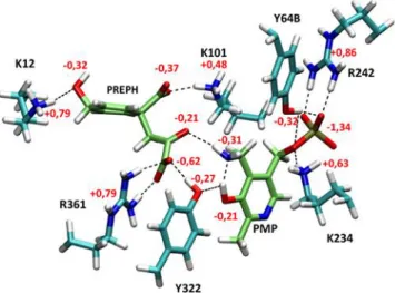
Figure 6: View of the QM active site around prephenate and PMP after QM/MM optimization
with Gaussian/Tinker. QM waters, W125, S127 and MM atoms are omitted for clarity. Hydrogen bonds between protein and ligands are indicated by black dashes. Partial charges of the different functional groups are indicated (PREPH, PMP and Tyr hydroxyls, PREPH carboxylates and carbonyl, Lys ammoniums, Arg guadinidium).
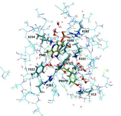
Figure 7: View of QM and QM/MM parts of the QM/MM simulation in an orientation close to Fig. 3. The QM part (shown in licorice) includes several residues around the active site with PREPH and PMP (shown in Fig. 5) plus W125 and S127 side chains and 14 water molecules shown in balls and sticks. All other represented atoms are MM atoms belonging to residues within a distance of 3 Å of the QM part and allowed to relax during geometry optimizations, while all other atoms of the enzyme were kept frozen.
Signature of prephenate competent 1AAT
17
Figure8: Evolution of dK-Prf vs. time during a constant Pressure and Temperature dynamics. The simulation starts with the old set of charges and NOE restraints until 20 ns (S0). Then, charges were modified according to QM/MM parameterization and the simulation was restarted for 4 ns. Finally, all NOE restraints were removed for the remaining 20 ns (24 to 44 ns).
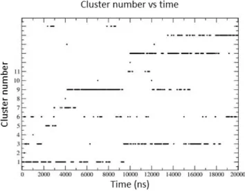
Figure 9: Evolution of cluster population versus time, following 16 20 ns RXSGLD simulations. Some clusters like cluster 1 or 7 appear at the beginning of the simulation and then disappear while others show up after 14 ns like cluster 15. Others like 2, 4, 8, 10, 12 or 14 appear only transiently. Clusters 1, 3, 6, 9, 13 and 15 are the most populated (>= 5%).
Figure 10: Evolution of dK-Prf vs. time during constant Pressure and Temperature dynamics simulations on selected clusters. The Noe restraints were removed after 5 ns. (Traces were smoothed by moving averages over a 20 point window). Most simulations show a rapid increase of dK-Prf just after the release of the restraints (5 ns). Although longer simulation times would be necessary to follow each of them, cluster 6 shows a decrease of dK-Prf down to less than 4 Å, already after 22 ns. During the 85 ns simulation of this cluster dK-Prf presents very large fluctuations from 2.6 to more than 21.6 Å.
Figure 11: Superimposition of the two structures with largest difference of dK-Prf (Lys12 NZ / prephenate O1 oxygen distance) in the RXSGLD simulation of cluster 6. Structure with dK-Prf= 2.6 Å is shown in cyan and structure with dK-Prf= 21.6 Å is shown in green. The conformational change of Lys12 is clearly visible (red arrow).
Figure 12: Molecular Phylogenetic analysis of 1AAT by Maximum Likelihood method
The evolutionary history was inferred by using the Maximum Likelihood method based on the JTT matrix-based model. The tree with the highest log likelihood (-16448,6813) is shown. Initial tree(s) for the heuristic search were obtained by applying the Neighbor-Joining method to a matrix of pairwise distances estimated using a JTT model. The tree is drawn to scale, with branch lengths measured in the number of substitutions per site. The analysis involved 36 amino acid sequences. All positions containing gaps and missing data were eliminated. There were a total of 361 positions in the final dataset. Evolutionary analyses were conducted in MEGA6. Nature of the amino acid residue in position 12 (according to Thermus thermophilus 1AAT) is represented.
Figure 13: SDSPAGE of the partial purification of recombinant Rhizobium meliloti 1AAT PAT (Q02635) wild type and K12G mutant.
MW: molecular weight; T: total protein extract; S: soluble protein extract; Q: Q sepharose pool; S200: S200 pool.
Figure 14: SDS-PAGE of EMD DEAE partial purification of recombinant Thermus thermophilus 1AAT Q56232 wild type and K12G mutant: T: total protein extract 20 µg; S: Soluble protein extract 20 µg. Arrows indicate the most active fraction used for kinetic analysis.
Signature of prephenate competent 1AAT
18
Figure 1. Tyrosine biosynthesis pathway. The alternative routes for the synthesis of tyrosine are
represented. Synthesis of phenylalanine from arogenate occurring in plants is also represented. Multiple enzymatic steps involved are shown by dotted lines. ADH: arogenate dehydrogenase; ADT: arogenate dehydratase; E4P: erythrose 4-phosphate; PAT: prephenate aminotransferase, PDH: prephenate dehydrogenase; PEP: phosphoenolpyruvate; TYR-AT: tyrosine aminotransferase
Figure 2: Three dimensional structure comparison of PAT competent and incompetent 1AAT.
A- 3D structure of A.thaliana PAT competent 1AAT dimer with PLP in stick. B- Superimposition of 3D structures of a monomer of A. thaliana PAT competent 1AAT (green), and R. meliloti PAT competent 1AAT (blue) and R. meliloti PAT incompetent 1AAT (red).