HAL Id: hal-00373744
https://hal.archives-ouvertes.fr/hal-00373744
Submitted on 7 Apr 2009HAL is a multi-disciplinary open access archive for the deposit and dissemination of sci-entific research documents, whether they are pub-lished or not. The documents may come from teaching and research institutions in France or abroad, or from public or private research centers.
L’archive ouverte pluridisciplinaire HAL, est destinée au dépôt et à la diffusion de documents scientifiques de niveau recherche, publiés ou non, émanant des établissements d’enseignement et de recherche français ou étrangers, des laboratoires publics ou privés.
Cyclic peptides bearing a side-chain tail: A tool to
model the structure and reactivity of protein zinc sites.
Olivier Sénèque, Emilie Bourlès, Vincent Lebrun, Estelle Bonnet, Pascal
Dumy, Jean-Marc Latour
To cite this version:
Olivier Sénèque, Emilie Bourlès, Vincent Lebrun, Estelle Bonnet, Pascal Dumy, et al.. Cyclic peptides bearing a side-chain tail: A tool to model the structure and reactivity of protein zinc sites.. Angewandte Chemie International Edition, Wiley-VCH Verlag, 2008, 47 (36), pp.6888-6891. �10.1002/anie.200800677�. �hal-00373744�
Cyclic Peptides Bearing a Side-Chain Tail: A Tool to Model the Structure
and Reactivity of Protein Zinc Sites
Olivier Sénèque,a
∗
Emilie Bourlès,a Vincent Lebrun,b Estelle Bonnet,a Pascal Dumy,c Jean-Marc Latourb∗
a) Laboratoire de Chimie et Biologie des Métaux, CNRS UMR 5249, 17, rue des Martyrs, 38054 Grenoble (France), Fax: (+33) 4 38 78 34 62.
b) CEA, iRTSV, LCBM, 17, rue des Martyrs, 38054 Grenoble (France).
c) Département de Chimie Moléculaire, UMR CNRS-UJF 5250, 301 Rue de la Chimie, Université Joseph Fourier, BP 53 38041 Grenoble Cedex 9 (France)
E-mail: olivier.seneque@cea.fr; jean-marc.latour@cea.fr
A recent bioinformatic study has evaluated to about 1000 (or ca 3 % of the total protein number) the number of human proteins possessing a tetracysteinate zinc site.[1] These sites were initially supposed to have a structural role since they were associated to zinc finger proteins[2] where they fold the protein chain in a conformation suitable to its binding to DNA. They were later found in several proteins and enzymes involved in demethylation processes such as the DNA repair protein Ada[3] and various transferases.[4] More recently, such sites were discovered in the heat shock protein Hsp33,[5] and the disulfide reductase Trx2[6] where their interaction with reactive oxygen species (ROS) contributes to the oxidative stress response. This is of special interest since tetracysteinate zinc sites, especially in zinc finger proteins, have been considered to be likely targets of ROS. Free cysteines are commonly involved in peroxide sensing and response[7] and their reactivity has been thoroughly studied over the past twenty years. A reasonable reactivity picture has emerged that points to the importance of hydrogen-bonding to increase the nucleophilic character of the cysteine sulfur. No such rationale is available for metal-bound cysteinates.
In order to get a better understanding of the reactivity of tetracysteinate zinc sites with ROS, we are developing a biomimetic approach based on de novo peptide synthesis. This approach is particularly suited to mimicking these sites and the potentially important hydrogen-bonding interactions, which is not possible with metalloorganic complexes in organic solvents. The validity of this approach has been demonstrated by Berg et al. in their modeling studies of zinc finger proteins with a mixture of cysteinate and histidine ligands[8] and further highlighted more recently by Gibney et
al.[9] Both groups used linear 16- to 26-mer peptides incorporating two CXnC (n = 2-4) zinc binding motif. Regan et al.[10] relied on self-assembling peptides to constitute a four-helix bundle orienting the cysteinates in the proper way to bind zinc. Nevertheless, this approach is generally weakened by the difficulty to obtain detailed structural characterization of metallopeptides. In addition, these two designs cannot reproduce the tetracysteinate arrangements that belong to β-hairpins such as that of Hsp33.[5] This prompted us to develop a totally new design based on introducing one CXnC motif into
a cyclic peptide and another one into a linear chain connected to the cycle via a glutamate or a lysine. In this communication, we show that this peptide design, with limited size and flexibility, allows to reproduce almost perfectly both the structure and the reactivity of the tetracysteinate zinc site of the protein Hsp33.
Figure 1. Model peptides design. Part of the crystallographic structure of Thermotoga maritima Hsp33[5] showing the Zn(Cys)4 site (upper left), its schematic representation (upper right) and the model peptides L1, L2
and L3 (bottom). Some of the amino-acids were changed in the models to prevent overlapping in the NMR spectra.
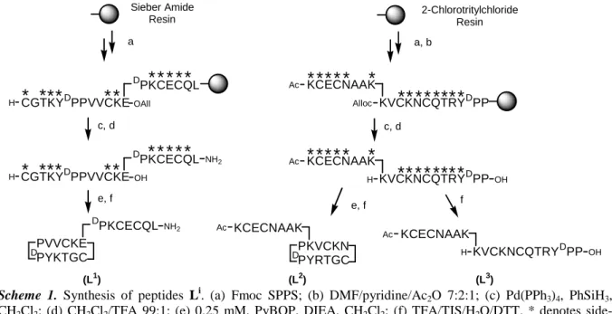
Figure 1 illustrates the tetracysteinate zinc site of Hsp33 that consists of a CXXC motif C263KWC266) located in a β-hairpin loop and a CXC motif (C231DC233). To reproduce the topology of this site, we designed a cyclic peptide to mimic the β-hairpin loop[11] and a linear tail was grafted on one of the side-chains of the loop to introduce the CXC motif. The D-Pro-L-Pro dipeptide template was used to preorganize the ten residues constituting the β-hairpin loop.[11] The linear tail was grafted on a Glu residue of the cycle via its N-end (peptide L1) or on a Lys residue via its C-end (peptide L2). The linear precursor of L1 was assembled on Sieber amide resin using Fmoc chemistry. Fmoc-Glu-OAll was used to create the branching point between the two parts of the peptide (scheme 1). After selective removal of the allyl group with Pd0, cleavage from the resin with 1% trifluoroacetic acid (TFA) in CH2Cl2 yielded a linear peptide amidated at the C-terminal leucine and still bearing the side-chain protecting groups. Cyclization between the only two unprotected functions (main side-chain Glu-COOH and Cys-NH2) in CH2Cl2 followed by removal of the side-chain protecting groups by TFA/tri-isopropylsilane (TIS)/H2O/dithiotreitol (DTT) and HPLC purification yielded L
1
. The peptide L2 was synthesized in a similar way on 2-chlorotrityl chloride resin using Alloc-Lys(Fmoc)-OH to introduce the branching point. To assess the value of the design described above, L3, the linear analogue of L2, was synthesized by suppressing the cyclization step. All peptides were identified by ESI/MS and 1H NMR.
DPKCECQL CGTKYDPPVVCKEOAll
H
*
*
*
*
*
*
*
*
*
c, d e, f DPKCECQL PVVCKE PYKTGC D a Sieber Amide Resin NH2 NH2 OH*
*
D PKCECQL CGTKYDPPVVCKE H*
*
*
*
*
*
*
*
*
*
*
(L1) KCECNAAK Alloc**
*
*
*
*
*
*
*
c, d e, f PKVCKN PYRTGC D a, b 2-Chlorotritylchloride Resin OH*
*
(L2) KVCKNCQTRY*
DPP*
*
Ac KCECNAAK H**
*
*
*
*
*
*
*
*
*
KVCKNCQTRY*
DPP*
*
Ac KCECNAAK Ac f OH KCECNAAK H KVCKNCQTRYDPP Ac (L3)Scheme 1. Synthesis of peptides Li. (a) Fmoc SPPS; (b) DMF/pyridine/Ac2O 7:2:1; (c) Pd(PPh3)4, PhSiH3,
CH2Cl2; (d) CH2Cl2/TFA 99:1; (e) 0.25 mM, PyBOP, DIEA, CH2Cl2; (f) TFA/TIS/H2O/DTT. * denotes
side-chain protecting groups.
Metal binding was investigated by UV/Vis, CD and fluorescence titrations at pH 7.0. Titrations with Co2+ show the formation of a unique 1:1 complex Co·Li for each of the three peptides. The LMCT and d-d transitions of these complexes are consistent with a Co2+ ion coordinated by four cysteinates in a tetrahedral geometry (Table 1).[12] The d-d transition patterns of the three Co·Li complexes differ quite significantly but the spectra of Co·L2 and Co·Hsp33[13] are strikingly similar (Figure S1). With Zn2+, the three peptides exhibit distinct behaviors. L2 forms only a 1:1 complex (Zn·L2) while L3 forms also a complex of undefined stoichiometry in excess of zinc, and L1 forms also a 1:2 Zn·(L1)2 complex, detected during CD titration in conditions of excess peptide. The LMCT band of the Zn·Li complexes around 210 nm indicates that the metal is coordinated by the four thiolates.[14] The apparent binding constants are 109.9(2) and 108.6(2) at pH 7.0 for Co·L2 and Co·L3, respectively, and are 1016.3(2) and 1015.2(2) for Zn·L2 and Zn·L3, respectively, at pH 7.5. Thus, the complexes formed by the cyclic peptide L2 are more stable than those of its linear analogue L3 by approximately one order of magnitude. The value measured for Zn·L2 is very similar to that reported for Zn·Hsp33 (1016.6) at pH 7.5.[15]
Table 1. UV-Vis characterizations of Co2+ and Zn2+ complexes of peptides Li at pH 7.0. Complex λ (∆ε) / nm (M-1cm-1) Co·L1 Co·L2 Co·L3 302 (6460), 360 (3980), 645 (630), 685 (835), 726 (930) 300 (4270), 347 (3200), 635 (369), 686 (769), 722 (697) 310 (4080), 356 (2860), 617 (408), 680 (570), 741 (407) Zn·L1 Zn·L2 Zn·L3 213 (19600) 204 (23700) 211 (20600)
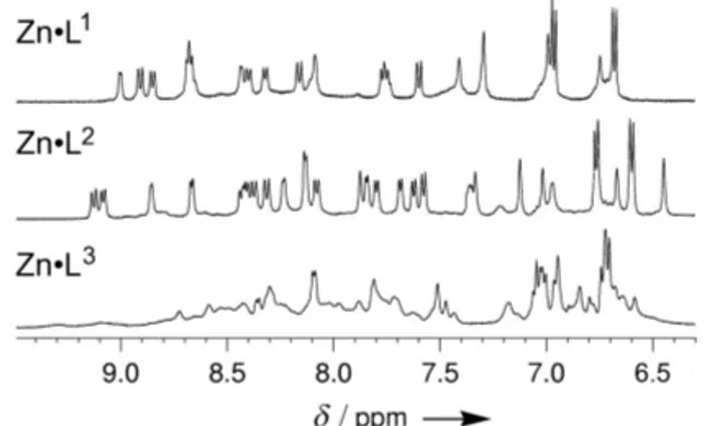
The structures of the zinc complexes were investigated by 1H NMR spectrometry. The 1H NMR spectra of complexes Zn·Li are depicted in figure 2. Whereas Zn·L1 and Zn·L2 display well
resolved spectra, Zn·L3 displays broad peaks characteristic of conformational motion. Thus only the cyclic peptide-based models Zn·L1 and Zn·L2 are suitable for a detailed structure determination. Their NH resonances are spread over the 7-9.5 ppm range, most of the 3JNH,Hα values are out of the 6-8 Hz range and a large number of NOE cross peaks are observed between the cycle and the tail (Supplementary information). This clearly indicates that Zn·L1 and Zn·L2 adopt a well defined conformation. The numerous cross-strand NOEs show that the cycle folds in a regular β-hairpin.[16] The NOE pattern of the four cysteines and the 3JHα,Hβ values show that the positions of the cysteine side-chains are well defined.[17] The structure was calculated using the program XPLOR 3.851[18] with 247 H-H distances constraints (81 intra-residue, 78 sequential and 88 medium-range) extracted from the NOESY spectra (200 ms) and 12 φ and 3 χ1 dihedral constraints for Zn·L
1
and with 255 H-H distances constraints (91 intra-residue, 66 sequential and 98 medium-range) and 12 φ and 4 χ1 dihedral constraints for Zn·L2. The superposition of the lowest energy structures with the zinc site of
Thermotoga maritima Hsp33 are depicted in figure 3. Both model peptides perfectly reproduce the
hairpin loop (the backbone root mean square deviation (r.m.s.d.) is 0.57 Å and 0.40 Å for Zn·L1 and Zn·L2 respectively). The linear tail of Zn·L1 is slightly tilted from the corresponding sequence in Zn·Hsp33 whereas a perfect match is noted for Zn·L2, with exactly the same relative orientation for the four cysteines side-chains. All of the seven hydrogen bonds involving the cysteine sulfurs in the X-Ray structure of Hsp33 are reproduced in the model compound Zn·L2 whereas Zn·L1 lacks one.
Figure 2. NH and aromatic region of 1H NMR spectra (500 MHz, 298 K, H2O/D2O 9:1, pH 6.2) of complexes
Figure 3. NMR Solution structures of Zn·L1 and Zn·L2. Left: Superposition of the 10 lowest energy NMR structures calculated using XPLOR. Middle: Lowest energy structure showing the hydrogen bond network (black). Right: Superposition of the lowest energy structure of Zn·Li (green) with the zinc site of Thermotoga maritima Hsp33 (pdb 1VQ0, blue).
In a preliminary reactivity study with ROS, we investigated the oxidation of the three Zn·Li complexes with H2O2. The reaction of Zn·L
i
with excess H2O2 was followed by ESI/MS, UV/Vis and fluorescence. Zn·Li, LiSS and Li2SS (the peptides presenting one and two disulfides, respectively) were observed by ESI/MS, the latter being the only species at the end of the reaction. Tyrosine fluorescence was recorded as a function of time (figure 4). The decrease of the fluorescence correlates with the decrease of the LMCT band at ca 210 nm monitored by UV/Vis. Fluorescence kinetic measurements allowed us to monitor the destruction of the Zn(Cys)4 centre and the formation of disulfides which are efficient quenchers of tyrosine fluorescence.[19] The kinetic traces were perfectly fitted with a single exponential. The apparent pseudo-first order constant kobs is proportional to [H2O2]. Therefore, the oxidation follows a second order kinetic law r = k×[Zn·Li]×[H2O2] showing that the rate determining step corresponds to the bimolecular reaction of Zn·Li with H2O2, with a highly ordered transition state as shown by the large negative value of ∆S≠ derived from Eyring plots (table 2). Interestingly, the kinetic constants at 303 K and 316 K for the best structural model Zn·L2 are in excellent agreement with those deduced from the data reported for Zn·Hsp33.[20] Moreover, it seems that the oxidation kinetics parallel the structural ordering of the zinc site. With one missing hydrogen bond, Zn·L1 is oxidized more rapidly than Zn·L2 and consistently, the fastest oxidation is observed for Zn·L3, which has no defined conformation and thus no defined hydrogen bond network.
Table 2. Kinetic parameters for the oxidation of Zn·Li by H2O2. Zn·L1 Zn·L2 Zn·L3 Zn·Hsp33[a] k303K / M-1 s-1 0.20 (1) 0.13 (1) 0.57 (2) 0.12 (1) k316K / M-1 s-1 0.45 (2) 0.28 (1) 1.2 (1) 0.31 (3) ∆H≠ / kJ mol-1 47.8 (5) 43.1 (5) 45.1 (5) - ∆S≠ / J mol-1 K-1 –101 (2) –120 (2) –101 (2) -
[a] Kinetic constants were obtained from ref. [20] by converting half-life reaction time of Zn·Hsp33 with 2 mM H2O2 using the
formula k = (ln2/t1/2)/[H2O2].
Figure 4. Normalized tyrosine fluorescence changes (λex = 280 nm, λem = 307 nm) observed during reaction of Zn·L2 (20 µM) with excess H2O2 at 298 K, pH 7.0. The numbers denotes the H2O2 concentrations in mM. The
inset shows the kobs obtained by fitting each kinetic trace with a single exponential as a function of [H2O2].
In summary, we have devised a new design based on branched cyclic peptides to mimic the tetracysteinate-zinc sites belonging to a β-hairpin such as the Zn(Cys)4 site of Hsp33. This is achieved with short peptides (≈ 20 amino-acids) which are easy to synthesize. Moreover their limited size allows a rapid NMR structural characterization, what can be scarcely achieved with linear peptides or multi-helix bundles. The differences between L2 and L3 highlight the interest of this design over the use of linear peptides for the modelling of structural properties and reactivity. The comparison between L1 and L2 shows that small changes in the structure and in the H-bonding pattern of theses complexes can influence their reactivity. The oxidation of these Zn(Cys)4 sites in condition of severe H2O2 stress (100µM-5mM) is very slow with half-life reaction times of several minutes, suggesting that H2O2 might not be the ROS actually responsible for the oxidation in vivo. We are further exploring the reactivity of these promising models towards H2O2 and other ROS.
Acknowledgments: The authors thank Mrs C. Lebrun for access to ESI-MS spectrometer and the Agence Nationale de la Recherche (ANR-06-JCJC-0018) for financial support.
Keywords: bioinorganic chemistry · cysteines · NMR spectroscopy · peptides · zinc
[1] C. Andreini, L. Banci, I. Bertini, A. Rosato, J. Proteome Res. 2006, 5, 196. [2] J. M. Berg, Curr. Op. Struct. Biol. 1993, 3, 11.
[3] H. Takinowaki, Y. Matsuda, T. Yoshida, Y. Kobayashi, T. Ohkubo, Protein Sci. 2006, 15, 487. [4] J. Penner-Hahn, Curr. Op. Chem. Biol. 2007, 11, 166.
[5] I. Janda, Y. Devedjiev, U. Derewenda, Z. Dauter, J. Bielnicki, D. R. Cooper, P. C. F. Graf, A. Joachimiak, U. Jakob, Z. S. Derewenda, Structure 2004, 12, 1901.
[6] J. F. Collet, J. C. D'Souza, U. Jakob, J. C. A. Bardwell, J. Biol. Chem. 2003, 278, 45325. [7] L. B. Poole, Arch. Biochem. Biophys. 2005, 433, 240.
[8] Y. G. Shi, J. M. Berg, Chem. Biol. 1995, 2, 83.
[9] A. K. Petros, A. R. Reddi, M. L. Kennedy, A. G. Hyslop, B. R. Gibney, Inorg. Chem. 2006, 45, 9941.
[10] L. Regan, N. D. Clarke, Biochemistry 1990, 29, 10878.
[11] M. Favre, K. Moehle, L. Y. Jiang, B. Pfeiffer, J. A. Robinson, J. Am. Chem. Soc. 1999, 121, 2679.
[12] A. B. P. Lever, Inorganic Electronic Spectroscopy, Elsevier, Amsterdam, 1984. [13] U. Jakob, M. Eser, J. C. A. Bardwell, J. Biol. Chem. 2000, 275, 38302.
[14] M. Vasak, J. H. R. Kagi, H. A. O. Hill, Biochemistry 1981, 20, 2852. [15] Jakob et al. measured a binding constant KZnHps33 of 10
17.4
for ZnHsp33 at pH 7.5 by competition experiments with TPEN. The binding constant they used for the ZnTPEN complex was the absolute binding constant (β11 = 10
16
). The apparent binding constant for ZnTPEN can be calculated by taking into account the protonation constants of TPEN. The actual value is 1015.2 at pH 7.5. Thus, KZnHps33 is rather 10
16.6
at pH 7.5.
[16] K. Wüthrich, NMR of Proteins and Nucleic Acids, Wiley, New York, 1986.
[17] G. Wagner, W. Braun, T. F. Havel, T. Schaumann, N. Go, K. Wuthrich, J. Mol. Biol. 1987, 196, 611.
[18] A. Brünger, A system for X-ray Crystallography and NMR. X-PLOR, version 3.1, Yale University Press, New Haven, CT, 1992.
[19] H. Szmacinski, W. Wiczk, M. N. Fishman, P. S. Eis, J. R. Lakowicz, M. L. Johnson, Eur.
Biophys. J. 1996, 24, 185.
[20] M. Ilbert, J. Horst, S. Ahrens, J. Winter, P. C. F. Graf, H. Lilie, U. Jakob, Nat. Struct. Mol. Biol. 2007, 14, 556.
![Figure 1. Model peptides design. Part of the crystallographic structure of Thermotoga maritima Hsp33 [5]](https://thumb-eu.123doks.com/thumbv2/123doknet/13428012.408573/3.892.280.624.238.493/figure-model-peptides-design-crystallographic-structure-thermotoga-maritima.webp)