Early enteral feeding in conservatively managed stage II
necrotizing enterocolitis is associated with a reduced risk
of catheter-related sepsis
Barbara Brotschi
1, Oskar Baenziger
1, Bernhard
Frey
1, Hans Ulrich Bucher
2and Jo¨rg Ersch
1,*
1
Department of Intensive Care and Neonatology,
University Children’s Hospital, Zurich, Switzerland
2
Division of Neonatology, Department of Obstetrics and
Gynecology, University Hospital Zurich, Switzerland
Abstract
Aims: To compare the effect of fasting period duration
on complication rates in neonates managed
conserva-tively for necrotizing enterocolitis (NEC) Bell stage II.
Methods: We conducted a multicenter study to analyze
retrospectively multiple data collected by standardized
questionnaire on all admissions for NEC between
Janu-ary 2000 and December 2006. NEC was staged using
modified Bell criteria. We divided the conservatively
man-aged neonates with NEC Bell stage II into two groups
(those fasted for -5 days and those fasted for )5 days)
and compared the complication rates.
Results: Of the 47 conservatively managed neonates
Bell stage II, 30 (64%) fasted for -5 days (range
1–4 days) and 17 (36%) for )5 days (range 6–16 days).
There were no significant differences for any of the
patient characteristics analyzed. One (3%) and four
(24%) neonates, respectively, developed post-NEC
bow-el stricture. One (3%) and two neonates (12%) suffered
NEC relapse. None and five (29%) neonates developed
catheter-related sepsis.
Conclusion: Shorter fasting after NEC appears to lower
morbidity after the acute phase of the disease. In
partic-ular, shorter-fasted neonates have significantly less
cath-eter-related sepsis. We found no benefit in longer fasting.
Keywords: Catheter-related sepsis; intestinal stricture;
necrotizing enterocolitis (NEC); neonate; nutrition.
*Corresponding author:Jo¨rg Ersch, MD
Department of Intensive Care and Neonatology University Children’s Hospital
Steinwiesstr. 75 CH-8032 Zurich Switzerland Tel.: q41 44 266 70 76 Fax: q41 44 266 71 68 E-mail: [email protected]
Introduction
Necrotizing enterocolitis (NEC) is the most common
acquired intraabdominal emergency in infants w10x and
the most common surgical emergency in the neonatal
intensive care unit (NICU) w11x. Incidences range from
3% to 28%, averaging 6%–10% in infants with birth
weights -1500 g, and incurring a mortality of 10%–30%
w
7x. Although the precise cause remains unclear,
incrim-inated factors include impaired immunity of the immature
gut, enteral feeding, and bacterial gut colonization,
although no specific pathogen has been identified w12,
16x.
Treatment of sonographically and radiologically
con-firmed NEC consists of nasogastric decompression,
anti-biotic therapy, total parenteral nutrition (TPN), and
surgery when necessary. There is no standard
recom-mendation for when to reinitiate enteral feeding in
non-surgically treated infants. Textbooks recommend resting
the bowel for 7–10 days w3, 7x. However, the scientific
basis for relatively prolonged fasting is unclear w13x. The
issue of fasting period duration is particularly important
in a NICU such as ours that manages most of its NEC
cases conservatively. We therefore conducted a
multi-center comparison of fasting period duration in
conser-vatively managed NEC Bell stage II to determine its effect
on complications such as bowel stricture, NEC relapse,
and catheter-related sepsis.
Patients and methods
We conducted a retrospective analysis of all term and preterm neonates with NEC (ns124) admitted to five Swiss NICUs between January 2000 and December 2006. All NICUs were tertiary centers, well equipped for managing sick neonates with pediatric surgical support. We used data collected by standard-ized questionnaire. Three NICU questionnaires were filled out by the same person in the study group and the other two by a NICU neonatologist. The questionnaires incorporated the inter-national definition of NEC and the Bell stages w2x. They covered the following items: fasting period duration (some NICUs fol-lowed a fixed 5-day regime, others a fixed 10-day regime, but all often tailored the period according to their individual experi-ence wfasting period range: 4–14 daysx and modified Bell criteria w8x); gestational age; birth weight; sex; antenatal steroids; cause of prematurity; umbilical catheterization; patent ductus
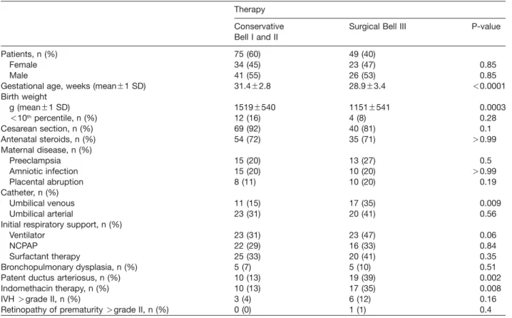
arterio-Table 1 Patient characteristics (ns124).
Therapy
Conservative Surgical Bell III P-value
Bell I and II
Patients, n (%) 75 (60) 49 (40)
Female 34 (45) 23 (47) 0.85
Male 41 (55) 26 (53) 0.85
Gestational age, weeks (mean"1 SD) 31.4"2.8 28.9"3.4 -0.0001
Birth weight g (mean"1 SD) 1519"540 1151"541 0.0003 -10thpercentile, n (%) 12 (16) 4 (8) 0.28 Cesarean section, n (%) 69 (92) 40 (81) 0.1 Antenatal steroids, n (%) 54 (72) 35 (71) )0.99 Maternal disease, n (%) Preeclampsia 15 (20) 13 (27) 0.5 Amniotic infection 15 (20) 10 (20) )0.99 Placental abruption 8 (11) 10 (20) 0.19 Catheter, n (%) Umbilical venous 11 (15) 17 (35) 0.009 Umbilical arterial 23 (31) 20 (41) 0.56
Initial respiratory support, n (%)
Ventilator 23 (31) 23 (47) 0.06
NCPAP 22 (29) 16 (33) 0.84
Surfactant therapy 25 (33) 20 (41) 0.35
Bronchopulmonary dysplasia, n (%) 5 (7) 5 (10) 0.51
Patent ductus arteriosus, n (%) 10 (13) 19 (39) 0.002
Indomethacin therapy, n (%) 10 (13) 17 (35) 0.008
IVH )grade II, n (%) 3 (4) 6 (12) 0.16
Retinopathy of prematurity )grade II, n (%) 0 (0) 1 (1) 0.4
nsnumber, SDsstandard deviation, NCPAPsnasal continuous positive airways pressure, IVHsintraventricular hemorrhage. sus; initial ventilatory support; surfactant replacement;
indo-methacin therapy; and concomitant disease (bronchopulmonary dysplasia, retinopathy of prematurity, intraventricular hemor-rhage (IVH), and periventricular leukomalacia). Retinopathy of prematurity and IVH were defined as significant if )grade II w9–11, 15, 17, 20x. Patent ductus arteriosus based on echocar-diologic criteria qualified only if it required treatment (indometh-acin or surgery). Diagnostic data for NEC related to clinical signs, cultures (blood and stool), and imaging (X-ray). Therapy and complication variables included circulatory and/or ventilatory support during the acute phase of NEC, surgery versus conser-vative management, antibiotic therapy, TPN duration, short bow-el syndrome, bowbow-el stricture (defined as significant if suspected clinically, confirmed radiographically and requiring surgery in the neonatal period), NEC relapse, and catheter-related sepsis. Catheter-related sepsis was diagnosed using the Centers for Disease Control criteria: clinical signs of sepsis, one positive peripheral blood culture, and negative urine and tracheal aspi-rate cultures w14x.
All neonates included in Bell stage II showed the same clas-sical clinical and radiologic signs. For that reason all neonates with radiologic pneumatosis intestinalis and/or portal venous gas were staged Bell II. Neonates with metabolic acidosis (pH-7.2), receiving inotropes or mechanical ventilation were staged Bell III. The retrospective design made subtle distinction between Bell stages IIa and IIb impossible.
Since conservatively managed neonates in Bell stage II were our primary focus, we subdivided the infants concerned into
those fasted for -5 days and those fasted for )5 days to permit comparison of complication rates, primarily bowel stricture, NEC relapse, and catheter-related sepsis. We chose the 5-day cut-off because it was half the duration of the longest standard regime (10 days) in any NICU. We focused on neonates in Bell stage II, because the criteria of Bell stage I (suspected NEC) are quite vague, with the potential for inconsistent reporting and classification. We also excluded Bell stage III neonates because all required surgery.
After different fasting periods in the individual NICUs, the same feeding algorithm was implemented. It comprised 10 mL/kg body weight (BW) solution by gastric tube in 12 or 8 por-tions/d on day 1 (D1), 10 mL/kg BW breast milk or formula milk on D2, increasing by 20 mL/kg BW/d to 140–150 mL/kg BW/d. Statistical analysis: we analyzed normally distributed contin-uous variables using the unpaired t-test and categorical variables using Fisher’s exact test.
Results
The total population of 124 infants with Bell stage I–III
NEC included five term and 119 preterm neonates
(-32 weeks, ns89; G32 weeks, ns30). Over the 7-year
study period, the five NICUs admitted 2005 inborn
pre-term neonates aged -32 weeks, giving an incidence of
NEC of 4.4% in this age group. Treatment was
conser-Table 2 Patient characteristics of conservatively managed necrotizing enterocolitis Bell stage II.
Fasting period
-5 days )5 days P-value
Patients, n (%) 30 (64) 17 (36)
Female 12 (40) 7 (41) )0.999
Male 18 (60) 10 (59) )0.999
Gestational age, weeks (mean"1 SD) 32.0"2.8 31.7"3.0 0.77
Birth weight g (mean"1 SD) 1717"560 1545"535 0.3 -10thpercentile, n (%) 3 (10) 0 0.29 Cesarean section, n (%) 28 (93) 16 (94) )0.999 Antenatal steroids, n (%) 24 (80) 12 (71) 0.49 Maternal disease, n (%) Preeclampsia 5 (17) 5 (29) 0.46 Amniotic infection 6 (20) 4 (24) )0.999 Placental abruption 5 (17) 2 (12) )0.999
Initial respiratory support, n (%)
Ventilator 8 (27) 6 (35) 0.74 NCPAP 8 (27) 4 (24) )0.999 Surfactant therapy 7 (23) 5 (29) 0.73 Bronchopulmonary dysplasia, n (%) 1 (3) 1 (6) )0.999 Catheter, n (%) Umbilical venous 4 (13) 3 (18) 0.68 Umbilical arterial 9 (30) 6 (35) 0.75
Patent ductus arteriosus, n (%) 5 (17) 2 (12) )0.999
Indomethacin therapy, n (%) 5 (17) 2 (12) )0.999
IVH )grade II, n (%) 1 (3) 0 )0.999
Retinopathy of prematurity )grade II, n (%) 1 (3) 1 (6) )0.999
Age at NEC onset, days (mean"1 SD) 8.6"7.8 9.4"9.0 0.74
Positive blood cultures at NEC diagnosis, n (%) 2 (7) 3 (18) 0.34
Blood in stool, n (%) 30 (100) 10 (59) 0.0003
Pneumatosis intestinalis, n (%) 30 (100) 17 (100) )0.999
TPN administered, days (mean"1 SD) 14.9"4.0 18.5"5.9 0.02
nsnumber, NCPAPsnasal continuous positive airways pressure, NECsnecrotizing enterocolitis, SDsstandard deviation, IVHs intraventricular hemorrhage, TPNstotal parenteral nutrition.
Table 3 Complications in conservatively managed necrotizing
enterocolitis Bell stage II.
Fasting period
-5 days )5 days P-value Catheter-related sepsis, n (%) 0 5 (29) 0.004 Early post-NEC stricture, n (%) 1 (3) 4 (24) 0.05
NEC relapse, n (%) 1 (3) 2 (12) 0.27
nsnumber, NECsnecrotizing enterocolitis.
vative in 75/124 neonates (60.5%) and surgical in 49
(39.5%). Surgically treated neonates were more seriously
ill and of lower gestational age (Table 1).
Of the 75 conservatively managed neonates, 28 (37%)
were classified as Bell stage I and 47 (63%) as Bell stage
II. Of the neonates classified as Bell stage II, 30 (64%)
fasted for -5 days (range 1–5 days; NICU1 ns28,
NICU2 ns2, NICU3–5 ns0) and 17 (36%) for )5 days
(range 6–16 days; NICU1 ns3, NICU2 ns7, NICU3
ns4, NICU4 ns3, NICU5 ns0). All were preterm except
for two and one neonates, respectively. Characteristics
of the groups did not differ (Table 2) except for ‘‘blood in
stool’’, which was less frequent in neonates with fasting
period )5 days (Ps0.0003). Ages at NEC symptom
onset (mean, SD) were 8.6"7.8 and 9.4"9.0 days
(Ps0.74). TPN was administered for 14.9"4.0 and
18.5"5.9 days (Ps0.02). Neonates in the -5 days
group needed 14.9"4.0 (range 8–25) days of fasting
until initiation of full enteral nutrition vs. 18.5"5.9 (range
10–36) days needed by those in the )5 days group
(Ps0.02).
Complications (Table 3)
None and five neonates (29%; NICU1 ns1, NICU2 ns2,
NICU3 ns2, NICU4/5 ns0) developed catheter-related
sepsis, a single episode in all cases (Ps0.004). Blood
cultures
yielded coagulase-negative Staphylococcus
(ns5). One (3%) and four neonates (24%) required
sur-gery for early post-NEC stricture (Ps0.05); all had
evi-dence of ileus with vomiting, abdominal distension, and
confirmatory radiology. One (3%) and two neonates
(12%) relapsed (Ps0.27), with symptom onset ranging
from 3 to 4 weeks after the initial episode; one was
treat-ed conservatively, the other two surgically. Stricture and
relapse rates tended to be lower in the shorter-fasted
group. No neonate developed short bowel syndrome and
none died.
Discussion
Despite many studies of enteral feeding before the onset
of NEC w18x, data are lacking on the timing of enteral
re-feeding after NEC, hence the absence of consensus
guidelines. A 1975 report associated relapse in ‘‘several
patients’’ with the reintroduction of enteral feeding
-10 days after NEC w8x. Textbooks offer the
unsubstan-tiated recommendation to rest the bowel for 7–10 days
w
3, 7x. However, a 2003 study concluded that early
enteral feeding after NEC was associated with significant
benefits and no apparent adverse effects w5x. Early
re-feeding may have a healing effect or, at least, helps
recovery of the intestinal mucosa. Local trophic nutrients
support and stimulate mucosal growth and may thus be
protective. In contrast, in the absence of enteral feeding,
mucosal villous atrophy may occur within one week,
pre-disposing to recurrent intestinal injury and feeding
intol-erance w4, 12x.
Our analysis of five Swiss neonatal units mirrors the
variability in the literature over post-NEC re-feeding.
Although each unit practices the same re-feeding regime,
they differ in their belief as to when re-feeding should
start, in particular, in conservatively managed stage II
NEC. Whereas one unit has already managed to
signifi-cantly shorten the fasting period, the other units, despite
the same severity of NEC, are still equivocal over the
introduction of advanced re-feeding. Individual
experi-ence is still much too often at work. This raises the
ques-tions: When is the right time to re-feed in stage II NEC
and are long fasting periods actually counterproductive?
Our retrospective study shows that early enteral
feed-ing in conservatively managed NEC stage II is associated
with less catheter-related sepsis, which may be related
to the shorter requirement for TPN, but our data also
reveal no adverse effects of shorter fasting. On the
con-trary, the rates of bowel stricture (3% vs. 24%) showed
only borderline significance and relapse (3% vs. 12%)
tended to be lower than in longer-fasted neonates.
Assuming an NEC relapse rate of roughly 6% (for
con-servatively managed neonates) in this study, 356 patients
would be required per treatment arm to have a modest
chance of detecting a doubling of the NEC relapse rate
(80% power, at 0.05 significance level). With only 17
patients in one of the treatment arms, this study was
underpowered for detecting this clinically important
outcome (it was only powered to detect a difference
in relapse rates of 6% vs. 45%).
Early stricture, within the neonatal period, is quite a
common complication of NEC. It occurs in conservatively
managed neonates as well as in those treated surgically,
and is presumably due to post-ischemic scarring w1x.
Treatment is always surgical. No specific (including
bac-teriological) criteria have succeeded in predicting which
patients are at risk of stricture after conservative
man-agement w6x. The incidences in our study, namely 3%
and 24%, were somewhat lower than the 16%–37%
reported in the literature w6, 19x.
The incidence of NEC in our overall population of
pre-term neonates -32 weeks, 4.4%, was within the
report-ed range w3, 7, 12x. Similarly, our relapse rates in
conservatively managed neonates, 3% and 12%, were
also consistent with the literature w3, 9x despite potential
inconsistencies in reporting this parameter: The five
NICUs differed in their interpretation of certain signs (e.g.,
blood in the stool), and did not always classify them as
relapse.
Despite our study’s limitations, e.g., its retrospective
design, our observations suggest that early enteral
feed-ing after conservatively managed NEC stage II may lower
morbidity after the acute phase of the disease. In
partic-ular, neonates fasting for a shorter period had
signifi-cantly less catheter-related sepsis. We failed to identify
any benefit in longer fasting.
Acknowledgements
We thank M. Lu¨cking-Famira, C. Buehrer and G. Zeilinger for making this survey possible and providing all the data.
References
w1x Bell MJ, Ternberg JL, Askin FB. Intestinal stricture in nec-rotizing enterocolitis. J Pediatr Surg. 1976;11:319–27. w2x Bell MJ, Ternberg JL, Feigin RD, Keating JP, Marshall R,
Barton L, et al. Neonatal necrotizing enterocolitis: thera-peutic decisions based upon clinical staging. Ann Surg. 1978;187:1–7.
w3x Berseth CL, Poenaru D. Necrotizing enterocolitis and short bowel syndrome. In: Taevsch HW, Ballard RA, Gleason CA, editors. Avery’s Diseases of the Newborn. Philadelphia: Elsevier Saunders; 2005. p. 1123–33.
w4x Bertolo RF, Chen CZ, Pencharz PB, Ball RO. Intestinal atrophy has a greater impact on nitrogen metabolism than liver by-pass in piglets fed identical diets via gastric, cen-tral venous or portal venous routes. J Nutr. 1999;129: 1045–52.
w5x Bohnhorst B, Mu¨ller S, Do¨rdelmann M, Peter CS, Petersen C, Poets CF. Early feeding after necrotizing enterocolitis in preterm infants. J Pediatr. 2003;143:484–7.
w6x Bu¨tter A, Flageole H, Laberge JM. The changing face of surgical indications for necrotizing enterocolitis. J Pediatr Surg. 2002;37:496–9.
w7x Caplan M. Neonatal necrotizing enterocolitis. In: Martin RJ, Fanaroff AA, Walsh MC, editors. Neonatal-Perinatal
Med-icine (Diseases of the Fetus and Infant). Philadelphia: Else-vier Mosby; 2006. p. 1403–10.
w8x Frantz ID, L’Heureux P, Engel RR, Hunt CE. Necrotizing enterocolitis. J Pediatr. 1975;86:259–63.
w9x Henry MC, Moss RL. Current issues in the management of necrotizing enterocolitis. Semin Perinatol. 2004;28:221– 33.
w10x Kafetzis DA, Skevaki C, Costalos C. Neonatal necrotizing enterocolitis: an overview. Curr Opin Infect Dis. 2003;16: 349–55.
w11x Kliegman RM. Necrotizing enterocolitis. N Engl J Med. 1984;310:1093–103.
w12x Kliegman RM, Walker WA, Yolken RH. Necrotizing ente-rocolitis: research agenda for a disease of unknown etiol-ogy and pathogenesis. Pediatr Res. 1993;34:701–8. w13x Motil KJ. Necrotizing enterocolitis. In: McMillan JA,
De-Angelis CD, Feigin RD, Warshaw JB, editors. Oski’s Pedi-atrics: Principles and Practice. Philadelphia: Lippincott Williams & Wilkins; 1999. p. 325–32.
w14x O’Grady NP, Alexander M, Dellinger EP, Gerberding JL, Heard SO, Maki DG, et al. Guidelines for the prevention of intravascular catheter-related infections. Centers for dis-ease control and prevention. MMWR Recomm Rep. 2002; 51(RR-10):1–29.
w15x Papile LA, Burstein J, Burstein R, Koffler H. Incidence and evolution of subependymal and intraventricular
hemor-rhage: a study of infants with birth weights less than 1500 g. J Pediatr. 1978;92:529–34.
w16x Peter CS, Feuerhahn M, Bohnhorst B, Schlaud M, Ziesing S, von der Hardt H, et al. Necrotizing enterocolitis: is there a relationship to specific pathogens? Eur J Pediatr. 1999; 158:67–70.
w17x Phelps DL. Retinopathy of prematurity. Pediatr Rev. 1995; 16:50–6.
w18x Pietz J, Achanti B, Lilien L, Stepka EC, Mehta SK. Preven-tion of necrotizing enterocolitis in preterm infants: a 20-year experience. Pediatrics. 2007;119:164–70.
w19x Schwartz MZ, Hayden CK, Richardson CJ, Toyson KR, Lobe TE. A prospective evaluation of intestinal stenosis following necrotizing enterocolitis. J Pediatr Surg. 1982; 17:764–70.
w20x The Committee for the Classification of Retinopathy of Prematurity. An international classification of retinopathy of prematurity. Arch Ophthalmol. 1984;102:1130–4.
The authors stated that there are no conflicts of interest regard-ing the publication of this article.
Received November 1, 2008. Revised June 5, 2009. Accepted July 20, 2009. Previously published online August 13, 2009.