HAL Id: inserm-00715876
https://www.hal.inserm.fr/inserm-00715876
Submitted on 5 Jul 2013
HAL is a multi-disciplinary open access
archive for the deposit and dissemination of sci-entific research documents, whether they are pub-lished or not. The documents may come from teaching and research institutions in France or abroad, or from public or private research centers.
L’archive ouverte pluridisciplinaire HAL, est destinée au dépôt et à la diffusion de documents scientifiques de niveau recherche, publiés ou non, émanant des établissements d’enseignement et de recherche français ou étrangers, des laboratoires publics ou privés.
Lipocalin 2, the TNF-like receptor TWEAKR and its
ligand TWEAK act downstream of NFAT1 to regulate
breast cancer cell invasion.
Benoît Gaudineau, Marjorie Fougère, Frédéric Guaddachi, Frédéric Lemoine,
Pierre de la Grange, Sébastien Jauliac
To cite this version:
Benoît Gaudineau, Marjorie Fougère, Frédéric Guaddachi, Frédéric Lemoine, Pierre de la Grange, et al.. Lipocalin 2, the TNF-like receptor TWEAKR and its ligand TWEAK act downstream of NFAT1 to regulate breast cancer cell invasion.: NFAT1/LCN2/TWEAKR/TWEAK regulate motility. Journal of Cell Science, Company of Biologists, 2012, 125 (Pt 19), pp.4475-86. �10.1242/jcs.099879�. �inserm-00715876�
1
Lipocalin 2 (LCN2), the TNF-like receptor TWEAKR and its
1
ligand TWEAK act downstream of NFAT1 to regulate breast
2
cancer cell invasion.
3 4
Benoît Gaudineau
1, Marjorie Fougère
1, Frédéric Guaddachi
1, Frédéric
5Lemoine
2, Pierre de la Grange
2and Sébastien Jauliac
1,*6 7
1CNRS UMR7212, INSERM U944, Université Paris Diderot, Institut d'Hématologie, Hôpital
8
Saint-Louis, 1 avenue Claude Vellefaux 75475 Paris Cedex 10, France. 2GENOSPLICE 9
TECHNOLOGY, Institut d'Hématologie, Centre Hayem, Hôpital Saint-Louis, 1 avenue 10
Claude Vellefaux 75475 Paris Cedex 10, France. 11
12
*
Author for correspondence (sebastien.jauliac@inserm.fr) 13
14
Running title: NFAT1/LCN2/TWEAKR/TWEAK regulate motility 15 16 Words count: 7442 17
Jo
u
rn
a
l
o
f
C
e
ll
Sci
e
n
ce
Acce
p
te
d
ma
n
u
scri
p
t
SUMMARY
18 19
NFAT1 is a transcription factor that elicits breast carcinoma cells to become invasive,
20
contributing thus to formation of metastasis. The molecular mechanisms by which
21
NFAT1 operates in this respect are still poorly known. Here, we report that NFAT1
22
increases Lipocalin 2 (LCN2) mRNA and protein expression by binding to specific sites
23
in the LCN2 gene promoter region. We show that the LCN2 protein is required
24
downstream of NFAT1 to increase breast cancer cell invasion. We demonstrate that the
25
NFAT1/LCN2 axis is sufficient to regulate expression of the TNF-like receptor
26
TWEAKR at the RNA level and of its ligand, TWEAK, at the protein level. We show,
27
however, that TWEAKR mediates an anti-invasive effect in breast cancer cells whereas,
28
depending on LCN2 expression, TWEAK has either anti- and pro-invasive capacities.
29
Thus, we identify LCN2 and TWEAKR/TWEAK as critical downstream effectors of
30
NFAT1 to regulate breast cancer cell motility and invasive capacity.
31 32 33
Jo
u
rn
a
l
o
f
C
e
ll
Sci
e
n
ce
Acce
p
te
d
ma
n
u
scri
p
t
Introduction
34
Metastases are the main cause of morbidity and mortality in breast cancer patients. 35
Their efficient dissemination is directly linked to the invasive behaviour of cancer cells 36
(Friedl and Wolf 2003), which requires the cells to destroy and reorganize the extra-cellular 37
matrix as well as the capacity to migrate. This is a complex process involving many players 38
among which NFAT transcription factors, especially NFAT1, are critical (Jauliac et al. 2002; 39
Yoeli-Lerner et al. 2005). 40
The family of NFAT transcription factors comprises five genes (NFAT1 to 5). NFAT1, 41
NFAT2, NFAT3 and NFAT4 were first identified as T cell transcription factors that bind IL-2 42
promoter after cell activation (Shaw et al. 1988), while NFAT5 is induced by osmotic stress 43
(Lopez-Rodriguez et al. 1999). Because of their role in critical signalling pathways that 44
control cell fate (Baksh et al. 2002; Chuvpilo et al. 2002; Xanthoudakis et al. 1996), one 45
would expect that disturbing NFAT signalling could impact carcinogenesis. Indeed, beside 46
their role in the migratory and invasive capacities of breast cancer cells (Fougère et al. 2010; 47
Jauliac et al. 2002; Yiu and Toker 2006; Yoeli-Lerner et al. 2005, Germann et al; 2012), there 48
is growing evidence for an active role of NFAT factors in carcinogenesis (Buchholz et al. 49
2006; Dejmek et al. 2006; Foldynová-Trantírková et al. 2010; Holzmann et al. 2004; Jönsson 50
et al. 2002). However, beside these reports of a “pro-cancer” role, others are showing the 51
“anti-cancer” capacities of NFAT factors (Glud et al. 2005; Lee et al. 2005; Wu et al. 2010). 52
In line with this duality of function, we have reported that NFAT3 is specifically expressed in 53
oestrogen receptor +-positive breast cancer cells and, contrary to NFAT1 and NFAT5, 54
inhibits these cells' motility by blocking LCN2 (Lipocalin 2) gene expression (Fougère et al. 55
2010). 56
LCN2 is a secreted protein of the Lipocalin family (Flower 1996), the high expression 57
of which is associated with malignancy in different types of cancers (Bartsch and Tschesche 58
1995; Furutani et al. 1998; Missiaglia et al. 2004; Nielsen et al. 1996), including breast cancer 59
(Stoesz et al. 1998), and is a predictor of breast cancer progression (Hu et al. 2008). Actually, 60
LCN2 regulates cell migration (Fougère et al. 2010; Leng et al. 2008; Playford et al. 2006) 61
and formation of metastases (Shi et al. 2008; Yang et al. 2009). LCN2 is induced under 62
diverse inflammatory conditions (Nielsen et al. 1996), and it is known that inflammation is 63
strongly linked to tumorigenesis (Balkwill and Mantovani 2001; Iliopoulos, Hirsch, and 64
Struhl 2009; Pierce et al. 2009) and metastasis formation. 65
Apart from LCN2, other inflammatory molecules, such as those of the 66
TWEAK/TWEAKR cytokine receptor axis, have been linked to cancer. TWEAK (TNF-67
related Weak Inducer of Apoptosis) belongs to the TNF family and is involved in many 68
diseases (Winkles 2008) including breast cancer (Willis et al. 2008). TWEAKR (TNF-related 69
Weak Inducer of Apoptosis Receptor, Fn14) expression has been observed in breast tumour 70
Jo
u
rn
a
l
o
f
C
e
ll
Sci
e
n
ce
Acce
p
te
d
ma
n
u
scri
p
t
samples, where it might regulate cancer cell migration (Michaelson et al. 2005; Willis et al. 71
2008). Indeed, the positive role of the TWEAK/TWEAKR axis on cancer cell migration has 72
been reported (Dai et al. 2009; Tran et al. 2006), but there are also reports that TWEAK 73
inhibits migration (Meighan-Mantha et al. 1999). Recently, a preclinical study has shown that 74
a humanized antibody directed against the TWEAKR can mediate anti-tumour effects by 75
signalling through TWEAKR (Culp et al. 2010). 76
Here, we report that signalling through NFAT1 up-regulates LCN2 expression by 77
NFAT1 direct binding on LCN2 promoter and that LCN2 is required for NFAT1 to foster 78
breast cancer cell invasion. Downstream of NFAT1 and LCN2, we identify the 79
TWEAKR/TWEAK axis as a key player for regulating the invasive process, and show that, 80
depending on LCN2 expression level, TWEAK displays both anti-invasive and pro-invasive 81
functions. Thus, we uncover a novel NFAT1/LCN2/TWEAKR/TWEAK axis critical to 82
regulate breast cancer cell invasion. 83 84
Jo
u
rn
a
l
o
f
C
e
ll
Sci
e
n
ce
Acce
p
te
d
ma
n
u
scri
p
t
Results
85 86
NFAT1 up-regulates LCN2 expression in breast cancer cells
87
Having recently shown that NFAT3 inhibits LCN2 transcription in breast cancer cells 88
(Fougère et al., 2010), we evaluated here the effect of NFAT1 on LCN2 expression. However, 89
since NFAT1 and LCN2 proteins are essentially not expressed in oestrogen receptor-positive 90
(ERA+) cancer cells (Fougère et al., 2010), this was done only using ERA-negative (ERA-) 91
cells in which, contrary to NFAT3, NFAT1 and LCN2 are highly expressed. First, we used 92
NFAT1-specific siRNA to silence endogenous NFAT1 in MDA-MB-231, which reduced its 93
expression by 90% (Fig. 1A, right panel) without affecting that of NFAT5 or NFAT4 (data 94
not shown). NFAT1 silencing (which did not result in cell apoptosis as assayed by annexin V 95
labelling data; not shown) reduced by 5-fold LCN2 mRNA expression relative to control cells 96
(Fig. 1A, left panel), and abrogated both intracellular (Fig. 1B, left panel) and secreted (Fig. 97
1B, middle panel) LCN2 protein expression. 98
We next examined whether transiently transfected ectopic NFAT1 affected LCN2 99
protein expression. Because only 10% of cell were actually transfected, empty vector control 100
(Fig. 1B, right panel, vector) and NFAT1 (Fig. 1B, right panel, NFAT1) were cotransfected 101
with a GFP-expressing vector in order to identify transfected cells. Thus, NFAT1 over-102
expression increased LCN2 protein expression relative to control cells as assessed by Western 103
blot analysis (Fig. 1B, right panel). 104
Thus, in contrast to NFAT3 (Fougère et al. 2010), NFAT1 up-regulates both LCN2 105
mRNA and protein expression in breast cancer cells. 106
107
Downstream of NFAT1, LCN2 is key to increase breast cancer cell invasion
108
LCN2 plays a critical role in regulating breast cancer cell motility (Fougère et al. 109
2010). In order to evaluate the physiological relevance of LCN2 modulation by NFAT1, we 110
tested whether LCN2 was involved in NFAT1-regulated chemoinvasion. Transiently 111
transfecting MDA-MB-231 cells with HA epitope-tagged LCN2 (Fig. 1C, left panel) 112
increased breast cancer cell chemoinvasion as compared with empty vector-transfected cells 113
(Fig. 1C, right panel), indicating LCN2 role in the process. To evaluate whether LCN2 114
expression downstream of NFAT1 was needed to regulate chemoinvasion, we transiently co-115
transfected the cells with T7 epitope-tagged NFAT1 together with either control or LCN2-116
targeted siRNA (Fig. 1D, right panel)and in combination with a betagalactosidase (!-gal) 117
Jo
u
rn
a
l
o
f
C
e
ll
Sci
e
n
ce
Acce
p
te
d
ma
n
u
scri
p
t
expressing vector. NFAT1 increased chemoinvasion in the presence of LCN2, whereas 118
silencing LCN2 precluded NFAT1 pro-invasive effect (si LCN2 / NFAT1), as compared with 119
cells transfected with both control and NFAT1 siRNAs (si ctrl / NFAT1), and was inhibitory 120
by itself (si LCN2) (Fig. 1D, left panel). These results indicate that LCN2 expression 121
downstream of NFAT1 is required to regulate breast cancer cell invasion. 122
123
NFAT1 binds to the LCN2 promoter region to up-regulate LCN2 expression
124
To more thoroughly analyse the relationship between LCN2 expression and NFAT1 125
signalling, we investigated the LCN2 promoter region where 6 potential NFAT-binding sites 126
(-881, -522, -501, -441, -409, -142) (Fig. 2A) have been identified (Fougère et al. 2010). 127
Using a Luciferase gene-fused LCN2 promoter, we found that NFAT1 ectopic expression 128
doubled the promoter activity relative to empty vector-transfected cells (Fig. 2B). To assess 129
whether endogenous NFAT1 interacted with LCN2 promoter region, chromatin 130
immunoprecipitation (ChIP) was performed in cells treated by control or NFAT1-specific 131
siRNAs: indeed, endogenous NFAT1 bound to LCN2 promoter whereas no significant signal 132
enrichment was noted when NFAT1 was silenced (Fig. 2C, si ctrl versus si NFAT1). To 133
confirm NFAT1 binding to LCN2 promoter region and determine whether NFAT1 putative 134
binding sites are functional in vivo, we individually mutated each of these. Measurement of 135
Luciferase activity after transient co-transfection of MDA-MB-231 cells with NFAT1 of 136
mutated LCN2 promoter revealed that the -409 binding site was required for NFAT1 to 137
increase transcriptional activity, the -501 and-142 sites being required for LCN2 promoter 138
basal activity (Fig. 2D). Electrophoretic mobility shift assays were then performed to confirm 139
that endogenous NFAT1 bound to these identified sites and, indeed, NFAT1 bound to the -140
501, -409, -142 wild-type, but not mutated, binding sites (Fig. 2E). NFAT1-specific binding 141
was confirmed by pre-incubating nuclear extracts with an anti-NFAT1 antibody that 142
supershifts the complex NFAT1/probe (Fig. 2E, arrows). As control, no supershift was 143
induced by pre-incubating nuclear extracts with control IgG. 144
These data demonstrate that endogenous NFAT1 binds directly to the LCN2 promoter 145
-501, -142 sites to modulate its basal expression, and to the -409 site to regulate its inducible 146
expression. 147
148
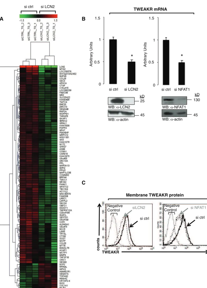
The NFAT1/LCN2 axis modulates TWEAKR expression
149
To assess the mechanisms by which the NFAT1/LCN2 axis up-regulates breast cancer 150
Jo
u
rn
a
l
o
f
C
e
ll
Sci
e
n
ce
Acce
p
te
d
ma
n
u
scri
p
t
cells in order to identify genes regulated by LCN2 potentially involved in the invasive 152
capacity. LCN2 silencing impacted 95 genes by at least 1.5 fold (95% confidence level) (Fig. 153
3A, and Supplementary Table 1), which was confirmed by RT-QPCR for most of these (data 154
not shown). Because we have shown that NFAT1 regulates LCN2 expression, we examined 155
whether modifying NFAT1 expression affected any of the 95 LCN2-regulated genes. Indeed, 156
siRNA silencing of NFAT1 reduced expression of two of them: TWEAKR (TNFSFR12A) 157
(Fig. 3B) and C6ORF55 (VTA1) (data not shown). Thus, out of the 95 identified genes that 158
putatively could be involved in the invasion process of breast cancer cells, only these two 159
were regulated in the same manner by NFAT1 and LCN2. Silencing VTA1 with siRNA had 160
no effect on cancer cell invasive capacity (data not shown). Because TWEAKR was already 161
known to participate in the motility of different cell types (Meighan-Mantha et al. 1999; 162
Willis et al. 2008) and was regulated in the same manner by NFAT1 and LCN2, we further 163
investigated this regulation of TWEAKR expression by LCN2 and NFAT1 and its role in the 164
invasive behaviour of breast cancer cells. Flow cytometry analysis confirmed that siRNA 165
silencing of either LCN2 or NFAT1 down-regulated TWEAKR protein expression in MDA-166
MB-231 (Fig. 3C) and SUM-159-PT (Supplementary Fig. S1) cells relative to control cells. 167
These data indicate that TWEAKR is regulated by NFAT1 and LCN2 at the mRNA 168
and protein levels in breast cancer cells. 169
170
Reciprocal regulation of the TWEAKR/TWEAK axis and LCN2 expression
171
In contrast to TWEAKR, silencing or overexpressing LCN2 in MDA-MB-231 cells 172
did not modify mRNA levels of its ligand, TWEAK (Supplementary Fig. S2A and B), which 173
was indeed not found among the 95 genes identified in Fig. 3A (see Supplementary Table 1). 174
Nonetheless, when LCN2 or NFAT1 were independently silenced with siRNA directed to 175
either, which reduced their expression by 90%, TWEAK protein levels in cell supernatants 176
increased relative to the control condition (Fig. 4A). To validate that TWEAK protein is 177
regulated by LCN2, cells were transiently transfected with a fixed amount of a TWEAK-178
expressing vector and increasing amounts of a LCN2-expressing vector, which resulted in 179
dose-dependent decrease of TWEAK levels (Fig. 4B). Moreover, TWEAKR depletion by 180
siRNA led to increased TWEAK protein levels without modifying its mRNA expression in 181
MDA-MB-231 and SUM-159-PT cells (Supplementary Fig. S3). 182
Because LCN2 regulated TWEAK protein amount, we verified whether TWEAK or 183
TWEAKR per se affected LCN2 protein levels by transiently transfecting MDA-MB-231 184
Jo
u
rn
a
l
o
f
C
e
ll
Sci
e
n
ce
Acce
p
te
d
ma
n
u
scri
p
t
cells with siRNA targeting TWEAKR or TWEAK. Indeed, silencing of either led to LCN2 185
level increase and this effect was cumulative (Fig. 4C). Moreover, TWEAK and TWEAKR 186
depletion up-regulated LCN2 mRNA expression (Fig. 4D), suggesting a transcriptional link. 187
To confirm this link, the cells were pre-treated by a transcriptional inhibitor (actinomycin D), 188
which inhibited LCN2 mRNA increase elicited by TWEAK and TWEAKR depletion. We 189
verified here that silencing TWEAKR and/or TWEAK did not modify cell apoptosis or 190
proliferation (Supplementary Fig. S4A). No effect of TWEAK and TWEAKR depletion on 191
NFAT1 transcriptional activity was noted, suggesting that other unidentified transcription 192
factors are involved in up-regulating LCN2 expression (data not shown). 193
Altogether, these results point to a novel axis by which LCN2 modulates TWEAKR at 194
the mRNA level and its cognate ligand TWEAK at the protein level, both of which 195
conversely regulate LCN2 expression at the transcriptional level. 196
197
TWEAK increases breast cancer cell invasion independently of the TWEAKR
198
TWEAKR signalling has already been implicated in the invasive process in different 199
cell models. To validate that TWEAKR was involved in the chemotactic invasive capacity of 200
MDA-MB-231 cells, chemoinvasion assays were performed in the presence or not of 201
recombinant TWEAK, whose presence was thus shown to increase invasion (Fig. 5A). 202
However, blocking TWEAKR with a specific neutralizing antibody also increased invasion as 203
compared to control IgG, and adding recombinant TWEAK potentiated this effect (Fig. 5A), 204
neither of which modifying cell apoptosis or proliferation (Supplementary Fig. S4B, C). 205
When we repeated the experiment in NFAT1-transfected cells, again pre-treatment with the 206
TWEAKR neutralising antibody enhanced chemoinvasion elicited by NFAT1 (Fig. 5B), 207
suggesting that TWEAKR mediates an anti-invasive effect in breast cancer cells in this assay. 208
As a confirmation, we repeated the experiment presented in Fig. 5A by using a siRNA 209
targeting TWEAKR. After verifying that the siRNA actually reduced TWEAKR expression 210
(Fig. 5C, right panel) and did not affect apoptosis or proliferation (Supplementary Fig. S4A), 211
we found again that chemoinvasion increased when TWEAKR was silenced and that this was 212
potentiated by adding recombinant TWEAK (Fig. 5C, left panel). 213
These findings were surprising in light of the report that, in breast cancer cells, 214
TWEAKR silencing led to loss of invasion potential (Willis et al. 2008). But in that case the 215
assay (Hauck et al. 2002; Hsia et al. 2003) assessed random cell invasion, no chemotactic 216
gradient being used, whereas ours is chemoinvasion assay using conditioned medium from 217
Jo
u
rn
a
l
o
f
C
e
ll
Sci
e
n
ce
Acce
p
te
d
ma
n
u
scri
p
t
NIH3T3 cells in order to create a chemotactic gradient (Albini and Benelli 2007). Therefore, 218
we examined if the different assays accounted for the discrepancy. To this end MDA-MB-231 219
cells, transiently transfected with either control or TWEAKR-specific siRNAs, were 220
comparatively assessed in both assays. Data from Fig. S5 indicate that cells were actually less 221
invasive and that TWEAKR silencing inhibited invasion in the radom invasion assay, whereas 222
increased invasion in presence was noted as expected in the chemoinvasion assay. Thus, 223
TWEAKR would differently influence random and directed cell invasion. 224
To evaluate the role of LCN2 in regulating invasion downstream of TWEAK, MDA- 225
MB-231 cells were transiently transfected with control or LCN2-specific siRNAs and 226
exposed to recombinant TWEAK. In this case, when LCN2 was silenced, TWEAK no longer 227
increased chemoinvasion (Fig. 5D). 228
These data identify TWEAKR as a receptor that antagonizes the chemotactic 229
invasion process of breast cancer cells. Importantly, they indicate that LCN2 is required for 230
TWEAK to increase chemotactic invasion, either directly or via another as yet unidentified 231
receptor different from TWEAKR. 232
233
Depending on LCN2 expression TWEAK displays either anti- and pro-invasive activities
234
in breast cancer cells
235
Inasmuch as TWEAK availability appeared to be regulated by TWEAKR, LCN2 and 236
NFAT1, we more precisely examined the role of TWEAK protein on breast cancer cell 237
invasion. MDA-MB-231 cells were transiently transfected with control siRNA or with 238
siRNAs targeting TWEAKR, LCN2, NFAT1 or TWEAK, alone or in combination (Fig. 6A). 239
As shown in Fig. 5C, TWEAKR silencing led to increased chemoinvasion, and either NFAT1 240
(Fig. 6A, si TWR si NFAT1) or LCN2 (Fig. 6A, si TWR si LCN2) depletion prevented this 241
increase, demonstrating that LCN2 increased expression elicited by TWEAKR knockdown 242
was responsible for this effect. Indeed, when NFAT1 was silenced, and by consequence 243
LCN2 too, again the increase of chemoinvasion induced by TWEAKR down-regulation was 244
abrogated (Fig. 6A, si TWR si NFAT1). Since we have shown that TWEAKR depletion 245
induces TWEAK protein up-regulation (Supplementary Fig. S3), we examined whether the 246
latter was involved in this increased invasion. Indeed, when TWEAK was silenced by siRNA 247
together with TWEAKR (Fig. 6A, si TWR si TW, -), increased invasion was abolished but it 248
was rescued by adding back recombinant TWEAK (Fig. 6A, si TWR si TW, +). The same 249
results were obtained with SUM-159-PT cells (Supplementary Fig. S6). 250
Jo
u
rn
a
l
o
f
C
e
ll
Sci
e
n
ce
Acce
p
te
d
ma
n
u
scri
p
t
These data indicate that TWEAK protein up-regulation is critical to increase invasion 251
and entails the presence of LCN2. 252
Interestingly, we have shown that, like TWEAKR down-regulation, that of either 253
LCN2 or NFAT1 induced increased TWEAK protein levels, albeit without increasing but 254
rather inhibiting chemoinvasion. Therefore, we hypothesized that, beside its pro-invasive role, 255
TWEAK might be anti-invasive in the absence of LCN2 or NFAT1. To clarify this 256
possibility, either LCN2 or NFAT1, alone or in combination with TWEAK, were silenced in 257
MDA-MB-231 cells. Depletion of endogenous LCN2 inhibited invasion (Fig. 6B, si LCN2) 258
and up-regulated TWEAK protein (from 27.4 to 73.3 ng/L; Fig. 6B lower panel). TWEAK 259
up-regulation was critical to blunt invasion when LCN2 was silenced since co-silencing 260
TWEAK with LCN2 abolished the inhibition of invasion induced by LCN2 knockdown (Fig. 261
6B, si LCN2 si TW, -), which could be rescued by adding recombinant TWEAK (Fig. 6B, si 262
LCN2 si TW, +). These results demonstrate that TWEAK, beside its pro-invasive role in the 263
presence of LCN2, possesses an anti-invasive function in the absence of LCN2. We repeated 264
the same experiment by depleting NFAT1 and obtained almost the same results as with LCN2 265
depletion (Fig. 6C), the sole difference being the lack of reversion of the inhibition of 266
invasion by depleting TWEAK and NFAT1 together (Fig. 6B, si NFAT1 si TW, -). This last 267
result is not surprising since NFAT1 might certainly target other genes with LCN2 and 268
TWEAK to regulate breast cancer cell chemoinvasion. Comparable data were obtained using 269
SUM-159-PT cells (Supplementary Fig. S6). 270
Altogether, these data demonstrate that, beside its pro-invasive action, TWEAK has an 271
anti-invasive action in breast cancer cells in the absence of LCN2. Thus, TWEAK is a pro-272
invasive factor in breast cancer cells in presence of LCN2 and anti-invasive in its absence. 273 274
Jo
u
rn
a
l
o
f
C
e
ll
Sci
e
n
ce
Acce
p
te
d
ma
n
u
scri
p
t
Discussion
275
The role of NFAT transcription factors in breast cancer cell motility has been 276
identified recently (Jauliac et al. 2002; Mancini and Toker 2009; Yoeli-Lerner et al. 2005) but 277
it is still poorly understood. Apart from studies indicating that either COX2 (Yiu and Toker 278
2006) or the autotaxin gene (Chen and O'Connor 2005) are targeted by NFAT1 to increase 279
invasion and migration, we do not know which other specific signalling pathways these 280
factors use to modulate breast cancer cell motility. 281
Recently, a new layer of complexity has been added by the observation that among the 282
members of the NFAT family, NFAT3 –expressed in less aggressive oestrogen receptor +-283
positive breast cancer cells– has anti-migratory and anti-invasive actions in contrast to 284
NFAT1 and NFAT5. One mechanism by which NFAT3 blunts migration is by inhibiting 285
LCN2 gene expression (Fougère et al. 2010). 286
Here, we show that LCN2 expression is required for NFAT1 to increase the invasive 287
capacity of breast cancer cells. We demonstrate that, contrary to NFAT3, NFAT1 binds to 288
LCN2 promoter to up-regulate LCN2 mRNA and, therefore, protein expression. Together 289
with our recent report (Fougère et al. 2010), this highlights the differing effects of NFAT 290
isotypes, despite their high sequence homology, which has already been indicated in specific 291
knockout murine models (Graef et al. 2001; Ranger et al. 1998; Xanthoudakis et al. 1996) or 292
by studies on tumour cell transformation (Buchholz et al. 2006; Neal and Clipstone 2003; 293
Robbs et al. 2008; Wu et al. 2010). These observations suggest that each NFAT isotype 294
regulates independent set of genes as well as, as demonstrated here for LCN2, differentially 295
modulates overlapping gene subsets critical for cell fate, and they underscore the idea that 296
depending on the cell type where they are expressed NFAT isotypes either suppress or 297
promote oncogenic transformation. 298
We also evaluated the mechanisms by which LCN2 modulates breast cancer cell 299
invasion downstream of NFAT1. To this end, we examined the genomic effects of LCN2 300
depletion in order to identify the genes that are similarly regulated by NFAT1 and LCN2. We 301
found that, among 95 genes affected by LCN2 down-regulation, two –TWEAKR 302
(TNFSFR12A) and C6ORF55 (VTA1)– were regulated in the same manner by NFAT1. We 303
focused on TWEAKR because this gene has already been implicated in the motility of 304
different cell types (Dai et al. 2009; Tran et al. 2006; Wiley et al. 2001), and silencing VTA1 305
had no effect in this respect. We found that, at both the mRNA and protein levels, either 306
NFAT1 or LCN2 down-regulation inhibited TWEAKR expression. We therefore reasoned 307
that TWEAKR disappearance was responsible for the inhibition of breast cancer cell invasion 308
Jo
u
rn
a
l
o
f
C
e
ll
Sci
e
n
ce
Acce
p
te
d
ma
n
u
scri
p
t
in light of reports showing its role in migration and invasion (Dai et al. 2009; Michaelson et 309
al. 2005; Tran et al. 2006; Willis et al. 2008). Indeed, treating the cells with recombinant 310
TWEAK elicited increase of their chemoinvasive capacity. Surprisingly, when we silenced 311
TWEAKR, invasion was increased and TWEAK could still potentiate this increase. We 312
showed that, in contrast to chemoinvasion, TWEAKR down-regulation inhibited random 313
invasion as reported (Willis et al., 2008). Therefore, we concluded that, in our chemoinvasion 314
model, TWEAKR was a receptor that mediated an anti-invasive effect and that its cognate 315
ligand, TWEAK, could promote invasion via another, as yet unidentified, receptor the 316
presence of which has already been suggested in RAW264.7 cells (Polek et al. 2003). Hence, 317
we hypothesised that TWEAK binding to its cognate receptor (TWEAKR) can inhibit 318
invasion and that blocking this association with a TWEAKR-specific neutralising antibody 319
enables TWEAK binding to the other receptor that promotes invasion, but it cannot be ruled 320
out that TWEAK directly enters the cells. Further studies are needed to identify this potential 321
second TWEAK receptor if it exists. We cannot rule out that NFAT1 and LCN2 participate in 322
the regulation of this potential unknown TWEAK receptor. 323
When we examined TWEAK expression in the absence of either LCN2 or NFAT1, we 324
found that it was up-regulated at the protein, but not the mRNA, level. This demonstrates that 325
NFAT1, via LCN2, regulates TWEAK protein expression or stability. In the same manner as 326
for LCN2, inhibiting or silencing TWEAKR elicited up-regulated TWEAK protein expression 327
or availability. This highlights the tight regulation between LCN2 and the 328
TWEAKR/TWEAK axis, all the more so that we found also that TWEAKR and TWEAK can 329
both modulate LCN2 expression at the mRNA and protein levels. Therefore, in breast cancer 330
cells, there exists a subtle equilibrium between LCN2, TWEAKR and TWEAK to modulate 331
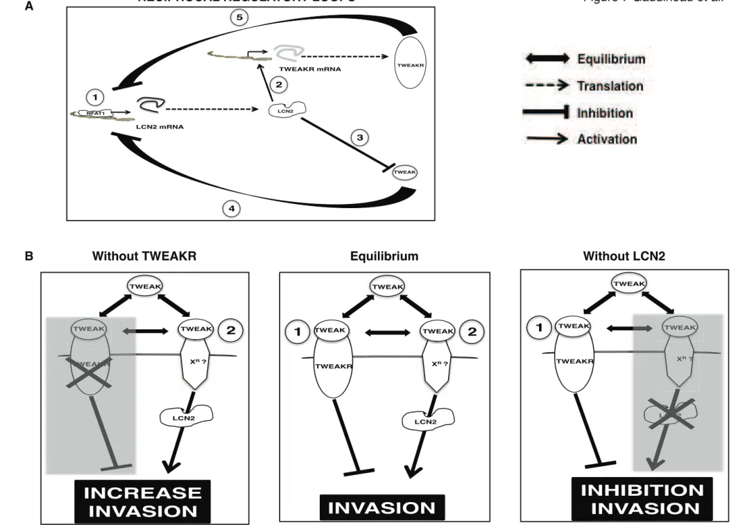
the availability of these key factors for the regulation of cell invasion (Fig. 7A). 332
Since we showed that adding recombinant TWEAK increased the cells' invasive 333
capacity, it was puzzling that elevated TWEAK levels induced by depleting either LCN2 or 334
NFAT1 correlated with a decrease rather than an increase of invasion. Indeed, our study 335
shows that presence of LCN2 is necessary for TWEAK to promote its pro-invasive effect 336
(Fig. 5D). Therefore, in the absence of LCN2, TWEAK can only signal by the TWEAKR, 337
and inhibits invasion (Fig. 7B, Without LCN2). Indeed, when TWEAKR is silenced, TWEAK 338
expression is required to increase invasion and needs the presence of LCN2 (Fig. 6A and 7B, 339
without TWEAKR). In contrast, in the absence of LCN2, when LCN2 is silenced, TWEAK is 340
an anti-invasive factor (Fig. 6B). Importantly, this indicates that TWEAK can be either pro-341
Jo
u
rn
a
l
o
f
C
e
ll
Sci
e
n
ce
Acce
p
te
d
ma
n
u
scri
p
t
Meighan-Mantha et al. 1999), but in our study these opposite effects occur in the same cells, 343
suggesting an equilibrium between the two functions (Fig. 7B, Equilibrium). Critically, we 344
show that LCN2 expression up-regulation by NFAT1 is necessary for TWEAK to increase 345
breast cancer cell invasion. Therefore, disturbing this tight equilibrium may be a new entry to 346
inhibit breast cancer cell invasion and ultimately metastasis formation. 347
Both LCN2 and TWEAK are able to activate the ERK pathway (Gwira et al. 2005; 348
Peternel et al. 2011; Vincent et al. 2009), ERK apparently being an important actor of breast 349
cancer cell migration (Irie et al. 2005; Krueger et al. 2001). One possibility would be that 350
promoting invasion downstream of TWEAK requires modulation of the ERK pathway in 351
association with LCN2, but this has yet to be determined. 352
In summary, we have shown that signalling through NFAT1 increases invasion 353
through a previously unknown LCN2/TWEAKR/TWEAK axis. We demonstrate that 354
TWEAK can have opposite effects on breast cancer cell invasion: anti-invasive via 355
TWEAKR, independently of LCN2 (Fig.7B, Without LCN2); pro-invasive via another 356
unidentified receptor (XR) or directly in association with LCN2 (Fig. 7B, Without TWEAKR). 357
Therefore, increased NF A T1 activation leads to up-regulation of LCN2 protein expression 358
that consequently enables TWEAK pro-invasive activity in breast cancer cells. These 359
findings underscore the importance of dissecting the mechanisms by which TWEAK and 360
LCN2 regulate breast cancer cell invasion, downstream of NFAT1, in order to be able to 361
attempt to therapeutically target this pathway to limit the dissemination of metastasis. 362 363 364
Jo
u
rn
a
l
o
f
C
e
ll
Sci
e
n
ce
Acce
p
te
d
ma
n
u
scri
p
t
Materials and Methods
365 366
Cell culture
367
The MDA-MB-231 cell line was from the American Type Culture Collection, The 368
SUM-159-PT cell line was provided by Alex Toker (Harvard Medical School). MDA-MB-369
231 and SUM-159-PT cells were maintained in Dulbecco Modified Eagle Medium (DMEM), 370
low glucose (1 g/L D-glucose), 10% Foetal Calf Serum. NIH-3T3 cells were maintained in 371
DMEM, high glucose (4.5 g/L D-glucose), 10% Newborn Calf Serum. All media were 372
supplemented with 2 mM L-Glutamine, 100 U/mL Penicillin and 100 μg/mL Streptomycin. 373
374
Antibodies and reagents
375
The following antibodies were used: Anti-T7 (Novagen; #69522), anti-FLAG (Sigma; 376
#F1804), anti-HA (Roche; #11867423), anti-NFAT1 from ABR Affinity BioReagent (MAl-377
025) or from Abcam for ChIP assays (ab2722), anti-Actin (Santa-Cruz Biotechnology; SC-378
1616), anti-LCN2 (Sigma-Aldrich; HPA002695), and anti-TWEAKR (Abcam; ab21359) for 379
flow cytometry. The mouse IgG was from Santa-Cruz Biotechnology (SC-2025) and the anti-380
mouse phycoerythrin-labelled antibody (F0102B) was from R&D systems. The blocking anti-381
TWEAKR antibody was from R&D systems (#AF1199). 382
The protein G Plus/Protein A Agarose suspension was from Calbiochem (IP05). 383
Recombinant human TWEAK (#1090TW/CF) was from R&D Systems. The ELISA used to 384
assess TWEAK (#DY1090) and LCN2 (#DLCN20) were from R&D systems. SiRNA were 385
from Dharmacon and their sequences are reported in Supplemental Table 2. Cells were 386
transiently transfected with the appropriate plasmids using Lipofectamine 2000 (Invitrogen) 387
or DharmaFECT (Dharmacon) for siRNA according to the manufacturer’s instructions. 388
389
Plasmids, siRNA and quantitative real time reverse transcriptase PCR (RT-QPCR)
390
NFAT1 was cloned by PCR in the pcDNA3 vector expressing a N-terminal T7-tag. 391
LCN2 promoter and the pCS4-(n)-,-galactosidase were previously described (Fougère et al. 392
2010). The potential NFAT-binding sites of LCN2 promoter were mutated by PCR using the 393
primers described in Supplementaray Table 2. LCN2 was cloned by PCR in the pcDNA3.1+ 394
vector with a HA tag from RNA isolated from MDA-MB-231 cells. The FLAG-TWEAK 395
expression vector was from Origene. All constructions were verified by sequencing. To 396
transiently silence TWEAKR, TWEAK, NFAT1 or LCN2, we used specific siRNAs 397
Jo
u
rn
a
l
o
f
C
e
ll
Sci
e
n
ce
Acce
p
te
d
ma
n
u
scri
p
t
(Dharmacon) at 30 nM, and transfected cells with DharmaFECT for 48 hr. Sequences of the 398
different siRNAs used are accessible in the Supplementary Table 2. 399
LCN2, TWEAKR and TWEAK mRNA expression was determined by RT-QPCR. 400
Total RNA was extracted using the RNeasy mini kit (QIAGEN) according to the 401
manufacturer’s instructions, and cDNA synthesis was prepared using the SuperScript Reverse 402
Transcriptase (RT) (Invitrogen). RT-QPCR was performed using SYBR Green PCR Master 403
Mix on Roche LightCycler as directed by the manufacturer. Cycling parameters were 10 404
minutes at 95°C followed by 45 cycles of 10 seconds at 95°C (denaturation) and 4 seconds at 405
68°C (annealing/extension). Primers are accessible in the Supplementary Information, 406 Supplementary Table 2. 407 408 Proteins detection 409
For all assays using transfected cells, expression or silencing of the protein were 410
verified by immunoblotting. Harvested cells were washed twice and resuspended in cold PBS. 411
Cells were lysed in SDS sample buffer containing ,-mercaptoethanol for 20 minutes at 95°C. 412
The lysates were resolved by SDS-PAGE and immunoblotted with the appropriate antibodies. 413
For flow cytometry, MDA-MB-231 or SUM-159-PT cells were stained with an anti-414
TWEAKR antibody and a secondary phycoerythrin-labelled antibody. As controls, MDA-415
MB-231 or SUM-159-PT cells were stained only by the secondary phycoerythrin-labelled 416
antibody, omitting the anti-TWEAKR antibody. Analyses were performed on a FACSCalibur 417
flow cytometer (Becton Dickinson). 418
419
ELISA for assessing LCN2 and TWEAK
420
The ELISA for LCN2 was performed as directed by the manufacturer on 50 -l of cell 421
supernatants. For TWEAK, cell supernatants were concentrated using the Centricon 422
(Millipore) prior to performing the ELISA as directed by the manufacturer on 50 μl of 423
concentrated supernatant. 424
425
Invasion assays (chemoinvasion and random invasion assays)
426
The chemoinvasion assay was performed essentially as described, using Transwell chambers 427
(Becton Dickinson) with 8--m pore membranes coated with Matrigel (Becton Dickinson). 428
Cells, co-transfected with the relevant expression plasmids and the pCS2-(n)-,-gal reporter 429
plasmid, were resupsended after 24 hours in serum-free medium containing 0.1% BSA, and 430
Jo
u
rn
a
l
o
f
C
e
ll
Sci
e
n
ce
Acce
p
te
d
ma
n
u
scri
p
t
cells were added to each well. Conditioned NIH-3T3 medium was added to the bottom wells 431
of the chambers. After 6 hours, cells that had not invaded were removed from the upper face 432
of the filters using cotton swabs, and cells that had invaded to the lower surface of the filters 433
were fixed for 30 min in 4% paraformaldehyde and then stained with PBS containing 1 mg/ml 434
bluo-gal, 2 mM MgCl2, 5 mM potassium ferrocyanide, and 5 mM potassium ferricyanide. 435
All cells in each Transwell were counted. All the counts were normalised by the efficiency of 436
transfection. The numbers of cells that invaded in each condition were compared with 437
the empty vector-transfected condition arbitrary set as a ratio of 1. When the assay was 438
performed with cells transiently transfected by siRNA, cells were stained with crystal violet 439
since 95% of the cells were effectively transfected. In some cases, 200 ng/mL recombinant 440
TWEAK was added for 6 hours to the cells during the assay. 441
The random invasion assay was performed as reported (Willis et al. 2008). 442
443
Luciferase assay
444
Cells were cotransfected with the appropriate LCN2 Luciferase reporter construct, 445
pCS2-(n)-,-galactosidase and NFAT1 expression vector or control vector using 446
Lipofectamine 2000. After 48 hr, cells were lysed with the Reporter Lysis Buffer (Promega) 447
and Luciferase and ,-gal activities were measured using the Luciferase Assay System 448
(Promega) and Galacton-plus (Tropix) on a luminometer. Luciferase activities were 449
normalized relative to the corresponding ,-gal activities. 450
451
Electrophoretic mobility shift assay
452
Harvested cells were washed in ice-cold PBS, 0.5 mM DTT, and lysed in buffer with 453
10 mM HEPES, 10 mM KCl, 15 mM MgCl2. Nuclei were isolated by centrifugation, and
454
nuclear proteins were extracted in buffer with 20 mM HEPES, 25% glycerol, 420 mM NaCl, 455
15 mM MgCl2, 0.2 mM EDTA. DTT (0.5 mM) and PMSF (0.2 mM). Binding of nuclear
456
extracts to 5’-end-IRDye700-labeled probes (Eurofins MWG Operon) was carried out by 457
incubating the extracts with 50 fmol of labelled probes in 20 μL at room temperature for 20 458
min in binding buffer (10 mM Tris-HCl (pH 7.5), 50 mM KCl, 3 mM DTT, 0.25% Tween20 459
and 50 ng/μL poly(dI-dC)). Unbound probes were separated from DNA-protein complexes in 460
a 5% polyacrylamide gel, and detected after migration on an infrared imaging system 461
ODYSSEY (Li-Cor Biosciences). Super-shift was obtained by incubating nuclear extracts 462
with 0.5 μg antibodies (anti-NFAT1 or IgG control) at room temperature for 20 min before 463
Jo
u
rn
a
l
o
f
C
e
ll
Sci
e
n
ce
Acce
p
te
d
ma
n
u
scri
p
t
465
ChIP assay
466
Crosslinking, 48 hr after siRNA transfection, was performed by incubating cells in 1% 467
formaldehyde for 5 min, and stopped by adding 1:7 volume of 1 M glycine for 5 min. After 468
washing, cells were scraped with 1 mL cold PBS. After cell lysis, nuclei were isolated in a 469
first buffer (20 mM HEPES, 10 mM EDTA, 0.5 mM EGTA, 0.25% TritonX100) and then a 470
second (50 mM HEPES, 150 mM NaCl, 1 mM EDTA, 0.5 mM EGTA), each for 10 min at 471
4°C followed by centrifugation. Nuclei pellets were suspended in buffer with 20 mM HEPES, 472
1 mM EDTA, 0.5 mM EGTA, protease inhibitors, and 0.05% SDS. Nuclei were sonicated for 473
15 min in a Bioruptor (Diagenode). Efficient sonication was verified on Agarose gel. Protein 474
A/G beads were blocked overnight at 4°C in incubation buffer (10 mM Tris pH 8.0, 150 mM 475
NaCl, 1 mM EDTA, 0.5 mM EGTA, 0.15% SDS, 1% TritonX100, 0.1% BSA and protease 476
inhibitors). For each condition, the same amount of sonicated chromatin was incubated 477
overnight at 4°C with 4 μg anti-NFAT1 antibody or control IgG and 20 μL of 50% 478
suspension blocked beads in 300 μL final. Afterward, beads were washed twice in buffer (10 479
mM Tris, 150 mM NaCl, 1 mM EDTA, 0.5 mM EGTA, 0.1% SDS, 0.1% DOC, 1% 480
TritonX100), once in the same buffer but with 500 mM NaCl, twice in a third buffer (10 mM 481
Tris, 1 mM EDTA, 0.5 mM EGTA, 0.25 M LiCl, 0.5% DOC, 0.5% NP-40), and last with the 482
final buffer (10 mM Tris, 1 mM EDTA, 0.5 mM EGTA). Chromatin was eluted in 400 μL 483
elution buffer (1% SDS, 0.1 M NaHCO3), and incubated for 30 min at room temperature.
484
Immunoprecipitated chromatin was reverse-crosslinked with 200 mM NaCl overnight at 485
65°C, and purified by phenol/chloroform extraction and ethanol precipitation. The pellet was 486
resuspended in 30 μL water. RT-QPCR was performed as described. The relative proportions 487
of coimmunoprecipitated gene fragments were determined based on the threshold cycle (Ct) 488
for each PCR product. Data sets were normalized according to 2 Ct(unspec. Ab = Ig) ) Ct(spec. ab = anti-489
NFAT1)
. The fold difference over background obtained for gene regions was further normalized 490
relative to the value obtained with a primer pair amplifying an intergenic region on 491
chromosome 10. Each sample was quantified in duplicate and from %3 independent ChIPs. 492
SEM was determined for each fold difference above the IgG control and intergenic control 493
region. Primer sequences are reported in Supplemental Table 2. 494 495 496 497 498
Jo
u
rn
a
l
o
f
C
e
ll
Sci
e
n
ce
Acce
p
te
d
ma
n
u
scri
p
t
Array hybridization, data analysis and clustering
499
RNA was purified on Qiagen columns, and its integrity was verified using a Bioanalyzer. 500
Total RNA was processed following Roche Nimblegen’s instructions to produce double-501
strand DNA and was sent to Roche Nimblegen. Labelling, hybridization,data collection, and 502
normalization were carried out according to NimbleGen protocols. The array used was the 503
human 2006-08-03_HG18_60mer_expression array. Normalised data were then filtered 504
according to their expression level: for a given comparison, average expression of at least one 505
of the two compared experimental conditions had to be % 100. For genes targeted by several 506
Nimblegen’s probes, the average of probe-normalised intensities was calculated to estimate a 507
single gene signal intensity. We then performed paired Student’s t-tests to compare gene 508
expression intensities. Genes were considered significantly differentially regulated when fold-509
change was % 1.5 and p-value $ 0.05. The distance from the gene signal in a given sample to 510
the corresponding average in the 6 samples (MDA-MB-231 cells transfected with si ctrl or si 511
LCN2) was calculated for each LCN2-regulated gene. Corresponding values were displayed 512
and clusterised with MeV4.6.2 from The Institute of Genome Research using Euclidean 513
distance and complete linkage clustering. Full array data are shown in Supplemental Table 1. 514
515 516
Supplementary online data
517
Supplementary data are available at Journal of Cell Science online. 518 519 520 521
Jo
u
rn
a
l
o
f
C
e
ll
Sci
e
n
ce
Acce
p
te
d
ma
n
u
scri
p
t
Acknowledgements
522
We thank H. de Thé and members of his laboratories for insightful discussions, J.C. 523
Gluckman and M. Rajaud (Genosplice) for revising the manuscript. We are grateful to N. 524
Setterblad and the Service Commun d'Imagerie Cellulaire et Moléculaire (supported by grants 525
from the Conseil Régional d’Ile-de-France and the Ministère de la Recherche). B. Gaudineau 526
was supported by a grant from the Cancéropole Ile-de-France and an ARC fellowship. M. 527
Fougère was supported by a doctoral grant from INSERM Région Ile-de-France and an ARC 528
fellowship. This work was supported by the INSERM AVENIR Program and grants from 529
Ligue Nationale contre le Cancer, the Comité de Paris of Ligue Nationale contre le Cancer, 530
ARC, and the Comité Tumeurs de la Fondation de France and the Cancéropole Ile-de-France. 531 532
Jo
u
rn
a
l
o
f
C
e
ll
Sci
e
n
ce
Acce
p
te
d
ma
n
u
scri
p
t
Figure Legends
533 534
Figure 1. NFAT1 up-regulates LCN2 expression to increase the invasive capacity of breast
535
cancer cells. (A) MDA-MB-231 cells were transiently transfected with control (si ctrl) or 536
NFAT1-specific (si NFAT1) siRNAs and assayed by RT-QPCR to quantify LCN2 and !-2 537
microglobulin mRNAs as arbitrary units relative to si ctrl-transfected cells. Immunoblots with 538
anti-NFAT1 or anti-actin antibodies are shown. (B) Cells were transfected as in (A). Left 539
panel: Immunoblots with anti-NFAT1, anti-LCN2 or anti-actin antibodies of total cell lysates. 540
Middle panel: LCN2 protein amount was assessed by ELISA in 50 μl conditioned 48-hr-541
culture medium from the cells. Right panel: Cells were transiently co-transfected with a GFP-542
expressing vector and either a control (vector) or a T7 epitope-tagged NFAT1 (NFAT1) 543
vector. After 24 hr, cell lysates of bulk unsorted cells were immunoblotted to probe for 544
NFAT1 (anti-T7) and actin (anti-actin) to avoid a too strong signal, while endogenous LCN2 545
(anti-LCN2) and actin (anti-actin) were assessed in lysates of their GFP+
FACS-sorted 546
counterparts. (C) Cells were transiently transfected with a control (vector) or a HA epitope-547
tagged LCN2 (LCN2) vector in combination with a ,)gal-expressing vector, and tested after 548
24 hr for the ability to invade using the chemoinvasion assay. Immunoblots with anti-HA 549
or anti-actin antibodies are shown. (D) Cells were transiently co-transfected with a T7 550
epitope-tagged NFAT1 (NFAT1) or control (vector) vector and either control (si ctrl) or 551
LCN2-specific (si LCN2) siRNAs in combination with a ,)gal-expressing vector, and tested 552
after 48 hr in the chemoinvasion assay. Immunoblots with anti-T7 (NFAT1), anti-LCN2 553
or anti-actin antibodies are shown. Statistical analyses were performed relative to si ctrl/ 554
vector-transfected cells. All data are representative of three independent experiments. Bars: 555
SE. ", P < 0.05. 556
557
Figure 2. NFAT1 binds to LCN2 promoter region and up-regulates LCN2 expression. (A)
558
Schematic representation of the LCN2 promoter. Six potential NFAT-binding sites have been 559
found using the TESS software: http://www.cbil.upenn.edu/cgi-bin/tess/tess; positions relative 560
to the +1 initiation site are indicated. (B) MDA-MB-231 cells were cotransfected with the 561
pCS4-(n)-!-gal and the LCN2 promoter plasmids, and with the NFAT1 (NFAT1) or control 562
(vector) vectors. After 48 hr, cells were analysed for Luciferase and !-gal activities. 563
Quantification of LCN2 promoter-mediated Luciferase activity was normalized relative to 564
!-gal. Immunoblots with anti-T7 (NFAT1) and anti-actin antibodies are shown. (C)
Jo
u
rn
a
l
o
f
C
e
ll
Sci
e
n
ce
Acce
p
te
d
ma
n
u
scri
p
t
Recruitment of endogenous NFAT1 on LCN2 promoter region was quantified by QPCR after 566
ChIP using an anti-NFAT1 antibody and comparing si NFAT1- and si ctrl-treated cells using 567
primers encompassing the -881 or -522 or -501, -441, -409 and -142 potential NFAT1-568
binding sites. Immunoblots with anti-NFAT1 or anti-actin antibodies are shown. (D) MDA-569
MB-231 cells were cotransfected with the pCS4-(n)-!-gal and wild-type or mutated (x
) LCN2 570
promoter (pLCN2) plasmids, and cotransfected with the NFAT1 (+) or control (-) vector. 571
Cells were analysed 48 hr later for Luciferase and !-gal activities. Quantification of LCN2 572
promoter Luciferase activity was normalized relative to that of !-gal. Immunoblots with anti-573
T7 (NFAT1) or anti-actin antibodies are shown. (E) Nuclear extracts from MDA-MB-231 574
cells were incubated with IRDye700labeled probes containing the wildtype or mutated (*) -575
142, -409, -501 predicted NFAT-binding sites. The NFAT1/probe complexes were separated 576
on a 5% polyacrylamide gel. A specific anti-NFAT1 antibody or control IgG were pre-577
incubated with nuclear extracts before incubation with the IRDye700-labeled probes. Arrows 578
indicate positions of the super-shifted NFAT antibody–probe complex and asterix position of 579
the non-specific bands. All data are representative of three independent experiments. Bars: 580
SE. ", P < 0.05. 581
582
Figure 3. The NFAT1/LCN2 axis modulates TWEAKR expression. (A) The distance from
583
the gene signal in a given sample to the corresponding average in the 6 samples (MDA-MB-584
231 cells transfected with si ctrl or si LCN2) was calculated for each LCN2-regulated gene. 585
Corresponding values were displayed and clusterised with MeV4.6.2 from The Institute of 586
Genome Research using Euclidean distance and complete linkage clustering. (B) MDA-MB-587
231 cells were transiently transfected with control (si ctrl), NFAT1-specific (si NFAT1) or 588
LCN2-specific (si LCN2) siRNAs, and assayed by RT-QPCR to quantify LCN2 and !-2 589
microglobulin mRNAs as arbitrary units relative to si ctrl-transfected cells. Immunoblots with 590
anti-NFAT1, anti-LCN2 or anti-actin antibodies are shown. (C) Cells, transfected as in (B), 591
were stained after 48 hr with an anti-TWEAKR antibody and analysed by flow cytometry. As 592
control, cells were stained only with the secondary phycoerythrin-labelled antibody, omitting 593
the anti-TWEAKR antibody. All data are representative of three independent experiments. 594
Bars: SE. ", P < 0.05. 595
596
Figure 4. Reciprocal regulation of the TWEAKR/TWEAK axis and LCN2 expression. (A)
597
TWEAK and LCN2 (grey line) were assessed by ELISA in 50 μl concentrated conditioned 598
Jo
u
rn
a
l
o
f
C
e
ll
Sci
e
n
ce
Acce
p
te
d
ma
n
u
scri
p
t
48-hr culture medium from MDA-MB-231 cells transiently transfected with control (si ctrl), 599
NFAT1-specific (si NFAT1) or LCN2-specific (si LCN2) siRNAs. Immunoblots with anti-600
NFAT1, anti-LCN2 or anti-actin antibodies are shown. (B) Both proteins were also assessed 601
in medium from the cells transiently transfected with increasing amounts of HA epitope-602
tagged LCN2 (LCN2) and a fixed amount of FLAG epitope-tagged TWEAK (TWEAK), 603
alone or in combination, or an empty vector control (vector). Immunoblots with anti-HA, 604
anti-FLAG or anti-actin antibodies are shown. (C) Upper panel: LCN2 was assessed by 605
ELISA in 50 μl conditioned 48-hr culture medium from the cells transiently transfected with 606
either a control (si ctrl) or TWEAKR-specific (si TWEAKR) siRNAs, and a TWEAK-specific 607
(si TWEAK) siRNA, alone or in combination. Lower panel: LCN2 expression was evaluated 608
in cell extracts with an anti-LCN2 or an anti-actin antibody as loading control. (D) LCN2 609
mRNA quantification in the cells transiently transfected with control (si ctrl) or TWEAKR- 610
and TWEAK-specific (si TWEAKR/si TWEAK) siRNAs; 36 hr post-transfection (t=0), 611
vehicle (-) or 2 μg/mL actinomycin D (+) were added to the cells. After 8-hr treatment, total 612
RNA was obtained and RT-QPCR was performed as described earlier to quantify LCN2 and 613
!-2 microglobulin mRNAs. All data are representative of three independent experiments. 614
Bars: SE. ", P < 0.05. 615
616
Figure 5. TWEAKR is an anti-invasive receptor in breast cancer cells. (A) MDA-MB-231
617
cells were pre-incubated with a TWEAKR-specific neutralizing antibody ( -TWEAKR) or 618
control IgG (Ig ctrl), and tested in the invasion assay in the presence of TWEAK (+) or 619
vehicle (-). (B) Cells were transiently transfected with a T7 epitope-tagged NFAT1 (NFAT1) 620
or control (vector) vector. After 24 hr, cells were pre-incubated with the TWEAKR-specific 621
neutralizing antibody ( -TWEAKR) or control IgG (Ig ctrl) and tested in the invasion assay 622
as in (A). Immunoblots with anti-T7 (NFAT1) or anti-actin antibodies are shown. (C) Cells 623
were transiently transfected with control (si ctrl) or TWEAKR-specific (si TWEAKR) 624
siRNAs. After 48 hr, cells were tested in the invasion assay as in (A). Cell membrane 625
TWEAKR expression monitored by flow cytometry is shown in the right panel. (D) Cells 626
were transiently transfected with control (si ctrl) or LCN2-specific (si LCN2) siRNAs, and 627
tested in the invasion assay after 48 hr as above. All data are representative of three 628
independent experiments. Bars: SE. ", P < 0.05. 629 630
Jo
u
rn
a
l
o
f
C
e
ll
Sci
e
n
ce
Acce
p
te
d
ma
n
u
scri
p
t
Figure 6. Depending on LCN2 expression TWEAK displays both anti- and pro-invasive
631
activities in breast cancer cells. (A) MDA-MB-231 cells were transiently transfected with 632
control (si ctrl) or TWEAKR- (si TWR), TWEAK- (si TW), NFAT1- (si NFAT1) or LCN2-633
specific (si LCN2) siRNAs, alone or in combination. Cells were tested 48 hr later in the 634
invasion assay in the presence of TWEAK (+) or vehicle (-). Cell membrane TWEAKR was 635
monitored by flow cytometry: geometric means of TWEAKR expression intensities are 636
indicated above the graph. TWEAK amounts in concentrated conditioned media of 637
transfected cells was monitored by ELISA: values (TWEAK (ng/L) are indicated below the 638
immunoblots with anti-NFAT1, anti-LCN2 and anti-actin antibodies shown at the bottom of 639
Fig. 6. (B) The cells were transiently transfected with control (si ctrl), TWEAK- (si TW) or 640
LCN2-specific (si LCN2) siRNAs, alone or in combination. After 48 hr, cells were tested in 641
the invasion assay as in (A). Expression of TWEAKR, TWEAK and LCN2 is presented as 642
described in (A). (C) The cells were transiently transfected with control (si ctrl), TWEAK- (si 643
TW) or NFAT1-specific (si NFAT1) siRNAs, alone or in combination. After 48 hr, cells were 644
tested in the invasion assay as in (A). Expression of TWEAKR, TWEAK and NFAT1 is 645
presented as in (A). All data are representative of three independent experiments. Bars: SE. , 646
P < 0.05. 647
648
Figure 7. A working model of the interactions between NFAT1, LCN2, the TWEAKR and
649
TWEAK. (A) Reciprocal regulatory loops between TWEAK/TWEAKR/LCN2 and NFAT1. 650
NFAT1 up-regulates LCN2 mRNA and protein expression (1). LCN2 protein regulates 651
TWEAKR expression (2) and modulates TWEAK protein expression (3). As a retro-control, 652
TWEAKR and TWEAK inhibit transcription of LCN2 mRNA (4 and 5). (B). In breast cancer 653
cells there is an equilibrium between inhibitory signalling mediated by TWEAK via the 654
TWEAKR independently of LCN2 (1) and activating signalling mediated by TWEAK 655
through binding to an unknown receptor (XR
) in cooperation with LCN2 (2). In the absence of 656
TWEAKR (Without TWEAKR), LCN2-dependent, TWEAK-mediated activating signalling 657
is promoted and invasion increases (left panel). On the contrary, in the absence of LCN2 658
(Without LCN2) LCN2-independent inhibitory signalling mediated by TWEAK via 659
TWEAKR is promoted and invasion is prevented (right panel). 660 661
Jo
u
rn
a
l
o
f
C
e
ll
Sci
e
n
ce
Acce
p
te
d
ma
n
u
scri
p
t
References
662 663
Albini, Adriana, and Roberto Benelli. 2007. “The chemoinvasion assay: a method to assess 664
tumor and endothelial cell invasion and its modulation.” Nature protocols 2 (3):504–511. 665
Baksh, Shairaz et al. 2002. “NFATc2-mediated repression of cyclin-dependent kinase 4 666
expression.” Molecular cell 10 (5):1071–1081. 667
Balkwill, F, and A Mantovani. 2001. “Inflammation and cancer: back to Virchow.” Lancet 668
357 (9255):539–545. 669
Bartsch, S, and H Tschesche. 1995. “Cloning and expression of human neutrophil lipocalin 670
cDNA derived from bone marrow and ovarian cancer cells.” FEBS letters 357 (3):255– 671
259. 672
Buchholz, Malte et al. 2006. “Overexpression of c-myc in pancreatic cancer caused by ectopic 673
activation of NFATc1 and the Ca2+/calcineurin signaling pathway.” The EMBO Journal 674
25 (15):3714–3724. 675
Chen, Min, and Kathleen L O'Connor. 2005. “Integrin alpha6beta4 promotes expression of 676
autotaxin/ENPP2 autocrine motility factor in breast carcinoma cells.” Oncogene 24 677
(32):5125–5130. 678
Chuvpilo, Sergei et al. 2002. “Autoregulation of NFATc1/A expression facilitates effector T 679
cells to escape from rapid apoptosis.” Immunity 16 (6):881–895. 680
Culp, Patricia A et al. 2010. “Antibodies to TWEAK receptor inhibit human tumor growth 681
through dual mechanisms.” Clinical cancer research : an official journal of the American 682
Association for Cancer Research 16 (2):497–508. 683
Dai, Lan, Liying Gu, Chuanwei Ding, Lihua Qiu, and Wen Di. 2009. “TWEAK promotes 684
ovarian cancer cell metastasis via NF-kappaB pathway activation and VEGF expression.” 685
Cancer Letters 283 (2):159–167. 686
Dejmek, Janna, Annette Säfholm, Christian Kamp Nielsen, Tommy Andersson, and Karin 687
Leandersson. 2006. “5a/Ca2+-induced NFAT activity is counteracted by Wnt-688
5a/Yes-Cdc42-casein kinase 1alpha signaling in human mammary epithelial cells.” 689
Molecular and cellular biology 26 (16):6024–6036. 690
Flower, D R. 1996. “The lipocalin protein family: structure and function.” The Biochemical 691
journal 318 (Pt 1):1–14. 692
Foldynová-Trantírková, Silvie et al. 2010. “Breast cancer-specific mutations in CK1epsilon 693
inhibit Wnt/beta-catenin and activate the Wnt/Rac1/JNK and NFAT pathways to decrease 694
cell adhesion and promote cell migration.” Breast cancer research : BCR 12 (3):R30. 695
Fougère, M et al. 2010. “NFAT3 transcription factor inhibits breast cancer cell motility by 696
targeting the Lipocalin 2 gene.” Oncogene 29 (15):2292–2301. 697
Friedl, Peter, and Katarina Wolf. 2003. “Proteolytic and non-proteolytic migration of tumour 698
Jo
u
rn
a
l
o
f
C
e
ll
Sci
e
n
ce
Acce
p
te
d
ma
n
u
scri
p
t
Furutani, M, S Arii, M Mizumoto, M Kato, and M Imamura. 1998. “Identification of a 700
neutrophil gelatinase-associated lipocalin mRNA in human pancreatic cancers using a 701
modified signal sequence trap method.” Cancer Letters 122 (1-2):209–214. 702
Germann S, Gratadou L, Zonta E, Dardenne E, Gaudineau B, Fougère M, Samaan S, Dutertre 703
M, Jauliac S, Auboeuf D. 2012. "Dual role of the ddx5/ddx17 RNA helicases in the 704
control of the pro-migratory NFAT5 transcription factor." Oncogene. 2012 Jan 23. doi: 705
10.1038/onc.2011.618. 706
Glud, Sys Zoffmann et al. 2005. “A tumor-suppressor function for NFATc3 in T-cell 707
lymphomagenesis by murine leukemia virus.” Blood 106 (10):3546–3552. 708
Graef, I A, F Chen, L Chen, A Kuo, and G R Crabtree. 2001. “Signals transduced by 709
Ca2+/calcineurin and NFATc3/c4 pattern the developing vasculature.” Cell 105 (7):863– 710
876. 711
Gwira, Jane A et al. 2005. “Expression of neutrophil gelatinase-associated lipocalin regulates 712
epithelial morphogenesis in vitro.” The Journal of biological chemistry 280 (9):7875– 713
7882. 714
Hauck, Christof R, Datsun A Hsia, Xose S Puente, David A Cheresh, and David D 715
Schlaepfer. 2002. “FRNK blocks v-Src-stimulated invasion and experimental metastases 716
without effects on cell motility or growth.” The EMBO Journal 21 (23):6289–6302. 717
Holzmann, Karlheinz et al. 2004. “Genomic DNA-chip hybridization reveals a higher 718
incidence of genomic amplifications in pancreatic cancer than conventional comparative 719
genomic hybridization and leads to the identification of novel candidate genes.” Cancer 720
Research 64 (13):4428–4433. 721
Hsia, Datsun A et al. 2003. “Differential regulation of cell motility and invasion by FAK.” 722
The Journal of cell biology 160 (5):753–767. 723
Hu, Min et al. 2008. “Regulation of in situ to invasive breast carcinoma transition.” Cancer 724
Cell 13(5):394–406. 725
Iliopoulos, Dimitrios, Heather A Hirsch, and Kevin Struhl. 2009. “An epigenetic switch 726
involving NF-kappaB, Lin28, Let-7 MicroRNA, and IL6 links inflammation to cell 727
transformation.” Cell 139 (4):693–706. 728
Irie, Hanna Y et al. 2005. “Distinct roles of Akt1 and Akt2 in regulating cell migration and 729
epithelial-mesenchymal transition.” The Journal of cell biology 171(6):1023–1034. 730
Jauliac, Sébastien et al. 2002. “The role of NFAT transcription factors in integrin-mediated 731
carcinoma invasion.” Nature cell biology 4 (7):540–544. 732
Jönsson, Marzieh, Janna Dejmek, Pär-Ola Bendahl, and Tommy Andersson. 2002. “Loss of 733
Wnt-5a protein is associated with early relapse in invasive ductal breast carcinomas.” 734
Cancer Research 62 (2):409–416. 735
Kaduka, Yuki et al. 2005. “TWEAK mediates anti-tumor effect of tumor-infiltrating 736
macrophage.” Biochemical and Biophysical Research Communications 331 (2):384–390. 737