Review
Modalities and future prospects of gene therapy in heart transplantation
§Giuseppe Vassalli
a,b,c,*
, Marc-Estienne Roehrich
b, Pierre Vogt
b, Giovanni B. Pedrazzini
a,
Francesco Siclari
a, Tiziano Moccetti
a, Ludwig K. von Segesser
baDepartments of Cardiology and Cardiovascular Surgery, Fondazione CardioCentro Ticino, Lugano, Switzerland
bDepartments of Cardiology and Cardiovascular Surgery, Centre Hospitalier Universitaire Vaudois (CHUV), University of Lausanne, Lausanne, Switzerland cGeneva-Lausanne Transplant Network, Hoˆpital Universitaire de Gene`ve (HUG) and CHUV, Geneva and Lausanne, Switzerland
Received 2 June 2008; received in revised form 28 January 2009; accepted 28 January 2009; Available online 21 March 2009
Summary
Heart transplantation is the treatment of choice for many patients with end-stage heart failure. Its success, however, is limited by organ shortage, side effects of immunosuppressive drugs, and chronic rejection. Gene therapy is conceptually appealing for applications in transplantation, as the donor organ is genetically manipulated ex vivo before transplantation. Localised expression of immunomodulatory genes aims to create a state of immune privilege within the graft, which could eliminate the need for systemic immunosuppression. In this review, recent advances in the development of gene therapy in heart transplantation are discussed. Studies in animal models have demonstrated that genetic modification of the donor heart with immunomodulatory genes attenuates ischaemia—reperfusion injury and rejection. Alternatively, bone marrow-derived cells genetically engineered with donor-type major histocompatibility complex (MHC) class I or II promote donor-specific hyporesponsiveness. Genetic engineering of naı¨ve T cells or dendritic cells may induce regulatory T cells and regulatory dendritic cells. Despite encouraging results in animal models, however, clinical gene therapy trials in heart transplantation have not yet been started. The best vector and gene to be delivered remain to be identified. Pre-clinical studies in non-human primates are needed. Nonetheless, the potential of gene therapy as an adjunct therapy in transplantation is essentially intact.
#2009 European Association for Cardio-Thoracic Surgery. Published by Elsevier B.V. All rights reserved.
Keywords: Heart transplantation; Gene therapy; Rejection; Graft vasculopathy; Tolerance
1. Introduction
Organ transplantation is the treatment of choice for many patients with end-stage organ failure. However, the success of organ transplantation is limited by several factors, including a severe shortage of donor organs, side effects of immunosuppressive drugs, and chronic allograft rejection. Multi-drug immunosuppressive regimens currently used in human transplant recipients are associated with an increased risk of malignancy and opportunistic infections, a metabolic syndrome characterised by insulin resistance and dyslipi-daemia, and drug-specific toxicity (e.g., cyclosporin-related nephrotoxicity). Most importantly, immunosuppressive regi-mens have been quite effective in preventing acute rejection, but have shown limited efficacy against chronic rejection. A retrospective analysis of the United Network for Organ Sharing (UNOS) dataset revealed that, among first-time cardiac transplant recipients (n = 14,401 between the
years 1999 and 2006), survival at 30 days, 1 year and 5 years was 94%, 87% and 75% for the younger group (<60 years of age), and 93%, 84% and 69% for the older group[1]. The drop in survival rates beyond the first year after transplantation is particularly frustrating and calls for the development of new strategies. Among them, gene therapy has attracted considerable attention over the past decade. Proof-of-principle studies in animal models have indicated that gene therapy in organ transplantation is feasible. However, encouraging experimental results have not translated into clinical applications so far. Several issues regarding both gene transfer vectors and the most effective gene to be delivered remain unanswered.
Transplantation may be uniquely amenable to gene therapy applications for several reasons. First, the ther-apeutic gene can be introduced into the donor organ under controlled ex vivo conditions immediately after organ procurement. The therapeutic factor is produced by the graft itself, which could maximise graft protection while minimising systemic side effects. Over the past few years, gene transfer vectors with a potential for long-term gene expression have been developed. As an example, we have observed myocardial expression of a green fluorescent protein (GFP) reporter gene 1 year after adeno-associated
www.elsevier.com/locate/ejcts
§
Financial support: Swiss Science Foundation, Swiss Cardiology Foundation, Lausanne Transplant Foundation, Teo Rossi di Montelera Foundation, Novartis Research Foundation.
* Corresponding author. Address: CardioCentro Ticino, via Tesserete, 6900 Lugano, Switzerland.
E-mail address:[email protected](G. Vassalli).
1010-7940/$ — see front matter # 2009 European Association for Cardio-Thoracic Surgery. Published by Elsevier B.V. All rights reserved. doi:10.1016/j.ejcts.2009.01.044
virus (AAV)-mediated gene transfer into mouse hearts [2]. Stable expression of the delivered gene in vivo opens new opportunities for gene therapy to prevent chronic rejection. The present review discusses various gene therapy approaches that have been tested with positive results in cardiac transplant models (Fig. 1). A discussion of gene therapy of xenotransplantation is beyond the scope of the present review. This topic has been recently reviewed elsewhere[3]. Gene transfer vectors most frequently used in gene therapy studies in heart transplantation will be discussed briefly in the following section.
2. Gene transfer vectors
Gene delivery vehicles include viral and nonviral vectors. Overall, viral vectors are more efficient than nonviral but they are also associated with more significant side effects. Recombinant adenovirus vectors of the first generation have been used in the vast majority of gene therapy studies in heart transplantation (Tables 1 and 2). These vectors efficiently deliver and express genes in the myocardium (Fig. 2) and, somewhat less efficiently, in vascular endoth-elium. However, first-generation adenovirus vectors encode immunogenic viral proteins that trigger cytotoxic immune responses leading to the elimination of the cells that express the foreign gene. To circumvent this problem, so-called ‘gutless’ (or helper-dependent, high-capacity) adenovirus vectors deleted in most or all of the viral genes have been developed. We have shown that a ‘gutless’ adenovector caused significantly less myocardial inflammation than a first-generation adenovector in rat hearts[4].
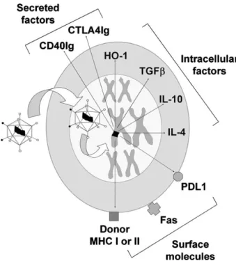
Fig. 1. Schematic illustration of the variety of modalities based on genetic modification of the donor organ. A viral vector carries the therapeutic gene (black) to the nucleus, where it is integrated in a chromosome when integrated factors such as retroviruses or lentiviruses are used (alternatively, the gene is not integrated but remains in an extra chromosomal state within the nucleus when using other viral vectors, e.g., adenovectors, or nonviral vehicles). Examples of therapeutic genes encoding secreted factors (e.g., CTLA4Ig), cell surface mole-cules (e.g., MHC class I or II), or intracellular factors (e.g., HO-1, immunosup-pressive cytokines) are shown. Alternatively, the vector may deliver antisense ODN or ribozyme that inhibit expression of pathogenic genes (abbreviations: see text).
Table 1
Selected examples of successful gene therapy studies for prevention of acute cardiac allograft rejection (non-exhaustive list). The likely mechanism of action has been indicated, although multiple mechanisms may be involved. (*) denotes a study carried out in a nontransplant ischaemia—reperfusion model (abbreviations: see text; Ad: adenovirus).
Mechanism Therapeutic gene Vector Model Ref.
Cytoprotection, anti-oxidant NFkB ODN decoy HVJ-liposomes Rat [24]
eNOS Liposomes, Ad Rabbit, rat [15,16]
Mn-SOD Ad Rabbit [16]
HO-1 Ad Rat [18]
Inhibition of adhesion molecules ICAM-1 ODN decoy (+anti-LFA-1 Ab) Liposomes Rat [14]
Cytokine inhibition TNFRp55-Ig Ad Rat [21]
IL-1R type 2-Ig Ad Rat [20]
IL-1RA (*) HVJ-liposomes Rat [19]
IL-17Ig Ad Rat [22]
IL-18 binding protein Ad Rat [23]
Immune deviation IL-4 Ad Rat [27]
IL-10 Ad Rabbit, rat [11,26]
vIL-10 Ad Rabbit [25]
IL-4 + IL-10 Liposomes Rabbit [29]
IL-13 Ad Rat [28]
TGFb1 Ad, liposomes Mouse, rabbit [10,25]
Chemokine inhibition vMIP-II, MC148 Liposomes Mouse [32]
8ND-RANTES/CCL5 Lentivirus Rat [9]
Inhibition of T cell costimulation CTLA4Ig Ad Rat [34]
CD40Ig Ad Rat [35]
CTLA4Ig + CD40Ig Ad Rat [36]
PDL1Ig Ad Rat [40]
Tolerance induction Donor MHC class I Retrovirus, Ad Mouse [43,44]
As mentioned above, AAV vectors have a potential for stable gene expression in many organs including the heart
[2]. These vectors are poorly immunogenic and have been safely used in clinical gene therapy trials in nontransplanted patients. Conventional AAV vectors derived from AAV serotype 2 have a broad tissue tropism, which results in the predominant transduction of the liver after systemic vector administration. Newer AAV vectors derived from serotypes 1, 4, 5, 6, and 9 allow enhanced gene transfer into the myocardium[5,6]. Ex vivo perfusion of an AAV-9 vector into the donor heart achieved up to 72% myocardial gene transfer efficiency 10 days after heart transplantation in rodents[6]. Cardiac expression levels of the LacZ reporter gene were unchanged 3 months after transplantation, with no overt evidence of tissue toxicity.
Retrovirus vectors have a potential for stable gene expression owing to chromosomal integration. Among retro-virus vectors, lentiretro-virus vectors have the unique ability to deliver genes to nondividing cells. We have shown that a self-inactivating, multiply attenuated lentivirus vector based on human immunodeficiency virus type 1 (HIV-1) efficiently delivered and expressed a GFP reporter gene in rat
myocardium and endothelial cells in vivo [7,8]. However, lentivirus vectors caused significant myocardial inflammation in our model.
Nonviral vectors including cationic lipids and liposomes, alone or in conjunction with fusogenic proteins that facilitate cell entry, such as those derived from the haemagglutinating virus of Japan (HVJ; Sendai virus) can carry large DNA sequences and are generally safer than viral vectors. Liposomes have been successfully used to introduce genes of interest into the donor heart[10,11]. The envelope of HVJ alone efficiently delivered proteins, genes, and oligode-oxynucleotides (ODN) to a majority of cultured neonatal cardiac myocytes, whereas the corresponding vector lacking the HVJ envelope delivered genes only to a few cells[12]. Recently, a multifunctional envelope-type nano-device (MEND) was shown to enter cells via macropinocytosis, escape lysosomal degradation, and mediate nuclear translo-cation [13]. Gene transfer efficiency of this vector approaches that of viral vectors.
A universal vector suitable for all gene therapy applica-tions does not exist. The most appropriate vector for a given application depends on both the therapeutic gene and the target tissue. The biological effect of the encoded protein determines whether transient or sustained gene expression is desirable.
3. Immune responses and graft rejection
Early damage to the donor organ may result from brain death of the donor, organ procurement, oxygen deprivation during the organ preservation time, the surgical procedure itself, and subsequent reperfusion injury. Graft endothelium is particularly vulnerable to ischaemia—reperfusion injury, which causes endothelial cell activation characterised by upregulation of cell adhesion molecules. As a result, leukocytes adhere to the endothelium and then accumulate in the graft. Factors of the complement system become activated, and neutrophils migrate into the graft, followed by natural killer (NK) cells and macrophages. Early non-specific inflammatory reactions are followed by non-specific alloimmune responses that culminate in massive graft infiltration by T cells, B cells, macrophages, and dendritic
Table 2
Selected examples of successful gene therapy studies for prevention of accelerated graft vasculopathy in arterial or cardiac allotransplant models (non-exhaustive list). (*) denotes a study in which the donor heart was first placed in a syngeneic recipient, followed by a second transplantation into an allogeneic recipient 4 days later (abbreviations: see text; Ad: adenovirus).
Mechanism Therapeutic gene Vector Model Ref.
Cytoprotection or anti-oxidant HO-1 Ad Rat aorta [55,56]
Bcl-2 (*) Ad Rat heart [58]
tPA Liposomes Rabbit heart [65]
SMC apoptosis, inhibition to cell proliferation Bcl-x ODN decoy Liposomes Mouse heart [57]
Soluble Fas Ad Rat aorta [60]
FasL Ad Rat carotid [61]
Anti-ERK1/2 decoy Liposomes Rat aorta [63]
CTLA4-FasL chimera Ad Mouse heart [64]
Anti-E2F decoy Liposomes Monkey heart [68]
Inhibition of extracellular matrix digestion Anti-MMP-2 ribozyme HVJ-liposomes Mouse heart [66]
Chemokine inhibition MCP-1 antagonist Liposomes Mouse heart [62]
Inhibition of T cell costimulation CD40Ig Ad Rat aorta [59]
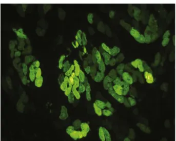
Fig. 2. Adenovirus-mediated in vivo transfer of a GFP reporter gene into rat myocardium. Myocytes represent the main cell type expressing GFP after gene transfer into the heart.
cells (DC). A direct and an indirect pathway of alloantigen recognition by the host immune system have been described. Donor-derived DC and monocytes/macrophages present in the donor heart are transplanted together with the donor heart. After transplantation, donor-derived DC leave the graft, migrate to recipient lymph nodes and spleen, and present donor antigen to recipient T cells directly. In contrast, the indirect pathway of allorecognition includes the migration of host DC into the graft, where they pick up and process donor antigen, which is subsequently presented to host T cells. It is believed that the direct pathway plays a dominant role in acute rejection, whereas the indirect pathway is more relevant to chronic rejection. Humoral immune responses also participate in chronic rejection. Full T cell activation by DC requires ‘costimulatory’ molecular interactions of pairs of ligands and receptors expressed on the surfaces of T cells and DC (see below).
A number of candidate genes that interfere with different mechanisms of ischaemia—reperfusion injury and graft rejection have been evaluated in cardiac transplant models. Selected examples of genes that have shown evidence for therapeutic benefit in these models are listed inTable 1. This table is not intended to provide an exhaustive list of the genes that have been tested, but to illustrate the variety of potential therapeutic targets in this context.
4. Gene transfer of anti-inflammatory and cytoprotective factors
Intercellular adhesion molecules (ICAM) are upregulated on graft endothelial cells during ischaemia and reperfusion. Hyperbaric introduction of antisense ODN with specific affinity for ICAM-1 into the donor heart inhibited ischae-mia—reperfusion injury and allograft rejection in a cardiac transplant model in the rat[14]. Nitric oxide (NO) generated by endothelial NO synthase (eNOS) is a key vasoprotective molecule. eNOS gene transfer into the donor heart attenuated ischaemia—reperfusion injury, leukocyte infiltra-tion, and allograft rejection in a cardiac transplant model in the rabbit[15]. Free radical scavengers neutralise reactive oxygen species (ROS) generated during ischaemia and reperfusion, as well as ROS produced by neutrophils and monocytes/macrophages that infiltrate the graft. Adeno-virus-mediated manganese-superoxide dismutase (SOD) gene transfer into the donor heart mitigated ischaemia—reperfu-sion injury after organ preservation and transplantation in a cardiac transplant model in the rabbit[16].
Haem oxygenase (HO) is a cytoprotective enzyme that catalyses the rate limiting step in haem degradation, which results in the formation of carbon monoxide (CO), iron, and biliverdin. The products of haem degradation have antiox-idant, anti-inflammatory, anti-proliferative, and anti-apop-totic effects. Local increase in CO may downregulate macrophage activity and prevents apoptosis in endothelial cells. The inducible isoform HO-1 is increased as an adaptive response in various stress conditions including exposure to haem, hyperoxia, and hypoxia. In these conditions, HO-1 activity contributes to diminishing the overall production of ROS. The HO-1 system has been associated with cytoprotec-tion in various pathological condicytoprotec-tions including
cardiovas-cular diseases, diabetes, inflammation and hypertension, as reviewed elsewhere [17]. Sustained HO-1 induction delays the onset of diabetes in NOD mice. Human HO-1 gene transfer attenuated angiotensin II- and tumour necrosis factor (TNF)-mediated DNA damage in endothelial cells. Systemic HO-1 gene delivery using an adenoviral vector allowed long-term allograft survival (>100 days) in up to 80% of cardiac transplant recipients in a mouse model[18]. By comparison, ex vivo HO-1 gene delivery to the donor heart was less beneficial than systemic gene transfer, possibly due to lack of immunosuppressive effects associated with HO-1 expression in the spleen after systemic gene delivery.
5. Gene transfer of inhibitors of pro-inflammatory cytokines
We and others have shown that gene transfer of inhibitors of pro-inflammatory cytokines, such as IL-1, TNFa, IL-17, and IL-18 moderately prolongs cardiac allograft survival in rodent models[19—23]. The transcriptional factor nuclear factor-kappaB (NFkB) is a key inflammatory mediator of endothelial activation. ODN with specific affinity for NFkB attenuated ischaemia—reperfusion injury in rat donor hearts preserved at 4 8C in Euro-Collins solution during 16 h and reperfused for 1 h[24].
6. Gene transfer of immunoregulatory cytokines
Production of interferon g (IFNg) and other Th1 cytokines has been associated with acute rejection. Conversely, ‘immune deviation’ towards Th2 cytokines, such as IL-4, IL-10, IL-13, and transforming growth factor b (TGFb) has been linked to improved graft survival. Consistent with this, gene transfer-based overexpression of IL-4, IL-10, IL-13 or TGFb1 in the donor heart moderately prolonged cardiac allograft survival in rodent models[10,25—29]. While over-expression of a single Th2 cytokine conferred modest protection upon the graft, combined IL-4 and IL-10 gene transfer allowed long-term cardiac allograft survival in rabbits [29]. These results indicate synergistic effects of the two immunosuppressive cytokines.
7. Gene transfer of chemokine antagonists
Chemoattractant cytokines (i.e., chemokines) play a central role in the mobilisation and activation of leukocytes in inflammatory conditions, including graft rejection [30]. Differential expression patterns of chemokines and chemo-kine receptors have been observed in acute rejection of cardiac and pancreatic islet allografts [31]. The RANTES/ CCL5 chemokine is expressed in cardiac grafts early after transplantation. We have shown that gene transfer of an NH2
-terminally deleted RANTES/CCL5 mutant gene acting as a C— C chemokine antagonist moderately prolonged cardiac allograft survival in rats [9]. Similar results were observed with gene transfer of the viral chemokine homologues MC148 and vMIP-II, which act as functional chemokine antagonists, in a cardiac transplant model in rodents[32].
8. Inhibition of T cell costimulatory activation
Full activation of host T cells, the central mechanism of allograft rejection, requires three distinct signals: (1) T cell receptor (TCR) activation upon recognition of the major histocompatibility complex (MHC)—peptide complex on the surfaces of antigen-presenting cells (APC); (2) costimulatory signals that arise from the interactions of ligands and their corresponding receptors expressed on T cells and APC, including CD28 and B7 (CD80/CD86), as well as CD40 ligand (CD154) and CD40; and (3) secretion of stimulatory cytokines, such as IL-12 [33]. Antigen pre-sentation in the absence of strong costimulatory T cell activation favours the development of a state of antigen-specific T cell unresponsiveness (anergy), or primes T cells for apoptosis. Cytotoxic T lymphocyte antigen-4 (CTLA4), which is upregulated on T cells upon activation, modulates T-cell costimulatory activation. Adenovirus-mediated ex vivo gene transfer of a soluble CTLA4-immunoglobulin fusion protein (CTLA4Ig) into the donor heart was associated with detectable CTLA4Ig serum levels 120 days after transplantation and long-term cardiac allograft survival (>100 days) in a rat model [34]. These results exemplify the notion that, while gene expression from first-generation adenovirus vectors usually is short lived due to immune responses to the vector, it may be markedly prolonged in the case when the delivered gene encodes an immunosuppressive peptide that inhibits these immune responses. Recipients of long-term surviving grafts trans-duced with the CTLA4Ig gene accepted a second cardiac graft from the original donor strain in the absence of pharmacological immunosuppression, while normally rejecting third-party grafts. These findings suggest that CTLA4Ig expressed in the graft induced donor-specific hyporesponsiveness. Gene transfer of a soluble CD40Ig fusion protein that blocks costimulatory CD40—CD154 interactions similarly prolonged cardiac allograft survival in the same model[35]. However, expression of CTLA4Ig or CD40Ig in the graft induced some degrees of general immunosuppression due to spill-over of the soluble chimeric protein into the systemic circulation. Interest-ingly, combined gene transfer of CTLA4Ig and CD40Ig was more effective than that of either gene alone [36].
CD40Ig treatment induced indefinite donor-specific allograft acceptance in a complete MHC-mismatched cardiac transplant model in the rat. The tolerogenic effect was associated with induction of CD8+CD45+RC+regulatory
T (Treg) cells, IFNg, and indoleamine 2,3-dioxygenase (IDO) [37]. IDO is an IFNg-inducible enzyme that catalyses the rate-limiting step in the catabolism of the essential amino acid tryptophan, which is important for normal T cell functions. Recent evidence suggests that IDO modulates T cell responses to self antigens (autoimmunity), alloanti-gens, and tumour antigens. Within cardiac allografts, CD40Ig treatment induced selective IDO expression in endothelial cells. Neutralisation of IFNg or IDO triggered acute allograft rejection in CD40Ig-treated recipients. These results suggest that inhibition of CD40—CD154 interactions results in the formation of allospecific Treg
cells that facilitate cardiac allograft acceptance via induction of IFNg and endothelial IDO expression. We have
shown that adenoviral IDO gene transfer into DC inhibits allogeneic T cell responses in vitro, and that IDO gene transfer into rat donor hearts moderately prolongs cardiac allograft survival in vivo[38].
Programmed death (PD) 1 is a CD28 homologue expressed on activated T cells, B cells, and myeloid cells
[39]. Engagement of PD1 by its ligands, PDL1 and PDL2, inhibits activated T cells. We have shown that adenoviral gene transfer of a soluble PDL1Ig fusion protein into the donor heart moderately prolongs cardiac allograft survival in rats [40]. Together, these results suggest that gene therapy modalities that interfere with T cell costimulatory activation may confer significant protection upon the graft. It should be noted, however, that these results likely reflect the development of donor-specific hyporesponsive-ness, rather than true immunological tolerance. In this regard, it has been noted that cardiac allografts in rodents, unlike in larger animals and humans, may provide direct tolerogenic effects that facilitate graft acceptance
[41].
9. Transfer of donor MHC class I and II genes
MHC mismatch between the donor and recipient is a major, albeit not exclusive mechanism of allograft rejection. To induce ‘molecular chimerism’ and unresponsiveness to donor MHC antigens, autologous cells genetically modified with donor MHC class I or II genes were introduced back into the recipient around the time of transplantation. Autologous fibroblasts expressing donor MHC class I or II genes allowed prolonged cardiac allograft survival in a mouse model[42]. Reconstitution of the host bone marrow with autologous haematopoietic stem cells genetically modified with donor MHC class I or II induced donor-specific T cell unresponsive-ness in cardiac transplant models in rodents [43—46]. A critical issue with these approaches, however, is stability of T cell unresponsiveness induced by ‘molecular chimerism’
[47].
10. Induction of Tregcells
Tregcells modulate T cell activation in various contexts,
including auto- and alloimmunity. These cells promote the development of antigen-specific immune tolerance, and therefore facilitate the acceptance of grafts that express their cognate antigens [48—50]. Foxp3 is the master transcriptional regulator of Tregcell development.
foxp3 gene transfer into naı¨ve CD4+CD25 precursor cells induced the formation of functional CD4+CD25+Foxp3+T
reg
cells that facilitated skin allograft acceptance in mice
[51]. Because Treg cells are antigen-specific, and because
the graft expresses multiple different antigens, induction of Tregcells by foxp3 gene transfer would be most effective
if transferred unresponsiveness to defined antigens were to expand to other antigens shared by the graft
[52]. This phenomenon, referred to as ‘infectious toler-ance’, has been observed in tolerant systems in which Treg cells constitute the central mechanism of tolerance [53].
11. Prevention of graft vasculopathy
An accelerated form of coronary artery disease char-acterised by concentric intimal thickening and widespread distribution of lesions across the coronary tree is the central manifestation of chronic rejection of cardiac allografts. Chronic immune reactions associated with perivascular inflammation induce persistent endothelial cell activation and secretion of growth factors, such as platelet-derived growth factor (PDGF), which stimulates the proliferation of vascular smooth muscle cells (SMC) leading to intimal thickening. Nonimmunological factors such as drug-related toxicity, metabolic abnormalities, and viral infections (particularly cytomegalovirus) play contributory roles in the pathogenesis of graft vasculopathy[54]. Selected studies of gene transfer-based approaches to prevent transplant vasculopathy are listed inTable 2.
Adenovirus-mediated gene transfer of the cytoprotective enzyme HO-1 partially prevented antibody-induced graft vasculopathy in aortic transplant models in rodents[55,56]. Antisense ODN with specific affinity for Bcl-x, a mitochondrial anti-apoptotic factor, also mitigated the development of graft vasculopathy [57]. The underlying mechanism pre-sumably involved increased apoptosis of SMC in intimal lesions, leading to decreased lesion formation. Paradoxically, overexpression of Bcl-2, a distinct mitochondrial anti-apoptotic factor, also attenuated the development of graft vasculopathy[58]. This was shown by first placing the donor heart transduced with the Bcl-2 gene in a syngeneic recipient to allow for Bcl-2 gene expression in the graft in the absence of alloimmune responses, with re-transplantation of the graft into an allogeneic recipient 4 days later. In other studies, adenoviral gene transfer of CD40Ig[59], soluble Fas receptor
[60], or Fas ligand[61]partially prevented the development of graft vasculopathy. Fas ligand acts by inducing apoptosis of activated T cells that express Fas receptor on their surfaces. Monocyte chemoattractant protein-1 (MCP-1) is a potent chemoattractant of monocytes-macrophages into graft arterial lesions. Gene transfer of an NH2-terminally deleted
MCP-1 mutant gene reduced intimal thickening in an aortic transplant model[62]. Other gene therapy approaches that have shown protective effects against graft vasculopathy include antisense ODN with specific affinity for mitogen-activated protein kinase ERK1/2[63], a signalling molecule involved in cell survival and proliferation, gene transfer of a chimeric CTLA4-FasL protein [64], and gene transfer of tissue-type plasminogen activator (tPA)[65], a vasoprotec-tive factor that is downregulated throughout the develop-ment of graft vasculopathy. Matrix metalloproteinases (MMP) digest extracellular matrix in the vessel wall, and therefore allow for SMC migration into the intima and neointimal formation. MMP-2 is persistently activated in the vessel wall throughout the progression of graft vasculopathy. A ribozyme against MMP-2 inhibited MMP-2 expression and the develop-ment of graft coronary artery lesions in a rodent model[66]. Ribozymes are a class of RNA molecules that possess RNA sequence-specific activity combined with catalytic activity. They hybridise with a target mRNA, and cleave the complementary mRNA[67].
Finally, double-stranded DNA with specific affinity for E2F, a transcription factor that plays a central role in cell cycle
progression, attenuated cell proliferation, intimal thicken-ing, and graft coronary artery lesions 8 weeks after heart transplantation in rhesus monkeys[68]. However, the long-term efficacy of these approaches remains to be established.
12. Challenges toward clinical applications
Despite encouraging results in animal models, progress toward clinical applications in gene therapy for transplanta-tion has been slow. This can be explained by several factors, including limitations of gene transfer vectors, an incomplete understanding of the mechanisms of alloimmune activation, and scarce data in non-human primate models. Immunosup-pressive drugs currently used in clinical transplantation are quite effective in preventing acute rejection, and therefore it will be difficult for gene therapy to achieve improvement on top of them.
Vector limitations include both poor gene transfer effi-ciency and side effects. As already mentioned, however, significant advances in vector development have been scored recently. Over the past decade, rare adverse events caused by viral vectors used in clinical trials in nontransplanted patients have set back the field of gene therapy. In a gene therapy trial of ornithine transcarbamylase (OCT) deficiency, a young man died after receiving the highest dose of adenovirus vector in the study[69]. The death was caused by an unusually strong, systemic inflammatory syndrome leading to multi-organ failure. It should be noted, however, that the first-generation adenovector used in this study was more immunogenic than last-generation adenovectors. More recently, the three youngest patients in a retrovirus-based gene therapy trial of X-linked severe combined immunodeficiency disorder (SCID-X1) developed leukaemia due to insertional mutagenesis caused by the retrovirus vector[70]. However, the leukaemia found in these patients might be unique to this particular disease because the genetic reconstitution of a very few precursor cells results in the selective proliferation of immune cells genetically corrected with the vector. Regardless, these adverse events have highlighted the potential risks of viral vectors. Clearly, gene transfer vehicles differ from pharma-cological agents in many ways, including the definition of the dose and biological activity, and the tissue distribution and kinetics of the transgenic protein in vivo. These aspects of gene therapy, together with the reproducibility of gene delivery protocols, need to be characterised more precisely. Little is known about the inter-individual variability in gene transfer efficiency and the duration of transgene expression in humans. This may depend on multiple factors, such as genetic factors and pre-existing antibodies to the vector (e.g., as a result of previous exposure to wild-type adenovirus).
While molecular mechanisms of immunological rejection and tolerance are relatively similar in non-human primates and humans, they differ in several important ways in rodents and primates[71]. Stable tolerance induction is not achieved routinely in large animals and humans. For practical reasons, the vast majority of gene therapy studies in transplantation have been carried out in rodents. These models are still useful for initial feasibility studies of new vectors, gene delivery protocols, and therapeutic genes. It will be important to compare different vectors and genes directly
in the same model in order to minimise confounding variables. Based on results in these models, the most promising modalities will be tested in more relevant monkey models, starting with small feasibility studies, followed by larger safety and efficacy studies. Prevention of chronic rejection and graft vasculopathy should be assessed long-term (e.g., 1—2 years after transplantation), not just after 2—3 months, as in previous studies[68]. Potential side effects of immunomodulatory genes including opportunistic infec-tions, cancer, and induction of autoimmune responses should be investigated. Obviously, gene therapy modalities must be compared directly with current immunosuppressive regimens in well-controlled pre-clinical studies in monkeys. Beneficial combinations of the two approaches must be tested. The development of a new gene therapy modality from small animal models to pre-clinical studies may well require a decade (or longer, if limited effort is devoted to achieving this goal).
13. Future prospects
The real challenge in transplantation is prevention of chronic rejection. The recent development of vectors capable of expressing a gene for extended periods of time has provided new tools to achieve this goal. AAV vectors offer several advantages, including high efficiency, differential tissue targeting by different AAV serotypes, stable gene expression, negligible tissue inflammation, and a good safety profile, as documented in several clinical trials in non-transplanted patients [72]. Newer nonviral vehicles repre-sent a valuable alternative, as they are nearly as efficient as, and potentially safer than, viral vectors. Desirable features of future vectors include regulation of gene expression levels (‘gene dosage’) to match clinical needs, tissue-specific gene expression, and multi-cistronic vectors expressing multiple genes. Regulatable vectors controlled by oral intake of doxicycline or other agents have been tested successfully in vivo [73] but still need to be optimised for clinical applications.
The most effective gene to be introduced into the donor heart, or to be employed in cell-based gene therapy using DC
[74], stem cells or other bone marrow-derived cells still needs to be identified. In previous studies, genes encoding immunomodulatory cytokines or inhibitors of pro-inflamma-tory cytokines and chemokines have shown only modest protective effects. Genes encoding inhibitors of T cell costimulatory activation, such as CTLA4Ig and CD40Ig, have provided more encouraging results, likely due to direct inhibition of alloreactive T cells. Simultaneous blockade of multiple T cell costimulatory pathways has shown additive or synergistic effects [75]. The emerging role of Treg cells in
antigen-specific tolerance induction has fostered the devel-opment of gene therapy approaches that focus on the generation and/or functional enhancement of these cells.
In conclusion, the conceptual advantage of gene therapy in transplantation is that localised overexpression of the protective gene within the graft may promote a state of local immune privilege, and therefore reduce the need for general immunosuppression. Alternatively, gene therapy may attenu-ate alloimmune responses through the generation of Tregcells
and regulatory DC. Because expression of a protective gene may last for extended periods of time, gene therapy has a potential for preventing chronic rejection. Improved efficacy may be achieved by combining different modalities, e.g., expression of multiple therapeutic genes from a multi-cistronic vector within the graft, with concomitant transfer of Tregcells and/or genetically modified DC. If clinical trials in
gene therapy of transplantation are started one day, it can be anticipated that the role of gene therapy will be that of an adjunct therapy. In fact, immunosuppressive drugs would still be needed, at least temporarily. A role for gene therapy as a sole treatment in heart transplantation is theoretically conceivable, but it cannot be envisioned in the foreseeable future.
References
[1] Weiss ES, Nwakanma LU, Patel ND, Yuh DD. Outcomes in patients older than 60 years of age undergoing orthotopic heart transplantation: an analysis of the UNOS database. J Heart Lung Transplant 2008;27:184—91. [2] Vassalli G, Bu¨eler H, Dudler J, von Seegsser LK, Kappenberger L. Adeno-associated virus (AAV) vectors achieve prolonged transgene expression in mouse myocardium and arteries in vivo: a comparative study with adenovirus vectors. Int J Cardiol 2003;90:229—38.
[3] Le Bas-Bernardet S, Anegon I, Blancho G. Progress and prospects: genetic engineering in xenotransplantation. Gene Ther 2008;15:1247—56. [4] Fleury S, Driscoll RA, Simeoni E, Dudler J, von Segesser LK, Kappenberger
L, Vassalli G. Helper-dependent adenovirus vectors devoid of all viral genes cause less myocardial inflammation compared with first-generation adenovirus vectors. Basic Res Cardiol 2004;99:247—56.
[5] Mu¨ller OJ, Leuchs B, Pleger ST, Grimm D, Franz WM, Katus HA, Kleinsch-midt JA. Improved cardiac gene transfer by transcriptional and transduc-tional targeting of adeno-associated viral vectors. Cardiovasc Res 2006; 70:70—8.
[6] Miyagi N, Rao VP, Ricci D, Du Z, Byrne GW, Bailey KR, Nakai H, Russell SJ, McGregor CG. Efficient and durable gene transfer to transplanted heart using adeno-associated virus 9 vector. J Heart Lung Transplant 2008;27: 554—60.
[7] Fleury S, Simeoni E, Zuppinger C, De´glon N, von Segesser LK, Kappen-berger L, Vassalli G. Multiply attenuated, self-inactivating lentiviral vectors efficiently deliver and express genes for extended periods of time in adult rat cardiomyocytes in vivo. Circulation 2003;107:2375—82. [8] Cefaı¨ D, Simeoni E, Ludunge KM, Driscoll R, von Segesser LK, Kappen-berger L, Vassalli G. Multiply attenuated, self-inactivating lentiviral vectors efficiently transduce human coronary artery cells in vitro and rat arteries in vivo. J Mol Cell Cardiol 2005;38:333—44.
[9] Vassalli G, Simeoni E, Li J, Fleury S. Lentiviral gene transfer of the chemokine antagonist RANTES 9-68 prolongs heart graft survival. Trans-plantation 2006;81:240—6.
[10] Chan SY, Goodman RE, Szmuszkovicz JR, Roessler B, Eichwald EJ, Bishop DK. DNA-liposome versus adenoviral mediated gene transfer of trans-forming growth factor beta1 in vascularized cardiac allografts: differ-ential sensitivity of CD4+ and CD8+ T cells to transforming growth factor beta1. Transplantation 2000;70:1292—304.
[11] Hong YS, Laks H, Cui G, Chong T, Sen L. Localized immunosuppression in the cardiac allograft induced by a new liposome-mediated IL-10 gene therapy. J Heart Lung Transplant 2002;21:1188—200.
[12] Tashiro H, Aoki M, Isobe M, Hashiya N, Makino H, Kaneda Y, Ogihara T, Morishita R. Development of novel method of non-viral efficient gene transfer into neonatal cardiac myocytes. J Mol Cell Cardiol 2005;39: 503—9.
[13] Akita H, Harashima G. Advances in non-viral gene delivery: using multi-functional envelope-type nano-devices. Expert Opin Drug Deliv 2008;5: 847—59.
[14] Poston RS, Mann MJ, Hoyt EG, Ennen M, Dzau VJ, Robbins RC. Antisense oligodeoxynucleotides prevent acute cardiac allograft rejection via a novel, nontoxic, highly efficient transfection method. Transplantation 1999;68:825—32.
[15] Iwata A, Sai S, Nitta Y, Chen M, deFries-Hallstrand R, Dalesandro J, Thomas R, Allen MD. Liposome-mediated gene transfection of endothelial
nitric oxide synthase reduces endothelial activation and leukocyte infil-tration in transplanted hearts. Circulation 2001;103:2753—9. [16] Abunasra HJ, Smolenski RT, Yap J, Sheppard M, O’Brien T, Yacoub MH.
Multigene adenoviral therapy for the attenuation of ischemia-reperfusion injury after preservation for cardiac transplantation. J Thorac Cardiovasc Surg 2003;125:998—1006.
[17] Abraham NG, Kappas A. Pharmacological and clinical aspects of heme oxygenase. Pharmacol Rev 2008;60:79—127. Erratum in: Pharmacol Rev 2008;60:242.
[18] Braudeau C, Bouchet D, Tesson L, Iver S, Re´my S, Buelow R, Anegon I, Chaveau C. Induction of long-term cardiac allograft survival by heme oxygenase-1 gene transfer. Gene Ther 2004;11:701—19.
[19] Suzuki K, Murtuza B, Smolenski RT, Sammut IA, Suzuki N, Kaneda Y, Yacoub MH. Overexpression of interleukin-1 receptor antagonist provides cardi-oprotection against ischemia-reperfusion injury associated with reduc-tion in apoptosis. Circulareduc-tion 2001;104(Suppl. 1):I308—13.
[20] Simeoni E, Dudler J, Fleury S, Li J, Pagnotta M, Pascual M, von Segesser LK, Vassalli G. Gene transfer of a soluble IL-1 type 2 receptor-Ig fusion protein improves cardiac allograft survival in rats. Eur J Cardiothorac Surg 2007;31:222—8.
[21] Ritter T, Schroder G, Risch K, Vergopoulos A, Shean MK, Kolls J, Brock J, Lehmann M, Volk HD. Ischemia/reperfusion injury-mediated down-reg-ulation of adenovirus-mediated gene expression in a rat heart transplan-tation model is inhibited by co-application of a TNFRp55-Ig chimeric construct. Gene Ther 2000;7:1238—43.
[22] Li J, Simeoni E, Fleury S, Dudler J, Fiorini E, Kappenberger L, von Segesser LK, Vassalli G. Gene transfer of soluble interleukin-17 receptor prolongs cardiac allograft survival in a rat model. Eur J Cardiothorac Surg 2006; 29:779—83.
[23] Dudler J, Simeoni E, Fleury S, Li J, Pagnotta M, Pascual M, von Segesser LK, Vassalli G. Gene transfer of IL-18 binding protein attenuates cardiac allograft rejection. Transplant Int 2007;20:460—6.
[24] Sakaguchi T, Sawa Y, Fukushima N, Nishimura M, Ichikawa H, Kaneda Y, Matsuda H. A novel strategy of decoy transfection against nuclear factor-kappaB in myocardial preservation. Ann Thorac Surg 2001;71:629—30. [25] Brauner R, Nonoyama M, Laks H, Drinkwater Jr SC, McCaffery S, Drake T,
Berk AJ, Sen L, Wu L. Intracoronary adenovirus-mediated transfer of immunosuppressive cytokine genes prolongs allograft survival. J Thorac Cardiovasc Surg 1997;114:923—33.
[26] David A, Chetritt J, Guillot C, Tesson L, Heslan JM, Cuturi MC, Soulillou JP, Anegon I. Interleukin-10 produced by recombinant adenovirus prolongs survival of cardiac allografts in rats. Gene Ther 2000;7:505—10. [27] Ke B, Ritter T, Kato H, Zhai Y, Li J, Lehmann M, Busuttil RW, Volk HD,
Kupiec-Weglinski JW. Regulatory cells potentiate the efficacy of IL-4 gene transfer by up-regulating Th2-dependent expression of protective mole-cules in the infectious tolerance pathway in transplant recipients. J Immunol 2000;164:5739—45.
[28] Ke B, Shen XD, Zhai Y, Gao F, Busuttil RW, Volk HD, Kupiec-Weglinski JW. Heme oxygenase 1 mediates the immunomodulatory and antiapoptotic effects of interleukin 13 gene therapy in vivo and in vitro. Hum Gene Ther 2002;13:1845—57.
[29] Furukawa H, Oshima K, Tung T, Cui G, Laks H, Sen L. Liposome-mediated combinatorial cytokine gene therapy induces localized synergistic immu-nosuppression and promotes long-term survival of cardiac allografts. J Immunol 2005;174:6983—92.
[30] Smith RN, Ueno T, Ito T, Tanaka K, Shea SP, Abdi R. Chemokines and chronic heart allograft rejection. Transplantation 2007;84:442—4. [31] Abdi R, Means TK, Ito T, Smith RN, Najafian N, Jurewicz M, Tchipachvili V,
Charo I, Auchincloss Jr H, Sayegh MH, Luster AD. Differential role of CCR2 in islet and heart allograft rejection: tissue specificity of chemokine/ chemokine receptor function in vivo. J Immunol 2004;172:767—75. [32] DeBruyne LA, Li K, Bishop DK, Bromberg JS. Gene transfer of virally
encoded chemokine antagonists vMIP-II and MC148 prolongs cardiac allograft survival and inhibits donor-specific immunity. Gene Ther 2000;7:575—82.
[33] Sayegh MH, Turka LA. The role of T-cell costimulatory activation pathways in transplant rejection. N Engl J Med 1998;338:1813—21.
[34] Guillot C, Mathieu P, Coathalem H, LeMauff B, Castro MG, Tesson L, Usal C, Laumonier T, Brouard S, Soulillou JP, Lo¨wenstein PR, Cuturi MC, Anegon I. Tolerance to cardiac allografts via local and systemic mechanisms after adenovirus-mediated CTLA4Ig expression. J Immunol 2000;164:5258—68. [35] Guillot C, Guillonneau C, Mathieu P, Gerder CA, Me´noret S, Braudeau C, Tesson L, Renaudin K, Castro MG, Lo¨wenstein PR, Anegon I. Prolonged blockade of CD40-CD40 ligand interactions by gene transfer of CD40Ig
results in long-term heart allograft survival and donor-specific hypore-sponsiveness, but does not prevent chronic rejection. J Immunol 2002;168:1600—9.
[36] Yamashita K, Masunaga T, Yanagida N, Takehara M, Hashimoto T, Kobaya-shi T, Echizenya H, Hua N, Fujita M, Murakami M, Furukawa H, Uede T, Todo S. Long-term acceptance of rat cardiac allografts on the basis of adenovirus mediated CD40Ig plus CTLA4Ig gene therapies. Transplanta-tion 2003;76:1089—96.
[37] Guillonneau C, Hill M, Hubert FX, Chiffoleau E, Herve´ C, Li XL, Heslan M, Usal C, Tesson L, Me´noret S, Saoudi A, LeMauff B, Josien R, Cuturi MC, Anegon I. CD40Ig treatment results in allograft acceptance mediated by CD8CD45RC T cells, IFN-gamma, and indoleamine 2,3-dioxygenase. J Clin Invest 2007;117:871—3.
[38] Li J, Meinhardt A, Roehrich ME, Golshayan D, Dudler J, Pagnotta M, Trucco M, Vassalli G. Indoleamine 2,3-dioxygenase gene transfer prolongs car-diac allograft survival. Am J Physiol Circ Physiol 2007;293:H3415—23. [39] Freeman GJ, Long AJ, Iwai Y, Bourque K, Chernova T, Nishimura H, Fitz LJ,
Malenkovich N, Okazaki T, Byrne MC, Horton HF, Fouser L, Carter L, Ling V, Bowman MR, Carreno BM, Collins M, Wood CR, Honjo T. Engagement of the PD-1 immunoinhibitory receptor by a novel B7 family member leads to negative regulation of lymphocyte activation. J Exp Med 2000;192:1027—34. [40] Dudler J, Li J, Pagnotta M, von Segesser LK, Vassalli G. Gene transfer of programmed death ligand-1.Ig prolongs cardiac allograft survival. Trans-plantation 2006;82:1733—7.
[41] Bagley J, Jacomini J. Gene therapy in organ transplantation. Gene Ther 2003;10:605—11.
[42] Madsen JC, Superina RA, Wood KJ, Morris PJ. Immunological unrespon-siveness induced by recipient cells transfected with donor MHC genes. Nature 1988;331:161—4.
[43] Wong W, Stranford SA, Morris PJ, Wood KJ. Retroviral gene transfer of a class I MHC gene to recipient bone marrow induces tolerance to alloanti-gens in vivo. Transplant Proc 1997;29:1130.
[44] Fry JW, Morris PJ, Wood KJ. Adenoviral transfer of a single donor-specific MHC class I gene to recipient bone marrow cells can induce specific immunological unresponsiveness in vivo. Gene Ther 2002;9:220—6. [45] Sonntag KC, Emery DW, Yasumoto A, Haller G, Germana S, Sablinski T,
Shimizu A, Yamada K, Shimada H, Arn S, Sachs DH, LeGuern C. Tolerance to solid organ transplants through transfer of MHC class II genes. J Clin Invest 2001;107:65—71.
[46] Bagley J, Tian C, Sachs DH, Iacomini J. Induction of T-cell tolerance to an MHC class I alloantigen by gene therapy. Blood 2002;99:4394—9. [47] Bagley J, Bracy JL, Tian C, Kang ES, Iacomini J. Establishing
immunolo-gical tolerance through the induction of molecular chimerism. Front Biosci 2002;7:1331—7.
[48] Karim M, Feng G, Wood K, Bushell A. CD25+ CD4+ regulatory T cells generated by exposure to a model protein antigen prevent allograft rejection: antigen specific reactivation in vivo is critical for bystander regulation. Blood 2005;105:4871—7.
[49] Lee M, Moore D, Jarrett B, Lian MM, Deng S, Huang X, Markmann JW, Chiaccio M, Barker CF, Caton AJ, Markmann JF. Promotion of allograft survival by CD4+ CD25+ regulatory T cells: evidence for in vivo inhibition of effector cell proliferation. J Immunol 2004;172:6539—44.
[50] Ochando J, Homma C, Yang Y, Hidalgo A, Garin A, Tacke F, Angeli V, Li Y, Boros P, Ding Y, Jessberger R, Trinchieri G, Lira SA, Randolph GJ, Bromberg JS. Alloantigen-presenting plasmacytoid dendritic cells mediate toler-ance to vascularized grafts. Nat Immunol 2006;7:652—62.
[51] Chai J, Xue S, Coe D, Addey C, Bartok I, Scott D, Simpson E, Strauss HJ, Hori S, Sakaguchi S, Dyson J. Regulatory T cells, derived from naive CD4+ CD25- T cells by in vitro Foxp3 gene transfer, can induce transplantation tolerance. Transplantation 2005;79:1310—6.
[52] Moore D, Markmann J, Deng S. Avenues for immunomodulation and graft protection by gene therapy in transplantation. Transplant Int 2006;19: 435—45.
[53] Waldmann H, Adams E, Fairchild P, Cobbold S. Infectious tolerance and the long-term acceptance of transplanted tissue. Immunol Rev 2006;212: 301—13.
[54] Vassalli G, Gallino A, Weis M, von Scheidt W, Kappenberger L, von Segesser LK, Goy JJ, Working Group Microcirculation of the European Society of Cardiology. Alloimmunity and nonimmunologic risk factors in cardiac allograft vasculopathy. Eur Heart J 2003;24:1180—8.
[55] Bouche D, Chauveau C, Roussel J, Mathieu P, Braudeau C, Tesson L, Soulillou JP, Iyer S, Buelow R, Anegon I. Inhibition of graft arteriosclerosis development in rat aortas following heme oxygenase-1 gene transfer. Transplant Immunol 2002;9:235—8.
[56] Chauveau C, Bouchet D, Roussel J, Mathieu P, Braudeau C, Renaudin K, Tesson L, Soulillou JP, Iyer S, Buelow R, Anegon I. Gene transfer of heme oxygenase-1 and carbon monoxide delivery inhibit chronic rejection. Am J Transplant 2002;2:581—92.
[57] Suzuki J, Isobe M, Morishita R, Nishikawa T, Amano J, Kaneda Y. Antisense Bcl-x oligonucleotide induces apoptosis and prevents arterial neointimal formation in murine cardiac allografts. Cardiovasc Res 2000;45:783—7. [58] Miniati D, Lijkwan M, Murata S, Martens J, Coleman CT, Hoyt EG, Robbins RC.
Effects of adenoviral up-regulation of bcl-2 on oxidative stress and graft coronary artery disease in rat heart transplants. Transplantation 2003;76: 382—6.
[59] Mathieu P, Guillot C, Gerdes C, Buzelin F, Lowenstein P, Castro M, Soulillou JP, Anegon I. Adenovirus-mediated CD40Ig expression attenuates chronic vascular rejection lesions in an aorta allotransplantation model. Trans-plant Proc 2002;34:743—4.
[60] Wang T, Dong C, Stevenson S, Herderick EE, Marshall-Neff J, Vasudevan SS, Moldovan NI, Michler RE, Movva NR, Goldschmidt-Clermont PJ. Over-expression of soluble fas attenuates transplant arteriosclerosis in rat aortic allografts. Circulation 2002;106:1536—42.
[61] Sata M, Luo Z, Walsh K. Fas ligand overexpression on allograft endothe-lium inhibits inflammatory cell infiltration and transplant-associated intimal hyperplasia. J Immunol 2001;166:6964—71.
[62] Saiura A, Sata M, Hiasa K, Kitamoto S, Washida M, Egashira K, Nagai R, Makuuchi M. Antimonocyte chemoattractant protein-1 gene therapy attenu-ates graft vasculopathy. Arterioscler Thromb Vasc Biol 2004;24:1886—90. [63] Dong C, Gong N, Chen Z, Chen X, Xu Q, Guo H, Zeng Z, Ming C, Chen ZK.
Antisense ERK1/2 oligodeoxynucleotide gene therapy attenuates graft arteriosclerosis of aortic transplant in a rat model. Transplant Proc 2006;38:3304—6.
[64] Feng Y, Jin Y, Zhang Q, Hao J, Wang G, Xie S. CTLA4-Fas ligand gene transfer mediated by adenovirus induce long-time survival of murine cardiac allografts. Transplant Proc 2005;37:2379—81.
[65] Scholl F, Sen L, Drinkwater D, Laks H, Ma XY, Hong YS, Chang P, Cui G. Effects of human tissue plasminogen gene transfer on allograft coronary atherosclerosis. J Heart Lung Transplant 2001;20:322—9.
[66] Tsukioka K, Suzuki J, Fujimori M, Wada Y, Yamaura K, Ito K, Morishita R, Kaneda Y, Isobe M, Amano J. Expression of matrix metalloproteinases in cardiac allograft vasculopathy and its attenuation by anti MMP-2 ribo-zyme gene transfection. Cardiovasc Res 2002;56:472—8.
[67] Asif-Ullah M, Le´vesque M, Robichaud G, Perreault JP. Development of ribozyme-based gene inactivations; the example of the hepatitis delta virus ribozyme. Curr Gene Ther 2007;7:205—16.
[68] Kawauchi M, Suzuki J, Morishita R, Wada Y, Izawa A, Tomita N, Amano J, Kaneda Y, Ogihara T, Takamoto S, Isobe M. Gene therapy for attenuating cardiac allograft vasculopathy using ex vivo E2F decoy transfection by HVJ-AVE-liposome method in mice and nonhuman primates. Circ Res 2000;87:1063—8.
[69] Verma IM. A voluntary moratorium? Mol Ther 2003;7:141.
[70] Hacein-Bey-Abina S, Von Kalle C, Schmidt M, McCormack M, Wulffraat PN, Leboulch P, Lim A, Osborne CS, Pawliuk R, Morillon E, Sorensen R, Forster A, Fraser P, Cohen JI, de Saint Basile G, Alexander I, Wintergerst U, Frebourg T, Aurias A, Stoppa-Lyonnet D, Romana S, Radford-Weiss I, Gross F, Valensi F, Delabesse E, Macintyre E, Sigaux F, Soulier J, Leiva LE, Wissler M, Prinz C, Rabbitts TH, Le Deist F, Fischer A, Cavazzana-Calvo M. LMO2-associated clonal T cell proliferation in two patients after gene therapy for SCID-X1. Science 2003;302:415—9.
[71] Salama AD, Remuzzi G, Harmon WE, Sayegh MH. Challenges to achieving clinical transplantation tolerance. J Clin Invest 2001;108:943—8. [72] Kay MA, Nakai H. Looking into the safety of AAV vectors. Nature 2003;
424:251.
[73] Ye X, Rivera VM, Zoltick P, Cerasoli Jr F, Schnell MA, Gao G, Hughes JV, Gilman M, Wilson JM. Regulated delivery of therapeutic proteins after in vivo somatic cell gene transfer. Science 1999;283:88—91.
[74] McCurry KR, Colvin BL, Zahorchak AF, Thomson AW. Regulatory den-dritic cell therapy in organ transplantation. Transplant Int 2006;19: 525—38.
[75] Larsen CP, Elwood ET, Alexander DZ, Ritchie SC, Hendrix R, Tucker-Burden C, Cho HR, Aruffo A, Hollenbaugh D, Linsley PS, Winn KJ, Pearson TC. Long-term acceptance of skin and cardiac allografts after blocking CD40 and CD28 pathways. Nature 1996;381:434—8.