HAL Id: hal-02469267
https://hal-univ-rennes1.archives-ouvertes.fr/hal-02469267
Submitted on 6 Feb 2020
HAL is a multi-disciplinary open access
archive for the deposit and dissemination of
sci-entific research documents, whether they are
pub-lished or not. The documents may come from
teaching and research institutions in France or
abroad, or from public or private research centers.
L’archive ouverte pluridisciplinaire HAL, est
destinée au dépôt et à la diffusion de documents
scientifiques de niveau recherche, publiés ou non,
émanant des établissements d’enseignement et de
recherche français ou étrangers, des laboratoires
publics ou privés.
Circulating neuregulin1-beta in heart failure with
preserved and reduced left ventricular ejection fraction
Camilla Hage, Eva Wärdell, Cecilia Linde, Erwan Donal, Carolyn S. P. Lam,
Claude Daubert, Lars H. Lund, Agneta Månsson-Broberg
To cite this version:
Camilla Hage, Eva Wärdell, Cecilia Linde, Erwan Donal, Carolyn S. P. Lam, et al.. Circulating
neuregulin1-beta in heart failure with preserved and reduced left ventricular ejection fraction. ESC
Heart Failure, Wiley, 2020, 7 (2), pp.445-455. �10.1002/ehf2.12615�. �hal-02469267�
Circulating neuregulin
1-β in heart failure with
preserved and reduced left ventricular ejection
fraction
Camilla Hage
1,3*, Eva Wärdell
2,3, Cecilia Linde
1,3, Erwan Donal
4, Carolyn S.P. Lam
5,6,7, Claude Daubert
4, Lars H.
Lund
1,3and Agneta Månsson-Broberg
2,31Department of Medicine, Karolinska Institutet– Solna, Stockholm, Sweden;2Department of Medicine, Karolinska Institutet– Huddinge, Huddinge, Sweden;3Heart and Vascular Theme, Karolinska University Hospital, SE-171 76, Stockholm, Sweden;4Département de Cardiologie & CIC-IT U804, Centre Hospitalier Universitaire de Rennes, Rennes, France;5National Heart Centre Singapore, Duke-National University of Singapore, Singapore, Singapore;6University Medical Centre Groningen, Groningen, The Netherlands;7The George Institute for Global Health, Sydney, Australia
Abstract
Aims Neuregulin1-β (NRG1-β) is released from microvascular endothelial cells in response to inflammation with compensa-tory cardioprotective effects. Circulating NRG1-β is elevated in heart failure (HF) with reduced ejection fraction (HFrEF) but not studied in HF with preserved EF (HFpEF).
Methods and results Circulating NRG1-β was quantified in 86 stable patients with HFpEF (EF ≥45% and N-terminal pro-brain natriuretic peptide>300 ng/L), in 86 patients with HFrEF prior to and after left ventricular assist device (LVAD) and/or heart transplantation (HTx) and in 21 healthy controls. Association between NRG1-β and the composite outcome of all-cause mortality/HF hospitalization in HFpEF and all-cause mortality/HTx/LVAD implantation in HFrEF with and without ischaemia assessed as macrovascular coronary artery disease was assessed. In HFpEF, median (25th–75th percentile) NRG1-β was 6.5 (2.1–11.3) ng/mL; in HFrEF, 3.6 (2.1–7.6) ng/mL (P = 0.035); after LVAD, 1.7 (0.9–3.6) ng/mL; after HTx 2.1 (1.4–3.6) ng/mL (overall P< 0.001); and in controls, 29.0 (23.1–34.3) ng/mL (P = 0.001). In HFrEF, higher NRG1-β was associated with worse outcomes (hazard ratio per log increase1.45, 95% confidence interval 1.04–2.03, P = 0.029), regardless of ischaemia. In HFpEF, the association of NRG1-β with outcomes was modified by ischaemia (log-rank P = 0.020; Pinteraction=0.553) such that only in ischaemic patients, higher NRG1-β was related to worse outcomes. In contrast, in patients without ischaemia, higher NRG1-β trended towards better outcomes (hazard ratio0.71, 95% confidence interval 0.48–1.05, P = 0.085).
Conclusions Neuregulin1-β was reduced in HFpEF and further reduced in HFrEF. The opposing relationships of NRG1-β with outcomes in non-ischaemic HFpEF compared with HFrEF and ischaemic HFpEF may indicate compensatory increases of cardioprotective NRG1-β from microvascular endothelial dysfunction in the former (non-ischaemic HFpEF), but this compen-satory mechanism is overwhelmed by the presence of ischaemia in the latter (HFrEF and ischaemic HFpEF).
Keywords HFpEF; HFrEF; Neuregulin1-β; Coronary artery disease; Prognosis
Received:5 September 2019; Revised: 22 November 2019; Accepted: 23 December 2019
*Correspondence to: Camilla Hage, Heart and Vascular Theme, Karolinska University Hospital, SE-171 76 Stockholm, Sweden. Tel: +46 8 517 700 00. Email: [email protected]
Clinical Trial Registration: ClinicalTrials.gov NCT00774709 https://clinicaltrials.gov
Introduction
Heart failure (HF) is categorized according to left ventricular ejection fraction (LVEF) as being reduced (HFrEF) or preserved (HFpEF), and lately also a mid-range (HFmrEF)1, all common and associated with poor prognosis. Pharmacological
treatments in HFrEF have decreased morbidity and mortality, but prognosis remains poor. In HFpEF, often including patients with HFmrEF, trials have been neutral suggesting different or heterogeneous underlying pathophysiologies.
In both HFpEF and HFrEF, endothelial dysfunction is as-sociated with incident HF, disease progression, and adverse
ESC Heart Failure (2020)
outcomes,2 but its role may differ. In HFrEF, direct cardio-myocyte injury is a key trigger, while in HFpEF, co-morbid-ity-driven endothelial dysfunction plays a dominant role.3
Neuregulin-1 (NRG1) is a membrane bound vasculo-active peptide and part of the epidermal growth factor family. It is released by proteolytic cleavage from endothe-lial cells in the microvasculature in several tissue types, in-cluding the heart, in response to inflammation, ischaemic, and oxidative stress.4–6 NRG1 affects cardiomyocytes by activating tyrosine kinase receptors, such as ERBB4 and ERBB2, activating downstream signalling through the phosphatidylinositol 3-kinase and mitogen-activated protein kinase pathways thereby inhibiting apo-ptosis and inducing cardiomyocyte proliferation.5,6 NRG1 has at least 31 isoforms with different epidermal growth factor-like domains, but NRG1-β is the most extensively studied.
In the HFrEF myocardium, both the ERBB2 and ERBB4 receptors are downregulated, whereas NRG-1 expression is upregulated,7 and circulating NRG1-β has been associated with HF severity and mortality.8 In HFpEF, NRG1-β and its prognostic impact have not been studied. Furthermore, the implications of NRG1-β may differ de-pending on the HF aetiology (ischaemic versus non-ischaemic).8
We aimed to explore circulating NRG1-β, its prognostic role in HFpEF and HFrEF, and the impact of is-chaemia in patients with HFrEF, before and after left ven-tricular assist device (LVAD) therapy, and heart transplantation (HTx).
Methods
Patients
Patients were recruited between 21 May 2007 and 29 De-cember 2011 at Karolinska University Hospital, Stockholm, Sweden. Patients with decompensated HFpEF (n = 86), N-terminal pro-brain natriuretic peptide (NT-proBNP) >300 ng/L, and LVEF≥45% were enrolled. Follow-up at the hospital was performed in stable condition after4–8 weeks including blood sampling and echocardiography (regarded as baseline in the present analysis). Patients with HFrEF (n =86) referred for assessment for LVAD or Tx and LVEF<40% were enrolled. Blood sampling was performed prior to LVAD/HTx (n =86), cross-sectionally 1 year after LVAD implantation (n = 26) or HTx (n =35).
Healthy individuals (n =21) with systolic blood pressure < 150 mmHg, body mass index < 35, free from hypertensive treatment, and known macrovascular coronary artery disease (CAD) were blood-sampled.
Laboratory analyses
Blood samples were collected in a fasting state in the morning in ethylenediaminetetraacetic acid tubes and cen-trifuged, and plasma was aliquoted and stored in 70 °C until analysis. NRG1-β was assessed with enzyme-linked im-munosorbent assay RAB0388, Sigma-Aldrich Sweden AB. It includes antibody pre-coated plate(s) and other compo-nents needed to perform the assay. This NRG1-β solid-phase, sandwich enzyme-linked immunosorbent assay detects the amount of the specific protein bound between a matched antibody pair. After incubation periods and wash steps, a substrate solution was added that produces a measurable signal. The intensity of this signal is propor-tional to the concentration of target present in the original specimen. The intensity was measured with a Microplate Reader (SpectraMax 250, Molecular Devices, USA) at 450 nm.
N-terminal pro-brain natriuretic peptide was analysed by proBNPII (Roche Diagnostics, Bromma, Sweden). In addition to insulin-like growth factor (IGF)-1 values with age adjusted standard deviation scores calculated from the regression of the IGF-1 concentrations of healthy adult subjects (standard deviation score = ((10lnIFG-1 observed +0.00693*age) 2.581)/0.120) were calculated. Insulin resistance was assessed according to homeostatic model assessment of insu-lin resistance calculated as ([glucose*Insuinsu-lin]/22.5; with glu-cose in mmol/L and insulin in mU/L) and estimated glomerularfiltration rate to the Modification of Diet in Renal Disease equation.
Doppler
–echocardiography
The echocardiographic assessment was performed on a ViVid 7 echo-platform (GE VingMed, Horten, Norway) and analysed in a dedicated core centre in Hôpital Pontchaillou-CHU, Rennes, France. Each examination was interpreted once, and measurements were performed three times and aver-aged by an echocardiographist (E. D.) blinded to the specific clinical history of the patient. Diastolic dysfunction was assessed as ratio of early transmittal velocity to mitral annu-lar early velocity (E/e′) >15 and structural heart disease as ei-ther left atrial volume index (LAVI) calculated as left atrial volume in millilitres divided by body surface area in m2)
>34 mL/m2 or left ventricular hypertrophy defined as left ventricular mass index ≥95 g/m2 in women and≥115 g/m2 in men, respectively.1
Endpoints
Patients with HFpEF were followed until30 September 2012 when vital status was assessed by telephone contact or by
the Swedish National Patient Register and Population Regis-ter. The primary composite endpoint was defined as time to mortality from any cause or first hospitalization due to HF. All HF hospitalizations were adjudicated and defined accord-ing to clinical judgement by the local investigator, and addi-tionally, centrality was validated to confirm the presence of HF at hospitalization.
In patients with HFrEF, implantation of LVAD or HTx was assessed by patient charts in December 2014 and vital status by the Swedish National Patient and Popu-lation Registers. In such patients, the primary composite endpoint was death from any cause, implantation of LVAD, or HTx.
Statistics
Descriptive data in Table 1 is expressed as median and quartiles (Q1;Q3) or number (%) and compared by the Wilcoxon rank-sum test and Fisher’s exact test in HFpEF versus HFrEF and Kruskal–Wallis test and χ2 between HFrEF, LVAD, and HTx. Difference in NRG1-β concentrations between HF-groups and controls and NYHA classes was de-termined by ANOVA and analysis of covariance. Bivariate correlations with plasma or serum biomarkers and clinical variables were established by linear and logistic regression analyses.
Association between NT-proBNP and NRG1-β concentra-tions in HFpEF and HFrEF were tested with ischaemia de-fined as the presence of macrovascular CAD as an interaction term.
Associations with outcome were determined with Kaplan– Meier and Cox proportional hazards models with ischaemia as an interaction term. In thefinal multivariable Cox regres-sion model, four clinically significant covariates, age, sex, esti-mated glomerular filtration rate, and NT-proBNP, were included. Due to non-normal distribution plasma and serum, biomarkers were analysed in log-transformed format. P-values were two-sided, and statistical significance was set at 0.05. Statistical analyses were performed using SAS software version9.3 (SAS Institute, Cary N. C, USA).
Ethics
The KaRen and MetAnEnd studies were conducted according to International Conference on Harmonization and Good Clin-ical Practice guidelines. The investigation conforms with the Declaration of Helsinki and was approved by the ethical re-view board at Karolinska Institutet. Written informed consent was obtained from all participants prior to study participation.
Results
Patients
Clinical characteristics of HFpEF, HFrEF, patients receiving LVAD or HTx, and healthy controls are presented in Table
1. Patients with HFpEF were median 73 (interquartile range
67;79) years old and 51% were women. NT-proBNP was 1000 [469;2330] ng/L, LVEF was 64% [58;68], E/e′ ratio was 10.8 [8.3;14.0], and LAVI 43.3 mL/m2 [37.2;53.8]. A proportion of 23% had increased filling pressures E/e′
>15; 67% had e′ <9; 89% had enlarged left atria, LAVI >34 mL/m2; and 61% fulfilled criteria for left ventricular hypertrophy. The median age of patients with HFrEF was 63 [52;68] years, and 19% were female. NT-proBNP concen-tration was3290 ([1430;5860] ng/L and LVEF 21 [15;28] %; both P-values versus HFpEF <0.001). Macrovascular CAD was present in HFpEF and HFrEF in 29 [34%] vs. 38 (44%), respectively (ns).
Neuregulin
1-β concentrations
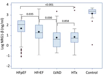
As depicted in Figure1 NRG1-β was higher in HFpEF, (6.5 [2.1-11.3]ng/mL compared to HFrEF 3.6 [2.1-7.6]ng/mL (P = 0.035). Concentrations decreased after LVAD treatment, 1.7 [0.9-3.6]ng/mL and HTx 2.1 [1.4-3.6]ng/mL (overall P < 0.001). NRG1-β was substantially lower in all groups, individ-ually and overall, compared to controls: 29.0 ng/mL [23.1-34.3] (P = 0.001).
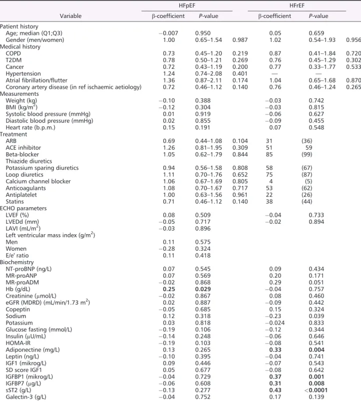
Neuregulin1-β did not correlate with age, sex, co-morbidities, and echocardiographic measures in either HFpEF or HFrEF (Table 2). Among biomarkers in HFpEF, only haemoglobin correlated with NRG1-β (β = 0.25; P = 0.029), and in HFrEF, ST2 (β = 0.43; P < 0.001), adiponectin (β = 0.33; P = 0.004), and IGFBP1 (β = 0.37;
P = 0.001).
In HFpEF, NRG1-β decreased with increasing NYHA class (overall P = 0.013) in contrast to HFrEF where NRG1-β was higher in NYHA class IV versus III (P-value = 0.030). NRG1-β stratified by NYHA class between HFpEF and HFrEF displayed no significant difference.
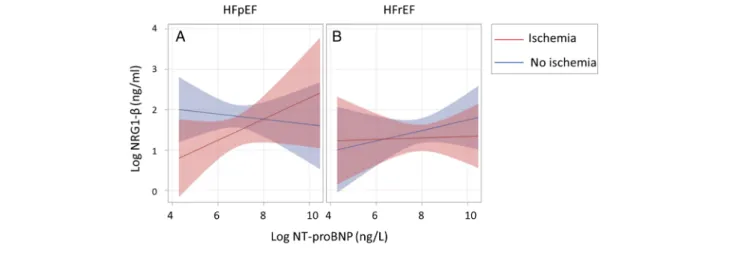
In ischaemic HFpEF, NRG1-β was insignificantly lower (5.2 [1.5–8.9] ng/mL) compared with non-ischaemic HFpEF (7.0 [2.3–11.9] ng/mL; P = 0.274), whereas there was no differ-ence in ischaemic HFrEF (3.3 [2.1–6.3]) and non-ischaemic HFrEF (3.9 [2.1–7.7]); P = 0.484). Interestingly, stratifying the HFpEF group by ischaemia, there was a trending association between increasing NRG1-β and NT-proBNP in ischaemic but not in non-ischaemic HFpEF (Figure 2A; P-interaction =0.589). This pattern was not seen in HFrEF
Table 1 Ba seline characteris tics in the 86 HFp EF, 86 HFrE F, 26 LVA D, 21 HTx patients, and 21 heal thy con trols Varia ble HFp EF (n = 86) HFrE F (n = 86) P -valu e (H FrEF versu s HFpEF) LVAD (n = 26) HT x (n = 35) P -valu e (overall HFrEF) Contr ols (n = 21) P -valu e (controls ver sus HFpE F) P -valu e (c ontrols ver sus HFrEF) Patient history Age (yea rs) 73 (67 ;79) 63 (52;68 ) < 0.001 53 (45 ;66) 51 (44;61 ) 0.001 67 (61;70) < 0.00 1 0.088 Gend er (females) 44 (51 %) 16 (19% ) < 0.001 6 (23 %) 6 (17% ) 0.832 12 (57%) 0.80 8 < 0.001 Medic al his tory COPD 17 (20 %) 10 (12% ) 0.208 1 (4% ) 1 (3%) 0.189 T2DM 28 (33 %) 25 (29% ) 0.741 6 (29 %) 6 (23% ) 0.710 Cancer 15 (17 %) 8 (9%) 0.178 2 (8% ) 1 (3%) 0.474 Hyperten sion 68 (79 %) —— — — — Atrial fi brilla tion /fl utte r 52 (60 %) 45 (52% ) 9 (35 %) 18 (51% ) 0.270 Coron ary artery disease 29 (34 %) 38 (44% ) 0.211 11 (42 %) 10 (29% ) 0.274 Whereof revascularized 17 (59 %) 30 (79% ) < 0.001 9 (82 %) 9 (90% ) Revascularized 17 (20 %) 33 (38% ) 0.011 12 (46 %) 11 (31% ) 0.050 NYHA class I 1 9 (22 %) 1 (1%) < 0.001 1 (4% ) 2 2 (63% ) < 0.001 NYHA class II 47 (55 %) 4 (5%) 14 (54 %) 10 (29% ) NYHA class III 20 (23 %) 67 (78% ) 7 (27 %) 2 (6%) NYHA class IV 0 (0% ) 1 4 (16% ) 1 (4% ) 1 (3%) Measureme nts Weigh t (kg) 83.5 (72 ;98) 83.8 (72;94 ) 0.951 80 (69 ;88) 84 (77;98 ) 0.310 BMI (kg/m 2 ) 28.5 (25 .0;32.9) 27. 3(23.2;30.1) 0.016 25 (23 ;27) 26 (23;30 ) 0.351 25 (23;26) 0.00 2 0.071 Systolic bl ood pressure (m mHg) 140 (13 0;150) 105 (96;12 0) < 0.001 103 (10 0;113) 118 (95;13 0) 0.495 125 (120;140) 0.00 2 < 0.001 Diastol ic blood pressure (m mHg) 80 (70 ;85) 70 (62;80 ) < 0.001 70 (63 ;73) 74 (65;83 ) 0.485 80 (75;83) 0.79 0 < 0.001 Heart rate (b. p.m.) 70 (60 ;80) 70 (60;74 ) 0.484 70 (64 ;71) 83 (74;88 ) 0.001 Treatm ent ARB 28 (33 %) 31 (36% ) 0.748 9 (35 %) 23 (37% ) 0.007 ACE inhi bitor 42 (49 %) 51 (59% ) 0.221 16 (62 %) 0 (0%) < 0.001 Beta-blocker 69 (80 %) 85 (99% ) < 0.001 23 (88 %) 15 (43% ) < 0.001 Thiazi de diure tics 14 (16 %) Potassium sparing diureti cs 18 (21 %) 58 (67% ) < 0.001 15 (58 %) 3 (9%) < 0.001 Loop diur etics 61 (71 %) 75 (87% ) 0.014 15 (58 %) 11 (31% ) < 0.001 Calcium chann el blocker 27 (31 %) 4 (5%) < 0.001 5 (19 %) 9 (26% ) 0.003 Anticoa gulant s 4 7 (55 %) 53 (62% ) 0.440 23 (88 %) 1 (3%) < 0.001 Antipla telet 29 (34 %) 22 (26% ) 0.317 19 (73 %) 31 (89% ) < 0.001 Statins 38 (44 %) 38 (44% ) 1.000 6 (23 %) 31 (41% ) < 0.001 ECHO para meters LVEF (%) 64 (58 ; 68) 21 (15;28 ) < 0.001 20 (13 ;30) 58 (55;63 ) < 0.001 LVEDd (m m) 47 (43 ;53) 66 (61;75 ) < 0.001 56 (52 ;66) 46 (42;50 ) < 0.001 LAVI (mL/m 2 ) 4 4 (38 ; 52) Left ve ntricu lar mass index (g/ m 2 ) 114 (95 ;143) Men 125 (10 2;157) (Co ntinu es )
Table 1 (c ontinued) Varia ble HFp EF (n = 86) HFrE F (n = 86) P -valu e (H FrEF versu s HFpEF) LVAD (n = 26) HT x (n = 35) P -valu e (overall HFrEF) Contr ols (n = 21) P -valu e (controls ver sus HFpE F) P -valu e (c ontrols ver sus HFrEF) Wom en 109 (94 ;136) E/A rati o 1.3 (0. 9;2.1) E/e ′ratio 10.8 (8. 3;14.0) E′ 8.0 (7. 0;10.0) IVRT (dia stole) 94 (77 ;113) Mitra l VTI 23 (16 ;30) E-wave decelera tion tim e (ms) 203 (15 6;228) Biochem istry NT-pro BNP (ng /L) 1000 (46 9;2330) 3290 (1430; 586 0) < 0.001 1115 (82 4;1760) 247 (166;469) < 0.001 67 (31;110) < 0.00 1 < 0.001 MR-p roANP (pmo l/ L) 313 (19 3;381) 449 (326;593 ) < 0.001 276 (22 4;301) 118 (96;15 8) < 0.001 MR-p roADM (nmol/ L) 1.22 (0. 93; 1.6 2) 1.33 (0.94; 1.9 5) 0.215 0.92 (0. 75;1.30 ) 0.67 (0.56; 0.86) < 0.001 Hb (g/ dL) 131 (12 2;142) 133 (122;144 ) 0.619 130 (11 1;138) 129 (115;138) 0.158 Creatinine (μ mol /L) 84 (73 ;107) 113 (98;14 3) < 0.001 104 (74 ;125) 96 (83;11 2) 0.004 eGFR (M DRD) (mL/ min/1 .73m 2 ) 70 (54 ;85) 59 (44;71 ) 0.004 65 (55 ;83) 77 (58;91 ) < 0.003 Copep tin (pmo l/L) 14 (9; 21) 28 (18;45 ) < 0.001 16 (7; 18) 12 (5;19) < 0.001 Suppre ssion of tumouri genic ity 2 (ng/mL) 23 (17 ;31) 35 (23;51 ) < 0.001 28 (26 ;32) 22 (17;26 ) < 0.001 Sodiu m (m mol/L) 141 (14 0;143) 138 (136;140 ) < 0.001 138 (13 6;139) 140 (138;142) < 0.001 Potassium (mmol /L) 3.9 (3. 7;4.2) 4.2 (3.9;4 .6) < 0.001 4.4 (4. 0;4.7) 3.9 (3.7;4 .2) 0.008 Glucos e fasting (mmol /L) 5.6 (5. 1;7.5) 5.5 (4.9;6 .9) 0.091 5.6 (5. 0;6.2) 5.3 (4.9;7 .2) 0.974 Insulin (μ U /mL) 11.2 (8. 4;16.9) 11.1 (6.7;1 6.7) 0.642 11.7 (6. 9;18.0) 9.5(6.8;14.0) 0.721 HOMA-IR 3.4 (2. 0;5.6) 2.8 (1.1;5 .1) 0.034 2.5 (1. 63;4.4) 2.2 (1.5;3 .1) 0.899 Adipon ectin (mg/L ) 11.7 (7. 8;20.1) 13.7 (7.0;2 1.1) 0.471 10.5 (96 ;272) 11.5 (6.8;1 5.9) 0.210 Leptin (ng/L) 24.1 (11 .7;51.7) 15.0 (6.2;3 3.2) 0.015 10.6 (7. 9;36.6) 10.4 (7.0;1 8.2) 0.531 IGF1 (m ikro g/L) 174 (13 7;206) 149 (105;219 ) 0.101 138 (96 ;272) 214 (170;306) 0.001 SD score IGF1 1.22 (0. 62;1.93) 0.09 ( 1.40 ;1.6 2) < 0.001 0.14 ( 1.4 ;1.1) 1.0 ( 0.4; 2.1) 0.053 IGFBP 1 (mikro g/L) 48 (28 ;78) 65 (29;10 1) 0.074 54 (35 ;77) 28 (20;39 ) < 0.001 ACE, ang iotens in con vertin g enzym e; ARB, ang iotens in II re cepto r bloc ker; BMI, body mass ind ex; COPD , chro nic ob structive p u lmonary di sease; eGF R , estim ated glom erula r fi ltration rate; Hb, haem oglo bin; HFp EF, heart failure w ith pres erved ejec tion frac tion; HFrEF, heart failure w ith re duced ejectio n fract ion; HOM A-IR, hom eo static model as sessmen t o f insuli n re-sistance; HT x, heart tran splantation; IGF1, ins ulin-lik e g rowth fact or 1; IVRT, isovol umi c relax ation tim e; LAV I, left atri al volu me ind ex; LVAD , left ventri cular assist device; LVED d, left ventri cular end dias tolic diam eter; LVEF, left ventri cular ejec tion fract ion; MDRD , Modi fi catio n o f Diet in Re nal Dise ase; MR-p roADM , mid-regi ona l pro -adrenom edu llin; MR -proAN P, mid-regi ona l pro atrial natr iuret ic peptide ; N T -pro BNP, N-term ina l pro-brain natr iuretic peptide; NYHA , New Yo rk Hear t Associat ion; SD , st andar d devi ation; T2DM, type 2 d iabetes mellitu s; VTI, velocit y-time int egral . Continu ous variables are present ed as med ian and lower and up per qu artiles (Q1 ;Q3) and catego rica l varia bles as num bers (n ) and perc entages .
Association with outcome
Median follow-up time in patients with HFpEF and in patients with HFrEF was522 days [238;1089] and 204 days [56;415], respectively. No patient was lost to follow-up. In HFpEF, the composite endpoint of all-cause death and HF hospitalization occurred in36 patients, whereof six were deaths. In HFrEF, the composite endpoint of all-cause death, LVAD implanta-tion, or HTx occurred in 56 patients out of which 28 were deaths.
In HFpEF, concentration of NRG1-β was not associated with the composite endpoint (hazard ratio (HR) per log in-crease0.75 [95% confidence interval (CI) 0.55–1.04; P-value = 0.083]; Figure 3A). When stratified by ischaemia, there was an association with outcome (log-rank P =0.020), reveal-ing that ischaemic HFpEF patients with NRG1-β above median had a worse outcome (Figure3C). The pattern was confirmed in the multivariable model but did not reach statistical signif-icance; ischaemic HFpEF (HR 1.07, 95% CI 0.52–2.17, P = 0.862) and non-ischaemic HFpEF (HR 0.71, 95% CI 0.48– 1.05, P = 0.085; Pinteraction=0.553).
In HFrEF, NRG1-β was prognostic (Figure3B) also in the multivariable model (HR 1.45 [95% CI 1.04–2.03; P-value = 0.029]). The association with outcome persisted when strati-fied for ischaemia (log-rank P = 0.003; Figure3D) After
adjust-ments in the multivariable model; in ischaemic HFrEF (HR 1.17, 95% CI 0.68–2.04, P = 0.569) and in non-ischaemic HFrEF (HR1.48, 95% CI 0.93–2.36, P = 0.098).
In addition, we analysed outcome stratified by above or below median NRG1-β and NT-proBNP. In HFpEF, worst out-come was in patients below median NRG1-β and above NT-proBNP (P = 0.049), while in patients with HFrEF, the strata above median NRG1-β and above median NT-proBNP had
the poorest prognosis (P < 0.001; Supporting Information,
Figure S1).
Discussion
Neuregulin1-β was reduced in both HFpEF and, even more, in HFrEF compared with healthy controls. In HFrEF, concentra-tions were further decreased after LVAD and HTx. In HFrEF and ischaemic HFpEF, higher levels of NRG1-β were associ-ated with worse outcomes. In contrast, in non-ischaemic HFpEF, it was reversed, and NRG1-β appeared to be poten-tially protective with higher concentrations tending to be as-sociated with better outcomes. The opposing relationship of NRG1-β with outcomes raises the possibility of a compensa-tory increase of cardioprotective NRG1-β from the endothe-lial microvasculature exposed to oxidative stress in non-ischaemic HFpEF, but this compensatory effect is overwhelmed in the presence of ischaemia in HFrEF and isch-aemic HFpEF.
Neuregulin
1-β concentrations
We confirm previous findings in HFrEF demonstrating an as-sociation between higher concentrations of circulating NRG1-β and HF disease severity.8,9 In addition, we demon-strate lower concentrations after LVAD and HTx. NRG1, like BNP, has been reported to be cardioprotective and part of the adaptive physiologic response in HF, that is, NRG1, like BNP, is a risk marker for more severe HFrEF and worse out-comes but not a risk factor that causes worse outout-comes.10 The most elevated concentrations of NRG1-β were found in healthy controls. This may be explained by a slightly lower age and absence of inflammation, contributing to a more pre-served endothelial function.
We found lower concentrations of circulating NRG1-β in HFrEF compared with HFpEF and controls which is counterin-tuitive as the neurohormonal activation is greater in HFrEF. Our patients with HFrEF all had severe HF referred for ad-vanced interventions such as LVAD or HTx reflecting signifi-cant pump failure in later stages of HF. Animal models suggest that concentric left ventricular hypertrophy and me-chanical wall strain initially increase NRG1 mRNA expres-sion.11At the same time, the ERBB2 and ERBB4 receptors are downregulated.7 As terminal HF approaches and pump failure occurs, NRG1 declines potentially due to increasing cir-culating levels of angiotensin II and epinephrine.11Although speculative, the lower NRG1-β concentrations in our patients with HFrEF after intervention could be mediated by a declin-ing neurohormonal activation accompanied by upregulation of the ERBB2 and ERBB4 receptors.7,12
Figure1 Concentrations of log NRG1-β in HFpEF, HFrEF, 1 year after left ventricular assist device implantation (LVAD),1 year after heart trans-plantation (HTx), and healthy controls. All groups individually and overall versus control P< 0.001.
Table 2 Correlates of NRG1-β in the 86 patients with HFpEF and the 86 patients with HFrEF from regression analyses.
Variable
HFpEF HFrEF
β-coefficient P-value β-coefficient P-value
Patient history Age; median (Q1;Q3) 0.007 0.950 0.05 0.659 Gender (men/women) 1.00 0.65–1.54 0.987 1.02 0.54–1.93 0.956 Medical history COPD 0.73 0.45–1.20 0.219 0.87 0.41–1.84 0.720 T2DM 0.78 0.50–1.21 0.269 0.76 0.45–1.29 0.302 Cancer 0.72 0.43–1.19 0.200 0.77 0.33–1.77 0.533 Hypertension 1.24 0.74–2.08 0.401 — — Atrialfibrillation/flutter 1.36 0.87–2.11 0.174 1.04 0.65–1.68 0.870
Coronary artery disease (in ref ischaemic aetiology) 0.72 0.46–1.12 0.140 0.76 0.46–1.24 0.265
Measurements
Weight (kg) 0.10 0.388 0.03 0.742
BMI (kg/m2) 0.12 0.304 0.03 0.815
Systolic blood pressure (mmHg) 0.01 0.919 0.06 0.627
Diastolic blood pressure (mmHg) 0.02 0.855 0.09 0.455
Heart rate (b.p.m.) 0.15 0.191 0.07 0.548 Treatment ARB 0.69 0.44–1.08 0.104 31 (36) ACE inhibitor 1.26 0.81–1.95 0.309 51 59 Beta-blocker 1.05 0.62–1.79 0.844 85 (99) Thiazide diuretics
Potassium sparing diuretics 0.94 0.56–1.58 0.808 58 (67)
Loop diuretics 1.11 0.70–1.76 0.652 75 (87)
Calcium channel blocker 1.06 0.67–1.69 0.805 4 (5)
Anticoagulants 1.08 0.70–1.67 0.717 53 (62) Antiplatelet 1.00 0.63–1.56 0.961 22 (26) Statins 0.71 0.46–1.12 0.140 38 (44) ECHO parameters LVEF (%) 0.08 0.509 0.04 0.733 LVEDd (mm) 0.05 0.717 0.02 0.894 LAVI (mL/m2) 0.03 0.896
Left ventricular mass index (g/m2)
Men 0.11 0.575 Women 0.28 0.324 E/e′ ratio 0.11 0.418 Biochemistry NT-proBNP (ng/L) 0.07 0.545 0.09 0.434 MR-proANP 0.07 0.569 0.20 0.171 MR-proADM 0.02 0.868 0.29 0.051 Hb (g/dL) 0.25 0.029 0.04 0.757 Creatinine (μmol/L) 0.02 0.867 0.08 0.460 eGFR (MDRD) (mL/min/1.73 m2) 0.02 0.887 0.09 0.442 Copeptin 0.05 0.685 0.15 0.324 Sodium 0.12 0.318 0.23 0.039 Potassium 0.03 0.818 0.024 0.833
Glucose fasting (mmol/L) 0.19 0.106 0.12 0.344
Insulin (μU/mL) 0.14 0.248 0.06 0.646 HOMA-IR 0.19 0.103 0.08 0.541 Adiponectine (mg/L) 0.13 0.265 0.33 0.004 Leptin (ng/L) 0.10 0.395 0.04 0.741 IGF1 (mikrog/L) 0.09 0.446 0.07 0.543 SD score IGF1 0.05 0.677 0.08 0.642 IGFBP1 (mikrog/L) 0.04 0.729 0.37 0.001 IGFBP7 (μg/L) 0.06 0.608 0.31 0.008 sST2 (g/L) 0.13 0.277 0.43 <0.0001 Galectin-3 (g/L) 0.04 0.752 0.17 0.139
ACE, angiotensin converting enzyme; ARB, angiotensin II receptor blocker; BMI, body mass index; COPD, chronic obstructive pulmonary disease; eGFR, estimated glomerularfiltration rate; Hb, haemoglobin; HFpEF, heart failure with preserved ejection fraction; HFrEF, heart failure with reduced ejection fraction; HOMA-IR, homeostatic model assessment of insulin resistance; IGF1, insulin-like growth factor 1; IVRT, isovolumic relaxation time; LAVI, left atrial volume index; LVEDd, left ventricular end diastolic diameter; LVEF, left ventricular ejection fraction; MDRD, Modification of Diet in Renal Disease; MR-proADM, mid-regional pro-adrenomedullin; MR-proANP, mid-regional pro atrial natriuretic peptide; NT-proBNP, N-terminal pro-brain natriuretic peptide; NRG1-β, neuregulin1-β; SD, standard deviation; T2DM, type 2 diabetes mellitus.
Prognostic implications of neuregulin
1-β
In concordance with previous findings, we found higher NRG1-β to be associated with increased mortality in HFrEF.8 In addition, Ky and colleagues suggest NRG1-β as a prognostic predictor in ischaemic but not in non-ischaemic HFrEF. In our study, it appeared prognostic in HFrEF regardless of the pres-ence of ischaemia defined as ischaemic HF aetiology. We add
new information on the role of NRG1-β in ischaemic versus non-ischaemic HFpEF. Patients with HFpEF with ischaemia de-fined as the presence of CAD and a high NRG1-β had the worst outcomes, whereas patients with HFpEF without CAD and high NRG1-β had the best outcomes.
In support of the association between outcome and NRG 1-β in ischaemia, it has been shown that NRG/erbB signalling is activated in the setting of ischaemia-reperfusion injury,13and
Figure3 (A–D) Kaplan–Meier analyses displaying increased survival free of HF hospitalization by median NRG1-β (A) in HFpEF displaying improved prognosis above median NRG1-β; (B) in HFrEF displaying worse prognosis above median NRG1-β; (C) in HFpEF displaying worse prognosis in the pres-ence of ischaemia above median NRG1-β but still improved prognosis in the absence of ischaemia; and (D) in HFrEF displaying worse prognosis above median NRG1-β regardless of the presence of ischaemia.
Figure2 Association between log NRG1-β concentrations and log NT-proBNP and interaction of ischaemia (A) in HFpEF and (B) in HFrEF. Curves depict, with95% confidence interval, presence of ischaemia in red and no ischaemia present in blue.
NRG1-β concentrations are initially elevated in patients with stress-induced ischaemia, but as HFrEF progresses, the endo-thelium is unable to release NRG1-β and concentrations dete-riorate. Both NRG1-β mRNA expression and ERBB2 and ERBB4 receptors are downregulated in hypoxic versus normoxic areas,14and NRG1 is inversely correlated with an-giographic severity.15
This raises the possibility, which needs further studies and confirmation, that NRG1-β has diverging roles in the presence and evolvement of ischaemia in HFpEF. In non-ischaemia, de-clining NRG1-β concentrations may rather reflect the disease-driving oxidative stress contributing to the microvascular en-dothelial dysfunction that is hypothesized to drive the HFpEF syndrome.3
Endothelial dysfunction, highly prevalent in HF regardless of LVEF, is associated with cardiovascular death and HF hospi-talization. In the myocardium, NRG1 is primarily produced by the microvascular endothelium in response to mechanical stretch, oxidative stress, and hypoxia.5NRG1/ErbB signalling and NRG1 are suggested to increase the number of microvessels in post-ischaemic animal models.16 Further, demonstrating the link between endothelial dysfunction and oxidative stress, we found that NRG1-β correlated with the oxidative stress marker IGFBP-1 in our patients with HFrEF.
Even if coronary microvascular inflammation and dysfunc-tion occur in both HFrEF and HFpEF, it is suggested to have a more prominent role in the pathophysiology and disease development in HFpEF.3 Supporting this hypothesis, we re-cently showed that coronary microvascular dysfunction as de-fined by reduced coronary flow reserve was present in 75% of patients with HFpEF and also correlated with peripheral en-dothelial dysfunction.17
Neuregulin1 is interesting in this aspect. It may act on both the ventricular and cardiomyocyte levels attenuating endo-thelial dysfunction and collagen synthesis thus potentially im-proving passive ventricular stiffness.18–20NRG1 has an impact on endothelial dysfunction reducing pro-inflammatory cyto-kines affecting the pro-fibrotic transforming growth factor beta 1 signalling pathway through ErbB4 receptor activa-tion.19It also acts on the cardiomyocyte calcium metabolism, at the level of the sarcoplasmatic reticulum through phos-phorylation of phospholamban and correct the altered titin phosphorylation.21,22
Limitations
There are limitations in this relatively small single centre co-hort study. Measured plasma NRG1-β reflects the circulation of NRG1 but not necessarily biologically active peptide or re-ceptor activation. The definition of the composite outcome differed between the groups. In HFrEF, a surrogate endpoint reflecting clinical deterioration, that is, HTx or LVAD, was in-cluded because these events reduce competing risk. In
HFpEF, HF hospitalization was a part of the composite end-point. We selected these most clinically relevant composite outcomes because we did not compare outcomes in HFrEF versus HFpEF, but we compared associations between NRG1-β and outcomes in HFrEF versus in HFpEF separately. The small sample size may hamper the results and does not allow extensively adjusted survival analyses, but we have ad-justed for a few important variables such as renal function, NT-proBNP, age, and sex. Further, we have measured circu-lating NRG1-β that does not necessarily reflect processes in the myocardium.
Conclusions
Neuregulin1-β was reduced in both HFpEF and, even more, in HFrEF compared with healthy controls. In HFrEF, concentra-tions were further decreased after LVAD and HTx. In HFrEF and ischaemic HFpEF, higher levels of NRG1-β were associ-ated with worse outcomes. In contrast, in non-ischaemic HFpEF, it was reversed, and NRG1-β appeared to be poten-tially protective with higher concentrations tending to be as-sociated with better outcomes. The opposing relationship of NRG1-β with outcomes raises the possibility of compensatory increases of cardioprotective NRG1-β from the endothelial microvasculature exposed to oxidative stress in non-ischaemic HFpEF, but this compensatory effect is overwhelmed in the presence of HFrEF and ischaemic HFpEF.
Con
flict of interest
There are no conflicts of interest related to this study. How-ever, to the extent that findings in KaRen and MetAnEnd may affect the use of heart failure drugs or devices, we dis-close the following. C.H. received consulting fees from Novartis and Roche and speaker honoraria from MSD; L.H.L. received research grants from AstraZeneca, Relypsa, Novartis, Boerhinger Ingelheim, and Boston Scientific and consulting or speaker honoraria from Novartis, Vifor, Bayer, Sanofi, Fresenius, and Merck; C.L. received research grants, speaker honoraria, and consulting fees from Medtronic and speaker honoraria and consulting fees from St. Jude Medical; E.D. received speaker honoraria and consulting fees from Novartis and AstraZeneca; J.C.D. received research grants, speaker honoraria, and consulting fees from Medtronic and St. Jude Medical; C.S.P.L. was supported by a Clinician Scien-tist Award from the National Medical Research Council of Singapore; has received research support from Boston Scien-tific, Bayer, Roche Diagnostics, AstraZeneca, Medtronic, and Vifor Pharma; has served as consultant or on the Advisory Board/Steering Committee/Executive Committee for Boston Scientific, Bayer, Roche Diagnostics, AstraZeneca, Medtronic,
Vifor Pharma, Novartis, Amgen, Merck, Janssen Research & Development LLC, Menarini, Boehringer Ingelheim, Novo Nordisk, Abbott Diagnostics, Corvia, Stealth BioTherapeutics, JanaCare, Biofourmis, Darma, Applied Therapeutics, WebMD Global LLC, and Radcliffe Group Ltd.
Funding
None of the funding sources had any role in the design and conduct of the study and in the collection, management, analysis, or interpretation of the data. The work was sup-ported by grants from Center for Gender Medicine Karolinska Institutet, Stockholm Sweden (to C.H. and A.M.B.), and The Prospective KaRen study was supported in part by grants from Fédération Française de Cardiologie/Société Française de Cardiologie, France and Medtronic Bakken Research Center, Maastricht, The Netherlands. L.H.L. was supported
by the Swedish Research Council (grant 2013-23897-104604-23), Swedish Heart Lung Foundation (grants 20080409, 20100419), and the Stockholm County Council (grants 00556-2009, 20110120). C.L. was supported by the Swedish Heart Lung Foundation (grants 20080498, 20110406] and the Stockholm County Council (grants 20090376, 20110610).
Supporting information
Additional supporting information may be found online in the Supporting Information section at the end of the article.
Figure S1.Survival free of HF hospitalization above and below
median of NRG and NT-proBNP in HFpEF and HFrEF.
References
1. Ponikowski P, Voors AA, Anker SD, Bueno H, Cleland JGF, Coats AJS, Falk V, González-Juanatey JR, Harjola V-P, Jankowska EA, Jessup M, Linde C, Nihoyannopoulos P, Parissis JT, Pieske B, Riley JP, Rosano GMC, Ruilope LM, Ruschitzka F, Rutten FH, van der Meer P. 2016 ESC guidelines for the diagnosis and treatment of acute and chronic heart failure. Eur J Heart Fail 2016;18: 891–975.
2. Marti CN, Gheorghiade M,
Kalogeropoulos AP, Georgiopoulou VV, Quyyumi AA, Butler J. Endothelial dys-function, arterial stiffness, and heart failure. J Am Coll Cardiol 2012; 60: 1455–1469.
3. Paulus WJ, Tschope C. A novel paradigm for heart failure with preserved ejection fraction: comorbidities drive myocardial dysfunction and remodeling through coronary microvascular endothelial in-flammation. J Am Coll Cardiol [Review] 2013;62: 263–271.
4. Timolati F, Ott D, Pentassuglia L, Giraud MN, Perriard JC, Suter TM, Zuppinger
C. Neuregulin-1 beta attenuates
doxorubicin-induced alterations of excitation-contraction coupling and re-duces oxidative stress in adult rat cardiomyocytes. J Mol Cell Cardiol 2006;41: 845–854.
5. Hedhli N, Huang Q, Kalinowski A, Palmeri M, Hu X, Russell RR, Russell KS. Endothelium-derived neuregulin protects the heart against ischemic in-jury. Circulation 2011;123: 2254–2262. 6. Brero A, Ramella R, Fitou A, Dati C,
Alloatti G, Gallo MP, Levi R.
Neuregulin-1β1 rapidly modulates nitric
oxide synthesis and calcium handling in rat cardiomyocytes. Cardiovasc Res 2010;88: 443–452.
7. Rohrbach S, Niemann B, Silber RE, Holtz J. Neuregulin receptors erbB2 and erbB4
in failing human myocardium
—de-pressed expression and attenuated acti-vation. Basic Res Cardiol [Research Support, Non-U.S. Gov’t] 2005; 100: 240–249.
8. Ky B, Kimmel SE, Safa RN, Putt ME, Sweitzer NK, Fang JC, Sawyer DB, Cappola TP. Neuregulin-1β is associated with disease severity and adverse out-comes in chronic heart failure.
Circula-tion 2009;120: 310–317.
9. Miao J, Huang S, Su YR, Lenneman CA, Wright M, Harrell FE Jr, Sawyer DB, Lenihan DJ. Effects of endogenous se-rum neuregulin-1β on morbidity and mortality in patients with heart failure and left ventricular systolic dysfunction.
Biomarkers 2018;23: 704–708.
10. Rupert CE, Coulombe KL. The roles of neuregulin-1 in cardiac development, homeostasis, and disease. Biomark
In-sights 2015;10: 1–9.
11. Lemmens K, Doggen K, De Keulenaer GW. Role of neuregulin-1/ErbB signal-ing in cardiovascular physiology and dis-ease: implications for therapy of heart failure. Circulation 2007;116: 954–960. 12. Uray IP, Connelly JH, Thomazy V,
Ship-ley GL, Vaughn WK, Frazier OH,
Taegtmeyer H, Davies PJ. Left ventricu-lar unloading alters receptor tyrosine ki-nase expression in the failing human heart. J Heart Lung Transplant [Re-search Support, U.S. Gov’t, P.H.S.] 2002;21: 771–782.
13. Kuramochi Y, Cote GM, Guo X,
Lebrasseur NK, Cui L, Liao R, Sawyer DB. Cardiac endothelial cells regulate reactive oxygen species-induced cardio-myocyte apoptosis through neuregulin-1β/erbB4 signaling. J Biol Chem [Re-search Support, Non-U.S. Gov’t Re-search Support, U.S. Gov’t, P.H.S.] 2004;279: 51141–51147.
14. Munk M, Memon AA, Goetze JP, Nielsen LB, Nexo E, Sorensen BS. Hypoxia changes the expression of the epidermal growth factor (EGF) system in human hearts and cultured cardiomyocytes.
PLoS ONE 2012;7: e40243.
15. Geisberg CA, Wang G, Safa RN, Smith HM, Anderson B, Peng XY, Veerkamp B, Zhao DX, Blakemore D, Yu C, Sawyer DB. Circulating neuregulin-1β levels vary according to the angiographic se-verity of coronary artery disease and is-chemia. Coron Artery Dis [Research Support, N.I.H., Extramural Research Support, Non-U.S. Gov’t] 2011; 22: 577–582.
16. Wu C, Gui C, Li L, Pang Y, Tang Z, Wei J. Expression and secretion of neuregulin-1 in cardiac microvascular endothelial
cells treated with angiogenic
factors. Exp Ther Med 2018; 15:
3577–3581.
17. Shah SJ, Lam CSP, Svedlund S, Saraste A, Hage C, Tan RS, Beussink-Nelson L, Ljung Faxen U, Fermer ML, Broberg MA, Gan LM, Lund LH. Prevalence and correlates of coronary microvascular dysfunction in heart failure with
pre-served ejection fraction:
PROMIS-HFpEF. Eur Heart J 2018; 39:
18. Mendes-Ferreira P, Maia-Rocha C, Adao R, Mendes MJ, Santos-Ribeiro D, Alves BS, Cerqueira RJ, Castro-Chaves P, Lourenco AP, De Keulenaer GW, Leite-Moreira AF, Bras-Silva C. Neuregulin-1 improves right ventricular function and attenuates experimental pulmonary ar-terial hypertension. Cardiovasc Res 2016;109: 44–54.
19. Vermeulen Z, Hervent AS, Dugaucquier L, Vandekerckhove L, Rombouts M,
Beyens M, Schrijvers DM, De
Meyer GRY, Maudsley S, De Keulenaer GW, Segers VFM. Inhibitory actions of the NRG-1/ErbB4 pathway in macro-phages during tissue fibrosis in the heart, skin, and lung. Am J Physiol
Heart Circ Physiol 2017; 313:
H934–H945.
20. Galindo CL, Kasasbeh E, Murphy A, Ryzhov S, Lenihan S, Ahmad FA, Wil-liams P, Nunnally A, Adcock J, Song Y, Harrell FE, Tran TL, Parry TJ, Iaci J, Ganguly A, Feoktistov I, Stephenson MK, Caggiano AO, Sawyer DB, Cleator JH. Anti-remodeling and anti-fibrotic ef-fects of the neuregulin-1β glial growth factor 2 in a large animal model of heart failure. J Am Heart Assoc 2014; 3: e000773.
21. Adao R, Mendes-Ferreira P, Maia-Rocha C, Santos-Ribeiro D, Rodrigues PG, Vidal-Meireles A, Monteiro-Pinto C,
Pimentel LD, Falcao-Pires I, De
Keulenaer GW, Leite-Moreira AF, Bras-Silva C. Neuregulin-1 attenuates right ventricular diastolic stiffness in experi-mental pulmonary hypertension. Clin
Exp Pharmacol Physiol 2018; 19:
1440–1681.
22. Hopf A-E, Andresen C, Kötter S, Isić M, Ulrich K, Sahin S, Bongardt S,
Röll W, Drove F, Scheerer N,
Vandekerckhove L, De Keulenaer GW, Hamdani N, Linke WA, Krüger M.
Diabe-tes-induced cardiomyocyte passive
stiffening is caused by impaired
insulin-dependent titin modification
and can be modulated by neuregulin-1. Circ Res 2018;123: 342–355.