Chromosome Intermingling: Mechanical
Hotspots for Genome Regulation
The MIT Faculty has made this article openly available. Please share how this access benefits you. Your story matters.
Citation Uhler, Caroline and G.V. Shivashankar. "Chromosome Intermingling: Mechanical Hotspots for Genome Regulation." Trends in Cell Biology 27, 11 (November 2017): P810-819. © 2017 Elsevier Ltd
As Published https://doi.org/10.1016/j.tcb.2017.06.005 Publisher Elsevier BV
Version Author's final manuscript
Citable link https://hdl.handle.net/1721.1/130126
Terms of Use Creative Commons Attribution-NonCommercial-NoDerivs License Detailed Terms http://creativecommons.org/licenses/by-nc-nd/4.0/
Chromosome Intermingling: Mechanical Hotspots for Genome Regulation
Caroline Uhler# and G.V.Shivashankar*
# Department of Electrical Engineering & Computer Science,
Institute for Data, Systems & Society, MIT, Cambridge, USA
* Mechanobiology Institute, National University of Singapore, Singapore &
FIRC Institute of Molecular Oncology (IFOM), Milan, Italy
Abstract
Cells sense physical and chemical signals from their local microenvironment and transduce them to the nucleus to regulate genomic programs. In this review, we first discuss different modes of mechanotransduction to the nucleus. Then we highlight the role of the spatial organization of chromosomes for integrating these signals. In particular, we emphasize the importance of chromosome intermingling for gene regulation. We also discuss various geometric models and recent advances in microscopy and genomics that have allowed accessing these nanoscale chromosome intermingling regions. Taken together, the recent work summarized in this review culminates in the hypothesis that the chromosome intermingling regions are mechanical hotspots for genome regulation. Maintenance of such mechanical hotspots is crucial for cellular homeostasis, and alterations in them could be precursors for various cellular reprogramming events including diseases.
Trends:
- Microenvironmental signals are transmitted to the cell nucleus via both, physical and biochemical intermediates.
- The spatial organization of chromosomes is critical to regulating microenvironmental control of gene expression.
- Intermingling regions between chromosomes are enriched with transcription factors and RNA Pol II.
- The functional clustering of genes is modulated by microenvironmental signals to exhibit differential gene expression programs.
Introduction
Cellular differentiation programs result in a few hundred different cell types with well-defined transcription profiles. In this process, cells adapt to the tissue micro-environment and simultaneously sculpt it, thereby defining its geometry, rigidity, and mechano-sensitivity (1-4). Cells seamlessly transition through the epigenetic landscape during differentiation and trans-differentiation programs in our body. These transitions are initiated at the molecular level: cells sense the geometry and stiffness of the micro-environment and perceive a number of biochemical signals via proteins on the cell membrane (5-7). These signals are transduced to the nucleus via a number of canonical signaling pathways (8-9). In addition, the
nucleus is linked with an elaborate meshwork of cytoskeletal filaments, including actin, microtubules, and intermediate filaments, that bridge the extracellular microenvironment with the nuclear envelope and the genome (10-12). The chemical and physical signals that reach the nucleus are translated via chromatin remodeling enzymes and the transcription machinery to facilitate spatial control of gene expression (13-15). Recent studies are beginning to reveal that the 3D neighborhoods of chromosomes, their degree of intermingling, and the clustering of genes within those intermingled regions, are optimized for cell-type specific regulatory programs (16-20).
In this review, we highlight our current understanding of how physical and chemical signals are transduced to the nucleus, how these signals are integrated within the 3d organization of chromosomes, and how cellular and nuclear responses to such signals remodel the nano-scale intermingling regions between chromosomes; see Figure 1. Access to such nano-scale chromosome intermingling regions is made possible through a number of technological innovations in super-resolution microscopy, chromosome capture methods, and large-scale genome sequencing, which are also discussed in this review. Our synthesis of the recent work presented in this review culminates in the hypothesis that the chromosome intermingling regions are mechanical hotspots for genome regulation that harbor cell-type specific gene clusters. Going forward, we highlight the importance of rigorous theoretical models to understand cell-type specific chromosome packing and how gene regulatory networks couple with such spatial hubs to bring about precise genomic programs when subjected to different micro-environmental inputs.
Mechano-transduction of extracellular signals to the nucleus
In addition to soluble signals, such as cytokines or growth factors, cells experience mechanical signals in the tissue microenvironment, such as stretch, compression or shear (21). These mechanical signals are either static or cyclic, ranging from seconds to hours (22). Force magnitudes can range from piconewtons for shear forces to nanonewtons for compressive loading in tissues. Mechanical and biochemical inputs are sensed via specialized membrane proteins, such as stretch activated channels, RTKs, Integrins, GPCRs and a host of other receptor proteins (5-9). Then the signals are relayed via second messengers and/or direct physical transmission from the membrane to the nucleus. This transmission can happen in two modes, namely via biochemical transmission and / or physical transmission (23-25).
Biochemical signaling mechanisms include the direct shuttling of transcription factors from the sites of cell-matrix or cell-cell contacts to the nucleus. This can happen for example through LIM domain proteins that dissociate from the cell-matrix (26, 27), through β-catenin that dissociates from cell-cell junctions (28), or through the cleavage of cytoplasmic domains from receptor proteins such as Notch (29). A second class of biochemical signaling mechanisms consists of a host of transcription factors that are shuttled between the cytoplasm and the nucleus upon activation, such as YAP (30). Finally, a third class of biochemical signaling mechanisms consists of the generation of secondary messengers, activation, and nuclear translocation of various cofactors to activate transcription factors that are localized in the nucleus, such as the MRTF-SRF pathway (31, 32). The typical timescales of such signaling from the membrane to the
nucleus ranges from minutes to hours, depending on whether the signal is transduced via diffusion and/or actively transported.
Physical signals can be transduced to the nucleus via two distinct modes and their combinations: For small mechanical perturbations experienced at the plasma membrane, stress is propagated via the direct physical links between the cytoskeleton and the nucleus. The most well characterized such links are the actin-nesprin links; and defects in these links lead to impaired nuclear mechanotransduction (33-35). Typical timescales for such force transduction is in the range of elastic transmission modes. On the other hand, large cellular mechanical perturbations lead to the remodeling of the cytoskeletal organization. This includes for example the calcium mediated remodeling of actin stress fibers, the tuning of acto-myosin contractility, or the transient reorganization of the perinuclear actin (36-38). In addition, microtubules and intermediate filaments can be reorganized to modulate the nuclear morphology and its deformability. Typical time scales for such remodeling events are in the range of seconds to minutes, reflecting cytoskeletal turnover rates. Finally, cyclic forces on the cell membrane can be transduced via a combination of both force modes. An in-depth review of mechanisms that regulate the assembly of cell-matrix and cell-cell junctions, the regulation of acto-myosin contractility, or the activation of mechano-sensing proteins is beyond the scope of this paper. The reader is directed to more recent comprehensive reviews of this aspect of mechano-signaling (1-9).
Non-Random 3D Chromosome Organization within the Cell Nucleus
Signals from the tissue microenvironment, transduced via a combination of biochemical and mechanical intermediates, eventually converge on transcription factors and cofactors. These factors then bind to regulatory DNA sites and determine which genes are upregulated or downregulated. However, processing of such micro-environmental signals in the nucleus poses a major challenge, since the meter-long DNA is packed within an approximately 10-50 μm diameter container.
Mechano-chemical signals have to be converted into precise gene expression programs, despite the highly crowded environment of the eukaryotic nucleus. The 3D nuclear architecture is determined by compaction of the double-stranded DNA, which has a typical contour length of ∼1m. Micromanipulation experiments have shown that DNA is a flexible polymer chain with a persistence length of ∼50nm, resulting in a typical radius of gyration of ∼200μm. This entropic coil is compacted within the cell nucleus, which is of about 10 to 50μm in diameter, depending on the cell type. This is achieved through the binding of histone and non-histone proteins to the DNA, forming a nucleosomal array that is ~11nm in diameter. This array is folded into a dynamic chromatin fiber through the interaction of linker histone proteins and stabilized by histone tail-tail interactions. Recent analysis of chromosome capture assays revealed that the chromatin is organized into topologically associated domains (i.e., TADs) of about 1MB in size (15). These topologically associated domains are further folded into higher-order chromatin structures composed of more condensed gene-poor heterochromatin and less condensed gene-rich euchromatin regions. This differential packaging is highly regulated by a number of epigenetic modifications on the amino acid residues of histone and non-histone proteins and their variants. For example, increased acetylation of core histone amino acid tail residues results in more open and
flexible chromatin structure; alternatively, methylation of amino acid residues of core histone proteins facilitates the binding of heterochromatin proteins to form condensed heterochromatin structures. In this way, as shown in Figure 2, the 1m long 1D sequence of DNA in an interphase nucleus is condensed into chromosomes of ∼1 to 2μm in diameter (39-42).
In addition to packaging DNA into a chromosome, the next level of packaging involves positioning chromosomes (i.e., 23 pairs of chromosomes in human cells or 20 pairs of chromosomes in mouse cells) within the cell nucleus (43). Because the contour length and folding fraction of each chromosome is different, the resulting chromosome volumes are variable. A number of nuclear proteins that reside in the nucleus, most importantly nuclear lamina (lamin A/C and lamin B), form a structural scaffold to anchor the chromosomes (44, 45). More precisely, lamin A/C proteins are organized as a sheet-like structure that connects the inner nuclear envelope to condensed heterochromatin regions of the chromosomes via a number of adaptor proteins (46, 47). Without such structural scaffolds, chromosomes would condense even further, as they do during mitosis. Thus, the interphase nuclei in living cells have to balance the outward entropic forces of the DNA fiber and the inward condensation forces of a metaphase chromosome, thereby imposing mechanical constraints on the nucleus (48-50).
Recent experiments have revealed that each chromosome occupies a specific territory and that the chromosomes are non-randomly positioned within the nucleus (43, 51, 52). For example, interphase chromosomes appear to have preferred 3D radial positions when mapped from the center of the nucleus to the periphery. Large or gene-poor chromosomes have been found to be preferentially located towards the nuclear periphery, whereas smaller or gene-rich chromosomes are positioned more towards the center of the nucleus. Recent evidence also suggests that the spatial organization of chromosomes is cell-type specific and may be evolutionary conserved (19, 53, 54). Such spatial chromosome organization requires mechanisms for physically anchoring the chromosomes in the nucleus. This is achieved through interactions between chromosomes and the tethering of chromosomes to the nuclear envelope via lamin proteins (55). Cytoplasmic to nuclear links and the transcription machinery provide further mechanisms to position chromosomes in differentiated cells. While chromosome neighborhoods are stable in differentiated cells, stem cell nuclei show a highly dynamic chromosome regulatory landscape and more random chromosome organizations as compared to somatic cells (56-58).
Chromosome Intermingling and Gene Regulation
To regulate gene expression, upon activation by micro-environmental signals the transcription factors and their co-factors need to find their respective target gene promoter sites that can be located on different chromosomes. The 3D organization of chromosome territories within the nucleus is non-random, highly structured, and cell-type specific. To obtain a better understanding of the spatial organization in the nucleus, the intermingling of chromosomes has been mapped using high-resolution imaging and FISH experiments (51, 59). The degree of intermingling can be used as a sensitive measure for the relative chromosome positions. This measure was shown to be more informative than the absolute chromosome positions, since the intermingling volume shows a smaller variability within a given cell type. Furthermore, earlier studies have shown that chromosomes with
similar average transcriptional activity tend to be close in space. Interestingly, the intermingling volumes correlate better with transcriptional activity than the distance between chromosome centroids, suggesting the importance of chromosome intermingling for the regulation of gene expression (59, 60). More recently, genome-wide contact maps using chromosome capture techniques have become available for both, mouse and human genomes. Notably, the FISH and chromosome capture experiments suggest that regions of chromosome intermingling potentially contain a higher density of regulatory sequences and could mediate interactions between chromosomes (51-54, 59-61).
Correlation analyses of sequences embedded within intermingling regions and whole-genome transcription and epigenetic profiles are beginning to reveal the spatial clustering of co-regulated genes (62-65). The intermingling regions can comprise of multiple topologically associated domains derived from different chromosomes. In addition, the intermingling regions have been shown to be enriched with epigenetic modifications of transcription activation and repression. For example, immunofluorescence analysis has revealed the preferential localization of transcriptionally active phosphorylated RNAPII in the intermingled regions (51, 59). Furthermore, these regions are also enriched with ATP-dependent chromatin remodeling enzymes. These tune the acetylation and methylation patterns to modulate local accessibility to regulatory sites on the chromatin fiber. These findings suggest that the nano-scale intermingling regions, which have also been described in the literature as transcription factories (14), are active sites of transcription. Recent experiments have also shown that the intermingling regions are structurally stable when inhibiting active phosphorylated RNAPII, but the intermingling volume is reduced upon inhibiting transcription factors (59). Cell-type specific chromosome intermingling needs to be established during differentiation and memorized during cell division. However, the molecular mechanisms underlying these processes are still unclear. Identification of RNA and protein based molecular interactions between chromosomes might be key to understanding the structural memory in chromosome organization and the structural stability of intermingling regions.
Packing Models: Linking Chromosome Organization to Gene Regulation
The spatial organization of chromosomes in the cell nucleus has been described by a number of different models, including polymer folding models, molecular dynamics approaches, statistical models using Hi-C data, and active mechanics models (41, 42, 66-69). To emphasize the important role of cell mechanical constraints on chromosome packing and gene regulation, we discuss a constrained optimization approach based on shape packing models (70, 71). Shape-packing problems have been a popular area of research in optimization and discrete mathematics over many years. Recent studies have revealed that chromosome territories are approximately ellipsoidal shaped. In addition, the shape of the nucleus strongly depends on the cellular mechanical constraints within the tissue microenvironment. Further, chromosomes are well-distributed in the nucleus with a certain amount of intermingling between them to facilitate the co-regulation of genes. This leads to an interesting model of the spatial arrangement of chromosomes obtained by solving a constrained optimization problem. We seek a minimal overlap arrangement of ellipsoids of a given size and
shape into an enclosing container that allows for the co-regulated genes to come close in space.
More precisely, each chromosome can be modeled by an ellipsoid εi, i=1,… ,n, of a
given size and shape. The nucleus, i.e. the enclosing container, is denoted by Ω and is assumed to be a convex body for computational reasons. Denoting the vector of pairwise overlaps between chromosomes by η, then finding an optimal chromosome arrangement amounts to minimizing the pairwise overlap η subject to the constraints that each ellipsoid εi lies in the container Ω. Furthermore, to find
configurations that reflect a cell-type specific gene expression pattern, the pairwise overlap ηij between ellipsoids εi and εj is differentially penalized by the
difference in average gene expression of the two chromosomes; see Figure 3. This reflects the above-described recent findings that chromosomes with similar activity tend to be spatially clustered. In addition, a number of studies have revealed that spatially clustered genes share transcription factors of specific signaling pathways. By also differentially penalizing chromosome overlap based on the amount of shared transcription factors, the solutions to the constrained optimization problem are chromosome configurations that link the nuclear architecture with gene regulation. This model can be used to analyze and predict the impact of tissue microenvironment signals to regulate genome programs in a cell-type specific manner (70, 71).
Mechanoregulation of gene expression
Cells perceive soluble and mechanical (rigidity and geometry) signals from the local micro-environment and adapt to these input signals by tuning their gene expression programs (21). The genetic response depends on cell-type; stem-cells use these micro-environmental signals to differentiate and establish cell-type specific chromosome organization that is optimized for a particular transcription program (56-57). While differentiated cells, use such signals to maintain cellular homeostasis (4-5, 24-25).
The cell nucleus is kept under prestress tension to maintain cell-type specific chromosome organization, which shows more cell-to-cell variability in stem cells versus terminally differentiated cells (34, 50). Microenvironmental signals result in remodelling the cytoskeleton, tuning nuclear deformability, and activating transcription factors (9-11). These signals then integrate into spatial hubs of functionally clustered genes – which we term “mechanical hotspots for transcription” (as depicted in Figure 1). In a given cell-type, the transcription factors, RNA Pol-II machinery, and post-translational chromatin modifications provide the necessary stickiness to stabilize chromosome organization and the mechanical hotspots of transcription (59).
Methods for analyzing these hotspots are coming from major breakthroughs in imaging and genomic technologies. The ability to map, at single-cell resolution, genome-wide chromosome contacts combined with whole genome analysis of epigenetic markers and transcriptomes, are starting to reveal 3D regulatory codes for transcription control (72). In addition, advances in multi-color super-resolution microscopy are enabling the nanoscale structural analysis of chromatin and its co-localization with active or repressive transcription marks in the
intermingled chromosome and gene neighborhoods (73-75). Furthermore, technologies to label proteins, genes (using Crispr-Cas systems) and transcripts (using in situ RNA FISH methods) are providing handles to analyze transcription at unprecedented spatial and temporal resolution (76-79). Such methods, combined with better ways to mechanically manipulate single cells (80), enable a quantitative analysis of chromosome intermingled regions. These experimental findings together with theoretical models describing the polymer physics of chromosome folding and the more recent geometrical models of chromosome packing combined with gene expression analysis are beginning to suggest 3D DNA folding patterns which are optimized for cell-type specific transcription.
Our hypothesis, that these nano-scale regions of functional gene clusters act as “mechanical hotspots for transcription control” has important implications for establishing cellular transcriptional memory. In dividing cells the nuclear membrane is disintegrated with the spindle formation and sister chromatids are separated to form two daughter nuclei. While this process involves the breakdown of cytoskeletal and nuclear architecture, the resulting transcriptional programs are maintained. A number of studies have revealed that the maintenance of such transcriptional memory is encoded at the level of posttranslational modifications on chromatin binding proteins (13). However, such mechanisms alone are insufficient to epigenetic inheritance. We argue that the spatial organization of chromosomes and the specific transcription dependent chromosome intermingling could provide additional mechanisms to maintain cellular transcription memory (51, 59-60). The local functional gene clusters could provide stickiness to specific chromosomes enabling the maintenance of their spatial organization in the two daughter nuclei (62, 65). We suggest that the relative arrangement of chromosomes and genes are more important to preserve cellular transcription memory. Specific pairwise chromosome FISH experiments as well as Hi-C contact maps provide beginning evidence for such mechanical hotspots necessary to maintain spatial memory in dividing cells (52-54, 58-59). Stem cells have been shown to have a highly dynamic cell nucleus, and as a consequence, there is a higher degree of variability in chromosome positions. In addition, stem cells possess a highly active transcriptome which has been observed to be correlated with heterogeneous relative chromosome positions. Mechano-chemical signals experienced by stem cells lead to their differentiation into specific lineages (81, 82). Time lapse studies of chromosome positions during the early onset of stem cell differentiation indeed revealed the emergence of stable relative chromosome positions (59). Collectively, these studies suggest that cellular differentiation is accompanied by the emergence of a tight coupling between cellular architecture, the spatial chromosome positions and functional gene clustering to stabilize cell-type-specific transcription programs (56). Terminally differentiated cells constantly use micro-environmental signals to reinforce and maintain their corresponding chromosome organization and gene expression (85).
Cells test their local microenvironment and activate specific downstream transcription factors to regulate gene expression patterns (28, 83-84). In fact, the
target genes of specific pathways have been observed to be spatially clustered, thereby providing an optimized mechanism for the integration of regulatory molecules, chromatin remodeling enzymes, and the RNA Pol II machinery (62-65, 75). In general, cells in physiology are in a quiescent state and maintain their gene expression programs throughout the cell-cycle. These expression programs are altered when local micro-environmental signals change either due to wounds or pathogen infections. Such alterations invariably involve cytoskeletal remodeling, reorganization of 3D nuclear architecture, and the activation and cytoplasmic to nuclear shuttling of transcription factors that eventually are integrated to functional gene clusters; see Figure 4. We speculate that the key to such expression changes is the ability to transiently remodel chromosome intermingling regions, i.e. the “mechanical hotspots”, but yet to maintain a structural memory of spatial chromosome organization to revert back into quiescent transcriptional states. Loss of such structural memory could be a precursor for trans-differentiation programs in tissues, the induction of cellular apoptosis, and the onset of a number of diseases, including cancer and fibrosis (1-3).
Concluding Remarks
Going forward, a number of major questions still remain unanswered. For example, it is yet to be understood, how cells establish cell-type specific chromosome intermingling that is optimized for its mechanical state. In addition, the molecular mechanisms that allow the maintenance of such intermingled regions through cell cycle are rather unclear. Another important question concerns the mechanical rewiring of such intermingling regions to form new functional gene clusters during trans-differentiation programs. Insights to such questions are enabled through single-cell chromosome capture assays combined with super-resolution imaging during reprogramming. In addition, it is desirable to develop a rigorous theoretical framework to describe the chromosome intermingling regions by combining genome-wide regulatory networks with chromosome packing models and polymer physics at the scale of functional gene clusters. Such a combination of experiments and modeling will lead to important new insights into how changes in cell-type specific chromosome intermingled regions are involved in the onset of diseases such as fibrosis and cancer, and open new ways for early disease diagnostics and therapeutic interventions.
Outstanding questions:
- How are cell type-specific transcription factors transduced to the nucleus by seemingly non-specific mechanical microenvironmental signals?
- Are there active mechanical links between cell matrix and functional gene clusters that can relay physical forces directly to the scale of genes?
- What is the nanoscale structural organization of chromosome intermingling regions?
- What are the molecular mechanisms that cluster genes depending on the microenvironmental signals?
- Can one predict the gene expression changes induced by alterations in nuclear shape and chromosome packing that are modulated by microenvironmental signals using theoretical models?
Acknowledgments
CU was partially supported by DARPA (W911NF-16-1-0551), NSF (1651995) and ONR (N00014-17-1-2147). GVS was funded by the Mechanobiology Institute, Singapore, MOE-Tier3 grant Singapore and IFOM, Milan Italy. We thank members of the Uhler and Shivashankar laboratories for useful discussions. We also thank Diego Pitta de Araujo for the schematic drawing.
References
1. Heller, E. and Fuchs, E. (2015) Tissue patterning and cellular mechanics. J. Cell Biol. 211, 219-231
2. Przybyla L., Muncie, J.M. and Weaver, V.M. (2016) Mechanical Control of Epithelial-to-Mesenchymal Transitions in Development and Cancer. Annu. Rev. Cell Dev. Biol. 32, 527-554
3. Fernandez-Sanchez, M.E. et al. (2015) Mechanotransduction's impact on animal development, evolution, and tumorigenesis. Annu. Rev. Cell Dev. Biol. 31, 373-397
4. Sun, Y., Chen, C.S. and Fu, J. (2012) Forcing stem cells to behave: a biophysical perspective of the cellular microenvironment. Annu. Rev. Biophys. 41, 519-542
5. Humphrey, J.D., Dufresne, E.R. and Schwartz, M.A.(2014)
Mechanotransduction and extracellular matrix homeostasis. Nat. Rev. Mol. Cell Biol. 15, 802-12
6. Shivashankar, G.V.(2011) Mechanosignaling to the cell nucleus and gene regulation. Annu. Rev. Biophys. 40, 361-378
7. Iskratsch, T., Wolfenson, H. and Sheetz, M.P.(2014) Appreciating force and shape—the rise of mechanotransduction in cell biology. Nat. Rev. Mol. Cell Biol. 15,825-33.
8. Sun, Z., Guo, S.S. and Fässler, R.(2016)
Integrin-mediated mechanotransduction. J. Cell Biol. 215, 445-456
9. Halder, G., Dupont, S. and Piccolo, S. (2012) Transduction of mechanical and cytoskeletal cues by YAP and TAZ. Nat. Rev. Mol. Cell Biol. 13, 591-600 10. Cho, S., Irianto, J. and Discher, D.E.(2017) Mechanosensing by the nucleus:
From pathways to scaling relationships. J. Cell Biol. 216, 305-315 11. Fedorchak, G.R., Kaminski, A. and Lammerding, J.(2014) Cellular
mechanosensing: getting to the nucleus of it all. Prog. Biophys. Mol. Biol. 115, 76-92.
12. Bustin, M. and Misteli, T.(2016) Nongenetic functions of the genome. Science 352, aad6933
13. Allis, C.D. and Jenuwein, T.(2016) The molecular hallmarks of epigenetic control. Nat. Rev. Genet. 17, 487-500
14. Feuerborn, A. and Cook, P.R. (2015) Why the activity of a gene depends on its neighbors.Trends Genet. 31, 483-90
15. Gonzalez-Sandoval, A. and Gasser, S.M.(2016) On TADs and LADs: Spatial Control Over Gene Expression. Trends Genet. 32, 485-495
16. Lanctôt, C. et al. (2007) Dynamic genome architecture in the nuclear space: regulation of gene expression in three dimensions. Nat. Rev. Genet. 8, 104-15. 17. Bulger, M. and Groudine, M. (2011) Functional and mechanistic diversity of
distal transcription enhancers. Cell 144, 327-339
18. Hnisz, D., Day, D.S. and Young, R.A. (2016) Insulated Neighborhoods: Structural and Functional Units of Mammalian Gene Control.Cell 167, 1188-1200
19. Schmitt, A.D., Hu, M. and Ren, B.(2016) Genome-wide mapping and analysis of chromosome architecture. Nat. Rev. Mol. Cell Biol. 17, 743-755
20. Furlan-Magaril, M. et al. (2015) 3D genome architecture from populations to single cells.Curr. Opin. Genet. Dev. 31, 36-41
21. Discher, D.E., Janmey, P., and Wang, Y.L. (2005) Tissue cells feel and respond to the stiffness of their substrate. Science 310, 1139-1143 22. Vogel, V. and Sheetz, M.P. (2009) Cell fate regulation by coupling
mechanical cycles to biochemical signaling pathways. Curr. Opin. Cell. Biol. 21, 38-46
23. Ingber, D.E., Wang, N. and Stamenovic, D. (2014) Tensegrity, cellular
biophysics, and the mechanics of living systems. Rep. Prog. Phys. 77, 046603
24. Mammoto, A., Mammoto, T. and Ingber, D.E. (2012) Mechanosensitive mechanisms in transcriptional regulation. J. Cell Sci. 125, 3061-3073 25. Graham, D.M. and Burridge, K.(2016) Mechanotransduction and nuclear
function. Curr. Opin. Cell Biol. 40, 98-105
26. Kadrmas, J.L. and Beckerle, M.C. (2004) The LIM domain: from the cytoskeleton to the nucleus. Nat. Rev. Mol. Cell Biol. 5, 920-931
27. Nakazawa, N. et al. (2016) Matrix mechanics controls FHL2 movement to the nucleus to activate p21 expression.Proc. Natl. Acad. Sci. USA 113, E6813-E6822
28. Fernández-Sánchez, M.E. et al. (2015) Mechanical induction of the
tumorigenic β-catenin pathway by tumour growth pressure. Nature 523, 92-95 29. Gordon, W.R. et al. (2015) Mechanical Allostery: Evidence for a Force
30. Dupont, S. et al. (2011) Role of YAP/TAZ in mechanotransduction. Nature 474, 179-183
31. Pawłowski, R. et al. (2010) An actin-regulated importin α/β-dependent extended bipartite NLS directs nuclear import of MRTF-A. EMBO J. 29, 3448-3458
32. Iyer, K.V. et al. (2012) Mechanical activation of cells induces chromatin remodeling preceding MKL nuclear transport. Biophys. J. 103, 1416-1428 33. Starr, D.A. and Fridolfsson, H.N. (2010) Interactions between nuclei and the
cytoskeleton are mediated by SUN-KASH nuclear-envelope bridges. Annu. Rev. Cell Dev. Biol. 26, 421-444
34. Ramdas, N.M. and Shivashankar, G.V. (2015) Cytoskeletal control of nuclear morphology and chromatin organization.J. Mol. Biol. 427, 695-706
35. Arsenovic, P.T. et al. (2016) Nesprin-2G, a Component of the Nuclear LINC Complex, Is Subject to Myosin-Dependent Tension. Biophys. J. 110, 34-43 36. Li, Q. et al. (2014) The regulation of dynamic mechanical coupling between
actin cytoskeleton and nucleus by matrix geometry. Biomaterials 35, 961-969 37. Kim, D.H. and Wirtz, D. (2015) Cytoskeletal tension induces the polarized
architecture of the nucleus. Biomaterials 48, 161-172
38. Shao, X. et al. (2015) Mechanical stimulation induces formin-dependent assembly of a perinuclear actin rim. Proc. Natl. Acad. Sci. USA 112, E2595-601
39. Li, G. and Reinberg, D. (2011) Chromatin higher-order structures and gene regulation. Curr. Opin. Genet. Dev. 21, 175-186
40. Killian, J.L. et al. (2012) Recent advances in single molecule studies of nucleosomes.Curr. Opin. Struct. Biol. 22, 80-87
41. Fudenberg, G. and Mirny, L.A. (2012) Higher-order chromatin structure: bridging physics and biology. Curr. Opin. Genet. Dev. 22, 115-124
42. Bascom, G. and Schlick, T. (2017) Linking Chromatin Fibers to Gene Folding by Hierarchical Looping. Biophys. J. 112, 434-445
43. Bolzer, A. et al. (2005) Three-dimensional maps of all chromosomes in human male fibroblast nuclei and prometaphase rosettes. PLoS Biol. 3, e157 44. Burke, B. and Stewart, C.L. (2013) The nuclear lamins: flexibility in function.
45. Gruenbaum, Y. and Foisner, R. (2015) Lamins: nuclear intermediate filament proteins with fundamental functions in nuclear mechanics and genome regulation. Annu. Rev. Biochem. 84, 131-164
46. Kind, J. et al. (2013) Single-cell dynamics of genome-nuclear lamina interactions. Cell 153, 178-192
47. Gesson, K. et al. (2016) A-type lamins bind both hetero- and euchromatin, the latter being regulated by lamina-associated polypeptide 2 alpha. Genome Res. 26, 462-473
48. Mazumder, A. and Shivashankar, G.V. (2007) Gold-nanoparticle-assisted laser perturbation of chromatin assembly reveals unusual aspects of nuclear architecture within living cells. Biophys. J. 93, 2209-2216
49. Mazumder, A. et al. (2008) Dynamics of chromatin decondensation reveals the structural integrity of a mechanically prestressed nucleus. Biophys. J. 95, 3028-3035
50. Mazumder, A. and Shivashankar, G.V. (2010) Emergence of a prestressed eukaryotic nucleus during cellular differentiation and development. J. R. Soc. Interface 7, S321-S330
51. Branco, M.R. and Pombo, A. (2006) Intermingling of chromosome territories in interphase suggests role in translocations and transcription-dependent associations. PLoS Biol. 4, e138
52. Lieberman-Aiden, E. et al. (2009) Comprehensive mapping of long-range interactions reveals folding principles of the human genome.Science 326, 289-293
53. Rao, S.S. et al. (2014) A 3D map of the human genome at kilobase resolution reveals principles of chromatin looping. Cell 159, 1665-1680
54. Chen, H. et al. (2015) Functional organization of the human 4D Nucleome. Proc. Natl. Acad. Sci. USA 112, 8002-8007
55. Kind, J. et al. (2015) Genome-wide maps of nuclear lamina interactions in single human cells. Cell 163, 134-147
56. Krijger, P.H. et al. (2016) Cell-of-Origin-Specific 3D Genome Structure Acquired during Somatic Cell Reprogramming. Cell Stem Cell 18, 597-610 57. Ji, X. et al. (2016) 3D Chromosome Regulatory Landscape of Human
Pluripotent Cells. Cell Stem Cell 18, 262-275
58. Bickmore, W.A. and van Steensel, B. (2013) Genome architecture: domain organization of interphase chromosomes. Cell 152, 1270-1284
59. Maharana, S. et al. (2016) Chromosome intermingling-the physical basis of chromosome organization in differentiated cells. Nucleic Acids Res. 44, 5148-5160
60. Iyer, K.V. et al. (2012) Modeling and experimental methods to probe the link between global transcription and spatial organization of chromosomes. PLoS One 7, e46628
61. Dekker, J. and Mirny, L. (2016) The 3D Genome as Moderator of Chromosomal Communication. Cell 164, 1110-1121
62. Thévenin , A. et al. (2014) Functional gene groups are concentrated within chromosomes, among chromosomes and in the nuclear space of the human genome. Nucleic Acids Res. 42, 9854-9861
63. Wang, S., Xu, J. and Zeng, J.(2015) Inferential modeling of 3D chromatin structure. Nucleic Acids Res. 43, e54
64. Zhu, Y. et al. (2016) Constructing 3D interaction maps from 1D epigenomes. Nat. Commun. 7, 10812
65. Capurso, D., Bengtsson, H. and Segal, M.R. (2016) Discovering hotspots in functional genomic data superposed on 3D chromatin configuration
reconstructions. Nucleic Acids Res. 44, 2028-2035
66. Grigoryev, S.A. et al. (2016) Hierarchical looping of zigzag nucleosome chains in metaphase chromosomes. Proc. Natl. Acad. Sci. USA 113, 1238-1243
67. Zhang, B. and Wolynes, P.G. (2015) Topology, structures, and energy landscapes of human chromosomes. Proc. Natl. Acad. Sci. USA 112, 6062-6067
68. Di Pierro, M. et al. (2016) Transferable model for chromosome architecture. Proc. Natl. Acad. Sci. USA 113, 12168-12173
69. Ganai, N., Sengupta, S. and Menon, G.I. (2014) Chromosome positioning from activity-based segregation. Nucleic Acids Res. 42, 4145-4159
70. Uhler C. and Wright, S.J. (2013) Packing ellipsoids with overlap. SIAM Rev. 55, 671-706
71. Uhler, C. and Shivashankar, G.V. (2016) Geometric control and modeling of genome reprogramming. Bioarchitecture 6, 76-84
72. Ramani, V. et al. (2017) Massively multiplex single-cell Hi-C. Nat. Methods, doi: 10.1038/nmeth.4155. [Epub ahead of print]
73. Boettiger, A.N. et al. (2016) Super-resolution imaging reveals distinct chromatin folding for different epigenetic states. Nature 529, 418-422
74. Dong, B. et al. (2016)
Superresolution intrinsic fluorescence imaging of chromatin utilizing
native, unmodified nucleic acids for contrast. Proc. Natl. Acad. Sci. USA 113, 9716-9721
75. Wang, Y., Ratna, P. and Shivashankar, G.V. (2017) Superresolution imaging of nanoscale chromosome contacts. Sci. Rep. 7, 42422
76. Chen, X. et al. (2016) Synthetic dual-input mammalian genetic circuits enable tunable and stringent transcription control by chemical and light. Nucleic Acids Res. 44, 2677-2690
77. Seo, D. et al. (2016) A Mechanogenetic Toolkit for Interrogating Cell Signaling in Space and Time. Cell 165, 1507-1518
78. Chen, B. et al. (2013)
Dynamic imaging of genomic loci in living human cells by an optimized CRISPR/Cas system. Cell 155, 1479-1491
79. Ma, H. et al. (2015) Multicolor CRISPR labeling of chromosomal loci in human cells. Proc. Natl. Acad. Sci. USA 112, 3002-3007
80. Mandal, K. et al. (2016) Mapping intracellular mechanics on micropatterned substrates. Proc. Natl. Acad. Sci. USA 113, E7159-E7168
81. Engler, A.J. et al. (2006) Matrix elasticity directs stem cell lineage specification. Cell 126:677-89
82. Le HQ, et al. (2016) Mechanical regulation of transcription controls
Polycomb-mediated gene silencing during lineage commitment. Nature Cell Biology 18(8):864-875
83. Dupont S. et al. (2011) Role of YAP/TAZ in mechanotransduction. Nature 474:179-83
84. Aarash Tajik. et al. (2016) Transcription upregulation via force-induced direct stretching of chromatin. Nature Materials 15, 1287–1296
85. Wang Y. et al. (2017) Orientation and repositioning of chromosomes correlate with cell geometry-dependent gene expression. Mol Biol Cell. E16-12-0825
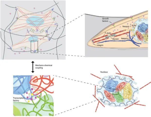
Figure 1: (A) Cells sense the geometry and rigidity of the tissue micro-environment and perceive a number of biochemical signals via proteins on the cell membrane. (B) These signals are transduced to the nucleus via a number of canonical signaling pathways. In addition, the nucleus is linked with the cell membrane through an elaborate meshwork of cytoskeletal filaments, including actin, microtubules, and intermediate filaments. (C) The spatial organization of chromosomes in the cell nucleus, in particular the spatial neighborhoods of chromosomes, their degree of intermingling, and the clustering of genes within those intermingled regions, are optimized for cell-type specific regulatory programs. (D) The chemical and physical signals that reach the nucleus are translated via chromatin remodeling enzymes and the transcription machinery to facilitate the regulation of particular genes. Our hypothesis is that these chromosome intermingling regions serve as mechanical hotspots that harbor cell-type specific gene clusters and integrate micro-environmental signals for genome regulation.
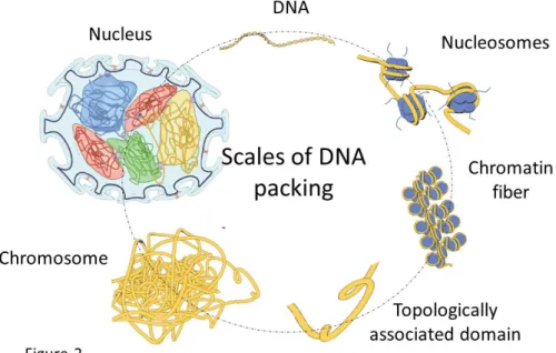
Figure 2: Scales of DNA packing in the cell nucleus. The ~1m long double-stranded DNA is packed into an approximately 10µm diameter cell nucleus. DNA is wrapped around histone and non-histone proteins to form a nucleosomal array of ~11nm in diameter. The resulting chromatin fiber is organized into topologically associated domains (i.e., TADs), which are further folded into a higher-order chromosome structure. The chromosomes (23 pairs for human cells) are then packed within the cell nucleus in a cell-type dependent manner.
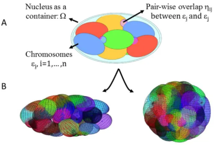
Figure 3: Geometric chromosome packing models. (A) Chromosomes are modeled as ellipsoids εi, i=1,… ,n, of a given size and shape. The nucleus is modeled as an
enclosing ellipsoid Ω. The chromosomes are packed within the cell nucleus by differentially penalizing the pairwise overlap ηij between ellipsoids depending on
the difference in average gene expression of the two chromosomes. (B) Simulation results of the optimized chromosome packing arrangements show that two distinct nuclear mechanical constraints result in different chromosome neighborhoods.
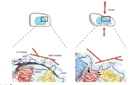
Figure 4: Mechanoregulation of chromosome intermingling. Cells objected to external microenvironmental signals result in a reversible reorganization of chromosome intermingling regions, i.e., the mechanical hotspots for