HAL Id: hal-02922090
https://hal.archives-ouvertes.fr/hal-02922090
Submitted on 25 Aug 2020
HAL is a multi-disciplinary open access archive for the deposit and dissemination of sci-entific research documents, whether they are pub-lished or not. The documents may come from teaching and research institutions in France or abroad, or from public or private research centers.
L’archive ouverte pluridisciplinaire HAL, est destinée au dépôt et à la diffusion de documents scientifiques de niveau recherche, publiés ou non, émanant des établissements d’enseignement et de recherche français ou étrangers, des laboratoires publics ou privés.
Experience and activity-dependent control of
glucocorticoid receptors during the stress response in
large-scale brain networks
Huzard Damien, Rappeneau Virginie, Onno Meijer, Touma Chadi, Margarita
Arango-Lievano, Michael Garabedian, Freddy Jeanneteau
To cite this version:
Huzard Damien, Rappeneau Virginie, Onno Meijer, Touma Chadi, Margarita Arango-Lievano, et al.. Experience and activity-dependent control of glucocorticoid receptors during the stress response in large-scale brain networks. Stress: The International Journal on the Biology of Stress, 2020, �10.1080/10253890.2020.1806226�. �hal-02922090�
Review: Special issue on Stress
Experience and activity-dependent control of glucocorticoid
receptors during the stress response in large-scale brain networks
HUZARD Damien 1, RAPPENEAU Virginie 2, MEIJER Onno C. 3, TOUMA Chadi 2 , ARANGO-LIEVANO Margarita 1, GARABEDIAN Michael J. 4, JEANNETEAU Freddy 1 *
1 Department of Neuroscience and Physiology, University of Montpellier, CNRS, INSERM, Institut de Génomique Fonctionnelle, Montpellier, France
2 Department of Behavioural Biology, University of Osnabrück, Osnabrück, Germany
3 Division of Endocrinology, Department of Internal Medicine, Leiden University Medical Center, Leiden University, Leiden, the Netherlands 4 New York University Langone Medical Center, New York, NY 10016, USA *Corresponding author: freddy.jeanneteau@igf.cnrs.fr Running title: Context-dependent glucocorticoid actions
Keywords: Allostery, epigenetic, plasticity, synapse, stress-related disorders, Bayesian brain
Abstract
The diversity of actions of the glucocorticoid stress hormones among individuals and within organs, tissues and cells is shaped by age, gender, genetics, metabolism, and the quantity of exposure. However, such factors cannot explain the heterogeneity of responses in the brain within cells of the same lineage, or similar tissue environment, or in the same individual. Here, we argue that the stress response is continuously updated by synchronized neural activity on large-scale brain networks. This occurs at the molecular, cellular and behavioral levels by crosstalk communication between activity-dependent and glucocorticoid signaling pathways, which updates the diversity of responses based on prior experience. Such a Bayesian process determines adaptation to the demands of the body and external world. We propose a framework for understanding how the diversity of glucocorticoid actions throughout brain networks is essential for supporting optimal health, while its disruption may contribute to the pathophysiology of stress-related disorders, such as major depression, and resistance to therapeutic treatments.
Introduction
Glucocorticoid hormones, such as cortisol and corticosterone, are secreted and used in a temporal- and organ-specific manner in response to internal and external stressors. They are ligands for the glucocorticoid receptor (GR) and the mineralocorticoid receptor (MR). Upon glucocorticoid binding, the receptors undergo structural alterations, revealing protein surfaces that interact with effectors of the signaling responses (Bledsoe et al., 2002; Li et al., 2005). The diversity of responses originates from a continuous exchange between the receptors and their diverse effector complexes, for which availability in time and space is defined by the environmental context. Agonist binding to GR and MR stabilizes an active conformational state, in which structural changes induce exchange of chaperone complexes for (transcriptionally) active machinery. These exchanges involve newly exposed surfaces that are dynamic and controlled by the timing and demands of the cellular environment (Weikum et al., 2017). The ultradian and circadian modes of glucocorticoid secretions support programmed rhythms of conformational changes in both the GR and MR receptors (Kalafatakis et al., 2016), but glucocorticoid-independent signals contingent to the environmental context are also involved (Galliher-Beckley et al., 2011; Hapgood et al., 2016).
Cellular history of excitation and exposure to stress mediators may define the accessibility of ligands (glucocorticoids, nucleic acids and co-regulators) to the core functional units. For the rapid non-genomic effects, the interactors of GR and MR are ill-defined. For transcriptional effects, the basic effector mechanism can be conceptually reduced to the ligand, the receptor, the DNA, and the transcriptional coregulators that interact directly with the receptors (Monczor et al., 2019). Of note, while the stress-induced glucocorticoid signal is predicted to mainly affect GR occupancy (Reul and de Kloet, 1985), GR and MR share many of their DNA binding sites (van Weert et al., 2017) and potential target genes, including often used readouts for receptor activity such as the Fkbp5 gene (van Weert et al., 2019). This implies that GR/MR signaling prior to a stressor will interact with stress-induced GR signaling. GR/MR and other nuclear receptors also share an important part of their coregulator repertoire (Broekema et al., 2018; Meijer et al., 2005). This points to intrinsic interactions between both GR and MR, and between glucocorticoids and other steroid signaling pathways. However, these may be considered stable over the time frame of hours, or often longer periods of time.
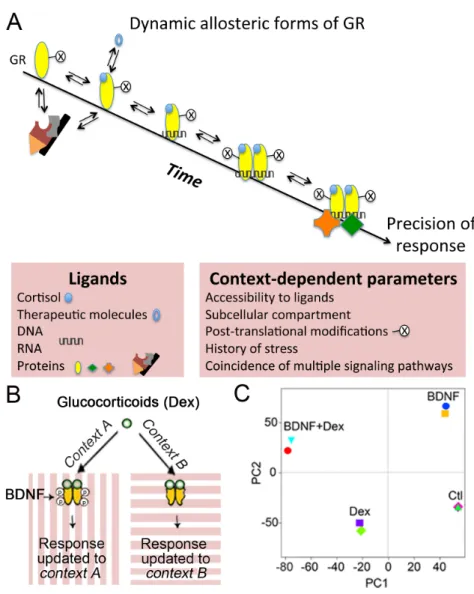
In contrast, the cellular biochemical state at the time of exposure to glucocorticoids is defining the context-dependent responses (Figure 1A). The post-translational modifications occurring on the glucocorticoid receptors are important allosteric determinants contributing to context-dependence (Faresse, 2014; Liberman et al., 2014). The range of effectors available for the different conformational states may depend on the organ, the cell type, the history of stress exposure, the ongoing cellular activity and the subcellular distribution of GRs and MRs. At the synapse, GR and MR convey glucocorticoid-elicited effects on neurotransmission within seconds that are inconsistent with genomic actions (Groeneweg et al., 2011). These effects can be initiated at the plasma membrane or in the cytoplasm via non-canonical signaling pathways that interact with the slow genomic
effects to organize long-term adaptive responses (Joels et al., 2013). Within the cytosol, the hormone-activated receptors can interact with protein partners to signal in non-genomic ways (Gutierrez-Mecinas et al., 2011). The nucleus and the mitochondrion are privileged locations for GR and MR, where they can bind to regulatory DNA elements (Du et al., 2009). These may be considered as ligands for GR and MR, affecting the conformation of the receptors via their DNA binding domain (Lapp et al., 2019; Meijsing et al., 2009). Availability of these “DNA ligands” is conferred by the interaction of glucocorticoid-dependent and glucocorticoid-independent signals that converge on chromatin (Bartlett et al., 2019). The binding of “RNA ligands” have also been described outside the nucleus, in P-granules where glucocorticoid receptors accelerate mRNAs turnover rates via a nuclear activated GR-mediated genomic action (Ishmael et al., 2011). The transcriptional effects are consistent with the rapid import of GR and MR into the mitochondria and into the nucleus upon binding of glucocorticoids (Conway-Campbell et al., 2012; Lapp et al., 2019).
Although glucocorticoids are known to produce differential changes in gene expression among individuals, within different tissues and even from cells of the same individual, a recent meta-analysis identified a core set of genes systematically regulated in the same direction (Kan et al., 2018). Such a gene module apparently represents constitutively available DNA binding sites, and its regulation upon ligand exposure depends on the availability of the constitutive effector complexes to determine the route of glucocorticoid signaling (Mahfouz et al., 2016; Meijer et al., 2019). By contrast, the diversity of actions to the other sets of glucocorticoid-responsive genes involves glucocorticoid-independent factors (e.g. cell specific chromatin landscape, subcellular local presence of RNA and protein coregulatory factors based on contextual cellular activity and biochemical makeup) to determine alternative routes of glucocorticoid signaling restricted in time and by the state of cellular activity (Clayton et al., 2019).
First, we discuss how the pairing of glucocorticoid signaling with neuronal activity tailors responsivity in brain cells, and unifies reactions of neurons that operate in functional networks. We then explore how the misalignment between activity-dependent and glucocorticoid-dependent actions may derail adaptation to stress, and promote the development of stress-related diseases. Finally, we provide a framework to understand how glucocorticoid-independent signals have the potential to increase the precision of glucocorticoid actions during stress by adopting specific allosteric states of MR and GR.
1. A molecular perspective
The precision of the glucocorticoid actions depends on multiple factors that are determined by specific cellular and physiological contexts, and integrated over time (Arango-Lievano and Jeanneteau, 2016). This section will explore how glucocorticoid receptors integrate multiple coincident signals via dynamic allosteric changes. Here, the term ‘allosteric’ refers to the conformational changes on GR and MR triggered by the specific cellular and physiological contexts –not to confuse with orthosteric conformational changes brought by ligand binding- that define the conformational modulation required to pick the effector complexes that guide an appropriate route of glucocorticoid signaling. Although the
conformational modifications of GR are known to convey gene, cell or physiological context information, it remains unclear whether each allosteric state corresponds to a unique response, whether allosteric states are dynamic and reversible, whether contextual signals can secure a specific allosteric state and finally, how these combinatorial effects are restricted in time. 1.1. Detection of coincidence signaling Transcription factors respond to changes of neuronal activity (Yap and Greenberg, 2018). Such regulatory mechanisms may also be shared by ligand-dependent transcription factors like the GR and MR. Classic studies have paved the way for a detailed understanding of how these receptors are switched-on by cognate and synthetic ligands in different cell types. However, little is known about the impact of cellular experience on GR and MR activities. Coincident neuronal activity could interfere with many attributes of the glucocorticoid receptor signaling, including ligand availability, ligand binding, transport across compartments, post-translational modifications, and transcriptional activity (Arango- Lievano et al., 2015b). Post-translational modifications of the ligand binding domain and N-terminal transactivation domain, dynamic and reversible in nature, are likely associated with particular conformational transition states of glucocorticoid receptors (Weikum et al., 2017). For example, site-specific phosphorylation in the transcriptional activation domain of GR is sufficient to fold its disordered structure, producing new receptor surfaces for docking new protein partners in a context-specific manner (Khan et al., 2017). GR and MR are targeted by post-translational modifications at distinct sites involved in all aspects of glucocorticoid actions (Faresse, 2014; Liberman et al., 2014; Vandevyver et al., 2014). Some of these modifications are directed by non-steroid factors organized by prior experience and by ongoing cellular activity that can overlap in time with the glucocorticoid signaling.
Glucocorticoid-independent site-specific phosphorylation of the glucocorticoid receptors occurs in response to various physiological contexts (e.g. KCL-induced depolarization, oxidative stress, sensory stimulation, motor learning, enriched housing environment, antidepressant treatment). These contexts depend, in part, on the secretion of the activity-dependent brain-derived neurotrophic factor (BDNF), a neurotrophic factor playing key roles in neuronal plasticity (Figure 1B and (Arango-Lievano et al., 2019; Arango-Lievano et al., 2015a)). These sites are conserved among humans, mice and rats and the phosphorylation was experimentally confirmed in each species (Arango-Lievano and Jeanneteau, 2016; Jeanneteau et al., 2019). A caspase-1 cleavage site resides near one of the BDNF-dependent phosphorylation sites in GR that is responsible for glucocorticoid resistance in humans with acute lymphoblastic leukemia suggests that healthy responses to glucocorticoids are in part determined by the conformational surfaces in the GR N-terminal domain and its dynamics to contextual post-translational modifications (Paugh et al., 2015). Interaction between contextual signals and glucocorticoid signaling per se was tested by co-stimulation of neurons with BDNF and glucocorticoids, which extended the duration of phosphorylation at these sites compared to a stimulation with BDNF only (Lambert et al., 2013). This can be explained by the activation of GR kinases (e.g. Erk1/2, [extracellular signal-regulated kinase 1/2], JNK [c-Jun N terminal kinase], P38) via the BDNF pathway and by the deactivation of the GR phosphatase PP5 [protein phosphatase 5] through the
glucocorticoid pathway (Arango-Lievano et al., 2015a). Other kinases (e.g. CDK5 [cyclin-dependent kinase 5], GSK3 [Glycogen Synthase Kinase-3]) and phosphatases (e.g. PP1 [protein phosphatase-1]) involved in the phosphorylation of GR sites (Galliher-Beckley and Cidlowski, 2009; Ismaili and Garabedian, 2004) are common to many signaling pathways modulated during behavioral experience and neuronal activity ((Yap and Greenberg, 2018).
Mutational analyses of phosphorylation sites in GR revealed the dependency between the BDNF and the glucocorticoid pathways for controlling the molecular determinants that dock specific effector complexes with the cAMP response element-binding protein 1 (CREB1), histone deacetylase 2 (HDAC2) and DNA (Lambert et al., 2013). Previous studies indicated the importance of such a coincidence-based interaction between the Erk1/2 kinase pathway and the glucocorticoid pathway to cope with stress on the molecular, cellular and behavioral scales (Arango-Lievano et al., 2015a; Chen et al., 2012a; Gutierrez-Mecinas et al., 2011; Revest et al., 2013). Accordingly, although BDNF-dependent GR phosphorylation occurred in the cytoplasm, its phospho-isoforms were detected in the nucleus, on the chromatin, in mitochondria as well as in synaptosomes. These findings explain the target-gene specific potentiation of GR actions that is observed after BDNF co-treatment (Lambert et al. 2013). It also defines GR as a detector of coincidence between the glucocorticoid and the BDNF pathways with the aim to link the external pressure of stress in the environment to the internal cellular state of activity. The subsequent unifying response is thus restrained within the temporal domain of GR phosphorylation (Arango-Lievano et al., 2015a). The biochemical half-life of GR phosphorylation ranges from minutes to hours – similarly, ligand dependent activation of the GR may range from approximately 30 minutes (for ultradian peaks) to hours or days, depending on acute or chronic stressors (Russell et al., 2015). Overlaps of timing between glucocorticoid-dependent and glucocorticoid-independent signals are expected to determine the diversity and precision of responses within cells and among individuals, which for example is the case during learning and memory (Arango-Lievano and Jeanneteau, 2016; Schwabe et al., 2012).
The human Bdnf gene has a polymorphism (substitution of valine (Val) to methionine (Met) at codon 66 [Val66Met]), which affects BDNF trafficking and diminishes the activity-dependent release of BDNF in the brain (Egan et al., 2003). Homozygote carriers of the BDNF-Met66 allele reduce GR phosphorylation (Arango-Lievano et al. 2019), and under basal non-stressed conditions, display stress-related behavioral impairments in mice (Chen et al., 2006; Notaras et al., 2017; Notaras et al., 2015; Soliman et al., 2010; Yu et al., 2012) and in human homo and heterozygotes (de Assis and Gasanov, 2019; Shalev et al., 2009). Further deletion of BDNF-dependent GR phosphorylation sites in the context of the BDNF-Val66Met polymorphism failed to alter stress-induced plasticity in mice (Arango-Lievano et al., 2019). This indicates that site-specific GR phosphorylation is important in stress regulation and stress-related behaviors, acting downstream of activity-dependent BDNF signaling in vivo.
Other post-translational modifications likely influenced by experience and neuronal activity such as acetylation, ubiquitination, sumoylation and nitrosylation were implicated in the regulation of GR and MR turnover, transport and binding to corticosterone and DNA (Arango-Lievano et al., 2015b; Weikum et al., 2017). However, none of these
post-translational modifications in GR were altered by BDNF stimulation of neuronal cells (Arango-Lievano and Jeanneteau, 2016). Consistently, mutations of BDNF-dependent phosphorylation sites did not interfere with GR turnover, transport, corticosterone binding or DNA binding (Arango-Lievano and Jeanneteau, 2016). This suggests that phosphorylation affords some level of contextual specificity that other modifications don’t. The allosterically induced context-dependent conformational states of GR, acquired by the post-translational modifications, do not necessarily set an overall responsiveness, but may instead increase the precision of response to serve cellular and organismal demands. They may also involve co-regulators of the steroid receptors to fine tune responses, as exemplified by the cell state dependent phosphorylation of the SRC2/GRIP1 effector of GR and MR (Rollins et al., 2017). More studies are necessary to understand the significance of GR and MR post-translational modifications and their dynamics, as well as their consequences on physiological stress responses and stress-related behaviors (Garabedian et al., 2017).
1.2 Epigenomic priming
The diversity of genome-wide occupancy of GR in different cell types is dictated by chromatin loci that were already opened or accessible de novo upon stimulation with glucocorticoids (John et al., 2011). This may be similar for MR that binds to DNA already exposed to transcriptional complexes of cell fate determination and plasticity like NeuroD and GR (Mifsud and Reul, 2016; van Weert et al., 2017). Most GRs interact with already-open chromatin before receptor activation while de novo opening of the chromatin was reported at a minority of loci (Burd et al., 2012). Interactions between GR and local chromatin occur directly and indirectly through the recruitment of histone/DNA-modifying enzymes (Bartlett et al., 2019). At resting state, most “potential sites” for GR binding in the genome reside in the inaccessible chromatin. Accessibility to these loci is gained by various stimuli coincident to glucocorticoid stimulation (e.g. neuronal activity, growth factors, neurotransmitters, steroid hormones) that create an opportunity for GR binding and gene regulation (Klemm et al., 2019). Opening the chromatin to expose new epigenomic landscapes before the nuclear translocation of GR, influences the frequency of GR–DNA binding events. Indeed, experience, neuronal activity and other conditioning factors can change the frequency of transcription factor binding to specific genome loci (Kitagawa et al., 2017), acting as potent modifiers of chromatin topology by enzymes working on DNA or histones (Yap and Greenberg, 2018). Prolonged exposure to exogenous glucocorticoid is an example of conditioning that can alter future stress responses by changing DNA methylation at target genes (Provencal et al., 2019). In the hippocampus, corticosterone regulated the expression of genes that overlapped by only 50% between unstressed controls and animals prior exposed to chronic stress (Polman et al., 2013). The social context, like the hierarchical rank in a group of individuals is another example of conditioning that can alter chromatin accessibility and transcription at glucocorticoid-responsive genes (Snyder-Mackler et al., 2019). These examples illustrate the interaction of glucocorticoid exposure and behavioral experience on the epigenome, and that stress responsivity may be shaped accordingly.
process intended to influence cell responses to subsequent events based on prior experience, like a memory. Methylation of DNA is supposed to serve this function (Bird, 2002). For example, methylation of Nr3c1 -the gene encoding GR- in the context of childhood trauma predicts changed physiological reactivity to stress in rodents (Liu et al., 1997), and was reported to be associated with major depressive episodes and suicide attempts at adulthood (McGowan et al., 2009). To avoid such maladaptive responses, the epigenomic priming of GR responsivity should be reversible (Yang et al., 2016). The DNA methylation is linked to changes of histone acetylation and phosphorylation that serve as a multi-layered code of epigenomic regulation used to update new information based on prior experience (Bousiges et al., 2013; Crosio et al., 2003). Such feed-forward mechanisms can operate in multiple environmental and behavioral contexts to regulate the precision and magnitude of responses to glucocorticoid receptors (Bartlett et al., 2019). For example, BDNF signaling promotes nitrosylation of HDAC2, which reduces its enzymatic activity and localization on the chromatin (Nott et al., 2008). The interconnectedness of regulatory mechanisms is exemplified by the fact that besides histones, one prominent substrate of HDAC2 is GR, whose deacetylation impairs the binding with protein transcription factors like nuclear factor kappa B (NF-κB) (Ito et al., 2006). GR deacetylation by HDAC2 has been implicated in the pathophysiology of glucocorticoid resistance in chronic obstructive pulmonary disease, which is also associated with impaired phosphorylation dynamics of GR by kinases pathways (Barnes, 2009, 2013). As an additional example of epigenomic regulation, GR-DNA binding frequency is significantly increased if chromatin loci are co-occupied by GR and CCAAT-enhancer-binding proteins (C/EBP), representing a cooperative mechanism involving again BDNF and glucocorticoid pathways (Grontved et al., 2013). In general, cell-type-specific gene regulation is supposed to involve the co-occurrence of GR and MR with its machinery and the cell-type-specific chromatin accessibility and DNA methylation (Datson et al., 2011; Gertz et al., 2013; Pooley et al., 2017; van Weert et al., 2017). Future ChIP-seq studies shall provide information regarding the genome-wide positioning of GR and MR in distinct cell types, and in different physiological contexts.
1.3 Transcription of gene networks
Less than 1% of chromatin loci to which glucocorticoid receptors bind in one cell type, is actually common to other cell types. The heterogeneity in transcriptional patterns that are important for programming the stress response is reduced when comparing cell types issued from the same tissue of the same mouse, and yet meta-analyses emphasize more differences than similarities (Crow et al., 2019; Sacta et al., 2016). Therefore, studies of particular cell subpopulations, in the context of synchronized neuronal activity or stimulation with activity-dependent factors like BDNF, are necessary to clarify what determines the direction and magnitude of transcriptional regulation by glucocorticoids in the genome. A co-stimulation of neurons with BDNF and the GR synthetic agonist dexamethasone lead to a distinct transcriptome from the one elicited by individual stimuli, and that reflected interactions, rather than merely additive effects between treatments (Figure 1C). Although similar gene modules are consistently repurposed across contexts, some gene clusters are specific to salient experiences, to stress and to coincident BDNF and glucocorticoid signaling (Lambert et al., 2013; Mukherjee et al., 2018). Overlap and dependency between gene expression modules containing DNA-binding elements for
activity-dependent transcription factors as well as for GR and MR is likely essential to determine the precision of a stress response gene network tailored to the contextual demands.
GR and MR use their DNA-binding domain zinc fingers to make contact with glucocorticoid responsive elements (GRE). The nature of GRE, in terms of sequence identity or orientation, varies across target genes in the genome, and instructs either positive or negative cooperative remodeling of the dimerization motif that facilitates (homo- or hetero) dimers, tetramers or monomers (alone or tethered to another transcription factor) to sit on DNA (Paakinaho et al., 2019; Schiller et al., 2014; Watson et al., 2013). Thus, the interaction between receptors and DNA response elements allosterically stabilizes particular conformational forms that guide the direction of responses (Hudson et al., 2013). These events on chromatin are very dynamic, consisting of association and dissociation exchanges within seconds, a time frame compatible with integrating coincident contextual signals at high frequencies (Weikum et al., 2017). This integration can be mediated by direct protein– protein interactions between the receptors and other transcription factors but also by indirect effects via inhibitory long noncoding RNAs, or by competition for nuclear coactivator proteins (Rapicavoli et al., 2013; Ratman et al., 2013). For example, glucocorticoid-activated GRs can be recruited to specific loci in the genome without binding directly to DNA (i.e., via protein–protein interactions with other DNA-bound factors likely responding to signaling elicited by neuronal activity). Both the direct and tethering modes are associated with distinct transcriptional attributes (Vockley et al., 2016). However, GR and MR were mostly detected on GRE-dependent motifs in ‘bulk’ hippocampal tissue under resting condition and after stress (Polman et al., 2013; Pooley et al., 2017; van Weert et al., 2017). This implies that the detection of coincidence, via interaction of GR with activity-dependent transcription factors, would take place in small subset of cells – in accordance to the mosaic-like activation of neurons, for example in learning processes (Rao-Ruiz et al., 2019).
Intracellular calcium (Ca2+) is a central mediator of transcriptional regulation and is controlled by neuronal activity that could gate GR and MR responsiveness either through the direct or indirect mode of transcriptional regulation (Zhang et al., 2009). Several phosphatases, proteases and cargo transporters operating as Ca2+-sensors are involved in the response to glucocorticoids in multiple cellular compartments. Actions on synaptic neurotransmission and on nuclear gene transcription, which are mediated by GR and MR, require coincident Ca2+ mobilization from the mitochondria and the endoplasmic reticulum (Chameau et al., 2007; Harris et al., 2019; Mayanagi et al., 2008; Simard et al., 1999). Mobilization of Ca2+ and cAMP are both required for the nuclear import of the CREB-regulated transcription coactivators (CRTC1/2/3), a family of cofactors for CREB and GR (Altarejos and Montminy, 2011; Hill et al., 2016). In particular, CRTC2 can integrate BDNF and glucocorticoid signals in the hypothalamus to control the direction and magnitude of transcription at the corticotropin-releasing hormone (Crh) promoter and the response of the hypothalamic-pituitary-adrenocortical (HPA) axis to stressors (Jeanneteau et al., 2012). This molecular pathway also regulates the glucose metabolism and the energetic adaptation to stress by controlling the expression of the rate-limiting enzymes glucose-6-phosphatase and phosphoenolpyruvate carboxykinase in the liver (Hill et al., 2016).
Future studies employing single-cell transcriptomics will be useful in capturing the impact of experience and activity on the refinement of gene expression modules, since it allows the dissection of global gene expression in specific cell types during defined stress environment and behavioral state. This method could lead to the identification of subpopulations of cells activated in certain contexts, such as learning a task or being exposed to psychostimulants. For example, differential gene expression profiles were induced by morphine in astrocytes and oligodendrocytes collected from the same tissue of the same animals (Avey et al., 2018). Similarly, the influence of ongoing cell excitation (e.g. during visual stimulation of the retina) can impact the expression of GR regulated gene modules that is consistent with the role of GR on the plasticity of the visual system (Muto et al., 2013). In conclusion, experience and activity-dependent control of GR and MR assembly with ligands, DNA and coregulators is essential to precisely update and maintain the diversity of the responses.
2. A cellular perspective
As outlined above, glucocorticoid responsiveness varies significantly across cell types, brain regions, neurocircuits, and physiological states (Chattarji et al., 2015; Franco et al., 2019; Joels et al., 2013). This section will explore how glucocorticoid receptors integrate multiple coincident signals, which are restricted to certain time domains and depend on neuronal experience, in order to modify cellular functions like migration, differentiation, connectivity and neurotransmission. Contextual signaling of glucocorticoid receptors is critical for understanding how both insufficient and excessive glucocorticoid signaling can have similar impact on neuronal functions (Yu et al., 2019). However, it remains unclear whether glucocorticoid action on a broad range of subcellular organelles (e.g. mitochondria, nucleus and synapse) is uniform or on the contrary multiform in time and space. Each site of action is likely to contribute to the overall adaptive program set by glucocorticoids. The following section focuses on the nuclear and synaptic actions but other actions on the mitochondria, the sarcoplasmic reticulum are described elsewhere (Choi et al., 2018; Lapp et al., 2019; Picard et al., 2014).
2.1 At the single cell level
Long range signaling from synapses to the nucleus is essential to store information in the epigenome and modify neuronal fate with a novel arsenal of transcripts (Deisseroth et al., 2003). Mobilization of Ca2+ at the synapse, via voltage-gated Ca2+ channels, sets in motion a series of Ca2+-sensing mechanisms driving synaptic effectors like nuclear factor of activated T-cells (NF-AT) directly into the nucleus (Hagenston and Bading, 2011; Wild et al., 2019). Acting as tethering co-activator of GR, NF-AT is expected to control the direction of transcription at selective glucocorticoid-responsive target genes in neurons as in immune cells (Petta et al., 2016). Long-range transport of cargoes between synapses and the nucleus is bidirectional, and may serve as tagging systems for promoting synapse-specific plasticity based on prior experience (Frey and Morris, 1997). Activity-dependent tags such as the BDNF-activated receptor Tropomyosin Related Tyrosine Kinase-B (TrkB) trap new mRNA transcripts from the soma with the protein synthesis machinery in specific synapses (Lu et
al., 2011). Long-range transport of signaling endosomes harboring phosphorylated TrkB from synapse to nucleus contributes to an important feedforward mechanism that prepares cellular responsiveness based on prior stimulation (Yamashita et al., 2017). GR can bind directly to TrkB (Numakawa et al., 2009) but it is unknown whether the phosphorylated isoforms of GR can traffic along with the activated TrkB cargoes (Barford et al., 2017). Cis/trans-isomerization of phosphorylated residues on GR increases its persistence overtime by preventing dephosphorylation (Poolman et al., 2013). Several cis/trans-isomerases (e.g peptidyl prolyl isomerases FKBP4, FKBP5 and PIN1) form complexes with GR and MR to regulate their activity, transport and signaling across organelles in the context of stress (Binder, 2009; Wochnik et al., 2005; Zhang et al., 2008). Cis/trans-isomerases of GR and MR complexes are often presented as important modifiers of future responsiveness to glucocorticoids and in diseases of stress sensitivity and responsiveness (Binder et al., 2004; Fries et al., 2015; Galigniana et al., 2012; Ratajczak et al., 2015).
Mechanisms of adaptive plasticity utilize immediate early genes as first line responders to glucocorticoid stimulation to prepare cells for a changing environment based on prior experience. For example, the MAPK specific phosphatases (Mitogen-Activated Protein Kinase Phosphatase 1/6 [MKP1/6]) are induced to terminate coincident growth factor signaling pathways converging on the GR phosphorylation code and the epigenome (Deinhardt and Jeanneteau, 2012; Jeanneteau and Deinhardt, 2011). Another example is the stress-induced transcription factors NR4A1/2/3, which shuttle in and out of the nucleus and mitochondria to coordinate metabolism and synapses number (Jeanneteau et al., 2018). Finally, actin binding proteins such as the Ca2+ sensor caldesmon and the cofilin kinase LIM-kinase 1 (LIMK1) are induced to reorganize the cytoskeleton supporting neuronal migration, differentiation and connectivity (Fukumoto et al., 2009; Mayanagi et al., 2008; Morsink et al., 2006). Altogether, these responses set in motion the adaptive machinery that updates many attributes of neuronal function to its environment based on prior experience.
The sequence in which an acute glucocorticoid pulse and neuronal activity follow each other further impacts the final response. In vitro, activation of GR prior to, or in conjunction with, cAMP signaling suppresses the transcription at the Crh gene promoter, a repressive effect that failed to develop when cAMP signaling preceded GR activation (van der Laan et al., 2009). In vivo, the GR-mediated control of Crh transcription is strikingly opposite in the hypothalamus and in the amygdala of the same mouse depending on the behavioral context (Jeanneteau et al., 2012; Zalachoras et al., 2016). Therefore, the direction and magnitude of glucocorticoid response may depend not only on the intrinsic presence of cell type specific mediators, but also on its timing with other contextual signals that change cAMP and/or Ca2+ concentrations, like during neuronal spiking activity.
The time-dependence of glucocorticoid effects on synapses would support the opposite effects of glucocorticoids on dendritic spines, reported to be branch-specific and distinct across brain regions and circuits (Arango-Lievano et al., 2016; Chattarji et al., 2015; Dias-Ferreira et al., 2009). Some of these effects rely on the remodeling of a major constituent of dendritic spines, the actin cytoskeleton, by rapid non-genomic and slow genomic glucocorticoid signaling (Stournaras et al., 2014). The dynamic rates of formation and elimination of dendritic spines increases throughout the cortex by exogenous
glucocorticoids whereas removal of endogenous glucocorticoids from the brain cancelled such a turnover (Liston and Gan, 2011). This was demonstrated by low-dose injection of dexamethasone, which does not penetrate the brain but suppresses endogenous corticosterone secretion via the pituitary axis (Karssen et al., 2005). New thin spines formed upon glucocorticoid exposure are immature and very unstable. Both the membrane bound GR and MR (mGR and mMR) previously located in the synapse (Johnson et al., 2005; Prager et al., 2010) regulate spine structure via rapid non-genomic mechanisms upon circadian and experience dependent fluctuations of corticosterone (Russo et al., 2016). However, upon elevation of corticosterone by acute or chronic injections, the nuclear GR and MR also take part in the process of stabilization and elimination of dendritic spines via genomic mechanisms (Liston et al., 2013; Liston and Gan, 2011). Maintenance of a significant proportion of new spines requires additional coincident contextual signals influencing Ca2+ spike activity to form functional synapses (Cichon and Gan, 2015). This means that the circadian/ultradian rhythms of glucocorticoid secretions provide a time-restricted opportunity for persistent remodeling of synaptic connectivity at active dendritic branches whereas other silent dendritic branches, out of behavioral context, are not affected over long time periods (Liston et al., 2013).
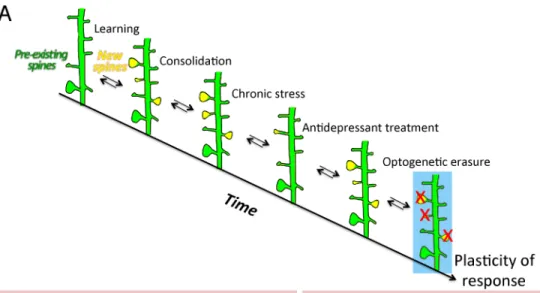
The consolidation of new dendritic spines formed at the time of learning requires a mechanism of coincidence detection of BDNF and glucocorticoid signaling pathways. It is restricted to the training period and it involves the retention of new glutamatergic AMPA receptors via GR phosphorylation (Arango-Lievano et al., 2019). Local dendritic synthesis of AMPA receptors increases their abundance at synapses and strengthens the new synapses associated with learning (Chater and Goda, 2014). This raises the threshold for the later induction of synaptic potentiation, which may help to consolidate experience-related information and prevent other context-unrelated information from being stored in these synapses (Sarabdjitsingh et al., 2014). Similar effects are anticipated with MR, as both MR and GR regulate synaptic transmission by modifying AMPA receptors (Conboy and Sandi, 2010; Groc et al., 2008) and the release of glutamate by the pre and or post-synapses (Karst et al., 1999; Tasker, 2006). Chronic stress exaggerates the elimination of certain pools of dendritic spines, in particular those associated with learning. Such effect can be reversed by a chronic treatment with antidepressant drugs. Further optogenetic erasure of this particular pool of dendritic spines impaired the physiological and behavioral effects of the antidepressant drugs (Moda-Sava et al., 2019), suggesting that information pertaining to behavioral experience can be stored in synaptic maps over extended periods of time (Figure 2A). Precision of glucocorticoid response is tailored to the time frame and brain area involved in contextual behavioral experience via the combinatorial effects of MR on basal firing frequency and spine elimination, and GR on neuronal activity and spine formation/maintenance (Groeneweg et al., 2011; Heshmati and Russo, 2013; Joels, 2008).
2.2 At the network level
Stress affects large-scale networks in the brain (e.g. executive, default mode, salience, valence and enteroceptive) to promote vigilance, attention, learning and adaptation (Chattarji et al., 2015; McEwen et al., 2015). For example, the salience and the default mode networks are upregulated by stress while, at the same time, the executive control network
is downregulated; such effects yet depend on the nature, intensity and duration of stressors (Clemens et al., 2017). The response of large-scale brain networks enables individuals to better reorient attention, detect salient external stimuli based on its enteroceptive state, and deal with the emotional and affective consequences of stress. Glucocorticoid receptors constantly interact with ongoing neural circuit activity of large-scale brain networks to prepare suitable physiological and behavioral response outcomes based on prior experience (Herman et al., 2016; Joels et al., 2006). The precision of responses to glucocorticoids at the neuronal network level depends on the organization of the circuit and types of cells involved. It also depends on glucocorticoid availability, which can be influenced by tissue- and context-specific expression of corticosterone/cortisol biosynthesis enzymes, like the hydroxysteroid dehydrogenases 1 and 2 (HSD1/2) (Myers et al., 2014; Ramamoorthy and Cidlowski, 2013). To date, it is not known whether the delivery of effective concentrations of glucocorticoids can be targeted locally to specific cellular microenvironments or even subpopulations of cells in a brain area at a given time (Figure 2B). However, it is well established that the GR/MR ratio varies across brain regions and cell types, and influences the diversity of responses to glucocorticoids in normal physiological and pathological conditions (De Kloet et al., 1998; Joels and de Kloet, 1994).
Despite GR apparent ubiquitous expression, its phosphorylation and nucleocytoplasmic ratio can vary across brain regions, cell types and neuronal network depending on the behavioral context (Adzic et al., 2009; Mitic et al., 2013). During learning, phosphorylation of GR at glucocorticoid-responding sites is uniform throughout the cortex whereas phosphorylation at BDNF-responding sites is increased during the learning period and enriched in specific cortical layers, cell types and co-expressed with c-Fos, a marker of neuronal activity (Figure 2C). Mutation of BDNF-dependent GR phosphorylation sites impairs the formation and stabilization of branch-specific dendritic spines only in the context of learning (Arango-Lievano et al., 2019). The stability of dendritic spines is predicted by its size more than by its activity (Quinn et al., 2019). Consistently, the head size enlargement of spines, at the pool of new synapses induced by learning, is blocked by the GR antagonist RU486 and by the mutation of BDNF-dependent GR phosphorylation sites, both of which are required for enhancing the post-learning glutamatergic synaptic response. By contrast, the pool of pre-existing spines, which are unrelated to learning, is not sensitive to either manipulation. Such findings are consistent with the impairment of long-term potentiation (LTP) at cortical synapses in the context of learning, in mice lacking BDNF-dependent GR phosphorylation sites, and with the absence of impairment in LTP between controls and mutant mice outside the learning context (Arango-Lievano et al., 2019). Changes in synapse strengths and spine dynamics during learning acquisition is selective of neuronal networks expressing BDNF-dependent GR phosphorylation and associated with significant improvement of behavioral performance during the consolidation and retention phases (Yu and Zuo, 2011). This illustrates how specific contextual events influence GR allosteric forms and its precision of actions across neuronal ensembles that respond to the same behavioral experience, acting like a functional network.
The precision of glucocorticoid action also varies across neuronal networks if the context of stress occurs during the ascending or descending phases of the ultradian glucocorticoid oscillations (Sarabdjitsingh et al., 2010a; Sarabdjitsingh et al., 2010b; Sarabdjitsingh et al.,
2010c). Impairment of glucocorticoid secretion rhythms, by chronic stress exposure, exaggerates the elimination of dendritic spines, most notably the new experience-associated pool but also a tiny fraction of the pre-existing pool (Arango-Lievano et al., 2019; Chen et al., 2017). Some of the consequences of such impaired rhythmicity, along with behavioral alterations, can be reversed by antidepressant treatments and physical exercise through a mechanism requiring BDNF/TrkB and downstream GR phosphorylation event (Arango-Lievano et al., 2019; Chen et al., 2017). The stress-induced synapse elimination is observed in cortical glutamatergic neurons and parvalbumin interneurons where GR phosphorylation at BDNF-responding sites is significantly enriched and, together with behavioral alterations, was prevented by chemogenetic tools, enriched housing conditions or ketamine used at dosage affording antidepressant effect (Arango-Lievano et al., 2015a; Chen et al., 2018; Ng et al., 2018). Mechanism of action may rely on the experience and activity-dependent co-clustering dynamics of inhibitory synapses and dendritic spines in the cortex (Boivin and Nedivi, 2018; Chen et al., 2012b). Other mechanisms may involve the retrograde trans-synaptic cannabinoid and opioid signaling to suppress the release of neurotransmitters at hypothalamic glutamatergic and GABAergic synapses in a time-period compatible with the remodeling of synaptic contacts (Di et al., 2009; Wamsteeker Cusulin et al., 2013). Inhibitory control of excitatory drive by interneurons, including the parvalbumin subclass, is critical to determine the direction and magnitude of responses to stress and glucocorticoids in hypothalamic, amygdalar, hippocampal and cortical neuronal networks. In the amygdala, both GR and MR are necessary to switch neuronal responses to glucocorticoids from excitatory to inhibitory (Karst et al., 2010). In the neurogenic region of the hippocampus, glucocorticoid receptors also regulate the balance of excitation and inhibition with consequences on newborn cells maturation, migration and integration into new networks (Saaltink and Vreugdenhil, 2014). Heterogeneity of neurogenic responses to glucocorticoid oscillations, acting through GR, determines the fate of progenitor cells between quiescence and proliferation by a mechanism involving a crosstalk between GR signaling and DNA methylation on several promoters of genes modulating the cell cycle (Schouten et al., 2019).
The dynamic activation and termination of the neuroendocrine HPA axis response to stress requires a fine balance of excitation and inhibition across multiple brain regions and cell types, which condition physiological and behavioral adaptation to environmental demands, based on prior experience (Herman et al., 2016). The stress response uses modulators, like noradrenaline, to integrate activity in a network of interconnected forebrain regions including the amygdala, the frontal cortex, the bed nucleus of the stria terminalis, the hypothalamus, and the hippocampus (Herman, 2013; Ulrich-Lai and Herman, 2009). Tissue and cell type specific conditional genetic deletion of glucocorticoid receptors further demonstrated that the diversity of glucocorticoid responses on physiological, neuroendocrine and behavioral outcomes depends on the organization and ongoing activity of the neuronal networks involved (Arango-Lievano and Jeanneteau, 2016; Arnett et al., 2016; Scheimann et al., 2018; Whirledge and DeFranco, 2018). This might be explained by a Bayesian process used by the brain to update current information based on prior experience through multiple iterations of neuronal network activation intended to minimize the entropy of various sensory states and predict future outcomes based on value coding (Doya et al., 2007; Ernst et al., 2006).
In conclusion of this part, the mMR and mGR via non-genomic mechanisms, and the genomic GR and MR integrate the physiological glucocorticoid pulse secretions with coincident cellular activity to shape, within time and space, the diversity of responses tailored by ongoing demands for future adaption based on prior experience.
3. A behavioral perspective
Failure to control the HPA axis according to the environmental demands is associated with pathological states (Holsboer and Ising, 2010; Kalafatakis et al., 2016; Sarabdjitsingh et al., 2010a; Sarabdjitsingh et al., 2010c). The functional dynamics and plasticity of the neurocircuits upstream and downstream of the HPA axis, determine the diversity of stressor-specific responses (Herman et al., 2003). Stress-coping strategies facilitate the adaptation to acute environmental challenges and to internal demands, but they can also become maladaptive when the price for maintaining physiological stability does not match with the real costs of adaptation, which represents the allostatic load (Picard et al., 2014). If chronic in nature, the maladaptive processes can decrease the diversity of glucocorticoid actions across neural networks, reduce the number of stress-coping options and lead to behavioral rigidity and stress-related disorders (Karatsoreos and McEwen, 2011; McEwen, 1998) (Figure 3). Therefore, promoting the diversity of glucocorticoid responsivity across brain cells and networks seems essential for maintaining optimal health. This section explores how variations in glucocorticoid-related molecular signaling pathways in neuronal networks of the brain, through gene variants or epigenetic mechanisms, impact stress coping, behavioral adaptation and stress-related disorders.
3.1. Stress-coping and behavioral adaptation
The diversity of strategies used to cope with environmental changes may be related to the individual differences in susceptibility to stress exposure. Stress-coping has been defined as the behavioral and physiological efforts to adjust with aversive stimuli or stressors (Lazarus and Folkman, 1984). Because the unpredictability of a situation is a stressor, coping may employ a predictive coding through the salience, executive and valence-coding neuronal networks in order to reduce the uncertainty about the selection of actions based on predicted consequences (Peters et al., 2017). More precisely, the neuroanatomical correlates of stress-coping involve the coordinated activities of the enteroceptive network (e.g. insular cortex), the default mode network at resting states, the salience network (e.g. orbitofrontal cortex, anterior cingulate cortex and the amygdala) and the executive network (e.g. frontostriatal network) (Joels et al., 2006). Stress-induced glucocorticoid signaling overlaps with the mesocorticolimbic dopaminergic pathway to bias decision-making of goal-directed behaviors in both humans and animals (Gourley et al., 2012; Peters et al., 2017; Soares et al., 2013). In terms of learning and memory, stress, via glucocorticoid signaling, enhances memory formation when the stressor is relevant and proximal to the learning context, but stress impairs retrieval of learned information when the stressor occurs out-of-context. In particular, stress experienced directly prior to learning may activate a salience network and promote emotional learning, whereas hours after stress, the activity of executive control networks is facilitated through a diversity of responses by
glucocorticoid receptors (Joels and Baram, 2009; McEwen, 2007). The diversity of stress-coping responses depends on the interaction of glucocorticoids with multiple neurotransmitter systems (i.e., dopaminergic, serotoninergic, glutamatergic and peptidergic) operating in large-scale synaptic networks of selective brain regions. They enable individuals to better reorient attention, detect salient external stimuli, and deal with the emotional and affective consequences of stress.
The coping strategies have been described as either proactive or reactive (also referred to as “active” and “passive” respectively). Proactive stress-coping behaviors in rodents are generally associated with decreased depression-like phenotypes after exposure to chronic stress paradigms (de Boer et al., 2017; Wood and Bhatnagar, 2015). Mice genetically selected for extremes in aggressive behavior show different coping styles that can predict the susceptibility to chronic social stressors in adulthood (Koolhaas et al., 2017; Veenema et al., 2004). Studies with rats, selected for low, intermediate and high glucocorticoid responsivity to stress, distinguish subjects based on singular coping strategies in response to various challenges (Huzard et al., 2019; Walker et al., 2018; Walker et al., 2017). Moreover, mouse lines selectively bred for high (HR) vs. low (LR) HPA axis reactivity to a psychological stressor (15-min restraint) show a hyperactive versus more passive behavioral response in tests assessing stress-coping behaviors. Interestingly, the different coping strategies of HR and LR mice are also associated with distinct alterations in emotional behaviors, cognitive performance, circadian activity patterns, sleep architecture, and neuronal functions resembling the symptomatology seen in patients suffering from the melancholic and atypical subtypes of depression, respectively (Fenzl et al., 2011; Heinzmann et al., 2014; Knapman et al., 2010; Touma et al., 2008; Touma et al., 2009). Looking at Gene × Environment interaction effects in this mouse model of affective disorders, the genetic predisposition for an increased neuroendocrine stress reactivity in HR mice amplified the deleterious effects of early-life adversity on stress-coping behavior and other relevant depression-like endophenotypes, possibly involving the BDNF signaling pathway (McIlwrick et al., 2017; McIlwrick et al., 2016).
Although the ability of an organism to cope with environmental challenges depends on brain-wide glucocorticoid receptor signaling (Deroche-Gamonet et al., 2003; Tronche et al., 1999), different populations of neurons may have unique roles in modulating stress-induced behaviors. To better understand how specific neuronal circuits and brain cell populations can influence stress-related behaviors, we and others have selectively inactivated the GR using the Cre/Lox or the CRISPR/Cas9 gene editing systems (Arango-Lievano and Jeanneteau, 2016; Arnett et al., 2016; Kellendonk et al., 2002; Whirledge and DeFranco, 2018). Under basal, non-stressed conditions, the deletion of Nr3c1 in dopamine-receptive neurons, but not in dopamine-releasing neurons, reduces drug-related behaviors without affecting HPA axis activity and anxiety-like and stress-coping behaviors (Ambroggi et al., 2009; Barik et al., 2013; Parnaudeau et al., 2014). Under stressful conditions, it also prevents the development of social avoidance behavior (Barik et al., 2013). Together, these studies confirm the importance of cell-type specific GR signaling in motivational and social reward behaviors (Douma and de Kloet, 2019). The cellular specificity could explain the differential modulation of GR activity in different areas of the mesolimbic and corticolimbic networks during reward-directed behaviors (Deroche et al., 1995; Gourley et al., 2012;
Piazza and Le Moal, 1996; Swanson et al., 2013). The knockdown of GR in the forebrain reduces anxiety-like and active-coping behaviors, and it increases HPA axis activity and impairs the glucocorticoid negative feedback (Boyle et al., 2005; Boyle et al., 2006; Jeanneteau et al., 2012; Laryea et al., 2013). Yet, the specific deletion of GR in glutamatergic neurons of the forebrain shows region-specific effects: the deletion of GR in the amygdala reduces the fear response while a deletion in the prelimbic prefrontal cortex increases proactive-coping behaviors in males (Hartmann et al., 2017; Scheimann et al., 2019). The HPA axis activity and the fear response in females, however, are enhanced by GR deletion in the prelimbic cortex, which is not observed in males (Scheimann et al., 2019). These findings show that the maintenance of normal HPA axis function and the regulation of stress-coping and anxiety-like behaviors can be ascribed to changes in GR signaling in particular regions of the forebrain, while sex-specific modulation of fear responses can be related to GR in glutamatergic circuits of the amygdala and prefrontal cortex. Although the manipulation of GR signaling in particular subpopulations and subregions of the brain allows a better understanding of the diversity of GR responses across large-scale neuronal networks, these approaches lack temporal precision of GR regulation (Myers et al., 2014). More work is needed to understand how each GR allosteric form regulates the expression of target genes involved in cytoskeleton dynamics, mitochondrial functions, and synapse formation mediating the described effects of stress (Arango-Lievano et al., 2019; Jeanneteau et al., 2018; Liston et al., 2013; Swanson et al., 2013).
The diversity of GR responses is conditioned, at the time of the event, by the activity of brain-wide neural networks. One example of a molecular mediator involved in orchestrating the diversity of GR behavioral responses is the activity-dependent neurotrophic factor BDNF (Mitre et al., 2017). Indeed, BDNF signaling is negatively regulated by glucocorticoids and the effects of glucocorticoids on synaptic plasticity in the brain are regulated by BDNF (Rothman and Mattson, 2013). The influence of BDNF on GRs may rely on an activity-dependent release of BDNF. Mouse carriers of the BDNF genetic variant Val66Met exhibit behaviors and stress-response improper under both basal and stressful conditions (Arango-Lievano et al., 2019; Autry and Monteggia, 2012; Chen et al., 2006; Nasca et al., 2015; Notaras et al., 2017; Notaras et al., 2015; Soliman et al., 2010; Yu et al., 2012). Glucocorticoids and BDNF exert complementary roles on behaviors (e.g. avoidance, fear, stress-coping and impulse control) as well as on synaptic plasticity (Barfield and Gourley, 2018; Jeanneteau et al., 2019; Jeanneteau and Chao, 2013). One mechanism operates via changes of GR phosphorylation and signaling (Arango-Lievano et al., 2015a; Lambert et al., 2013; McEwen, 2015). The interdependent actions of BDNF and GR allow the formation of new memories of contextual fear, the maintenance of procedural memories and stress-coping strategies (Arango-Lievano et al., 2019; Blugeot et al., 2011; Chen et al., 2012a; Gourley et al., 2012; Revest et al., 2013). This was shown by conditioning GR responses in experience-dependent neuronal networks, while preserving the diversity of GR responses in networks unrelated to the task (Jeanneteau and Chao, 2013). Thus, the specific effects of BDNF on GR signaling could produce diverse GR allosteric phospho-isoforms and chromatin landscapes in a mosaic of cell assemblies, which could contribute to a diversity of stress-coping behaviors.
As previously outlined, the individual variability in susceptibility to stress-related disorders results from a complex interplay between stress perception, context-dependent factors (e.g. genetic predisposition, history of stress such as early-life trauma or stressful life events) and changes in BDNF and GR signaling pathways across large-scale brain networks. The capacity of an organism to deal with a chronic activation of the HPA axis and therefore with an increased allostatic load, determines the extent of behavioral, physiological, neuroendocrine and neuronal damages in brain regions regulating reward, fear, emotionality and social behavior (Figure 3).
3.2. A disease perspective
A normally well-tolerated stressor can become pathogenic if the glucocorticoid reactivity is inappropriate (Selye, 1956). The chronic deviation of the regulatory systems, away from homeostasis, by prolonged exposure to environmental or psychological stressors, can produce maladaptive allostatic loads (Myers et al., 2014; Picard et al., 2014). For example, in the context of drug abuse, the allostatic state is the deviation of a reward set-point, due to a dysregulation in brain reward mechanisms and stress responses, enhancing the vulnerability to drug-related behaviors (Koob and Le Moal, 2001).
3.2.1. At the molecular level
Pathological shifts in key mediators of the allostatic load include dysregulation of the HPA axis, decreased GR responsiveness, dysfunctional immune responses, chronic inflammation, increased oxidative stress and decreased neuronal survival due in part, to a reduction in neurotrophic support (McEwen, 2006). Behavioral and physiological functions orchestrated by the brain during stress highly depend on the complementary actions of glucocorticoid and BDNF signaling pathways in the mesolimbic and corticolimbic neural networks. Maintenance of the activity in the reward- and emotion-related networks has been shown to guide behavioral responses that are in line with the costs and benefits of the contextual demands (Vinckier et al., 2018). For example, lack of motivation and inflexible maladaptive responses likely develop when prefrontal BDNF-TrkB signaling is compromised and GR-mediated actions are desensitized. On the contrary, sensitization to reward or aversive conditioning results when mesolimbic BDNF-TrkB signaling and GR-mediated responses are upregulated (Barfield and Gourley, 2018; Jeanneteau et al., 2019). Desynchronization of BDNF-TrkB and glucocorticoid-activated GR signaling pathways, in multiple neural networks, may contribute to increasing the vulnerability to stress-induced disorders. Desensitization of GR viewed as a decrease of ligand binding or a reduction in its ability to act as a detector of coincident BDNF signaling is likely a key determinant in promoting pathological disorders advanced by inappropriate stress responses. 3.2.2. At the network level It is not fully understood why the stress response can become maladaptive and how it could be reversed. One difference between adaptive and maladaptive pathological responses may involve the duration of exposure to stressors and its context. Indeed, the interaction of the stressor with the activity of large-scale brain networks may predict future outcomes and avoid or reduce uncertainty. Although alterations in BDNF signaling vary from region to region in the brain depending on the stressor paradigms, coherence between several brain
regions emerged (for review (Barfield and Gourley, 2018; Jeanneteau et al., 2019; McEwen et al., 2016)). For example, BDNF levels, TrkB activation, GR responsiveness and glucocorticoid spine plasticity decreased in both the hippocampus and prefrontal cortex, or increased in both the amygdala and ventral tegmental area (VTA) depending on the nature and intensity of the stressor (Chattarji et al., 2015). A physical stressor producing depression-like phenotypes and impairing fear responses is associated with significant alterations in BDNF levels and trafficking as well as synaptic plasticity and neurotransmitter release (Biggio et al., 2019; Deng et al., 2017; Liu et al., 2018; Mallei et al., 2019; Nasrallah et al., 2019; Tornese et al., 2019). A social stressor producing depression-like phenotypes and impairing fear responses is associated with elevated BDNF levels, TrkB activity and GR responsiveness as well as synaptic plasticity in the amygdala and in the VTA-projections of the nucleus accumbens (Der-Avakian et al., 2014; Rattiner et al., 2004a; Rattiner et al., 2004b; Wook Koo et al., 2016). New insights in the correlated functional and structural changes in both the hippocampus and prefrontal cortex or in both the amygdala and VTA could be gained, for example, by investigating the behavioral, neuroendocrine, and neuronal phenotypes of mice lacking the BDNF-sensitive GR phosphorylation sites in these critical brain areas. Indeed, diversity in responses from large-scale brain networks may influence the GR responsivity to environmental changes potentially leading to pathological states.
3.3.3. Susceptibility vs. Resilience
Similar to humans (Charney, 2004), not all animals exposed to chronic stressors develop depression-like phenotypes (i.e., anhedonia, passive coping strategies or social avoidance). The heterogeneity in individual behaviors may mask the effects of chronic stress on BDNF and brain plasticity. For example, low or high hippocampal BDNF levels predict susceptibility or resilience to develop depression-like symptomatology in response to inescapable chronic stress paradigms (Blugeot et al., 2011). Indeed, although both susceptible and resilient animals typically exhibit alterations in HPA axis activity and increased anxiety-like behaviors following exposure to chronic or social defeat stressors, only the susceptible animals display increased depression-like behaviors and show alterations in BDNF signaling and synaptic plasticity in the hippocampus and prefrontal cortex (Akimoto et al., 2019; Bergstrom et al., 2008; Farhang et al., 2014; Mallei et al., 2019; Stepanichev et al., 2018). Interestingly, both the susceptible and resilient animals to social subordination show alterations in BDNF signaling in the VTA mesolimbic circuitry (Der-Avakian et al., 2014). Yet, only susceptible animals had increased BDNF levels in the nucleus accumbens (Krishnan et al., 2007), which may be due to enhanced activity-dependent BDNF release from dopaminergic neurons (Krishnan et al., 2007). Together, these studies highlight an important role of hippocampal, prefrontal and accumbal BDNF in the functionality of neural circuits controlling stress adaptation and emotional stability (Taliaz et al., 2011). BDNF is a key transducer of antidepressant treatment effects, including the regulation of GR sensitivity, HPA axis reactivity to stress, glutamate neurotransmission, synaptic strength, brain connectivity and behavior (Bjorkholm and Monteggia, 2016; Castren and Antila, 2017; Castren and Rantamaki, 2010; Duman et al., 2016; Lee et al., 2016). Therefore, impairments in the crosstalk between BDNF and GR signaling pathways may be at the foundation for vulnerability or resiliency to stress-induced pathologies (Castren and Antila, 2017; Pittenger and Duman, 2008). Further research is needed in order
to establish a better picture of how the vulnerability or resilience to adversity might be predicted by the diversity of BDNF or GR actions.
3.2.4. Gene × Environment interactions
The diversity of stress responses between individuals results from the complex interplay between the genome and the environment (Heim and Binder, 2012). Genetic predispositions to stress-related disorders have been related to polymorphisms in genes involved in the feedforward or feedback modulators of glucocorticoid receptors (Derijk, 2009). Variations in behavioral profiles depend on the history of gene expression and its dynamics to changes in the environment based on prior experience (Clayton et al., 2019). For example, dexamethasone challenge of blood immune cells from healthy subjects or post-traumatic stress disorder patients revealed an altered GR responsivity and signaling in the disease (Breen et al., 2019). Similar results were obtained from blood leukocytes of patients with major depression compared to healthy controls (Menke et al., 2012). In addition to the mechanisms previously mentioned, the diversity of stress responses likely involves epigenetic mechanisms, which mediate the gene expression changes induced by stressful experiences and the subsequent behavioral responses. For example, in the rat brain, stress generates epigenetic signatures linked to changes in HPA axis activity and emotional phenotypes heritable to subsequent generations (Faraji et al., 2017). Moreover, the epigenetic regulation of Nr3c1 by exposure to early-life stress is associated with increased risk for major depression and suicide in humans, and impaired behavioral and neuroendocrine responses in rats (McGowan et al., 2009; Suderman et al., 2012). In the context of early-life adversity, the epigenetic reprogramming of Nr3c1, and other regulators of stress sensitivity, is stable, persistent, and can be transmitted to the progeny (Franklin et al., 2010; Jawaid et al., 2018; Nestler, 2016). In contrast, alterations in GR expression in the adult brain upon stress exposure are not stable after the cessation of the stressor, thus, making it unlikely to be responsible for chronic depression-like phenotypes (Alt et al., 2010; Barfield and Gourley, 2018). The susceptibility to stress-related disorders is particularly associated with changes in epigenetic mechanisms such as DNA methylation and histone modifications primarily localized in the Nr3c1, Nr3c2 and Fkbp5 gene, but also in other components of the HPA axis (e.g. the Crh/Crhr1 system and the Arginine Vasopressin/Avpr1b system) (Ebner and Singewald, 2017).
4. Conclusion and Future directions
Here, we promote the idea that the diversity of glucocorticoid actions, which depends on context, is essential for maintaining the adaptive responsiveness of select brain networks based on prior experience. This diversity is maintained under the control of the HPA axis, including its physiological glucocorticoid rhythms, and under the control of contextual events across cells and networks, both of which converge to make ligands available to GR and MR in time and space. The response diversity owes a lot to the dynamics of glucocorticoid receptor allostery in a living brain. Although there is a significant advance in our understanding of how glucocorticoid receptors change temporarily and spatially, many questions remain unanswered. Studying glucocorticoid functions in isolation is not