HAL Id: tel-01202454
https://tel.archives-ouvertes.fr/tel-01202454
Submitted on 21 Sep 2015
HAL is a multi-disciplinary open access archive for the deposit and dissemination of sci-entific research documents, whether they are pub-lished or not. The documents may come from teaching and research institutions in France or abroad, or from public or private research centers.
L’archive ouverte pluridisciplinaire HAL, est destinée au dépôt et à la diffusion de documents scientifiques de niveau recherche, publiés ou non, émanant des établissements d’enseignement et de recherche français ou étrangers, des laboratoires publics ou privés.
Piwi function and piRNA cluster regulation : Drosophila
melanogaster
Adrien Le Thomas
To cite this version:
Adrien Le Thomas. Piwi function and piRNA cluster regulation : Drosophila melanogaster. Develop-ment Biology. Université Pierre et Marie Curie - Paris VI; California institute of technology, 2014. English. �NNT : 2014PA066688�. �tel-01202454�
Université Pierre et Marie Curie
California Institute of Technology
Ecole doctorale Complexité du Vivant
Alexei Aravin Laboratory
Piwi function and piRNA cluster regulation
Drosophila melanogaster
Par Adrien Le Thomas
Thèse de doctorat de Biologie
Dirigée par Dé
borah Bourc’His / Alexei Aravin
Présentée et soutenue publiquement le 11 septembre 2014
Devant un jury composé de : Stéphane Ronsseray, DR, HDR Hervé Le Hir, DR, HDR Hervé Seitz, CR, HDR
Alain Pélisson, DR, HDR, rapporteur Olivier Voinnet, DR, HDR, rapporteur
Acknowledgments
I would like to thank first my two thesis supervisors, Dr. Déborah Bourc’His and Dr. Alexei Aravin, who gave me the opportunity to achieve this PhD after three years of work. I thank Déborah for her support, her help, her advices, and her availability. I thank Alexei for welcoming me in his team at Caltech and for the trust he granted me from the beginning. He has been an invaluable mentor all along through, I quote, “the dark days of hard PhD work”. Working at his side made me discover the daily work of scientific research and its outcomes, both exciting and extremely rich intellectually. He was also particularly great outside the scientific environment, whether it is to discover the nature around Los Angeles and the United-States, or to learn how to drink vodka shots like a Russian man.
I would like to thank all the members of my thesis committee and the defense jury who accepted to participate and took their time to evaluate my work. I am very honored by their presence and for the interest they give to my project.
I would like to address a special thanks to Dr. Katalin Fejes-Tóth for guiding me while taking my first steps into PhD and until the last. Kata’s knowledge and guidance resulted in the publication of my first paper: her help was crucial and very precious. Besides, Kata, and Alexei, never left me alone while isolated from family during Thanksgiving, Christmas and Easter, I will never forget those moments.
A big thank to the now Doctors Ivan Olovnikov, for his vast knowledge and skills that he shared unconditionally, and Georgi Marinov, who taught me everything I know about computational analysis. Both were outstanding teachers.
I’d like to also heartily thank professors and teachers, who welcomed me in their team, over the years prior to my PhD: Pr. Bernard Dujon, Dr. Romain Koszul, Pr. Susan Gerbi, and Dr. Lionel Navarro. Internships I performed in their labs made me discover different research approaches that helped me determine my choices.
I would like to thank everyone in my lab for their assistance and constant advices: Dubi, Sergei, Alex, Evelyn, Ariel, Hamady, Junho, Ken, Svetlana, Alicia, Yicheng, Irina and Katya. You all had a tremendous impact on the success of my work.
I’d like to thank all my friends stayed in France, for their absolute moral support far from home. I am very grateful to have them around me.
Finally, I would like thank my parents, my sister, and my brother. My family has always been supporting throughout my life and I would never have made it that far without them. Despite the distance, new means of communication that internet provide, allowed their presence to be comforting and irreplaceable.
2
Contents
Introduction ... 3
Chapter 1 - The role of Piwi in Drosophila germline nuclei ... 11
Chapter 2 - piRNA cluster promoter and chromatin ... 12
Chapter 3 - Selection of piRNA cluster precursors ... 27
Part 1 – Drosophila melanogaster ... 28
Part 2 – Drosophila virilis ... 43
Discussion ... 52
Materials & Methods ... 59
References ... 65
Annexes – Supplementary Tables ... 71
Introduction
Throughout the eukaryotic lineage, to ensure cellular division and differentiation, various intracellular mechanisms exist to regulate gene expression and protect the genome against the deleterious influence of exogenous and endogenous selfish genetic elements. One of those regulatory mechanisms is RNA silencing, which provides highly specific RNA transcripts inhibition or cleavage, through complementary recognition of RNA targets by small non-coding RNA molecules1. Small silencing RNAs in metazoans can be divided into three classes: microRNAs
(miRNAs), small interfering RNAs (siRNAs) and Piwi-interacting RNAs (piRNAs). They are defined by their short length (~20–34 nucleotides), and their association with members of the Argonaute (AGO) family of proteins, which guide these short RNAs to their targets. These three classes differ in their biogenesis, their modes of target regulation and in the biological pathways they are involved in2. The piRNA pathway was discovered and started to be characterized a few
years ago3-5. piRNAs represent a distinct class of small non-coding RNAs whose processing
mechanism is different from that of siRNAs and microRNAs. It was found that piRNAs function to repress transposable elements, specifically in the germline cells of animal gonads.
Oogenesis
Each new generation of a multicellular organism, undergoing sexual reproduction, requires the intervention of the female germline and the male germline. The male germline (sperm) fertilizes the female germline (egg), producing a multicellular embryo which becomes a new individual. Drosophila is an excellent model system to study germ cells development and especially the female gonads. The female reproductive organ, the ovary, produces eggs constantly over the span of its life, and all stages of the oogenesis are present in sub-structures called ovariole6. There are around 15 to 20 ovarioles per ovary, which comprise two major cell types: the germline cells surrounded by the somatic follicular cells supporting and maintaining the germline cells. These two cell types are dispatched over the different developmental stages in the ovariole from the germarium up to the final egg (12 stages total). Oogenesis debuts at the anterior tip of the ovariole, in the germline stem cell (GSCs) niche of the germarium. The GSC niche consists of necessary somatic cells-terminal filament cells, cap cells, escort cells, and
4 other cells which function is to maintain the GSCs. There are 2 GSCs in the GSC niche, which are directly attached to somatic cap cells and escort stem cells. The asymmetrical division of a GSC leads to the formation of two daughter cells: a new GSC and a differentiated cystoblast which then undergoes four rounds of incomplete mitosis until it eventually gives rise to a mature egg chamber consisting of 15 nurse cells, and one oocyte, the future egg. The nurse cells actively transcribe their genome and provide the oocyte with cytoplasmic components. In contrast, the oocyte enters meiosis, and is mostly inactive transcriptionally.
Figure: Cartoon depicting Drosophila melanogaster ovariole.
The germline cells’ genome being the only one to be transmitted to the next generation, it is therefore of outmost importance to protect them from internal and external threats. Any flaws in the integrity of the germ cells genome can lead to sterility, or strong morphological defects and lethality of the progeny. It is essential for the oocyte to be able to transmit a correct genome, so the next generation can in turn ensure survival of the species.
One of the major internal threat to germline cells genome stability are transposable elements (TEs). These mobile genetic elements are repetitive parts of DNA that can move to insert at new locations in the genome7,8. They are very abundant in the Drosophila
melanogaster genome, which consist of 15%–20% of TEs. Uncontrolled activity of TEs
triggers DNA breaks causing cell cycle arrest, insertional mutagenesis causing gene disruption, and illegitimate recombination9,10.
RNA interference
To protect themselves against such invasive genetic elements, Drosophila germline have established a very effective defense mechanism that involves one of the major small RNA interference pathway: the piRNAs pathway11. The piRNA pathway has a memory of past transposon invasions as well as an ability to mount acute responses to active transposons.
In animals, three major small RNA interference pathways have been identified: the microRNA (miRNA) pathway , the small interfering RNA (siRNA) pathway, and the Piwi interacting RNA (piRNA) pathway2. At the core of these three pathways lies a ribonucleoprotein complex consisting of a small RNA, responsible for the target recognition, and a member of the Argonaute protein family carrying the effector function. miRNAs and siRNAs are ubiquitously expressed and play important roles in post-transcriptional regulation of gene expression and defense against exogenous viral agents, respectively12. The microRNA pathway is believed to work mainly through prevention of translation of mRNAs which have complementarity to miRNAs. First encoded in the genome as primary miRNAs (pri-miRNA) and then cleaved first by Drosha inside the nucleus into a precursor miRNA (pre-miRNA). Secondly, after export to the cytoplasm, pre-miRNAs are cleaved into miRNA by Dicer (DCR-1), and the duplex is then loaded in AGO1 for further translational repression of target RNAs. The siRNA pathway responds to the presence of a double stranded RNA in the cell, which can be of either exogenous (e.g. viral) or endogenous (so called endo-siRNA) nature. It is active in most tissues and, besides being an object of extensive research, can be used as a powerful research tool: one can down-regulate expression of specific genes in a given tissue by expressing a dsRNA against these genes. In Drosophila, the siRNA pathway is first triggered by cleavage of a double strand (ds) RNA precursor (exogenous or endogenous origin) by a Dicer protein (DCR-2) into siRNA duplexes containing guide and passenger strands. They are then loaded into AGO2 to direct target cleavage.
miRNAs and siRNAs are processed from double-stranded or hairpin precursors by the type III ribonucleases Drosha and Dicer. In opposite, piRNAs are expressed exclusively in the gonads of animals, are processed from single-stranded precursors, and do not require the action of Dicer proteins. Three Argonaute effector proteins, the Piwi proteins, are active in the
Drosophila melanogaster piRNA pathway, from which two, Aubergine (AUB) and Argonaute
3 (AGO3), are known to use their endonucleolytic activity to destroy transposon transcripts in the cytoplasm4,13. The third one, PIWI itself, is nuclear, but the molecular mechanism of its nuclear function remained unknown.
piRNA clusters
The biogenesis of piRNAs is far less elucidated than siRNAs and miRNAs. piRNAs are very diverse: hundreds of thousands of unique piRNAs are found that do not show any structure or sequence motif similarities4. In Drosophila, mapping of these piRNA sequences to the
6 genome revealed that piRNAs are coming from two types of genomic locations: the first and main source are discrete genomic loci, called piRNA clusters, while a much smaller fraction of piRNAs map to a hand-full of protein coding genes14. piRNA clusters are strongly enriched in repeats, transposon remnants, and are devoid of protein coding genes. Transposon remnants have accumulated enough mutations over time to lose their ability to move across the genome and represent a database of transposons that are to be recognized and silenced by the pathway: invasion of naïve host populations by a new transposon leads to uncontrolled jumping of the TE in the genome until a random integration occurs in a cluster leading to the generation of TE-derived piRNAs. Thus, the sequence of piRNA clusters is in constant flux to maintain up-to-date information about TE invasions15. Clusters can span up to 200kb and are located in pericentromeric and subtelomeric regions. Clusters are arranged in two constellations based on the orientation of their transcription: uni-directional clusters are transcribed in only one direction, while bi-directional clusters are transcribed convergently from two ends. Interestingly, the two cluster types differ in their expression pattern. Uni-directional clusters are mainly expressed in the somatic follicular cells of the Drosophila ovary, while bi-directional clusters are only transcribed in germline-derived nurse cells. P-element insertion at the beginning of the uni-directional Flamenco cluster disrupts piRNA expression up to 200kb downstream of the insertion sites, advocating for the presence of a single promoter unit responsible for the transcription of the whole cluster4.
The second source of piRNAs, much lower in yield production, is protein coding genes. Some piRNAs are indeed mapping to the 3’ untranslated region (UTR) of genes. The most prominent genic piRNAs come from the gene encoding for the transcription factor Traffic Jam (tj)16.
Two distinct biogenesis pathways have been proposed for piRNAs: the primary piRNA processing pathway and the secondary pathway, or also called the ping-pong amplification loop.
The Primary pathway
As piRNAs can originate from single-stranded RNA precursors, their processing differs from that of siRNAs and miRNAs. It was proposed that long single-stranded precursor transcripts are cleaved by an endonuclease to generate the 5' end of the mature piRNAs17. Recently, a combination of genetic, biochemical and structural data identified Zucchini as a
candidate nuclease for piRNA 5' end processing18,19. Zucchini is a member of the phospholipase D family of phosphodiesterases and resides in the outer membrane of mitochondria. It cleaves the RNA leaving a 5' phosphate residue characteristic of piRNAs. Following this cleavage, which generates the 5' end of piRNAs, the piRNA precursors are thought to be loaded into Piwi proteins before their 3' end is trimmed by a yet unidentified factor that was named trimmer20. Interestingly, each Piwi protein is associated with piRNAs of a distinct size range4,21. It was proposed that trimmer stops when it reaches the region of the RNA protected by the footprint of the Piwi protein, generating piRNAs of different lengths specific to each Piwi protein20. After piRNAs are trimmed to the final size, the 3' end of the RNA is 2'-O-methyl-modified by Hen1, resulting in mature piRNAs22. piRNAs have a strong bias for a uridine residue as their 5' nucleotide. Currently, the origin of this bias is unknown: it can be caused by preferential cleavage by Zucchini or due to selective incorporation of piRNAs containing a 5' uridine into Piwi proteins. The latter model is supported by the observation that the 5' binding pocket of many Argonaute proteins can discriminate the first base23,24.
The Ping-Pong loop
Many proteins that were implicated in piRNA biogenesis localize to nuage granules, cytoplasmic granular structures that are tightly associated with the outer nuclear membrane25. Accordingly, it is believed that many or all steps of piRNA biogenesis happen in nuage granules. In Drosophila, nuage granules contain two cytoplasmic Piwi proteins, AUB and AGO3. Computational analysis of piRNAs present in these two proteins revealed that Piwi proteins themselves can serve as nucleases that generate the 5' end of new piRNAs, in a process that was named the Ping-Pong amplification cycle4,26. AUB-loaded piRNAs recognize complementary transcripts (derived from active TEs or the opposite strand of the same piRNA cluster) and AUB cleaves them 10 nucleotides from the 5' end of the original piRNA. This forms the 5' end of a new piRNA, which is then incorporated into AGO3 and trimmed as described above. AGO3-associated piRNAs can in turn recognize complementary transcripts and cleave them, resulting in generation of new piRNAs that are identical in sequence to the initial piRNA that started the circle. Importantly, the Ping-Pong amplification loop is sensitive to target transposon expression and therefore might lead to amplification of piRNAs that target active elements.
8
PIWI function and piRNA cluster regulation
Piwi proteins and the associated piRNAs expressed in the maternal germline during oogenesis are deposited into the developing egg and are present in the early embryo before the start of zygotic transcription27. In Drosophila, such maternally inherited piRNAs are essential for effective piRNA-mediated silencing and fertility of the progeny. Recent studies of a long-known phenomenon called hybrid dysgenesis showed that the absence of piRNAs inherited from the previous generation causes a failure to silence TEs in the progeny28.
Piwi proteins are essential to carry the effector function of the pathway, and are maternally transmitted. However, while a lot is known about the two cytoplasmic Piwis, AUB and AGO3, in post-transcriptional silencing, the mechanism of PIWI mediated transposons silencing in the nucleus is unknown. Loaded with piRNAs that are antisense to TE transcripts, defect in PIWI lead to up-regulation of TEs and sterility of the organism.
piRNA cluster expression is central for piRNA biogenesis, thus some elements should exist within that differentiate them from non-piRNA producing loci. Clusters precursor transcripts are channeled into the piRNA biogenesis pathway through an unknown mechanism. In comparison to miRNA or siRNA pathways, for which the biogenesis of small RNAs is clearly identified and described, it is currently not known, how piRNA cluster transcripts are selected for piRNA production.
The fact that only a very specific subset of transcripts is processed into piRNAs indicates that some features of either the genomic locus or the transcript itself must differentiate piRNA precursors from other cellular RNAs. Three hypotheses can be proposed on how piRNA-producing loci are identified. First, distinct sequence or structure motifs in precursor transcripts or DNA inside or around clusters could signal the biogenesis machinery to process the transcript into piRNAs. Second, the chromatin structure of piRNA clusters could be marking these genomic regions for differential processing of their transcripts. Finally, piRNAs that are inherited from the previous generation could be responsible for selection of regions for piRNA processing in the progeny. It is not excluded however that all these system work in concert.
10
PhD projects
Critical questions, regarding the role of the PIWI protein in the nucleus for TE silencing and the definition of piRNA producing loci (piRNA clusters), remain unanswered. Accordingly, the research projects I carried out during these three years of PhD aimed to characterize the mechanism of PIWI function in the nucleus, and aimed to uncover the specific features that make a piRNA cluster different from any other RNA producing locus. The work involved the use of several different techniques and different model organisms, Drosophila
melanogaster and Drosophila virilis, in order to answer these central questions:
a) What is role of PIWI in the germline cells nuclei?
b) How piRNA clusters are transcribed and what is their chromatin structure? c) How cluster transcripts are selected for piRNA biogenesis?
Chapter 1 - The role of Piwi in Drosophila germline nuclei
The Piwi protein is essential for the ovary integrity and was first discovered in a screen for factors affecting stem cell maintenance in Drosophila germ line29. Mutants have very small gonads, and were therefore named P-element Induced WImpy testis. Among all three Drosophila Piwis, PIWI itself, is the only one nuclear. Although its cleavage activity is not required to achieve silencing30, in contrast to other Piwis AUB and AGO3, loss of PIWI results in transposons up-regulation, and a general arrest in the ovary development. PIWI function is therefore necessary for the survival of the species. However, how Piwi is carrying its role in silencing transposons in the nucleus, is not yet known.Piwi induces piRNA-guided transcriptional
silencing and establishment of a repressive
chromatin state
Adrien Le Thomas,1,2,3Alicia K. Rogers,1,3Alexandre Webster,1,3Georgi K. Marinov,1,3Susan E. Liao,1
Edward M. Perkins,1Junho K. Hur,1Alexei A. Aravin,1,4and Katalin Fejes To´th1,4
1California Institute of Technology, Pasadena, California 91125, USA; 2Universite´ Pierre et Marie Curie, Ecole Doctorale Complexite´ du Vivant, 75005 Paris, France
In the metazoan germline, piwi proteins and associated piwi-interacting RNAs (piRNAs) provide a defense system against the expression of transposable elements. In the cytoplasm, piRNA sequences guide piwi complexes to destroy complementary transposon transcripts by endonucleolytic cleavage. However, some piwi family members are nuclear, raising the possibility of alternative pathways for piRNA-mediated regulation of gene expression. We found that Drosophila Piwi is recruited to chromatin, colocalizing with RNA polymerase II (Pol II) on polytene chromosomes. Knockdown of Piwi in the germline increases expression of transposable elements that are targeted by piRNAs, whereas protein-coding genes remain largely unaffected. Derepression of transposons upon Piwi depletion correlates with increased occupancy of Pol II on their promoters. Expression of piRNAs that target a reporter construct results in a decrease in Pol II occupancy and an increase in repressive H3K9me3 marks and heterochromatin protein 1 (HP1) on the reporter locus. Our results indicate that Piwi identifies targets complementary to the associated piRNA and induces transcriptional repression by establishing a repressive chromatin state when correct targets are found.
[Keywords: piRNA; Piwi; chromatin; RNA polymerase II; transcription; transposon] Supplemental material is available for this article.
Received November 8, 2012; revised version accepted January 14, 2013.
Diverse small RNA pathways function in all kingdoms of life, from bacteria to higher eukaryotes. In eukaryotes, several classes of small RNA associate with members of the Argonaute protein family, forming effector complexes in which the RNA provides target recognition by se-quence complementarity, and the Argonaute provides the repressive function. Argonaute–small RNA complexes have been shown to regulate gene expression both transcrip-tionally and post-transcriptranscrip-tionally. Post-transcriptional re-pression involves cleavage of target RNA through either the endonucleolytic activity of Argonautes or sequester-ing targets into cytoplasmic ribonucleoprotein (RNP) granules (Hutvagner and Simard 2008).
The mechanism of transcriptional repression by small RNAs has been extensively studied in fission yeast and plants. Several studies showed that Argonaute–small RNA complexes induce transcriptional repression by tether-ing chromatin modifiers to target loci. In fission yeast,
the effector complex containing the Argonaute and the bound siRNA associates with the histone H3 Lys 9 (H3K9) methyltransferase Clr4 to install repressive H3K9-dimethyl marks at target sites (Nakayama et al. 2001; Maison and Almouzni 2004; Sugiyama et al. 2005; Grewal and Jia 2007). Methylation of histone H3K9 leads to recruitment of the heterochromatin protein 1 (HP1) homolog Swi6, enhanc-ing silencenhanc-ing and further promotenhanc-ing interaction with the Argonaute complex. The initial association of Ago with chromatin, however, requires active transcription (Ameyar-Zazoua et al. 2012; Keller et al. 2012). Plants also use siRNAs to establish repressive chromatin at repetitive regions. Contrary to yeast, heterochromatin in plants is marked by DNA methylation, although repression also depends on histone methylation by a Clr4 homolog (Soppe et al. 2002; Onodera et al. 2005). Although siRNA-mediated gene silencing is predominant on repetitive sequences, it is not limited to these sites. Constitutive expression of dsRNA mapping to promoter regions re-sults in production of corresponding siRNAs, de novo DNA methylation, and gene silencing (Mette et al. 2000; Matzke et al. 2004).
In metazoans, small RNA pathways are predominantly associated with post-transcriptional silencing. One class
3These authors contributed equally to this work. 4Corresponding authors
E-mail kft@caltech.edu E-mail aravin@caltech.edu
Article published online ahead of print. Article and publication date are online at http://www.genesdev.org/cgi/doi/10.1101/gad.209841.112.
390 GENES & DEVELOPMENT 27:390–399 Ó 2013 by Cold Spring Harbor Laboratory Press ISSN 0890-9369/13; www.genesdev.org
Cold Spring Harbor Laboratory Press
on April 10, 2013 - Published by
genesdev.cshlp.org
of small RNA, microRNA, regulates expression of a large fraction of protein-coding genes (Friedman et al. 2009). In Drosophila, siRNAs silence expression of transposable elements (TEs) in somatic cells (Chung et al. 2008; Ghildiyal et al. 2008) and target viral genes upon infection (Galiana-Arnoux et al. 2006; Wang et al. 2006; Zambon et al. 2006). Another class of small RNAs, Piwi-interact-ing RNAs (piRNAs), associates with the Piwi clade of Argonautes and acts to repress mobile genetic elements in the germline of both Drosophila and mammals (Siomi et al. 2011). Analysis of piRNA sequences in Drosophila revealed a very diverse population of small RNAs that primarily maps to transposon sequences and is derived from a number of heterochromatic loci called piRNA clusters, which serve as master regulators of transposon repression (Brennecke et al. 2007). Additionally, a small fraction of piRNAs seems to be processed from the mRNA of several host protein-coding genes (Robine et al. 2009; Saito et al. 2009). The Drosophila genome encodes three piwi proteins: Piwi, Aubergine (AUB), and Argonaute3 (AGO3). In the cytoplasm, AUB and AGO3 work together to repress transposons through cleavage of transposon transcripts, which are recognized through sequence com-plementarity by the associated piRNAs (Vagin et al. 2006; Agger et al. 2007; Brennecke et al. 2007; Gunawardane et al. 2007).
In both Drosophila and mammals, one member of the Piwi clade proteins localizes to the nucleus. Analogously to small RNA pathways in plants, the mouse piRNA pathway is required for de novo DNA methylation and silencing of TEs (Carmell et al. 2007; Aravin et al. 2008; Kuramochi-Miyagawa et al. 2008); however, the exact mechanism of this process is unknown. In Drosophila, DNA methylation is absent; however, several studies in-dicate that elimination of Piwi from the nucleus causes changes in histone marks on TEs (Klenov et al. 2011; Po¨yho¨nen et al. 2012), yet a genome-wide analysis of Piwi’s effect on chromatin marks and transcription is lacking.
Here we show that Piwi interacts with chromatin on polytene chromosomes in nurse cell nuclei. We found that Piwi exclusively represses loci that are targeted by piRNAs. We show that Piwi-mediated silencing occurs
through repression of transcription and correlates with installment of repressive chromatin marks at targeted loci.
Results
To analyze the role of Piwi in the nucleus, we generated transgenic flies expressing a GFP-tagged Piwi protein (GFP-Piwi) under the control of its native regulatory re-gion. GFP-Piwi was expressed in the ovary and testis in a pattern indistinguishable from the localization of native Piwi and was able to rescue the piwi-null phenotype as indicated by ovarian morphology, fertility, transposon expression, and piRNA levels. GFP-Piwi was deposited into the mature egg and localized to the pole plasm; how-ever, contrary to a previous observation (Brower-Toland et al. 2007), we did not detect Piwi expression outside of the ovary and testis in third instar larvae or adult flies. We also did not observe the association of Piwi with polytene chromosomes in salivary gland cells of third instar larvae. In both follicular and germline cells of the Drosophila ovary, GFP-Piwi localized exclusively in the nucleus, with slightly higher concentrations apparent in regions enriched for DAPI, indicating a possible interaction with chromatin. To gain further insight into Piwi localization in the nucleus, we took advantage of the fact that nurse cell chromosomes are polytenized and can be visualized on the otu mutant background (Mal’ceva et al. 1997). Analysis of polytene chromosomes from nurse cells demonstrated that GFP-Piwi associates with chromatin in a specific banding pattern. Interestingly, coimmuno-staining showed that a GFP-Piwi signal on polytene chromosomes generally overlaps with the RNA poly-merase II (Pol II) signal, which marks sites of active transcription (Fig. 1A).
In order to identify factors that might be responsible for targeting Piwi to chromatin, we immunoprecipitated Piwi complexes from the Drosophila ovary and analyzed Piwi interaction partners by mass spectrometry. We purified Piwi complexes from ovaries of three different transgenic lines expressing GFP-Piwi, myc-Piwi, or Flag-Piwi using antibodies against each respective tag. As a control, we used flies expressing free GFP in the ovary.
Figure 1. Piwi associates with chromatin and nuclear transcripts. (A) Polytene chromosomes from Drosophila nurse cells expressing GFP-Piwi on the otu[7]/otu[11] background. Piwi pattern on chromosomes correlates with Pol II staining. (B) Mass spectrometry analysis of Piwi interaction partners. Piwi complexes were pre-cipitated in the presence and absence of RNase A. The outer circle represents classification of Piwi-associated proteins based on GO term analysis. The inner pies represent the fraction of each group whose association with Piwi depends on RNA (percentage indicated). Note that chromatin, splice, and mRNA export factors are virtually absent after RNase A treatment.
Piwi represses piRNA target transcription
Cold Spring Harbor Laboratory Press
on April 10, 2013 - Published by
genesdev.cshlp.org
We identified >50 factors that showed significant enrich-ment in all three Piwi purifications but were absent in the control. We were unable to identify chromatin-associated factors that directly associate with Piwi but identified several RNA-binding proteins that associate with na-scent transcripts, such as splicing (Rm62, Pep, Ref1, Yps, CG9684, CG31368, CG5728, and Mago) and nuclear ex-port (Tho2 and Hpr1) factors (Fig. 1B). Upon RNase A treatment prior to immunoprecipitation, the presence of most of these RNA-binding proteins in purified Piwi complexes was eliminated.
Piwi proteins are believed to find their targets through sequence complementarity of the associated piRNA. In fact, it has been proposed that lack of the associated piRNA leads to destabilization of piwi proteins and to Piwi’s inability to localize to the nucleus (Saito et al. 2009; Haase et al. 2010; Olivieri et al. 2010; Handler et al. 2011; Ishizu et al. 2011). On the other hand, Piwi has been proposed to have functions that are independent of its role in transposon control by regulating stem cell niche development (Cox et al. 1998; Klenov et al. 2011). To ad-dress the role of piRNA in translocation of Piwi into the nucleus and its function, we generated transgenic flies expressing a point mutant Piwi—referenced as Piwi-YK— that is deficient in piRNA binding due to a substitution of two conserved amino acid residues (Y551L and K555E) in the 59 phosphate-binding pocket (Kiriakidou et al. 2007; Djuranovic et al. 2010). The Piwi-YK mutant was expressed in Drosophila follicular and germ cells at levels similar to that of wild-type Piwi but was completely devoid of associated piRNA (Fig. 2A). In contrast to wild-type Piwi, Piwi-YK could be found in the cytoplasm, supporting the existence of a quality control mechanism that prevents entrance of unloaded Piwi into the nucleus (Ishizu et al. 2011). Nevertheless, a significant amount of piRNA-deficient Piwi localized to the nucleus (Fig. 2B). Similar to wild-type Piwi, Piwi-YK seemed to associate with chromatin, as indicated by its localization in DAPI-stained regions of the nuclei, and this is consistent with fluorescence loss in photobleaching (FLIP) experiments that demonstrated reduced nuclear mobility compared with free diffusion (Supplemental Fig. S1). Based on ster-ility and ovarian morphology, the piwi-YK transgene was unable to rescue the piwi-null phenotype despite its nuclear localization (Fig. 2C), indicating that while piRNA binding is not absolutely essential for stability and nuclear localization of Piwi, it is required for Piwi function.
To directly test the function of Piwi in the nucleus, we analyzed the effect of Piwi deficiency on gene expression and chromatin state on a genome-wide scale. Piwi mu-tant females have atrophic ovaries caused by Piwi defi-ciency in somatic follicular cells (Lin and Spradling 1997; Cox et al. 1998), which precludes analysis of Piwi func-tion in null mutants. Instead, we used RNAi knockdown to deplete Piwi in germ cells while leaving it functionally intact in somatic follicular cells. The Piwi knockdown flies did not exhibit gross morphological defects in the ovary; however, they showed drastic reduction in GFP-Piwi expression in germ cells and were sterile (Fig. 3A,B).
To analyze the effect of Piwi deficiency on the steady-state transcriptome as well as the transcription machin-ery, we performed RNA sequencing (RNA-seq) and Pol II chromatin immunoprecipitation (ChIP) combined with deep sequencing (ChIP-seq) experiments from Piwi knock-down and control flies.
In agreement with previous observations that impli-cated Piwi in transposon repression (Saito et al. 2006; Aravin et al. 2007; Brennecke et al. 2007), we found that steady-state transcript levels of several TEs were increased
Figure 2. Piwi function, but not its nuclear localization, re-quires piRNA association. (A) The Piwi-YK mutant does not associate with piRNA. Immunoprecipitation of Piwi–piRNA complexes was performed with GFP antibody on ovaries from GFP-Piwi and GFP-Piwi-YK transgenic flies and a control strain. Small RNAs were isolated, 59-labeled, and resolved on a de-naturing gel. The same amount of 42-nucleotide RNA oligonu-cleotides was spiked into all samples prior to RNA isolation to control for loss of RNA during isolation and labeling. piRNAs (red arrow) are absent in the Piwi-YK complex. (B) GFP-Piwi-YK is present in the nuclei of nurse cells and colocalizes with chromatin (DAPI-stained areas). (C) The Piwi-YK mutant does not rescue the morphological changes caused by the piwi-null mutation. Dark-field images of ovaries where either the wild-type piwi or the piwi-YK transgene has been backcrossed onto the piwi-null background.
Le Thomas et al.
392 GENES & DEVELOPMENT
Cold Spring Harbor Laboratory Press
on April 10, 2013 - Published by
genesdev.cshlp.org
upon Piwi knockdown in germ cells (Fig. 3C,D; Supple-mental Fig. S2). We found little to no change of RNA levels for transposons whose activity is restricted to
changes are indeed due to loss of Piwi in the germline (Supplemental Fig. S2). The analysis of Pol II ChIP-seq showed that Pol II occupancy increased over promoters of
Figure 3. Piwi transcriptionally represses TEs. (A) Piwi knockdown is efficient and specific to ovarian germ cells as indicated by GFP-Piwi localization. GFP-GFP-Piwi; Nanos-Gal4-VP16 flies were crossed to control shRNA (shWhite) or shGFP-Piwi lines. GFP-Piwi is specifically depleted in germ cells and not in follicular cells, consistent with expression of the Nanos-Gal4-VP16 driver. (B) Piwi expression as measured by RNA-seq in the Piwi knockdown and control lines. Note that Piwi expression is unaffected in follicular cells, leading to relatively weak apparent knockdown in RNA-seq libraries from whole ovaries. (C) Effect of Piwi knockdown on the expression of TEs. Two biological replicate RNA-seq experiments were carried out, and differential expression was assessed using DESeq. Transposons that show significant change (P < 0.05) are indicated by dark-red circles. Out of 217 individual RepeatMasker-annotated TEs, 15 show a significant increase in expression upon Piwi knockdown. (D) The change in the levels of TE transcripts and Pol II occupancy on their promoters upon Piwi knockdown. Twenty up-regulated and 10 down-regulated transposons with the most significant changes in expression level are shown. Note the low statistical significance for down-regulated transposons. For a complete list of transposons, see Supplemental Figure S2. (E) Pol II signal over the Het-A retrotransposon in control flies (shWhite; red) and upon Piwi knockdown (shPiwi; blue). (F) Increased abundance of transposon transcripts upon Piwi depletion correlates with increased Pol II occupancy over their promoters (r2=0.21). Note that the majority of elements do not show significant change in either RNA abundance or Pol II occupancy.
Piwi represses piRNA target transcription
Cold Spring Harbor Laboratory Press
on April 10, 2013 - Published by
genesdev.cshlp.org
the change in steady-state levels of transposon transcripts upon Piwi depletion correlated with changes of Pol II occupancy (Fig. 3F). This result demonstrates that Piwi ensures low levels of transposon transcripts through a repressive effect on the transcription machinery.
To test whether Piwi-mediated transcriptional repres-sion is accompanied by a corresponding change in chroma-tin state, we used ChIP-seq to analyze the genome-wide distribution of the repressive H3K9me3 mark in the ovary upon Piwi knockdown. We identified 705 genomic loci at which the level of H3K9me3 significantly decreased. More than 90% of the regions that show a decrease in the H3K9me3 mark upon Piwi depletion overlapped TE se-quences, compared with the 33% that is expected from random genome sampling (Fig. 4A). Furthermore, these regions tend to be located in the heterochromatic por-tions of the genome that are not assembled on the main chromosomes (Fig. 4B). Only 20 of the identified regions localized to the euchromatic parts of the genome. Of these, 15 (75%) contained potentially active annotated copies of transposons. Taken together, our results indicate that Piwi is required for installment of repressive H3K9me3 chromatin marks on TE sequences of the genome.
While the vast majority of protein-coding host genes did not show significant changes in transcript level or Pol II occupancy upon Piwi knockdown, the expression of a small set of protein-coding genes (150 genes with a
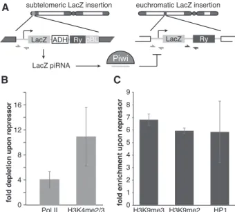
P-value <0.05) was significantly increased (Fig. 5A; Sup-plemental Table 1). There are several possible explanations for Piwi’s effect on host gene expression. First, failure in the piRNA pathway might cause up-regulation of several genes that generate piRNAs in wild-type ovaries (Robine et al. 2009; Saito et al. 2009). However, the genes up-regulated in Piwi-deficient ovaries were not enriched in piRNAs compared with other genes. Second, H3K9me3 marks installed on TE sequences in a Piwi-dependent manner might spread into neighboring host genes and repress their transcription, as was recently demonstrated in a follicular cell culture model (Sienski et al. 2012). To address this possibility, we analyzed genomic positions of the genes whose expression was increased upon Piwi knockdown relative to genomic regions that showed a decrease in H3K9me3 marks. We found that up-regulated genes did not show a significant change in the H3K9me3 mark (Fig. 5B; Supplemental Fig. S4). Furthermore, the few genes located close to the regions that show a de-crease in H3K9me3 signal had unaltered expression levels upon Piwi knockdown. Next, we analyzed the functions of up-regulated genes using gene ontology (GO) term classifications and found significant enrichment for pro-teins involved in protein turnover and stress and DNA damage response pathways (Fig. 5C). Particularly, we found that 31 subunits of the proteasome complex were overexpressed. Therefore, our analysis indicates that up-regulation of specific host genes is likely a secondary re-sponse to elevated transposon levels and genomic damage. In contrast to host genes, transcripts of TEs are targeted by piRNA. To directly address the role of piRNA in Piwi-mediated transcriptional silencing, we took advantage of a fly strain that expresses artificial piRNAs against the lacZ gene, which are loaded into Piwi complexes and are able to repress lacZ reporter expression in germ cells (Fig. 6A; Josse et al. 2007; Muerdter et al. 2012). Expres-sion of piRNAs that are antisense to the reporter gene caused transcriptional silencing of the lacZ gene as measured by Pol II occupancy (Fig. 6B). Furthermore, we found that piRNA-induced silencing of the reporter gene was associated with an increase in the repressive H3K9me3 mark and HP1 occupancy and a decrease in the abundance of the active H3K4me2/3 marks at the re-porter locus (Fig. 6C). This result is in good agreement with the genome-wide effect of Piwi depletion on distri-bution of the H3K9me3 mark and suggests that tran-scriptional silencing correlates with the establishment of a repressive chromatin structure and is mediated by piRNAs that match the target locus.
Discussion
Little is known about the function of nuclear piwi pro-teins. The nuclear piwi in mice (Miwi2) affects DNA methylation of TEs (Carmell et al. 2007; Aravin et al. 2008; Kuramochi-Miyagawa et al. 2008). Several recent reports implicate Drosophila Piwi in regulation of chro-matin marks on transposon sequences (Lin and Yin 2008; Klenov et al. 2011; Wang and Elgin 2011; Sienski et al. 2012). The mechanism of these processes is unknown in
Figure 4. Piwi-induced transcriptional repression correlates with establishment of a repressive chromatin state. (A) Overlap between genomic regions of H3K9me3 depletion upon Piwi knockdown and TEs. Two replicates of H3K9me3 ChIP-seq ex-periments were carried out on control and Piwi-depleted ova-ries, and enriched regions were identified using DESeq (see the Materials and Methods for details). A total of 705 regions show significant (P < 0.05) decrease in H3K9me3 occupancy upon Piwi knockdown, while only 30 regions showed a similarly signi-ficant increase. Out of the 705 regions that show a decrease in H3K9me3 marks upon Piwi knockdown, 91% (646) overlap with TE sequences compared with the 33% expected from random genome sampling. (B) Genomic positions of H3K9me3-depleted regions upon Piwi depletion (outer circle) and RepeatMasker-annotated transposons (inner circle). Note that almost all re-gions are localized in heterochromatic and repeat-rich portions of the genome (Het, chrU, and chrUExtra chromosomes). Le Thomas et al.
394 GENES & DEVELOPMENT
Cold Spring Harbor Laboratory Press
on April 10, 2013 - Published by
genesdev.cshlp.org
both organisms. Previously, Piwi was shown to associate with polytene chromosomes in salivary gland cells and colocalize with HP1, a chromodomain protein that binds to heterochromatin and a few loci in euchromatin, sug-gesting that HP1 mediates Piwi’s interaction with chro-matin (Brower-Toland et al. 2007). However, recent results showed that the putative HP1-binding site on Piwi is dispensable for Piwi-mediated transposon silencing (Wang and Elgin 2011).
We did not detect Piwi expression outside of the ovary
Piwi transgene expressed under native regulatory ele-ments. We detected GFP-Piwi on polytene chromosomes in ovarian nurse cells that have a germline origin; how-ever, it localizes in a pattern that largely does not overlap with HP1. FLIP experiments with GFP-Piwi indicated a relatively fast rate of fluorescence redistribution as compared with histone H2A (Supplemental Fig. S1), im-plying a transient interaction of Piwi with chromatin. Our proteomic analysis of Piwi complexes isolated from Drosophilaovaries did not identify chromatin-associated factors but revealed several RNA-binding proteins, such as splicing and nuclear export factors that bind nascent RNA transcripts (Fig. 1B). Importantly, the interaction of most of these RNA-binding proteins with Piwi was dependent on RNA, indicating that Piwi associates with nascent transcripts. As Piwi itself lacks DNA- and RNA-binding domains (beyond the piRNA-RNA-binding domain),
Figure 5. Piwi does not directly repress protein-coding genes. (A) Effect of Piwi knockdown on the expression of genes. Two replicate RNA-seq experiments were carried out, and differen-tial expression was assessed using DESeq. Genes that show significant change (P < 0.05) are indicated by black circles. The vast majority of genes does not change significantly upon germ-line Piwi knockdown (shPiwi) compared with control (shWhite). (B) H3K9me3 mark density does not change over genes that show a significant change in expression upon Piwi knockdown (see Fig. 3C). Up-regulated and down-regulated genes are plotted separately. Signal indicated is after background subtraction. (C) Functional analysis of up-regulated genes by the Database for Annotation, Visualization, and Integrated Discovery (DAVID) reveals activation of the protein degradation and DNA damage response pathways. Percentages of all up-regulated genes are indicated.
Figure 6. piRNA-dependent targeting of Piwi to a reporter locus leads to establishment of a repressive chromatin state and transcriptional silencing. (A) The mechanism of trans-silencing mediated by artificial piRNA and a schematic repre-sentation of the repressor and reporter lacZ constructs. The repressor construct is inserted in a subtelomeric piRNA cluster, leading to generation of piRNA from its sequence. Primers mapping to both constructs used for the Pol II and H3K4me2/3 ChIP-quantitative PCR (qPCR) are shown by light-gray arrows; primers specific to the reporter locus used for the H3K9me3, H3K9me2, and HP1 ChIP-qPCR are indicated by dark-gray arrows. (B) piRNAs induce transcriptional repression of the lacZ reporter. Pol II and H3K4me2/3 signals decreased on the lacZ promoter in the presence of artificial piRNAs as measured by ChIP-qPCR. Shown is the fold depletion of signal in flies that carry both repressor and reporter constructs compared with control flies that have only the reporter construct. The signal was normalized to RP49. (C) piRNAs induce an increase in H3K9me3 and H3K9me2 marks and HP1 binding as measured by ChIP-qPCR. Shown is the fold increase of corresponding ChIP signals downstream from the lacZ reporter in flies that carry both repressor and reporter constructs compared with control flies that have only reporter construct. The signal was Piwi represses piRNA target transcription
Cold Spring Harbor Laboratory Press
on April 10, 2013 - Published by
genesdev.cshlp.org
it is likely that the recruitment of Piwi to chromatin is through interactions with other RNA-binding proteins or sequence-specific interactions between Piwi-bound piRNA and nascent transcripts.
Using specific Piwi knockdown in germ cells of the Drosophilaovary, we analyzed the effect of Piwi deple-tion on gene expression, the transcripdeple-tion machinery, and H3K9me3 chromatin marks genome-wide. In agree-ment with previous results (Klenov et al. 2011), we found up-regulation of several TEs upon Piwi knock-down (Fig. 3C). The TEs that did not change their ex-pression upon germline knockdown of Piwi might be expressed exclusively in somatic follicular cells of the ovary, such as the gypsy retrotransposon. Alternatively, some elements present in the genome might not have transcriptionally active copies, or the cytoplasmic AUB/ AGO3 proteins may efficiently silence them at the post-transcriptional level.
The increase in steady-state levels of RNA upon Piwi depletion strongly correlates with an increase in Pol II occupancy on the promoters of transposons (Fig. 3D,F; Supplemental Fig S2). This result suggests that Piwi re-presses transposon expression at the transcriptional level, although we cannot completely exclude the possibility of an additional post-transcriptional effect. It was shown previously that depletion or mutation of Piwi leads to depletion of the repressive H3K9me3 mark and an in-crease in the active H3K4me2/3 marks on several trans-poson sequences (Klenov et al. 2011; Wang and Elgin 2011). Our ChIP-seq data extend these results to a genome-wide scale, proving that transposons are indeed the sole targets of Piwi, and demonstrate that changes in histone marks directly correlate with transcriptional repression.
Piwi depletion in the germline does not affect expres-sion of the majority of host genes, although a small frac-tion of genes changes expression (Fig. 5A). One possible mechanism of the effect Piwi has on host genes is the spreading of repressive chromatin structure from trans-poson sequences to adjacent host genes. Indeed, such a spreading and the resulting repression of host gene tran-scription were observed in an ovarian somatic cell (OSC) culture model (Sienski et al. 2012). However, we did not find significant changes in the H3K9me3 mark for genes that are up-regulated upon germline depletion of Piwi, arguing against this mechanism playing a major role in host gene regulation. Instead, we found that the majority of host genes whose expression is increased as a result of Piwi depletion participate in protein turnover (e.g., pro-teasome subunits) and stress and DNA damage response pathways, indicating that they might be activated as a secondary response to cellular damage induced by trans-poson activation. The different effect of Piwi depletion on host gene expression in ovary and cultured cells might be explained by the fact that silencing of host genes due to transposon insertion would likely have a strong negative effect on the fitness of the organism but could be tolerated in cultured cells. Accordingly, new transposon insertions that cause repression of adjacent host genes should be eliminated from the fly population but can be detected
in cultured cells. In agreement with this explanation, the majority of cases of repressive chromatin spreading in OSCs were observed for new transposon insertions that are absent in the sequenced Drosophila genome. Indeed, it was shown that the vast majority of new transposon insertions is present at a low frequency in the Drosophila population, likely due to strong negative selection (Petrov et al. 2003). Such selection was primarily attributed to the ability of TE sequences to cause recombination and ge-nomic rearrangements. We propose that in addition to the effects on recombination, the selection against transpo-sons can be driven by their negative impact on host gene expression in the germline linked to Piwi-mediated chro-matin silencing.
How does Piwi discriminate its proper targets— transposons—from host genes? In the case of cytoplas-mic Piwi proteins AUB and AGO3, recognition and post-transcriptional destruction of TE transcripts is guided by associated piRNAs. Our results indicate that piRNAs provide guidance for transcriptional silencing by the nu-clear Piwi protein as well. First, in contrast to host genes that are not targeted by piRNAs, TE transcripts, which are regulated by Piwi, are recognized by antisense Piwi-bound piRNA (Brennecke et al. 2007). Second, a Piwi mutant that is unable to bind piRNA failed to rescue the piwi-null mutation despite its ability to enter the nu-cleus. Finally, expression of artificial piRNAs that target a reporter locus induced transcriptional silencing associ-ated with an increase in repressive H3K9me3 and HP1 chromatin marks and a decrease in the active H3K4me2/3 marks (Fig. 6B,C). In contrast, the tethering of Piwi to chromatin in a piRNA-independent fashion by fusing Piwi with the lacI DNA-binding domain that recognizes lacO sequences inserted upstream of a reporter gene did not lead to silencing of the reporter (data not shown). Together, our results demonstrate that piRNAs are the essential guides of Piwi to recognize its targets for tran-scriptional repression.
It is tempting to propose that, similar to Argonautes in fission yeast, Drosophila Piwi directly recruits the enzy-matic machinery that establishes the repressive H3K9me3 mark on its targets. Establishment of repressive marks can lead to stable chromatin-based transcriptional silenc-ing that does not require further association of Piwi with target loci. This model explains why we found that Piwi is relatively mobile in the nucleus, indicative of only a transient interaction with chromatin. The Piwi-mediated transcriptional silencing has an interesting parallel in Caenorhabditis elegans, where the Piwi protein PRG-1 and associated 21U RNAs are able to induce stable trans-generational repression that correlates with formation of silencing chromatin marks on target loci. Interestingly, PRG-1 and 21U RNAs are necessary only for initial es-tablishment of silencing, while continuing repression depends on siRNA and the WAGO group of Argonautes (Ashe et al. 2012; Bagijn et al. 2012; Buckley et al. 2012; Shirayama et al. 2012). Future studies should reveal the pathway that leads to transcriptional repression down-stream from Piwi in Drosophila and the differences from and similarities to other species.
Le Thomas et al.
396 GENES & DEVELOPMENT
Cold Spring Harbor Laboratory Press
on April 10, 2013 - Published by
genesdev.cshlp.org
Materials and methods
Drosophila stocks
Nanos-Gal4-VP16 (BL4937), shWhite (BL33623), UASp-shPiwi (BL 33724), and Chr. I and II Balancer (BL7197) were purchased from the Bloomington Stock Center. GFP-Piwi-expressing flies (see below) were backcrossed onto the piwi1/ piwi2(available from Bloomington Stock Center) background or the otu7/otu11 (available from Bloomington Stock Center) background, respectively. LacZ reporter lines were a generous gift from S. Ronsseray.
Generation of transgenic fly lines
The GFP-Piwi, 3xFlag-HA-Piwi, and myc-Piwi constructs were generated using bacterial recombineering (Gene Bridges Counter Selection kit) to insert the respective tag after the start codon of the Piwi genomic region cloned in BAC clone BACN04M10. The KpnI–XbaI genomic fragment that contains the Piwi gene and flanking sequences was transferred to corresponding sites of the pCasper4 vector to create pCasper4/tagged Piwi.
The pCasper4/GFP-Piwi construct was used to generate pCasper4/GFP-Piwi-YK with two point mutations, Y551I and K555E. Mutations were introduced by PCR, amplifying products corresponding to a 3.1-kb upstream fragment and a 2.58-kb down-stream fragment. The updown-stream fragment included a unique XbaI site at the 59 end of the amplicon and overlapped 39 base pairs (bp) with the downstream fragment, which included a unique BamHI site at its 39 end. The single XbaI–BamHI fragment was generated by overlap PCR with outside primers and cloned into corresponding sites of pCasper4/GFP-Piwi to replace the wild-type fragment. Transgenic flies were generated by P-element-mediated transformation (BestGene).
Immunoprecipitation of Piwi proteins and RNA gel of piRNA Dissected ovaries were lysed in lysis buffer (20 mM HEPES at pH 7.0, 150 mM KCl, 2.5 mM MgCl, 0.5% Triton X-100, 0.5% Igepal, 100 U/mL RNasin [Promega], EDTA-free Complete Pro-tease Inhibitor Cocktail [Roche]) and supernatant clarified by centrifugation. Supernatant was incubated with anti-eGFP poly-clonal antibody (Covance) conjugated to Protein-G Dynabeads at 4°C. Beads were spiked with 5 pmol of synthesized 42-nucleotide RNA oligomer to assess purification efficiency, proteinase K-digested, and phenol-extracted. Isolated RNA was CIP-treated, radiolabeled using PNK and g-P32-labeled ATP, and run on a 15% urea-PAGE gel. Western blots of ovary lysate and anti-eGFP immunoprecipitates were obtained from 8% SDS-PAGE gels and probed with polyclonal rabbit anti-eGFP antibody to confirm expression of the full-length transgene.
Mass spectrometric analysis of Piwi interaction partners Lysis and clarification of ovary samples were performed as de-scribed above using lysis buffer with reduced detergent (0.1% Triton X-100, 0.1% Igepal). Piwi proteins with Flag, Myc, or GFP tag were purified from Drosophila ovaries using correspond-ing antibodies covalently coupled to M-270 epoxy Dynabeads (Invitrogen) (Cristea et al. 2005). Immunoprecipitation of free GFP from GFP-expressing ovaries was used as a negative control. Immunoprecipitations were performed in the presence or ab-sence of RNase A (100 mg/mL; 30 min at 25C). Piwi and copurified interacting proteins were resolved on NuPAGE Novex 4%–12% Bis-Tris gels and stained with colloidal Coomassie blue. Gel
trypsinized, and the peptides were extracted following the standard protocol of the Proteome Exploration Laboratory at California Institute of Technology. Peptide analyses were per-formed on an LTQ-FT Ultra (Thermo Fisher Scientific) equipped with a nanoelectrospray ion source (Thermo Fisher Scientific) connected to an EASY-nLC. Fractionation of peptides was per-formed on a 15-cm reversed-phase analytical column (75-mm internal diameter) in-house-packed with 3-mm C18 beads (ReproSil-Pur C18-AQ medium; Dr. Maisch GmbH). Acquired spectra were searched against the Drosophila melanogaster proteome using the search engine Mascot (Matrix Science, version 2.2.06), and protein inferences were performed using Scaffold (Proteome Software, version 3). For an Excel file of Piwi interaction partners, see the Supplemental Material.
ChIP, ChIP-seq, and RNA-seq
ChIP was carried out using standard protocols (Moshkovich and Lei 2010). ChIP-seq and RNA-seq library construction and se-quencing were carried out using standard protocols following the general principles described by Johnson et al. (2007) and Mortazavi et al. (2008), respectively. Data analysis was carried out using a combination of publicly available software tools and custom-written python scripts. Additional details regarding high-throughput data analysis are described in the Supplemental Material. For quantitative PCR (qPCR) primers, see Supplemen-tal Table 2. GO term analysis of genes up-regulated upon Piwi knockdown was performed using the Database for Annotation, Visualization, and Integrated Discovery (DAVID) (Huang et al. 2009a,b) and FlyBase for additional assignment of GO terms. Sequencing data is available through Gene Expression Omnibus (accession no. GSE43829).
Antibodies
eGFP antibody (rabbit polyclonal serum; Covance) was affinity-purified in our laboratory. Anti-myc (Millipore), anti-Flag (Sigma), Pol II (ab5408), and Pol II pSer5 (ab5131) are commer-cially available.
Imaging of ovaries
Ovaries were fixed in 4% PFA in PBS for 20 min, permeabilized in 1% Triton X-100 in PBS, DAPI-stained (Sigma-Aldrich), washed, and mounted in 50% glycerol/PBS. Images were captured using an AxioImager microscope; an Apotome structured illumi-nation system was used for optical sections (Carl Zeiss).
FLIP
FLIP time series were captured on an LSM510 confocal micro-scope equipped with a 403/0.9 NA Imm Corr multi-immersion objective. Ovaries were dissected into halocarbon 700 oil (Sigma) and mounted under a 0.17-mm coverslip (Carl Zeiss) immedi-ately before imaging. Two initial baseline images were captured, followed by 80–100 iterations consisting of two bleach iterations at 100% laser power (488 nm or 543 nm for GFP- and RFP-tagged proteins, respectively), followed by two images with reduced illumination intensity. FLIP series were cropped and median-filtered with a 2-pixel radius to reduce noise using FIJI (Schindelin et al. 2012) and the ‘‘Rigid Body’’ function of the StackReg plugin (The´venaz et al. 1998) to correct drift when needed. Using Matlab software (The Mathworks), images were background-subtracted and corrected for acquisition bleach-ing. A value representing the true loss of intensity relative to Piwi represses piRNA target transcription
Cold Spring Harbor Laboratory Press
on April 10, 2013 - Published by
genesdev.cshlp.org
intensity and 1 represents complete photobleaching, was calcu-lated for each pixel and each bleach/capture cycle and plotted with a color lookup table and calibration bar. Scale bars and annotations were made in Inkscape (http://inkscape.org).
Preparation of polytene squashes for immunofluorescence Flies carrying the GFP-Piwi BAC construct were backcrossed onto the otu[7] and otu[11] background. Progeny from the cross of the two lines were grown at 18°C. Stage 7–12 egg chambers were separated and transferred to a polylysine-coated micro-scopic slide into PBST. From here, the ‘‘smush’’ protocol was followed (Johansen et al. 2009), but PFA cross-linking was re-duced to 10 min. Slides were imaged using an AxioImager mi-croscope and a 633 oil immersion objective (Carl Zeiss).
Acknowledgments
We are grateful to Evelyn Stuwe from the Aravin laboratory for purifying the GFP antibody; I. Antoshechkin of the Millard and Muriel Jacobs Genetics and Genomics Laboratory for sequenc-ing; D. Trout, H. Amrhein, and S. Upchurch for computational assistance; the Bloomington Stock Center for fly stocks; and S. Hess, B. Graham, and M Sweredoski from the Proteome Ex-ploration Laboratory at the Beckmann Institute, California In-stitute of Technology, for assistance with the mass spectrometry experiments. We thank members of the Aravin laboratory for critical comments on the manuscript. We thank Barbara Wold and members of the Wold laboratory for helpful discussions on ChIP protocols and analysis. A.K.R. and E.M.P. are supported by the Institutional Training Grant NIH/NRSA 5T32 GM07616, and E.M.P. is additionally supported by the Gordon Ross Medical Foundation. G.K.M. is supported by The Beckman Foundation, the Donald Bren Endowment, and NIH grant U54 HG004576. This work was supported by grants from the National In-stitutes of Health (R01 GM097363, R00 HD057233, and DP2 OD007371A to A.A.A.), the Searle Scholar Award (to A.A.A.), and the Ellison Medical Foundation New Scholar in Aging Award (to K.F.T.).
References
Agger K, Cloos P, Christensen J, Pasini D, Rose S, Rappsilber J, Issaeva I, Canaani E, Salcini A, Helin K. 2007. UTX and JMJD3 are histone H3K27 demethylases involved in HOX gene regulation and development. Nature 449: 731–734. Ameyar-Zazoua M, Rachez C, Souidi M, Robin P, Fritsch L,
Young R, Morozova N, Fenouil R, Descostes N, Andrau J-C et al. 2012. Argonaute proteins couple chromatin silencing to alternative splicing. Nat Struct Mol Biol 19: 998–1004. Aravin A, Hannon G, Brennecke J. 2007. The Piwi–piRNA
path-way provides an adaptive defense in the transposon arms race. Science 318: 761–764.
Aravin AA, Sachidanandam R, Bourc’his D, Schaefer C, Pezic D, Toth KF, Bestor T, Hannon GJ. 2008. A piRNA pathway primed by individual transposons is linked to de novo DNA methylation in mice. Mol Cell 31: 785–799.
Ashe A, Sapetschnig A, Weick E-M, Mitchell J, Bagijn M, Cording A, Doebley A-L, Goldstein L, Lehrbach N, Le Pen J et al. 2012. piRNAs can trigger a multigenerational epige-netic memory in the germline of C. elegans. Cell 150: 88–99. Bagijn M, Goldstein L, Sapetschnig A, Weick E-M, Bouasker S, Lehrbach N, Simard M, Miska E. 2012. Function, targets, and evolution of Caenorhabditis elegans piRNAs. Science 337: 574–578.
Brennecke J, Aravin A, Stark A, Dus M, Kellis M, Sachidanandam R, Hannon G. 2007. Discrete small RNA-generating loci as master regulators of transposon activity in Drosophila. Cell128: 1089–1103.
Brower-Toland B, Findley S, Jiang L, Liu L, Yin H, Dus M, Zhou P, Elgin S, Lin H. 2007. Drosophila PIWI associates with chromatin and interacts directly with HP1a. Genes Dev 21: 2300–2311.
Buckley B, Burkhart K, Gu S, Spracklin G, Kershner A, Fritz H, Kimble J, Fire A, Kennedy S. 2012. A nuclear Argonaute promotes multigenerational epigenetic inheritance and germline immortality. Nature 489: 447–451.
Carmell M, Girard A, van de Kant H, Bourc’his D, Bestor T, de Rooij D, Hannon G. 2007. MIWI2 is essential for spermato-genesis and repression of transposons in the mouse male germline. Dev Cell 12: 503–514.
Chung W-J, Okamura K, Martin R, Lai E. 2008. Endogenous RNA interference provides a somatic defense against Dro-sophilatransposons. Curr Biol 18: 795–802.
Cox D, Chao A, Baker J, Chang L, Qiao D, Lin H. 1998. A novel class of evolutionarily conserved genes defined by piwi are essential for stem cell self-renewal. Genes Dev 12: 3715–3727. Cristea IM, Williams R, Chait BT, Rout MP. 2005. Fluorescent proteins as proteomic probes. Mol Cell Proteomics 4: 1933– 1941.
Djuranovic S, Zinchenko M, Hur J, Nahvi A, Brunelle J, Rogers E, Green R. 2010. Allosteric regulation of Argonaute proteins by miRNAs. Nat Struct Mol Biol 17: 144–150.
Friedman R, Farh K, Burge C, Bartel D. 2009. Most mammalian mRNAs are conserved targets of microRNAs. Genome Res 19: 92–105.
Galiana-Arnoux D, Dostert C, Schneemann A, Hoffmann J, Imler J-L. 2006. Essential function in vivo for Dicer-2 in host defense against RNA viruses in Drosophila. Nat Immunol7: 590–597.
Ghildiyal M, Seitz H, Horwich M, Li C, Du T, Lee S, Xu J, Kittler E, Zapp M, Weng Z et al. 2008. Endogenous siRNAs derived from transposons and mRNAs in Drosophila somatic cells. Science320: 1077–1081.
Grewal S, Jia S. 2007. Heterochromatin revisited. Nat Rev Genet 8: 35–46.
Gunawardane L, Saito K, Nishida K, Miyoshi K, Kawamura Y, Nagami T, Siomi H, Siomi M. 2007. A slicer-mediated mechanism for repeat-associated siRNA 59 end formation in Drosophila. Science 315: 1587–1590.
Haase A, Fenoglio S, Muerdter F, Guzzardo P, Czech B, Pappin D, Chen C, Gordon A, Hannon G. 2010. Probing the ini-tiation and effector phases of the somatic piRNA pathway in Drosophila. Genes Dev 24: 2499–2504.
Handler D, Olivieri D, Novatchkova M, Gruber F, Meixner K, Mechtler K, Stark A, Sachidanandam R, Brennecke J. 2011. A systematic analysis of Drosophila TUDOR domain-containing proteins identifies Vreteno and the Tdrd12 family as essential primary piRNA pathway factors. EMBO J 30: 3977–3993. Huang DW, Sherman B, Lempicki R. 2009a. Bioinformatics
enrichment tools: Paths toward the comprehensive func-tional analysis of large gene lists. Nucleic Acids Res 37: 1–13.
Huang DW, Sherman B, Lempicki R. 2009b. Systematic and integrative analysis of large gene lists using DAVID bioin-formatics resources. Nat Protoc 4: 44–57.
Hutvagner G, Simard M. 2008. Argonaute proteins: Key players in RNA silencing. Nat Rev Mol Cell Biol 9: 22–32. Ishizu H, Nagao A, Siomi H. 2011. Gatekeepers for Piwi–piRNA
complexes to enter the nucleus. Curr Opin Genetic Dev 21: 484–490.
Le Thomas et al.
398 GENES & DEVELOPMENT
Cold Spring Harbor Laboratory Press
on April 10, 2013 - Published by
genesdev.cshlp.org
![Figure 1. Piwi associates with chromatin and nuclear transcripts. (A) Polytene chromosomes from Drosophila nurse cells expressing GFP-Piwi on the otu[7]/otu[11]](https://thumb-eu.123doks.com/thumbv2/123doknet/14321014.497072/15.918.108.522.877.1099/figure-associates-chromatin-transcripts-polytene-chromosomes-drosophila-expressing.webp)