HAL Id: hal-02465771
https://hal.archives-ouvertes.fr/hal-02465771
Submitted on 19 Feb 2020
HAL is a multi-disciplinary open access
archive for the deposit and dissemination of
sci-entific research documents, whether they are
pub-lished or not. The documents may come from
teaching and research institutions in France or
abroad, or from public or private research centers.
L’archive ouverte pluridisciplinaire HAL, est
destinée au dépôt et à la diffusion de documents
scientifiques de niveau recherche, publiés ou non,
émanant des établissements d’enseignement et de
recherche français ou étrangers, des laboratoires
publics ou privés.
Distributed under a Creative Commons Attribution| 4.0 International License
Peripheral Routes to Neurodegeneration: Passing
Through the Blood–Brain Barrier
Patrizia Giannoni, Sylvie Claeysen, Francesco Noe, Nicola Marchi
To cite this version:
Patrizia Giannoni, Sylvie Claeysen, Francesco Noe, Nicola Marchi. Peripheral Routes to
Neurodegen-eration: Passing Through the Blood–Brain Barrier. Frontiers in Aging Neuroscience, Frontiers, 2020,
12, pp.3. �10.3389/fnagi.2020.00003�. �hal-02465771�
fnagi-12-00003 February 3, 2020 Time: 16:6 # 1 REVIEW published: 04 February 2020 doi: 10.3389/fnagi.2020.00003 Edited by: Daniel Ortuño-Sahagún, Universidad de Guadalajara, Mexico Reviewed by: Annadora Bruce-Keller, Pennington Biomedical Research Center, United States Berislav Zlokovic, University of Southern California, United States *Correspondence: Nicola Marchi [email protected] Received: 30 July 2019 Accepted: 08 January 2020 Published: 04 February 2020 Citation: Giannoni P, Claeysen S, Noe F and Marchi N (2020) Peripheral Routes to Neurodegeneration: Passing Through the Blood–Brain Barrier. Front. Aging Neurosci. 12:3. doi: 10.3389/fnagi.2020.00003
Peripheral Routes to
Neurodegeneration: Passing
Through the Blood–Brain Barrier
Patrizia Giannoni
1, Sylvie Claeysen
2, Francesco Noe
3and Nicola Marchi
2*
1Laboratoire CHROME (EA 7352), Université de Nîmes, Nîmes, France,2CNRS, INSERM U1191, Institut de Génomique
Fonctionnelle, University of Montpellier, Montpellier, France,3HiLIFE – Neuroscience Center, University of Helsinki, Helsinki,
Finland
A bidirectional crosstalk between peripheral players of immunity and the central nervous
system (CNS) exists. Hence, blood–brain barrier (BBB) breakdown is emerging as a
participant mechanism of dysregulated peripheral–CNS interplay, promoting diseases.
Here, we examine the implication of BBB damage in neurodegeneration, linking it
to peripheral brain-directed autoantibodies and gut–brain axis mechanisms. As BBB
breakdown is a factor contributing to, or even anticipating, neuronal dysfunction(s), we
here identify contemporary pharmacological strategies that could be exploited to repair
the BBB in disease conditions. Developing neurovascular, add on, therapeutic strategies
may lead to a more efficacious pre-clinical to clinical transition with the goal of curbing
the progression of neurodegeneration.
Keywords: blood–brain barrier, neurodegeneration, peripheral immunity, traumatic brain injury, status epilepticus, autoantibodies, gut–brain axis, inflammation
BRAIN BARRIERS’ PATHS, LEAKS, AND
NEURODEGENERATION
The term neurodegenerative describes a progressive deterioration of the central nervous system
(CNS) that is frequently associated with abnormal accumulation of proteins. Importantly,
neurofibrillary tau-protein tangles are not only a major sign of Alzheimer’s disease (AD) but
are reported in temporal lobe epilepsy and post-traumatic encephalopathies (
Tai et al., 2016
).
Among the emerging disease mechanisms, a peripheral–CNS pathological interplay is proposed
to contribute to the neurodegenerative process (
Marchi et al., 2014
;
Engelhardt et al., 2017
;
Fung
et al., 2017
;
Pavlov and Tracey, 2017
;
Prinz and Priller, 2017
;
Le Page et al., 2018
). Accordingly,
harmful events occurring at the cerebrovascular interface are being examined as key determinants
partaking to or even preceding neurodegeneration (
Zlokovic, 2011
;
Nation et al., 2019
;
Sweeney
et al., 2019
). At the cerebrovasculature, specialized endothelial cells, mural cells, and astroglia
constructs (
Abbott et al., 2010
;
Giannoni et al., 2018
;
Sweeney et al., 2019
) provide physical and
biological properties governing the homeostatic–immune interactions between peripheral blood
cells, or molecules, and brain neuroglia. The physiological parenchymal milieu composition ensures
a healthy neuronal transmission, attainable because of the tightness of the blood–brain barrier
(BBB;
Zlokovic, 2008
;
Giannoni et al., 2018
;
Nation et al., 2019
). At the pial arterial and venous level,
the cerebrovasculature is permissive to blood cells or molecules, while it becomes impermeable at
the arteriole–capillary level where barriers’ properties are fully established (
Abbott et al., 2010
).
fnagi-12-00003 February 3, 2020 Time: 16:6 # 2
Giannoni et al. Neurodegeneration and Blood–Brain Interfaces
BBB vessels also contribute to cerebrospinal and interstitial fluid
movements and the elimination of waste products from the
interstitial and perivascular spaces (
Noé and Marchi, 2019
).
It is increasingly recognized that a BBB pathological
imprint can provoke a brain pro-inflammatory disequilibrium
sufficient to modify neuronal activity in the long term
(
Marchi et al., 2007, 2014
;
Nation et al., 2019
).
Vascular-dependent mechanisms of neurodegeneration can rapidly elicit
as a consequence of peripheral infections, head trauma,
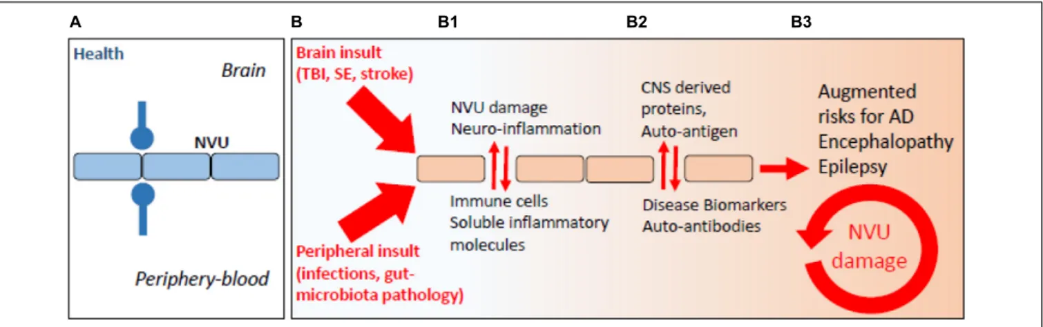
ischemic stroke, or status epilepticus (Figure 1;
Nation et al.,
2019
;
Sweeney et al., 2019
). These are risk factors for the
development of long-term neurodegenerative sequelae and
encephalopathies (e.g., post-concussion or head trauma-related
chronic traumatic encephalopathy, CTE), cerebral amyloid
angiopathy (CAA), AD, and epilepsy. Under conditions of
increased BBB permeability, an aberrant bidirectional exchange
between the neurovascular unit and the peripheral blood occurs,
compounding to neurodegenerative modifications (Figure 1;
Marchi et al., 2014
;
Engelhardt et al., 2017
;
Fung et al., 2017
;
Pavlov and Tracey, 2017
;
Prinz and Priller, 2017
;
Le Page et al.,
2018
). Completing a vicious cycle, beta-amyloid deposition in the
brain can provoke capillaries dysfunction (
Thomas et al., 1996
;
Zhang et al., 1997
;
Iadecola et al., 1999
;
Deane et al., 2003, 2012
;
Nortley et al., 2019
). As an example, reactive oxygen species and
endothelin-1 production were proposed to elicit vasoconstriction
at pericyte locations (
Nortley et al., 2019
). A question remains
regarding whether the endothelin-1 mechanism can directly drive
neurodegeneration.
AUTOANTIBODIES AND
NEURODEGENERATION: BAD, GOOD,
OR NIL?
The communication between the peripheral blood and the brain
occurs at preferential cerebrovascular sites (
Zlokovic, 2011
;
Noé
and Marchi, 2019
), e.g., at post-capillary venules or pial vessels,
and by a system of lymphatic vessels draining the cerebrospinal
and interstitial fluids to cervical lymph nodes (
Aspelund et al.,
2015
;
Louveau et al., 2015a,b, 2018
). At the intravascular level,
moving leukocytes shape a peripheral–brain immune dialog
where endothelium activation or permeability, perivascular
immune cell homing, and brain entry of immune soluble factors
prompt and sustain neuroglia inflammation [Figure 1; see
Engelhardt et al. (2017)
and
Ransohoff (2016)
for fundamental
aspects of endothelial–leukocyte adhesion]. The implication of
the cerebrovascular interface to innate and adaptive modalities of
immunity is central (
Schwartz and Shechter, 2010
;
Sommer et al.,
2017
). Adaptive immunity to the brain requires T- and B-cell
stimulation at extra-CNS lymphatic organs and by professional
antigen-presenting cells (
Janeway et al., 2001
), thus implying the
existence of a peripheral–brain immune dialog, e.g., via the CNS
vascular and lymphatic routes (
Noé and Marchi, 2019
).
A question exists on whether neurodegeneration may result
from autoimmune-like processes (Table 1). Contingent to a
prolonged or recurrent BBB permeability, specific antigens could
exit the brain to reach the bloodstream, mounting a peripheral
humoral response. Newly formed brain-directed autoantibodies
could be neuropathological upon their entry into the brain across
a continuously damaged BBB (
Levin et al., 2010
). Importantly,
autoantibodies and autoreactive T cells were reported in the
cerebrospinal fluid (CSF), sera, as well as in the brain of AD
patients and experimental models of disease (Table 1;
Kronimus
et al., 2016
;
Wu and Li, 2016
). Anti-A
β antibodies (Ig type
G) correlated with scores of dementia (
Wilson et al., 2009
).
Intrathecal antibodies against tau filaments were reported in
AD patients (
Mruthinti et al., 2004
) and were proposed as
contributors of disease progression (
Bartos et al., 2012
).
Anti-tau autoantibodies are not specific to AD as they are increased
in patients suffering from other neurodegenerative diseases, e.g.,
multiple sclerosis (
Fialová et al., 2011
).
The significance of peripheral autoantibodies as biomarkers
of neurodegenerative conditions also remains to be established.
Autoantibodies against the glutamate receptor
N-methyl-
D-aspartate receptor (NMDAR) were detected in plasma of AD
patients (
Davydova et al., 2007
). Levels of antibodies were
shown to correlate with clinical severity, as patients affected by
moderate and severe dementia presented a twofold autoantibody
increase compared with patients suffering from mild dementia
(
Davydova et al., 2007
). The presence of autoantibodies against
5-HT was also reported (
Myagkova et al., 2001
), with levels
increasing during the mild phase of the disease, subsequently
reaching a plateau (
Myagkova et al., 2001
). Similar findings
were reported for autoantibodies directed against the receptor
for advanced glycation end products (
Wilson et al., 2009
).
In a transgenic model of AD, autoantibodies against the
sphingolipid ceramide correlated with amyloid plaque increase
(
Posse de Chaves and Sipione, 2010
;
Dinkins et al., 2015
).
Autoantibodies against ATP synthase (
Vacirca et al., 2012
),
α(1)-adrenergic, and the
β(2)-adrenergic receptors were also reported
(
Karczewski et al., 2012
). Autoantibodies against the
α(1)-adrenergic and the
β(2)-adrenergic receptors may contribute to
vascular lesions and increased plaque formation in AD patients
(
Karczewski et al., 2012
).
Importantly, not all autoantibodies are harmful.
Brain-reactive natural autoantibodies (NAbs) are protective (
Britschgi
et al., 2009
;
Kellner et al., 2009
;
Dodel et al., 2011
;
Bach and Dodel,
2012
). NAbs are mostly IgM and are spontaneously produced.
NAbs are polyreactive with low affinity for self-antigens
(
Casali and Schettino, 1996
). Physiologically, NAbs facilitate
phagocytosis of apoptotic cells, inhibit inflammatory pathways,
and have a role in maintaining immune tolerance (
Elkon and
Silverman, 2012
). NAbs to Aβ can inhibit plaque aggregation,
block A
β toxicity, and catalyze Aβ clearance (
Lindhagen-Persson
et al., 2010
). Immunotherapies using specific, or aspecific,
autoantibodies were tested. Bapineuzumab is the humanized
form of a monoclonal anti-A
β antibody targeting the N-terminus
of Aβ. In phase II trials, Bapineuzumab administration reduced
A
β plaques in AD brains (
Salloway et al., 2009
;
Rinne et al.,
2010
) and was associated with decreased total and
phospho-tau levels in the CSF (
Asuni et al., 2007
). Bapineuzumab
was, however, discontinued after a phase III trial and showed
no beneficial effects on cognitive or functional outcomes
(
U.S. National Library of Medicine, 2019a,b
). Aducanumab
fnagi-12-00003 February 3, 2020 Time: 16:6 # 3
Giannoni et al. Neurodegeneration and Blood–Brain Interfaces
FIGURE 1 | The periphery–brain interplay and CNS disease: the neurovascular pathological imprint. (A) Proper peripheral–brain segregation under healthy conditions (neurovascular unit, NVU; blue lines). (B) Pathological insult(s) elicited in the periphery or in the brain (traumatic brain injury, TBI; status epilepticus, SE) converge to NVU damage (e.g., BBB permeability) and neuro-inflammation, leading to temporary or prolonged loss of brain homeostatic control (B1, red arrows). (B2) Under conditions of BBB permeability, concentration gradients favor brain-derived proteins to extravasate into the peripheral blood. Under this condition, a peripheral auto-immune reaction may mount leading to the production of autoantibodies, possibly re-entering into the CNS if BBB damage endures (B3).
(BIIB037) is a human monoclonal antibody selectively targeting
aggregated Aβ (oligomers and fibrils) (
Sevigny et al., 2016
).
An Aducanumab phase III trial was terminated as endpoints
were not meet. The analysis of a larger data set is ongoing.
Tau immunotherapies are also being developed, attenuating or
preventing functional impairment in experimental models, as
reviewed in
Sigurdsson (2018)
.
AUTOANTIBODIES AND
POST-TRAUMATIC ENCEPHALOPATHY
Resulting from repeated head trauma and BBB damage, chronic
traumatic encephalopathy (CTE) presents with accumulation of
neurofibrillary tau-protein tangles. In TBI subjects, blood and
CSF autoantibodies were suggested as etiological components
or as possible biomarkers of neurodegeneration (
Raad et al.,
2014
;
Kobeissy, 2015
; Table 1). Anti-glial fibrillary acidic protein
(GFAP) fragments were found in the sera of TBI patients (
Zhang
et al., 2014
). Serum autoantibodies against S100B were reported
in American football players when repeated sub-concussive
events were associated with BBB damage (
Marchi et al.,
2013
). Autoantibodies against the neuronal
α7-subunit of the
acetylcholine receptor (
Sorokina et al., 2011
) as well as AMPA and
NMDA receptors (
Goryunova et al., 2007
) were detected in TBI
subjects, while IgG autoantibodies to neurons and basal lamina
were reported in rat serum following experimental head trauma
(
Rudehill et al., 2006
). Autoantibodies to the pituitary gland were
reported in TBI subjects 3 years after the trauma (
Tanriverdi
et al., 2008, 2010
). Damage to the pituitary gland is distinctive
of the TBI pathology with 20–50% of patients showing some
degrees of pituitary dysfunction, which affects growth hormone
production (
Aimaretti et al., 2005
;
Tanriverdi et al., 2006
). An
association between anti-pituitary autoantibodies and pituitary
dysfunction was reported in patients suffering from mild TBI,
including repetitive concussions (
Tanriverdi et al., 2010
).
Autoreactive antibodies have been proposed for the treatment
of TBI sequelae. The presence of hyper-phosphorylated tau
accumulating in neurofibrillary tangles is a characteristic of CTE
(
Omalu et al., 2010
). Even if phospho-tau is detectable only at
low levels acutely after TBI (
Smith et al., 2003
;
Blennow et al.,
2012
;
Goldstein et al., 2012
;
Mannix et al., 2013
), a specific form
of phospho-tau can be produced in response to TBI (cis P-tau)
(
Kondo et al., 2015
). This protein spreads throughout the brain,
harming cells and leading to post-traumatic neurodegeneration
and dementia. In two animal models of TBI, administration of
a monoclonal antibody discriminating between the
cis and the
trans forms of the protein and blocking cis P-tau prevented the
onset of tauopathy and cortical atrophy. These accumulating
evidence supports the possible involvement of autoantibodies in
post-TBI neurodegenerative conditions, perhaps providing new
disease biomarkers and therapeutic entry points.
THE GUT–BRAIN AXIS AND
NEURODEGENERATION: IS THERE A
BARRIER IMPLICATION?
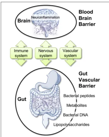
Here, we discuss a specific framework where alterations of the
gut microbiota (GM) could impact BBB permeability, promote
neuro-inflammation, and favor neurodegenerative modifications
(Figure 2;
Braniste et al., 2014
;
Cerovic et al., 2019
;
Parker
et al., 2019
;
Wang et al., 2019
). Bacteria, viruses, parasites, and
non-pathogenic fungi constitute the intestinal microbiota. These
complex communities of microbes colonizing the gastrointestinal
tract are major players in health. Modern life and diets have
progressively induced changes in the composition of the GM,
perhaps for the worse, as this can contribute to chronic illnesses
(
Lozupone et al., 2012
;
Myles, 2014
;
Kumar and Forster, 2017
;
Shanahan et al., 2017
;
Cryan et al., 2019
;
Pagliai et al., 2019
;
Reza et al., 2019
). Intestinal microbes can influence brain
function through a continuous dialog involving the immune,
fnagi-12-00003 February 3, 2020 Time: 16:6 # 4
Giannoni et al. Neurodegeneration and Blood–Brain Interfaces
FIGURE 2 | Gut–brain axis: communication routes and physiological barriers. A double, peripheral, and brain homeostatic control is performed by the intestinal–epithelial and blood–brain barriers under healthy conditions. Rupture of one barrier (e.g., gut) may impact the other (e.g., brain), with the blood stream and the immune system being the facilitators or the arbitrators of the pathological spread and neuro-inflammation.
the vascular, and the nervous systems (Figure 2;
Schroeder
and Bäckhed, 2016
;
Cox and Weiner, 2018
;
Butler et al.,
2019
;
Cryan et al., 2019
). Modifications in the composition
of the GM was reported in brain disorders, such as autism
(
Adams et al., 2011
;
Kang et al., 2019
), depression (
Kelly et al.,
2016
;
Zheng et al., 2016
), Parkinson’s disease (
Scheperjans
et al., 2015
;
Sampson et al., 2016
), and AD (
Cattaneo et al.,
2017
;
Vogt et al., 2017
;
Zhuang et al., 2018
). Intriguingly, the
extent of the amyloid pathology in AD mice appears to be
dependent of the microbial status, which is specific to the
animal housing facility. APP/PS1 mice bred in a germ-free
facility displays decreased amyloid plaque number compared
to mice housed in non-germ-free conditions (
Harach et al.,
2017
). Moreover, the administration of broad-spectrum,
combinatorial antibiotics to APP/PS1 mice, either during the
peri-natal or the adult stage, reduced brain Aβ deposition
(
Minter et al., 2016, 2017
).
Existing reports support the hypothesis of a possible infectious
origin of AD. A
β was proposed as an antimicrobial peptide
(
Soscia et al., 2010
;
Moir et al., 2018
) responding to pathogens
(
Kumar et al., 2016
;
Eimer et al., 2018
). Infectious agents, such
as
Chlamydia pneumonia, Proprionibacterium acne, Helicobacter
pylori, Porphyromonas gingivalis, or spirochetes, are associated
with AD (
Kornhuber, 1996
;
Balin et al., 1998
;
Kountouras et al.,
2006
;
Miklossy, 2011
;
Poole et al., 2015
). A microbial hypothesis
is supported by evidence describing the presence of viruses, such
as Herpes simplex virus type I, in the brains of AD patients (
Lin
et al., 2002
;
Alonso et al., 2014
;
Itzhaki et al., 2016
).
Within the complex interplay between the gut microbiome
and the CNS, a role for brain barriers and neuroinflammation is
becoming important (
Braniste et al., 2014
;
Cerovic et al., 2019
;
Parker et al., 2019
;
Wang et al., 2019
). The impact of the gut
microbiome composition on CNS health was reported (
Amedei
and Boem, 2018
;
Chu et al., 2019
;
Sherwin et al., 2019
;
Virtue
et al., 2019
). Recent work demonstrated that GM composition
controls BBB development and permeability in mice (
Braniste
et al., 2014
). In AD, increased gut permeability due to GM
dysbiosis was reported during prolonged stress. In this condition,
molecules that are normally secluded in the intestine, e.g.,
inflammatory mediators, bacteria, or bacterial-derived agents,
could leak out and reach the peripheral blood. Bacterial DNA,
metabolites, or proteins circulating in the blood stream could,
in turn, modify BBB permeability (
Braniste et al., 2014
;
Myles,
2014
;
Kumar and Forster, 2017
;
Cerovic et al., 2019
;
Parker et al.,
2019
;
Wang et al., 2019
). Existing reports indicated bacterial DNA
in human blood with a possibility for brain access (
Lelouvier
et al., 2016
;
Païssé et al., 2016
;
Schierwagen et al., 2018
). Brain
entry of
P. gingivalis, a bacterium associated with periodontal
disease, has been described (
Dominy et al., 2019
). Gingipain
inhibitors reduced the bacterial load and the bacteria-induced
neuro-inflammation in a mouse model (
Dominy et al., 2019
).
Among Spirochetes,
Borrelia burgdorferi is a strain associated
with Lyme dementia that could enter the brain. In humans, this
specific strain can form biofilms similar to senile plaques. A
β and
bacterial DNA appear as important constituents of these biofilms,
suggesting that amyloid plaques may originate in association with
or from the spirochetal colonies (
Allen, 2016
;
Miklossy, 2016
).
These examples highlight the need of tightly regulated
intestinal and brain barriers (
Rahman et al., 2018
). In AD, a
dysbiotic GM may enhance gut permeability and alter BBB
integrity, allowing the access of infectious agents or associated
molecules into the brain (
Martin et al., 2018
). Significantly,
intestinal and brain barriers are reactive to analogous
pro-inflammatory triggers. Circulating pro-inflammatory cytokines IL-17,
interferon-gamma (IFN-
γ), and the small intestine epithelium
protein zonulin can damage the intestinal–epithelia and BBBs
(
Rahman et al., 2018
).
GUT MICROBIOTA AND
AUTOANTIBODIES: INITIAL CLUES
Hypotheses linking modifications of the GM and production of
autoantibodies are emerging (
Petta et al., 2018
). Some evidence
supports the concept that specific dietary components may
affect B-cell maturation and activity, ultimately leading
to the formation of autoantibodies (
Petta et al., 2018
).
Obesity was associated with a systemic pro-inflammatory
state, characterized by changes in the frequency of B-cell
fna gi-12-00003 Fe bruary 3, 2020 T ime: 16:6 # 5 Giannoni et al. Neur odegeneration and Blood–Brain Interfaces
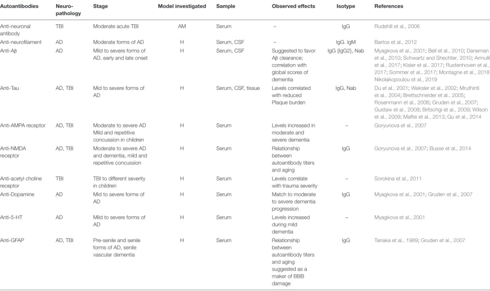
TABLE 1 | Autoantibodies reported in neurodegenerative disease and post-TBI.
Autoantibodies
Neuro-pathology
Stage Model investigated Sample Observed effects Isotype References
Anti-neuronal antibody
TBI Moderate acute TBI AM Serum – IgG Rudehill et al., 2006
Anti-neurofilament AD Moderate forms of AD H Serum, CSF – IgG. IgM Bartos et al., 2012
Anti-Aβ AD Mild to severe forms of
AD, early and late onset
H Serum, CSF Suggested to favor
Aβ clearance; correlation with global scores of dementia
IgG {IgG2}, Nab Myagkova et al., 2001;Bell et al., 2010;Daneman et al., 2010;Schwartz and Shechter, 2010;Armulik et al., 2017;Kisler et al., 2017;Rustenhoven et al., 2017;Sommer et al., 2017;Montagne et al., 2018;
Nikolakopoulou et al., 2019
Anti-Tau AD, TBI Mid to severe forms of
AD
H Serum, CSF, tissue Levels correlated with reduced Plaque burden
IgG, Nab Du et al., 2001;Weksler et al., 2002;Mruthinti et al., 2004;Brettschneider et al., 2005;
Rosenmann et al., 2006;Gruden et al., 2007;
Gustaw et al., 2008;Britschgi et al., 2009;Wilson et al., 2009;Maftei et al., 2013;Qu et al., 2014
Anti-AMPA receptor AD, TBI Moderate to severe AD Mild and repetitive concussion in children
H Serum Levels increased in
moderate and severe dementia
– Goryunova et al., 2007
Anti-NMDA receptor
AD, TBI Moderate to severe AD and dementia, mild and repetitive concussion
H Serum Relationship
between autoantibody titers and aging
IgG Goryunova et al., 2007;Busse et al., 2014
Anti-acetyl choline receptor
TBI TBI to different severity in children
H Serum Levels correlate
with trauma severity
– Sorokina et al., 2011
Anti-Dopamine AD Mid to severe forms of
AD
H Serum Match to moderate
to severe dementia progression
IgG Myagkova et al., 2001;Gruden et al., 2007
Anti-5-HT AD Mild to severe forms of
AD
H Serum Levels increased
during mild dementia
– Myagkova et al., 2001
Anti-GFAP AD, TBI Pre-senile and senile
forms of AD, senile vascular dementia H Serum Relationship between autoantibody titers and aging suggested as a maker of BBB damage
IgG Tanaka et al., 1989;Gruden et al., 2007
(Continued) Fr ontiers in Aging Neur oscience | www .fr ontiersin.org 5 February 2020 | V olume 12 | Article 3
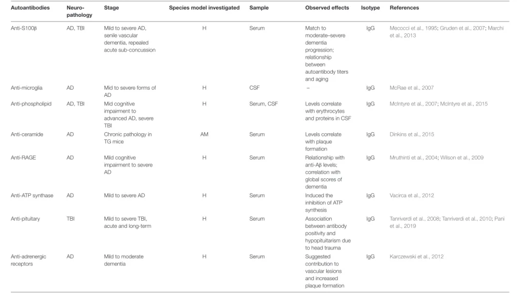
fna gi-12-00003 Fe bruary 3, 2020 T ime: 16:6 # 6 Giannoni et al. Neur odegeneration and Blood–Brain Interfaces TABLE 1 | Continued Autoantibodies Neuro-pathology
Stage Species model investigated Sample Observed effects Isotype References
Anti-S100β AD, TBI Mild to severe AD,
senile vascular dementia, repealed acute sub-concussion H Serum Match to moderate–severe dementia progression; relationship between autoantibody titers and aging
IgG Mecocci et al., 1995;Gruden et al., 2007;Marchi et al., 2013
Anti-microglia AD Mid to severe forms of
AD
H CSF – IgG McRae et al., 2007
Anti-phospholipid AD, TBI Mid cognitive impairment to advanced AD, severe TBI
H Serum, CSF Levels correlate
with erythrocytes and proteins in CSF
IgG McIntyre et al., 2007;McIntyre et al., 2015
Anti-ceramide AD Chronic pathology in
TG mice
AM Serum Levels correlate
with plaque formation
IgG Dinkins et al., 2015
Anti-RAGE AD Mild cognitive
impairment to severe AD
H Serum Relationship with
anti-Aβ levels; correlation with global scores of dementia
IgG Mruthinti et al., 2004;Wilson et al., 2009
Anti-ATP synthase AD Mild to severe AD H Serum Induced the
inhibition of ATP synthesis
IgG Vacirca et al., 2012
Anti-pituitary TBI Mild to severe TBI,
acute and long-term
H Serum Association
between antibody positivity and hypopituitarism due to head trauma
IgG Tanriverdi et al., 2008;Tanriverdi et al., 2010;Pani et al., 2019 Anti-adrenergic receptors AD Mild to moderate dementia H Serum Suggested contribution to vascular lesions and increased plaque formation
IgG Karczewski et al., 2012
AM, data derives from animal models only.
Fr ontiers in Aging Neur oscience | www .fr ontiersin.org 6 February 2020 | V olume 12 | Article 3
fnagi-12-00003 February 3, 2020 Time: 16:6 # 7
Giannoni et al. Neurodegeneration and Blood–Brain Interfaces
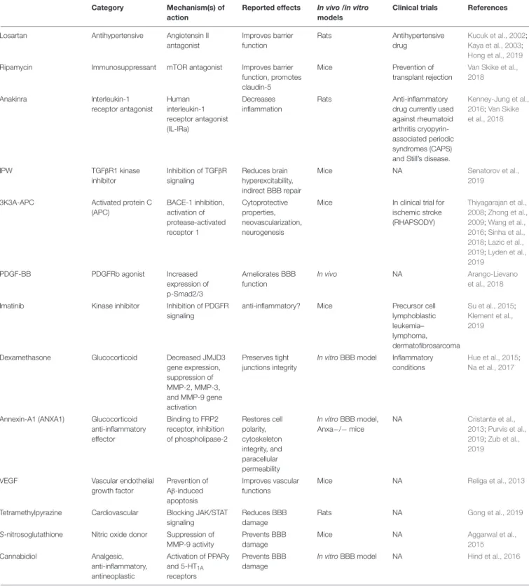
TABLE 2 | Available molecules exerting BBB repairing and anti-inflammatory effects.
Category Mechanism(s) of
action
Reported effects In vivo /in vitro models
Clinical trials References
Losartan Antihypertensive Angiotensin II
antagonist Improves barrier function Rats Antihypertensive drug Kucuk et al., 2002; Kaya et al., 2003; Hong et al., 2019
Ripamycin Immunosuppressant mTOR antagonist Improves barrier
function, promotes claudin-5
Mice Prevention of
transplant rejection
Van Skike et al., 2018 Anakinra lnterleukin-1 receptor antagonist Human interleukin-1 receptor antagonist (IL-IRa) Decreases inflammation Rats Anti-inflammatory
drug currently used against rheumatoid arthritis cryopyrin-associated periodic syndromes (CAPS) and Still’s disease.
Kenney-Jung et al., 2016;Van Skike et al., 2018 IPW TGFβR1 kinase inhibitor Inhibition of TGFβR signaling Reduces brain hyperexcitability, indirect BBB repair
Mice NA Senatorov et al.,
2019
3K3A-APC Activated protein C
(APC) BACE-1 inhibition, activation of protease-activated receptor 1 Cytoprotective properties, neovascularization, neurogenesis
Mice In clinical trial for
ischemic stroke (RHAPSODY) Thiyagarajan et al., 2008;Zhong et al., 2009;Wang et al., 2016;Sinha et al., 2018;Lazic et al., 2019;Lyden et al., 2019 PDGF-BB PDGFRb agonist Increased expression of p-Smad2/3 Ameliorates BBB function In vivo NA Arango-Lievano et al., 2018
Imatinib Kinase inhibitor Inhibition of PDGFR
signaling
anti-inflammatory? Mice Precursor cell
lymphoblastic leukemia– lymphoma, dermatofibrosarcoma Su et al., 2015; Klement et al., 2019
Dexamethasone Glucocorticoid Decreased JMJD3
gene expression, suppression of MMP-2, MMP-3, and MMP-9 gene activation Preserves tight junctions integrity
In vitro BBB model Inflammatory conditions
Hue et al., 2015;
Na et al., 2017
Annexin-A1 (ANXA1) Glucocorticoid anti-inflammatory effector Binding to FRP2 receptor, inhibition of phospholipase-2 Restores cell polarity, cytoskeleton integrity, and paracellular permeability In vitro BBB model, Anxa−/− mice NA Cristante et al., 2013;Purvis et al., 2019;Zub et al., 2019
VEGF Vascular endothelial
growth factor Prevention of Aβ-induced apoptosis Improves vascular functions
Mice NA Religa et al., 2013
Tetramethylpyrazine Cardiovascular Blocking JAK/STAT signaling
Reduces BBB damage
Rats NA Gong et al., 2019
S-nitrosoglutathione Nitric oxide donor Suppression of MMP-9 activity
Prevents BBB damage
Mice NA Aggarwal et al.,
2015 Cannabidiol Analgesic, anti-inflammatory, antineoplastic Activation of PPARy and 5-HT1A receptors Prevents BBB damage
In vitro BBB model NA Hind et al., 2016
subpopulation [e.g., reduction of the anti-inflammatory
IL-10
+regulatory B cell (
Nishimura et al., 2013
)] and by
an increase in autoantibody levels (
Kosaraju et al., 2017
).
Diets rich in polyunsaturated fatty acid are associated
with the suppression of pro-inflammatory responses and a
reduction of circulating autoantibodies (
Pestka et al., 2014
;
Tomasdottir et al., 2014
). Dietary components impact the
composition of the gastrointestinal bacterial populations:
fnagi-12-00003 February 3, 2020 Time: 16:6 # 8
Giannoni et al. Neurodegeneration and Blood–Brain Interfaces
consumption of prebiotics increases the intestinal levels
of
Bifidobacterium and Lactobacillus (
Singh et al., 2017
),
with a possible link to B-cell differentiation, maturation,
and activation (
Ouwehand et al., 2002
). Diet can impact
autoantibody
production,
directly
by
promoting
pro-inflammatory conditions and indirectly by altering the GM.
In experimental autoimmune encephalomyelitis (EAE) it was
demonstrated that the commensal microbiota composition
is a pivotal factor for disease development (
Lee et al.,
2011
) and that modifying the GM impacts the levels of
T and B cells or the levels of circulating autoantibodies
(
Ochoa-Repáraz et al., 2009, 2010
).
BBB REPAIRING PHARMACOLOGY:
AVAILABLE OPTIONS
The multi-level implication of BBB damage in neurodegenerative
disorders has prompted the quest for pharmacological repairing
strategies, either directed at the endothelium or by indirect
targeting of the cellular players of peripheral and
neuro-inflammation. Currently tested drugs are either repurposed or
new (Table 2). Examples include losartan, an anti-hypertensive
molecule acting as an angiotensin II antagonist. Losartan
was shown to reduce BBB permeability in a rat model of
hypertension (
Kucuk et al., 2002
;
Kaya et al., 2003
) and
following pilocarpine-induced status epilepticus (
Hong et al.,
2019
). BBB protection by losartan depends on angiotensin
receptor type 1 (AT1) blockade. Another drug is rapamycin,
a specific inhibitor of the mammalian target of rapamycin
(mTOR) pathway. Rapamycin improved cerebrovascular and
cognitive function in a mouse model of AD (
Van Skike et al.,
2018
). Inhibition of mTOR preserved BBB integrity through
the upregulation of tight junction proteins and downregulation
of matrix metalloproteinase-9. A third option is anakinra,
which is the recombinant form of the human IL-1 receptor
antagonist (IL1-Ra) that inhibits IL-1
α and IL-1β binding to
the IL-1 receptor type 1. As inflammation comprises BBB
dysfunction, the inhibition of IL-1 as proposed is a strategy
enabling cerebrovascular protection (
Marchi et al., 2009, 2011
;
Vezzani et al., 2011
;
Kenney-Jung et al., 2016
). Recent strategies
include the development of IL-1Ra molecules fused with a
cell-penetrating peptide to enhance brain access (
Zhang et al.,
2017
). After transient middle cerebral artery occlusion in rats,
IL-1Ra-PEP reduced neuro-inflammation and ischemia (
Zhang
et al., 2017
). The fourth option is IPW-5371, a small molecule
blocking the transforming growth factor
β receptor (TGFβR)
signaling. In a recent study (
Senatorov et al., 2019
), IPW
reduced hyperexcitability in a mouse model, protecting BBB
functions. The activated protein C (APC) therapeutic analog
3K3A-APC is a fifth option. This compound has BBB and
neuro-protective properties (
Thiyagarajan et al., 2008
;
Zhong
et al., 2009
;
Wang et al., 2016
;
Sinha et al., 2018
;
Lazic et al.,
2019
;
Lyden et al., 2019
) and it is in clinical trial for stroke
treatment (
Lyden et al., 2019
). Next is platelet-derived growth
factor subunits BB (PDGF-BB). Following an acute vascular
insult, activation of the PDGF receptor beta (PDGFR
β) by
PDGF-BB is beneficial, protecting the endothelium–pericyte
structures. The latter was reported in mouse models of status
epilepticus (
Arango-Lievano et al., 2018
) and cerebral ischemia
(
Marushima et al., 2019
). Conversely, in chronic disease settings
(e.g., AD, epilepsy, etc.), activation of PDGFR
β may participate
to inflammation (
Rustenhoven et al., 2017
;
Klement et al., 2019
).
Under this circumstance, blocking PDGFR
β signaling by using
the tyrosine kinase inhibitor Imatinib could represent an
anti-inflammatory strategy (
Rustenhoven et al., 2017
;
Klement et al.,
2019
). In general, reducing PDGFR
β signaling could lead to
contrasting effects, e.g., pericyte deficiency and BBB breakdown
(
Bell et al., 2010
;
Daneman et al., 2010
;
Armulik et al., 2017
;
Kisler et al., 2017
;
Montagne et al., 2018
;
Nikolakopoulou
et al., 2019
) or anti-inflammatory (
Rustenhoven et al., 2017
;
Klement et al., 2019
), depending on disease stage (acute vs.
chronic). Another option, Dexamethasone, is a glucocorticoid
effective in the formation and maintenance of endothelial tight
junctions (
Hue et al., 2015
;
Na et al., 2017
). Dexamethasone
was proposed to decrease the expression of the Jumonji Domain
Containing 3 gene (JMJD3) and metallo-proteinases (MMP-2,
MMP-3, and MMP-9). Finally, there is the vascular endothelial
growth factor (VEGF). Amyloid accumulation is associated with
endothelial apoptosis (
Religa et al., 2013
) in Alzheimer’s patients
as well as in mouse models. In AD mice, VEGF administration
rescued memory deficits by preventing A
β-induced vascular
apoptosis (
Religa et al., 2013
). See Table 2 for complete drug
listing, mechanisms and bibliography.
PERSPECTIVES AND CHALLENGES
The
importance
of
cerebrovascular
dysfunction
in
neurodegenerative disorders is twofold: BBB damage is
pathophysiological and it allows a diagnostic window, the latter
by exploiting specific proteins that shed from the damaged or
vascular wall cells to appear into accessible fluids, e.g., blood
or CSF. For instance, by dosing soluble PDGFR
β in CSF and
by using dynamic contrast-enhanced magnetic resonance
imaging, a recent study demonstrated BBB breakdown as an
early biomarker of human cognitive dysfunction (
Montagne
et al., 2015
;
Nation et al., 2019
).
Tackling the complex neurodegenerative puzzle requires
a continuous sharpening of pharmacological tools. This is
important because no efficacious disease-modifying strategy is
available to meaningfully delay or prevent disease progression.
The problematics here presented may stem from semantic habits
as the term
neuro- indicates, for most, neurons only. Revisiting
nomenclature(s) may benefit, if not legitimize, holistic, and
neurovascular approaches to CNS disorders since it is evident
that considering neuronal circuits insulated from the influence
of glio-vascular cells is excessively reductionist.
AUTHOR CONTRIBUTIONS
NM planned, drafted, and corrected most of the manuscript,
including figures and tables. FN wrote the parts on
auto-immunity and created the table. SC was responsible for the
fnagi-12-00003 February 3, 2020 Time: 16:6 # 9
Giannoni et al. Neurodegeneration and Blood–Brain Interfaces
section “The Gut-Brain Axis and Neurodegeneration: Is There
a Barriers’ Implication?”. PG contributed to the section on BBB
drugs and to the table, and also contributed to the sections “Gut
Microbiota and Autoantibodies Production: Initial Clues” and
“References.”
ACKNOWLEDGMENTS
This work was supported by the Epicyte,
ANR-HepatoBrain, Era-Net/ANR Neu-Vasc, Fondation de France,
FRC, and Muse Grants to NM.
REFERENCES
Abbott, N. J., Patabendige, A. A. K., Dolman, D. E. M., Yusof, S. R., and Begley, D. J. (2010). Structure and function of the blood-brain barrier.Neurobiol. Dis. 37, 13–25.
Adams, J. B., Johansen, L. J., Powell, L. D., Quig, D., and Rubin, R. A. (2011). Gastrointestinal flora and gastrointestinal status in children with autism–
comparisons to typical children and correlation with autism severity.BMC
Gastroenterol. 11:22. doi: 10.1186/1471-230X-11-22
Aggarwal, A., Khera, A., Singh, I., and Sandhir, R. (2015). S-nitrosoglutathione prevents blood-brain barrier disruption associated with increased matrix metalloproteinase-9 activity in experimental diabetes.J. Neurochem. 132, 595– 608. doi: 10.1111/jnc.12939
Aimaretti, G., Ambrosio, M. R., Di Somma, C., Gasperi, M., Cannavò, S., Scaroni, C., et al. (2005). Residual pituitary function after brain injury-induced hypopituitarism: a prospective 12-month study.J. Clin. Endocrinol. Metab. 90, 6085–6092. doi: 10.1210/jc.2005-0504
Allen, H. B. (2016). Alzheimer’s disease: assessing the role of spirochetes, biofilms, the immune system, and amyloid-β with regard to potential treatment and prevention.J. Alzheimers Dis. 27, 1271–1276. doi: 10.3233/jad-160388
Alonso, R., Pisa, D., Marina, A. I., Morato, E., Rábano, A., and Carrasco, L. (2014). Fungal infection in patients with Alzheimer’s disease.J. Alzheimers Dis. 41, 301–311.
Amedei, A., and Boem, F. (2018). I’ve gut a feeling: microbiota impacting the conceptual and experimental perspectives of personalized medicine.Int. J. Mol. Sci. 19:E3756. doi: 10.3390/ijms19123756
Arango-Lievano, M., Boussadia, B., De Terdonck, L. D. T., Gault, C., Fontanaud, P., Lafont, C., et al. (2018). Topographic reorganization of cerebrovascular mural cells under seizure conditions.Cell Rep. 24, 1045–1059. doi: 10.1016/j.celrep. 2018.03.110
Armulik, A., Genové, G., Mäe, M., Nisancioglu, M. H., Wallgard, E., Niaudet,
C., et al. (2017). Pericytes regulate the blood-brain barrier. Nature 468,
557–561.
Aspelund, A., Antila, S., Proulx, S. T., Karlsen, T. V., Karaman, S., Detmar, M., et al. (2015). A dural lymphatic vascular system that drains brain interstitial
fluid and macromolecules.J. Exp. Med. 212, 991–999. doi: 10.1084/jem.2014
2290
Asuni, A. A., Boutajangout, A., Quartermain, D., and Sigurdsson, E. M. (2007). Immunotherapy targeting pathological tau conformers in a tangle mouse model reduces brain pathology with associated functional improvements.J. Neurosci. 27, 9115–9129. doi: 10.1523/jneurosci.2361-07.2007
Bach, J.-P., and Dodel, R. (2012). Naturally occurring autoantibodies against β-Amyloid. Adv. Exp. Med. Biol. 750, 91–99. doi: 10.1007/978-1-4614-3461-0_7
Balin, B. J., Gérard, H. C., Arking, E. J., Appelt, D. M., Branigan, P. J., Abrams, J. T., et al. (1998). Identification and localization ofChlamydia pneumoniae in
the Alzheimer’s brain.Med. Microbiol. Immunol. 187, 23–42.
Bartos, A., Fialová, L., Svarcová, J., and Ripova, D. (2012). Patients with Alzheimer disease have elevated intrathecal synthesis of antibodies against tau protein and
heavy neurofilament.J. Neuroimmunol. 252, 100–105. doi: 10.1016/j.jneuroim.
2012.08.001
Bell, R. D., Winkler, E. A., Sagare, A. P., Singh, I., LaRue, B., Deane, R., et al. (2010). Pericytes control key neurovascular functions and neuronal phenotype in the adult brain and during brain aging.Neuron 68, 409–427. doi: 10.1016/j.neuron. 2010.09.043
Blennow, K., Hardy, J., and Zetterberg, H. (2012). The neuropathology and
neurobiology of traumatic brain injury.Neuron 76, 886–899. doi: 10.1016/j.
neuron.2012.11.021
Braniste, V., Al-Asmakh, M., Kowal, C., Anuar, F., Abbaspour, A., Tóth, M., et al. (2014). The gut microbiota influences blood-brain barrier permeability in mice. Sci. Transl. Med. 6:263ra158. doi: 10.1126/scitranslmed.3009759
Brettschneider, S., Morgenthaler, N. G., Teipel, S. J., Fischer-Schulz, C., Bürger, K., Dodel, R., et al. (2005). Decreased serum amyloid beta(1-42) autoantibody levels in Alzheimer’s disease, determined by a newly developed
immuno-precipitation assay with radiolabeled amyloid beta(1-42) peptide. Biol.
Psychiatry 57, 813–816. doi: 10.1016/j.biopsych.2004.12.008
Britschgi, M., Olin, C. E., Johns, H. T., Takeda-Uchimura, Y., LeMieux, M. C., Rufibach, K., et al. (2009). Neuroprotective natural antibodies to assemblies of amyloidogenic peptides decrease with normal aging and advancing Alzheimer’s
disease.Proc. Natl. Acad. Sci. U.S.A. 106, 12145–12150. doi: 10.1073/pnas.
0904866106
Busse, S., Busse, M., Brix, B., Probst, C., Genz, A., Bogerts, B., et al. (2014). Seroprevalence of N-methyl-D-aspartate glutamate receptor (NMDA-R) autoantibodies in aging subjects without neuropsychiatric disorders and in dementia patients.Eur. Arch. Psychiatry Clin. Neurosci. 264, 545–550. doi: 10.1007/s00406-014-0493-9
Butler, M. I., Cryan, J. F., and Dinan, T. G. (2019). Man and the microbiome: a new theory of everything?Annu. Rev. Clin. Psychol. 15, 371–398. doi: 10.1146/ annurev-clinpsy-050718-095432
Casali, P., and Schettino, E. W. (1996). Structure and function of natural antibodies. Curr. Top. Microbiol. Immunol. 210, 167–179. doi: 10.1007/978-3-642-85226-8_17
Cattaneo, A., Cattane, N., Galluzzi, S., Provasi, S., Lopizzo, N., Festari, C., et al. (2017). Association of brain amyloidosis with pro-inflammatory gut bacterial taxa and peripheral inflammation markers in cognitively impaired elderly. Neurobiol. Aging 49, 60–68. doi: 10.1016/j.neurobiolaging.2016.08.019 Cerovic, M., Forloni, G., and Balducci, C. (2019). Neuroinflammation and the gut
microbiota: possible alternative therapeutic targets to counteract Alzheimer’s disease?Front. Aging Neurosci. 11:284. doi: 10.3389/fnagi.2019.00284 Chu, C., Murdock, M. H., Jing, D., Won, T. H., Chung, H., Kressel, A. M., et al.
(2019). The microbiota regulate neuronal function and fear extinction learning. Nature 574, 543–548. doi: 10.1038/s41586-019-1644-y
Cox, L. M., and Weiner, H. L. (2018). Microbiota signaling pathways that influence
neurologic disease.Neurother. J. Am. Soc. Exp. Neurother. 15, 135–145. doi:
10.1007/s13311-017-0598-8
Cristante, E., McArthur, S., Mauro, C., Maggioli, E., Romero, I. A., Wylezinska-Arridge, M., et al. (2013). Identification of an essential endogenous regulator of blood-brain barrier integrity, and its pathological and therapeutic implications. Proc. Natl. Acad. Sci. U.S.A. 110, 832–841. doi: 10.1073/pnas.120936 2110
Cryan, J. F., O’Riordan, K. J., Cowan, C. S. M., Sandhu, K. V., Bastiaanssen, T. F. S.,
Boehme, M., et al. (2019). The microbiota-gut-brain axis.Physiol. Rev. 99,
1877–2013.
Daneman, R., Zhou, L., Kebede, A. A., and Barres, B. A. (2010). Pericytes are
required for blood-brain barrier integrity during embryogenesis.Nature 468,
562–566. doi: 10.1038/nature09513
Davydova, T. V., Voskresenskaya, N. I., Fomina, V. G., Vetrile, L. A., and Doronina, O. A. (2007). Induction of autoantibodies to glutamate in patients
with Alzheimer’s disease.Bull. Exp. Biol. Med. 143, 182–183. doi: 10.1007/
s10517-007-0044-8
Deane, R., Du Yan, S., Submamaryan, R. K., LaRue, B., Jovanovic, S., Hogg, E., et al. (2003). RAGE mediates amyloid-beta peptide transport across the blood-brain
barrier and accumulation in brain.Nat. Med. 9, 907–913. doi: 10.1038/nm890
Deane, R., Singh, I., Sagare, A. P., Bell, R. D., Ross, N. T., LaRue, B., et al.
(2012). A multimodal RAGE-specific inhibitor reduces amyloidβ-mediated
brain disorder in a mouse model of Alzheimer disease.J. Clin. Invest. 122,
1377–1392. doi: 10.1172/jci58642
fnagi-12-00003 February 3, 2020 Time: 16:6 # 10
Giannoni et al. Neurodegeneration and Blood–Brain Interfaces
Dinkins, M. B., Dasgupta, S., Wang, G., Zhu, G., He, Q., Kong, J. N., et al. (2015). The 5XFAD mouse model of Alzheimer’s disease exhibits an age-dependent increase in anti-ceramide IgG and exogenous administration of ceramide further increases anti-ceramide titers and amyloid plaque burden.J. Alzheimers Dis. 46, 55–61. doi: 10.3233/jad-150088
Dodel, R., Balakrishnan, K., Keyvani, K., Deuster, O., Neff, F., Andrei-Selmer, L.-C., et al. (2011). Naturally occurring autoantibodies against beta-amyloid: investigating their role in transgenic animal and in vitro models of Alzheimer’s disease.J. Neurosci. 31, 5847–5854. doi: 10.1523/jneurosci.4401-10.2011 Dominy, S. S., Lynch, C., Ermini, F., Benedyk, M., Marczyk, A., Konradi, A.,
et al. (2019).Porphyromonas gingivalis in Alzheimer’s disease brains: evidence for disease causation and treatment with small-molecule inhibitors.Sci. Adv. 5:eaau3333. doi: 10.1126/sciadv.aau3333
Du, Y., Dodel, R., Hampel, H., Buerger, K., Lin, S., Eastwood, B., et al. (2001). Reduced levels of amyloid beta-peptide antibody in Alzheimer disease. Neurology 57, 801–805. doi: 10.1212/wnl.57.5.801
Eimer, W. A., Vijaya Kumar, D. K., Navalpur Shanmugam, N. K., Rodriguez, A. S., Mitchell, T., Washicosky, K. J., et al. (2018). Alzheimer’s disease-associated β-amyloid is rapidly seeded by herpesviridae to protect against brain infection. Neuron 11, 56.e3–63.e3.
Elkon, K. B., and Silverman, G. J. (2012). Naturally occurring autoantibodies to apoptotic cells.Adv. Exp. Med. Biol. 750, 14–26. doi: 10.1007/978-1-4614-3461-0_2
Engelhardt, B., Vajkoczy, P., and Weller, R. O. (2017). The movers and shapers in
immune privilege of the CNS.Nat. Immunol. 18, 123–131. doi: 10.1038/ni.3666
Fialová, L., Bartos, A., Svarcová, J., and Malbohan, I. (2011). Increased intrathecal high-avidity anti-tau antibodies in patients with multiple sclerosis.PLoS One 6:e27476. doi: 10.1371/journal.pone.0027476
Fung, T. C., Olson, C. A., and Hsiao, E. Y. (2017). Interactions between the
microbiota, immune and nervous systems in health and disease.Nat. Neurosci.
20, 145–155. doi: 10.1038/nn.4476
Giannoni, P., Badaut, J., Dargazanli, C., De Maudave, A. F., Klement, W., Costalat, V., et al. (2018). The pericyte-glia interface at the blood-brain barrier.Clin. Sci. Lond. Engl. 14, 361–374. doi: 10.1042/CS20171634
Goldstein, L. E., Fisher, A. M., Tagge, C. A., Zhang, X.-L., Velisek, L., Sullivan, J. A., et al. (2012). Chronic traumatic encephalopathy in blast-exposed military
veterans and a blast neurotrauma mouse model.Sci. Transl. Med. 4:134ra60.
Gong, P., Zhang, Z., Zou, Y., Tian, Q., Han, S., Xu, Z., et al. (2019).
Tetramethylpyrazine attenuates blood-brain barrier disruption in
ischemia/reperfusion injury through the JAK/STAT signaling pathway. Eur. J. Pharmacol. 854, 289–297. doi: 10.1016/j.ejphar.2019.04.028
Goryunova, A. V., Bazarnaya, N. A., Sorokina, E. G., Semenova, N. Y., Globa, O. V., Semenova, Z. B., et al. (2007). Glutamate receptor autoantibody concentrations in children with chronic post-traumatic headache.Neurosci. Behav. Physiol. 37, 761–764. doi: 10.1007/s11055-007-0079-3
Gruden, M. A., Davidova, T. B., Malisauskas, M., Sewell, R. D. E., Voskresenskaya, N. I., Wilhelm, K., et al. (2007). Differential neuroimmune markers to the onset of Alzheimer’s disease neurodegeneration and dementia: autoantibodies
to Abeta((25-35)) oligomers, S100b and neurotransmitters.J. Neuroimmunol.
186, 181–192. doi: 10.1016/j.jneuroim.2007.03.023
Gustaw, K. A., Garrett, M. R., Lee, H.-G., Castellani, R. J., Zagorski, M. G., Prakasam, A., et al. (2008). Antigen-antibody dissociation in Alzheimer disease:
a novel approach to diagnosis.J. Neurochem. 106, 1350–1356. doi: 10.1111/j.
1471-4159.2008.05477.x
Harach, T., Marungruang, N., Duthilleul, N., Cheatham, V., Mc Coy, K. D., Frisoni, G., et al. (2017). Reduction of Abeta amyloid pathology in APPPS1 transgenic mice in the absence of gut microbiota.Sci. Rep. 08:41802.
Hind, W. H., England, T. J., and O’Sullivan, S. E. (2016). Cannabidiol protects an in vitro model of the blood-brain barrier from oxygen-glucose deprivation via
PPARγ and 5-HT1A receptors. Br. J. Pharmacol. 173, 815–825. doi: 10.1111/
bph.13368
Hong, S., JianCheng, H., JiaWen, W., ShuQin, Z., GuiLian, Z., HaiQin, W., et al. (2019). Losartan inhibits development of spontaneous recurrent seizures by preventing astrocyte activation and attenuating blood-brain barrier permeability following pilocarpine-induced status epilepticus.Brain Res. Bull. 149, 251–259. doi: 10.1016/j.brainresbull.2019.05.002
Hue, C. D., Cho, F. S., Cao, S., Dale Bass, C. R., Meaney, D. F., and Morrison, B. (2015). Dexamethasone potentiates in vitro blood-brain barrier recovery
after primary blast injury by glucocorticoid receptor-mediated upregulation
of ZO-1 tight junction protein.J. Cereb. Blood Flow Metab. 35, 1191–1198.
doi: 10.1038/jcbfm.2015.38
Iadecola, C., Zhang, F., Niwa, K., Eckman, C., Turner, S. K., Fischer, E., et al. (1999). SOD1 rescues cerebral endothelial dysfunction in mice overexpressing amyloid precursor protein.Nat. Neurosci. 2, 157–161. doi: 10.1038/5715
Itzhaki, R. F., Lathe, R., Balin, B. J., Ball, M. J., Bearer, E. L., Braak, H., et al. (2016). Microbes and Alzheimer’s disease.J. Alzheimers Dis. 51, 979–984.
Janeway, C. A., Travers, P., Walport, M., and Shlomchik, M. J. Jr. (2001). Immunobiology, 5th Edn. New York, NY: Garland Science.
Kang, D.-W., Adams, J. B., Coleman, D. M., Pollard, E. L., Maldonado, J., McDonough-Means, S., et al. (2019). Long-term benefit of microbiota transfer therapy on autism symptoms and gut microbiota.Sci. Rep. 9:5821. doi: 10.1038/ s41598-019-42183-0
Karczewski, P., Hempel, P., Kunze, R., and Bimmler, M. (2012). Agonistic
autoantibodies to the α(1) -adrenergic receptor and the β(2) -adrenergic
receptor in Alzheimer’s and vascular dementia.Scand. J. Immunol. 75, 524–530. doi: 10.1111/j.1365-3083.2012.02684.x
Kaya, M., Kalayci, R., Küçük, M., Arican, N., Elmas, I., Kudat, H., et al. (2003). Effect of losartan on the blood-brain barrier permeability in diabetic hypertensive rats.Life Sci. 73, 3235–3244. doi: 10.1016/j.lfs.2003.06.014 Kellner, A., Matschke, J., Bernreuther, C., Moch, H., Ferrer, I., and Glatzel,
M. (2009). Autoantibodies against beta-amyloid are common in Alzheimer’s disease and help control plaque burden.Ann. Neurol. 65, 24–31. doi: 10.1002/ ana.21475
Kelly, J. R., Borre, Y., O’ Brien, C., Patterson, E., El Aidy, S., Deane, J., et al. (2016). Transferring the blues: depression-associated gut microbiota induces neurobehavioural changes in the rat.J. Psychiatr. Res. 82, 109–118. doi: 10.1016/ j.jpsychires.2016.07.019
Kenney-Jung, D. L., Vezzani, A., Kahoud, R. J., LaFrance-Corey, R. G., Ho, M.-L., Muskardin, T. W., et al. (2016). Febrile infection-related epilepsy
syndrome treated with anakinra.Ann. Neurol. 80, 939–945. doi: 10.1002/ana.
24806
Kisler, K., Nelson, A. R., Rege, S. V., Ramanathan, A., Wang, Y., Ahuja, A., et al. (2017). Pericyte degeneration leads to neurovascular uncoupling and
limits oxygen supply to brain.Nat. Neurosci. 20, 406–416. doi: 10.1038/nn.
4489
Klement, W., Blaquiere, M., Zub, E., deBock, F., Boux, F., Barbier, E., et al. (2019). A pericyte-glia scarring develops at the leaky capillaries in the hippocampus during seizure activity.Epilepsia 60, 1399–1411. doi: 10.1111/epi.16019
Kobeissy, F. H. (2015).Brain Neurotrauma: Molecular, Neuropsychological, and
Rehabilitation Aspects. Boca Raton, FL: CRC Press.
Kondo, A., Shahpasand, K., Mannix, R., Qiu, J., Moncaster, J., Chen, C.-H., et al. (2015). Antibody against early driver of neurodegeneration cis P-tau
blocks brain injury and tauopathy.Nature 523, 431–436. doi: 10.1038/nature
14658
Kornhuber, H. H. (1996). Propionibacterium acnes in the cortex of patients with Alzheimer’s disease.Eur. Arch. Psychiatry Clin. Neurosci. 246, 108–109. doi: 10.1007/bf02274902
Kosaraju, R., Guesdon, W., Crouch, M. J., Teague, H. L., Sullivan, E. M., Karlsson, E. A., et al. (2017). B cell activity is impaired in human and mouse obesity and is responsive to an essential fatty acid upon murine influenza infection. J. Immunol. 198, 4738–4752. doi: 10.4049/jimmunol.1601031
Kountouras, J., Tsolaki, M., Gavalas, E., Boziki, M., Zavos, C., Karatzoglou, P., et al. (2006). Relationship betweenHelicobacter pylori infection and Alzheimer
disease.Neurology 66, 938–940. doi: 10.1212/01.wnl.0000203644.68059.5f
Kronimus, Y., Albus, A., Balzer-Geldsetzer, M., Straub, S., Semler, E., Otto, M., et al. (2016). Naturally occurring autoantibodies against tau protein are reduced
in Parkinson’s disease dementia.PLoS One 11:e0164953. doi: 10.1371/journal.
pone.0164953
Kucuk, M., Kaya, M., Kalayci, R., Cimen, V., Kudat, H., Arican, N., et al. (2002). Effects of losartan on the blood-brain barrier permeability in long-term nitric oxide blockade-induced hypertensive rats.Life Sci. 71, 937–946. doi: 10.1016/ s0024-3205(02)01772-1
Kumar, D. K. V., Choi, S. H., Washicosky, K. J., Eimer, W. A., Tucker, S., Ghofrani, J., et al. (2016). Amyloid-β peptide protects against microbial infection in
mouse and worm models of Alzheimer’s disease.Sci. Transl. Med. 8:340ra72.
doi: 10.1126/scitranslmed.aaf1059
fnagi-12-00003 February 3, 2020 Time: 16:6 # 11
Giannoni et al. Neurodegeneration and Blood–Brain Interfaces
Kumar, N., and Forster, S. C. (2017). Genome watch: microbiota shuns the modern
world.Nat. Rev. Microbiol. 15, 710–710. doi: 10.1038/nrmicro.2017.136
Lazic, D., Sagare, A. P., Nikolakopoulou, A. M., Griffin, J. H., Vassar, R., and Zlokovic, B. V. (2019). 3K3A-activated protein C blocks amyloidogenic BACE1
pathway and improves functional outcome in mice.J. Exp. Med. 216, 279–293.
doi: 10.1084/jem.20181035
Le Page, A., Dupuis, G., Frost, E. H., Larbi, A., Pawelec, G., Witkowski, J. M., et al. (2018). Role of the peripheral innate immune system in the development of Alzheimer’s disease.Exp. Gerontol. 01, 59–66.
Lee, Y. K., Menezes, J. S., Umesaki, Y., and Mazmanian, S. K. (2011). Proinflammatory T-cell responses to gut microbiota promote experimental
autoimmune encephalomyelitis. Proc. Natl. Acad. Sci. U.S.A. 108(Suppl. 1),
4615–4622. doi: 10.1073/pnas.1000082107
Lelouvier, B., Servant, F., Païssé, S., Brunet, A.-C., Benyahya, S., Serino, M., et al. (2016). Changes in blood microbiota profiles associated with liver fibrosis in obese patients: a pilot analysis.Hepatology 64, 2015–2027. doi: 10.1002/hep. 28829
Levin, E. C., Acharya, N. K., Han, M., Zavareh, S. B., Sedeyn, J. C., Venkataraman, V., et al. (2010). Brain-reactive autoantibodies are nearly ubiquitous in human sera and may be linked to pathology in the context of blood-brain barrier breakdown.Brain Res. 1345, 221–232. doi: 10.1016/j.brainres.2010.05.038 Lin, W.-R., Wozniak, M. A., Cooper, R. J., Wilcock, G. K., and Itzhaki, R. F.
(2002). Herpesviruses in brain and Alzheimer’s disease.J. Pathol. 197, 395–402. doi: 10.1002/path.1127
Lindhagen-Persson, M., Brännström, K., Vestling, M., Steinitz, M., and Olofsson,
A. (2010). Amyloid-β oligomer specificity mediated by the IgM isotype–
implications for a specific protective mechanism exerted by endogenous
auto-antibodies.PLoS One 5:e13928. doi: 10.1371/journal.pone.0013928
Louveau, A., Harris, T. H., and Kipnis, J. (2015a). Revisiting the mechanisms of
CNS immune privilege.Trends Immunol. 36, 569–577. doi: 10.1016/j.it.2015.
08.006
Louveau, A., Herz, J., Alme, M. N., Salvador, A. F., Dong, M. Q., Viar, K. E., et al. (2018). CNS lymphatic drainage and neuroinflammation are regulated by
meningeal lymphatic vasculature.Nat. Neurosci. 21, 1380–1391. doi: 10.1038/
s41593-018-0227-9
Louveau, A., Smirnov, I., Keyes, T. J., Eccles, J. D., Rouhani, S. J., Peske, J. D., et al. (2015b). Structural and functional features of central nervous system lymphatic vessels.Nature 523, 337–341. doi: 10.1038/nature14432
Lozupone, C. A., Stombaugh, J. I., Gordon, J. I., Jansson, J. K., and Knight, R. (2012). Diversity, stability and resilience of the human gut microbiota.Nature 489, 220–230. doi: 10.1038/nature11550
Lyden, P., Pryor, K. E., Coffey, C. S., Cudkowicz, M., Conwit, R., Jadhav, A., et al. (2019). Final results of the RHAPSODY trial: a multi-center, phase 2 trial using a continual reassessment method to determine the safety and tolerability of 3K3A-APC, a recombinant variant of human activated protein C, in combination with tissue plasminogen activator, mechanical thrombectomy or both in moderate to severe acute ischemic stroke.Ann. Neurol. 85, 125–136. doi: 10.1002/ana.25383
Maftei, M., Thurm, F., Schnack, C., Tumani, H., Otto, M., Elbert, T., et al. (2013).
Increased levels of antigen-bound β-amyloid autoantibodies in serum and
cerebrospinal fluid of Alzheimer’s disease patients.PLoS One 8:e68996. doi:
10.1371/journal.pone.0068996
Mannix, R., Meehan, W. P., Mandeville, J., Grant, P. E., Gray, T., Berglass, J., et al. (2013). Clinical correlates in an experimental model of repetitive mild brain injury.Ann. Neurol. 74, 65–75. doi: 10.1002/ana.23858
Marchi, N., Angelov, L., Masaryk, T., Fazio, V., Granata, T., Hernandez, N., et al. (2007). Seizure-promoting effect of blood-brain barrier disruption.Epilepsia 48, 732–742. doi: 10.1111/j.1528-1167.2007.00988.x
Marchi, N., Bazarian, J. J., Puvenna, V., Janigro, M., Ghosh, C., Zhong, J., et al. (2013). Consequences of repeated blood-brain barrier disruption in football
players.PLoS One 8:e56805. doi: 10.1371/journal.pone.0056805
Marchi, N., Fan, Q., Ghosh, C., Fazio, V., Bertolini, F., Betto, G., et al. (2009). Antagonism of peripheral inflammation reduces the severity of status epilepticus.Neurobiol. Dis. 33, 171–181. doi: 10.1016/j.nbd.2008.10.002 Marchi, N., Granata, T., Freri, E., Ciusani, E., Ragona, F., Puvenna, V., et al.
(2011). Efficacy of anti-inflammatory therapy in a model of acute seizures and in a population of pediatric drug resistant epileptics.PLoS One 6:e18200. doi: 10.1371/journal.pone.0018200
Marchi, N., Granata, T., and Janigro, D. (2014). Inflammatory pathways
of seizure disorders. Trends Neurosci. 37, 55–65. doi: 10.1016/j.tins.2013.
11.002
Martin, C. R., Osadchiy, V., Kalani, A., and Mayer, E. A. (2018). The brain-gut-microbiome axis.Cell Mol. Gastroenterol. Hepatol. 6, 133–148. doi: 10.1016/j. jcmgh.2018.04.003
Marushima, A., Nieminen, M., Kremenetskaia, I., Gianni-Barrera, R., Woitzik, J., von Degenfeld, G., et al. (2019). Balanced single-vector co-delivery of VEGF/PDGF-BB improves functional collateralization in chronic cerebral
ischemia. J. Cereb. Blood Flow Metab. 9:271678X18818298. doi: 10.1177/
0271678X18818298
McIntyre, J. A., Chapman, J., Shavit, E., Hamilton, R. L., and Dekosky, S. T. (2007). Redox-reactive autoantibodies in Alzheimer’s patients’ cerebrospinal
fluids: preliminary studies. Autoimmunity 40, 390–396. doi: 10.1080/
08916930701421020
McIntyre, J. A., Ramsey, C. J., Gitter, B. D., Saykin, A. J., Wagenknecht, D. R., Hyslop, P. A., et al. (2015). Antiphospholipid autoantibodies as blood biomarkers for detection of early stage Alzheimer’s disease.Autoimmunity 48, 344–351. doi: 10.3109/08916934.2015.1008464
McRae, A., Martins, R. N., Fonte, J., Kraftsik, R., Hirt, L., and Miklossy, J. (2007). Cerebrospinal fluid antimicroglial antibodies in Alzheimer disease: a putative
marker of an ongoing inflammatory process.Exp. Gerontol. 42, 355–363. doi:
10.1016/j.exger.2006.10.015
Mecocci, P., Parnetti, L., Romano, G., Scarelli, A., Chionne, F., Cecchetti, R., et al. (1995). Serum anti-GFAP and anti-S100 autoantibodies in brain aging,
Alzheimer’s disease and vascular dementia.J. Neuroimmunol. 57, 165–170.
doi: 10.1016/0165-5728(94)00180-v
Miklossy, J. (2011). Alzheimer’s disease - a neurospirochetosis. Analysis of the evidence following Koch’s and Hill’s criteria.J. Neuroinflamm. 8:90. doi: 10. 1186/1742-2094-8-90
Miklossy, J. (2016). Bacterial amyloid and DNA are important constituents of senile plaques: further evidence of the spirochetal and biofilm nature of senile plaques. J. Alzheimers Dis. 13, 1459–1473. doi: 10.3233/JAD-160451
Minter, M. R., Hinterleitner, R., Meisel, M., Zhang, C., Leone, V., Zhang, X., et al. (2017). Antibiotic-induced perturbations in microbial diversity during
post-natal development alters amyloid pathology in an aged APPSWE/PS11E9
murine model of Alzheimer’s disease.Sci. Rep. 7:10411.
Minter, M. R., Zhang, C., Leone, V., Ringus, D. L., Zhang, X., Oyler-Castrillo, P., et al. (2016). Antibiotic-induced perturbations in gut microbial diversity influences neuro-inflammation and amyloidosis in a murine model of Alzheimer’s disease.Sci. Rep. 21:30028.
Moir, R. D., Lathe, R., and Tanzi, R. E. (2018). The antimicrobial protection hypothesis of Alzheimer’s disease.Alzheimers Dement. J. Alzheimers Assoc. 14, 1602–1614. doi: 10.1016/j.jalz.2018.06.3040
Montagne, A., Barnes, S. R., Sweeney, M. D., Halliday, M. R., Sagare, A. P., Zhao, Z., et al. (2015). Blood-brain barrier breakdown in the aging human hippocampus. Neuron 85, 296–302. doi: 10.1016/j.neuron.2014.12.032
Montagne, A., Nikolakopoulou, A. M., Zhao, Z., Sagare, A. P., Si, G., Lazic, D., et al. (2018). Pericyte degeneration causes white matter dysfunction in
the mouse central nervous system.Nat. Med. 24, 326–337. doi: 10.1038/nm.
4482
Mruthinti, S., Buccafusco, J. J., Hill, W. D., Waller, J. L., Jackson, T. W., Zamrini, E. Y., et al. (2004). Autoimmunity in Alzheimer’s disease: increased levels
of circulating IgGs binding Abeta and RAGE peptides.Neurobiol. Aging 25,
1023–1032. doi: 10.1016/j.neurobiolaging.2003.11.001
Myagkova, M. A., Gavrilova, S. I., Lermontova, N. N., Kalyn, Y. B., Selezneva, N. D., Zharikov, G. A., et al. (2001). Autoantibodies to beta-amyloid and neurotransmitters in patients with Alzheimer’s disease and senile dementia of the Alzheimer type.Bull. Exp. Biol. Med. 131, 127–129.
Myles, I. A. (2014). Fast food fever: reviewing the impacts of the Western diet on
immunity.Nutr. J. 13:61. doi: 10.1186/1475-2891-13-61
Na, W., Shin, J. Y., Lee, J. Y., Jeong, S., Kim, W.-S., Yune, T. Y., et al. (2017). Dexamethasone suppresses JMJD3 gene activation via a putative negative glucocorticoid response element and maintains integrity of tight junctions in
brain microvascular endothelial cells.J. Cereb. Blood Flow Metab. 37, 3695–
3708. doi: 10.1177/0271678X17701156
Nation, D. A., Sweeney, M. D., Montagne, A., Sagare, A. P., D’Orazio, L. M., Pachicano, M., et al. (2019). Blood-brain barrier breakdown is an early