HAL Id: inserm-00529033
https://www.hal.inserm.fr/inserm-00529033
Submitted on 24 Oct 2010
HAL is a multi-disciplinary open access
archive for the deposit and dissemination of
sci-entific research documents, whether they are
pub-lished or not. The documents may come from
teaching and research institutions in France or
abroad, or from public or private research centers.
L’archive ouverte pluridisciplinaire HAL, est
destinée au dépôt et à la diffusion de documents
scientifiques de niveau recherche, publiés ou non,
émanant des établissements d’enseignement et de
recherche français ou étrangers, des laboratoires
publics ou privés.
Cardiac Function Estimation Using Multislice
Computed Tomography: A Comparison to Speckle
Tracking Imaging
Antoine Simon, Régis Delaunay, Erwan Donal, Christophe Leclercq, Mireille
Garreau
To cite this version:
Antoine Simon, Régis Delaunay, Erwan Donal, Christophe Leclercq, Mireille Garreau. Cardiac
Func-tion EstimaFunc-tion Using Multislice Computed Tomography: A Comparison to Speckle Tracking Imaging.
Computers in Cardiology, Sep 2009, Park City, UT, United States. pp.781 - 784. �inserm-00529033�
Cardiac Function Estimation Using Multislice Computed
Tomography: A Comparison to Speckle Tracking Imaging
A Simon
1,2, R Delaunay
1,2,3, C Leclercq
1,2,3, E Donal
1,2,3, M Garreau
1,21
INSERM, U642, Rennes, F-35000, France
2Universit´e de Rennes 1, LTSI, Rennes, F-35000, France
3
CHU Rennes, Service de cardiologie et maladies vasculaires, Rennes, F-35000, France
Abstract
The goal of this work is to evaluate the capabilities of Multislice Computed Tomography (MSCT) to assess lo-cal cardiac function. MSCT dynamic acquisition provides datasets representing the whole cardiac cycle, enabling to describe the left ventricle both anatomically and function-ally. In order to estimate the cardiac function, a motion estimation method has been proposed, based on a multi-resolution surface matching. It provides the means to pute functional parameters and to display them using com-pact representation modes. In order to evaluate the esti-mated displacements, a comparison to US Speckle Track-ing ImagTrack-ing (STI) has been carried out. Results are pre-sented for two patients, using apical 4- and 2-chambers long-axis views, and short-axis views. They highlight a similitude in transverse and radial displacements. A qual-itative evaluation shows how MSCT is powerful, providing the means to characterize the local cardiac function in 3D.
1.
Introduction
Multislice Computed Tomography (MSCT), combining ultra-fast rotating gantries, multi-rows detectors and retro-spective ECG-gated reconstruction, provides datasets rep-resenting the whole cardiac cycle with a high spatial res-olution. It enables to have in prospect an anatomical de-scription of the cardiac system (ventricles and coronary vessels) but also a functional description (cardiac motion in three dimensions), both obtained in one single examina-tion.
The estimation of the cardiac function has to help for the diagnosis and therapy planning of pathological situ-ations such as atherosclerosis (related to hypokinesia) or asynchronism. Indeed, such a description would be a great mean to improve Cardiac Resynchronization Ther-apy (CRT). Actually, if CRT is accepted as a therapeutic option in heart failure patients who remain highly
symp-tomatic despite optimized medical treatment [1], one third of the patients do not respond to the therapy [2]. The ma-jor ways to improve CRT remain both the identification of the most effective pacing sites and the left ventricular lead positioning.
This work is part of the IMOP project (IMaging for Op-timisation of biventricular Pacing) which purpose is to de-fine a CRT optimization method based on the fusion of mechanical, electrical and anatomical data. Our objec-tive is to better plan the placement of CRT leads using the Multislice-CT (MSCT) capabilities in imaging the heart. With this modality, transvenous path finding methods [3] and motion estimation approaches have been previously proposed [4,5]. The goal of this work is to compare the dis-placements extracted from MSCT data to those obtained with US Speckle Tracking Imaging in order to discuss the capabilities of MSCT to assess local cardiac function.
This paper is organized as follows: first, the cardiac mo-tion estimamo-tion method that we have proposed and applied to MSCT imaging is resumed. Speckle Tracking Imag-ing is then introduced. The method of comparison is then described. Results obtained on two patients databases are presented and discussed in a qualitative way, and followed by some conclusions.
2.
Methods
2.1.
Cardiac motion estimation in
multi-slice computed tomography
The method we have previously proposed [4, 5] for car-diac function estimation relies on three steps: (i) segmen-tation of the left endocardium along the whole temporal sequence; (ii) reconstruction of the surface meshes corre-sponding to the segmented surfaces; (iii) temporal surface matching to estimate the displacements of the mesh nodes. The segmentation process is based on a fuzzy connect-edness algorithm. The comparison of computed left ven-tricular volume to clinical measurements (with MSCT and
Figure 1. Two representation modes of the cardiac func-tion: maximal displacement (mm) represented in color with a 3D surface (left oblique anterior view) (left) and with a classic bull-eye representation (right).
MRI), such as the comparison of the segmentation with an expert segmentation of reference, have provided satisfying results [6]. The segmented surfaces are then reconstructed using the Marching Cubes algorithm.
The motion estimation relies on the multi-resolution matching of each pair of surfaces (S1, S2) corresponding
to two following moments. It is based on the definition of a Markov Random Field F , whose sites are the nodes of S1and labels are the nodes of S2. The most probable
real-ization of F , according to a global energy U , leads to the estimated motion field. The matching is guided by mean and Gaussian curvatures. In order to take advantage of the spatiotemporal regularity of the motion field, the energy U includes terms privileging low amplitude and spatially reg-ular displacements. A multi-resolution scheme is used in order to optimize the minimization process. At the lowest resolution, the energy minimization is performed using a simulated annealing algorithm while, at higher resolutions, an iterated conditional mode (ICM) algorithm is used.
The application of this method to each pair of successive surfaces S1 and S2 results to one motion field associated
to each instant of the cardiac cycle and defined on the set of nodes of the corresponding endocardial mesh.
From the obtained motion fields, motion descriptors can be extracted. For instance, maximal displacement (accord-ing to the first time-instant of the sequence), time-instant corresponding to this maximal displacement and total dis-placement can be computed and displayed using different representation modes (e.g. 3D endocardial surface, bull-eye representation based on the 17 cardiac anatomical seg-ments (cf. Fig. 1)).
2.2.
Cardiac motion estimation using 2D
speckle tracking
Speckle tracking is an image processing method that en-ables to assess cardiac motion from 2D dynamic echocar-diographic images [7]. Routine B-mode grayscale images are analyzed for frame-by-frame movement of stable pat-terns of natural acoustic markers, or speckles, present in
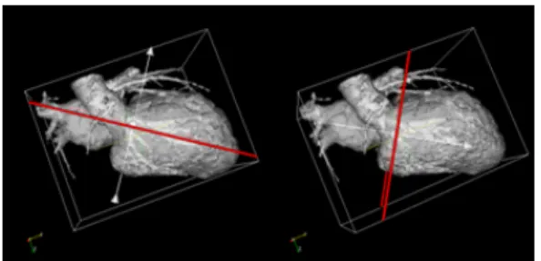
Figure 2. Selection of the echographic plane in the CT data (apical 4-chambers long axis plane (left) and short axis plane (right)).
ultrasound tissue images over the cardiac cycle. The track-ing relies on the manual selection of the endocardium con-tour. It is then extended to consider the whole myocardium and decomposed in segments (about 60 in long-axis view, 40 in short-axis view). Speckle tracking enables to track these segments and therefore to estimate cardiac motion.
The extracted motion is then decomposed, for each seg-ment, in different components used in clinical routine, i.e. transverse and longitudinal components in long-axis view and radial component in short-axis view. Finally, a baseline correction is applied to each motion component in order to correct global displacement caused by patient breathing or probe displacement.
2.3.
Comparison of the extracted motions
Because MSCT and STI provide motion descriptors with different characteristics (3D vs. 2D, different spatial and temporal resolutions), a multimodal registration stage is required. It is based on four steps:• Selection of the echographic plane in the CT data and extraction of the associated motion;
• Temporal interpolation of the extracted CT motion;
• Spatial registration of the CT and US contours;
• Association, to each US data point, of the corresponding
CT point.
These four steps are described in the following paragraphs. Selection of the echographic plane in the CT data. The selection of the plane associated to the echographic acqui-sition in the CT data is realized interactively. It relies on the selection of three points on the first reconstructed en-docardial surface. Once the plane has been selected, it is used to cut through the reconstructed surface to generate a contour corresponding to the echographic plane (cf. fig. 2). For each point of this contour, the neighboring nodes (with a distance lower than 1.5mm) in the original surface mesh are selected and their displacements are averaged. Finally, displacements are decomposed according to the echographic plane.
Temporal interpolation of the extracted CT motion. In order to obtain the same temporal resolution in CT and US
Figure 3. Representation mode used to display motion components.
data, a cubic spline interpolation is applied to each motion component and for each point of the CT contour. Similarly to US data, a baseline correction is applied to the interpo-lated data.
Spatial registration of CT and US contours.A rigid reg-istration process is used to align CT and US extracted con-tours. Firstly, an ICP (Iterative Closest Point) algorithm as-sociating rotation and translation is applied to the CT con-tour. Because the spatial resolution of CT is higher than US resolution, a scaling factor is applied (initialized at 1 and iteratively decreased until the sum of the squared Eu-clidean distances between closest points stops decreasing). Finally, a second ICP is applied to refine the alignment. Association between CT and US data points. Because the CT spatial resolution is higher than the US one, each US data point is associated to several CT data points. There-fore, for each US data point, the nearest CT data points are selected and their motion components averaged.
After these four steps, one set of points is obtained (cor-responding to the US data points) and, for each point, mo-tion components estimated from CT and US data.
In order to display these motion components in a com-mon way, we used a classic US representation (Fig. 3). It is based on the path followed from the base of the septal wall to the base of the lateral wall. For each point, the am-plitude of the motion component is displayed according to time. It results to spatiotemporal maps used by physicians to detect intraventricular delays.
3.
Results
For two patients, CT data acquisition has been realised with a 64-slices General Electric Lightspeed VCT 64 (GE, Milwaukee, WI, USA), providing 20 3D data volumes (resolution: 0.3x0.3x0.5 mm) representing a whole car-diac cycle. Transthoracic echocardiography has been per-formed using a GE Vivid 7 system. Two acquisitions have been realised in apical (long axis) view (4 and 2-chambers views) and three in parasternal short-axis views (basal left ventricle (LV), mid-LV and apical-LV). The frame rate is
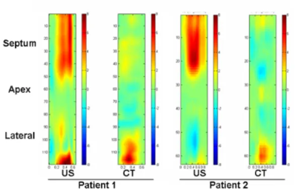
Figure 4. Longitudinal displacements from apical 4-chambers view (patients 1 and 2).
Figure 5. Transverse displacements from apical 4-chambers view (patients 1 and 2).
greater than 65 fr/s. Measurements of myocardial displace-ments were performed offline in the longitudinal, trans-verse and radial axes, using a dedicated software package (GE Echopac).
Motion estimated from long axis data is illustrated by figures 4, 5 and 6. Figures 4 and 5 represent, respec-tively, longitudinal and transverse displacements estimated from 4-chambers acquisitions of the two patients. Figure 6 shows longitudinal and transverse displacements from 2-chambers acquisition of the first patient. The fig-ure 4 shows that longitudinal components significantly differ between CT and US, especially in the septal seg-ments. This is explained by the well-known aperture phe-nomenon, highlighting a more difficult estimation of mo-tion components parallel to the considered surface, which is the case of the longitudinal motion in the basal and me-dial segments of the left ventricle.
For transverse displacements (Fig. 5), results are bet-ter, especially in the septal and lateral segments. However, transverse motion estimated in the apical segment show important differences. This can be explained by another well-known problem: the difficulty to visualize the apical area in echography. In the segments with good displace-ments coherence, a temporal delay appears: CT data seems to have an advance of about 100 ms. It is caused by the
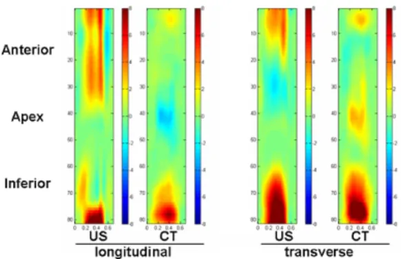
dif-Figure 6. Longitudinal and transverse displacements from apical 2-chambers view, from the anterior to the inferior walls (patient 1).
Figure 7. Radial motion from short-axis view (patient 1).
ference in the mode of ECG synchronization: US acquisi-tion starts at the beginning of the QRS complex, while CT acquisition is synchronized on the QRS peak. This results to a temporal difference of about 50 to 100 ms that can be noticed on the spatiotemporal maps.
Results obtained on 2-chambers acquisition (Fig. 6) confirm those obtained on 4-chambers data: longitudinal motion shows very important differences in the anterior segments while transverse displacements highlight an im-portant coherence, excepted in the apical segment. The figure 7 represents radial component in short axis view. The coherence of the estimation of this component is good, even if the delay previously exposed remains.
4.
Discussion and conclusions
Assessing local cardiac function with Multislice Com-puted Tomography would be a great advance towards the anatomical and functional description of the cardiac sys-tem within one single examination. In this paper, we pro-pose to compare local motion descriptors estimated with MSCT imaging to those obtained with US Speckle Track-ing ImagTrack-ing. In order to follow clinical routine, displace-ments are decomposed in different components before be-ing compared.
Results vary greatly according to the represented mo-tion component: transverse and radial displacements show
an important coherence while longitudinal displacements differ significantly.
The coherence shown in transverse and especially radial components is very satisfying because it has been proved that radial component study is the most useful motion com-ponent for dyssynchrony characterization [8].
Therefore, this study shows very promising results for the characterization of local cardiac function from MSCT imaging. Moreover, new MSCT generation will provide data with better temporal resolution allowing more precise motion estimation.
Future works will deal with the search for 3D dyssyn-chrony descriptors from MSCT and on the fusion of these descriptors with venous and myocardial anatomical infor-mation.
Acknowledgements
This work is supported by the French Research Minister (n 04 T 187 - 188 - 189 - 190 CITH).
References
[1] Leclercq C, Kass DA. Re-timing the failing heart: princi-ples and current clinical status of cardiac resynchronization. J.Am.Coll.Cardiol 2002;39:194-201.
[2] Mehra MR, Greenberg BH. Cardiac resynchronization therapy: caveat medicus!. J.Am.Coll.Cardiol. 2004;43:1145-1148. [3] Coatrieux JL, Hernandez AI, Mabo P, Garreau M, Haigron P.
Transvenous path finding in cardiac resynchronization therapy. In Proc. of the FIMH Conference. 2005; 236-45.
[4] Garreau M, Simon A, Boulmier D, Coatrieux, JL, Le Breton H. Surface and motion extraction in cardiac MSCT imaging for the assessment of left ventricular function. International Journal of Biomedical Imaging 2006; Article ID 37607.
[5] Simon A, Garreau M, Boulmier D, Coatrieux JL, Le Breton H. Cardiac Motion Extraction Using 3D Surface Matching in Multi-slice Computed Tomography. In Proc. of the MICCAI Conference. 2004; 1057-59.
[6] Fleureau J, Garreau M, Simon A, Hachemani R, Boulmier, D. As-sessment of global cardiac function in MSCT imaging using fuzzy connectedness segmentation. In Computers in Cardiology. 2008; 725-28.
[7] Langeland S, et al. Comparison of time-domain displacement es-timators for two-dimensional RF tracking. Ultrasound Med Biol 2003;29(8):1177-86.
[8] Suffoletto MS, et al. Novel speckle-tracking radial strain from rou-tine black-and-white echocardiographic images to quantify dyssyn-chrony and predict response to cardiac resynchronization therapy. Circulation 2006;113(7):960-8.
Address for correspondence: Antoine SIMON
LTSI, Campus de Beaulieu, Universit´e de Rennes 1, 263 Avenue du General Leclerc - CS 74205 - 35042 Rennes Cedex, France [email protected]