HAL Id: tel-03091963
https://tel.archives-ouvertes.fr/tel-03091963
Submitted on 1 Jan 2021
HAL is a multi-disciplinary open access archive for the deposit and dissemination of sci-entific research documents, whether they are pub-lished or not. The documents may come from teaching and research institutions in France or abroad, or from public or private research centers.
L’archive ouverte pluridisciplinaire HAL, est destinée au dépôt et à la diffusion de documents scientifiques de niveau recherche, publiés ou non, émanant des établissements d’enseignement et de recherche français ou étrangers, des laboratoires publics ou privés.
Efficient neuronal reprogramming into corticofugal
projection neurons at embryonic and postnatal stages in
the mouse
Torsten Felske
To cite this version:
Torsten Felske. Efficient neuronal reprogramming into corticofugal projection neurons at embryonic and postnatal stages in the mouse. Cellular Biology. COMUE Université Côte d’Azur (2015 - 2019), 2019. English. �NNT : 2019AZUR4020�. �tel-03091963�
Reprogrammation efficace en neurones de
projection corticofuges aux stades
embryonnaires et postnatals chez la souris
Torsten FELSKE
Institut de Biologie Valrose (IBV)
Présentée en vue de l’obtention du grade de docteur en Intéraction moléculaire et cellulaire de l’Université Côte d’Azur
Dirigée par : Michèle STUDER Soutenue le : 29 mars 2019
Devant le jury, composé de :
Thomas LAMONERIE, Professeur, Université Côte d’Azur Fanny MANN, DR1 CNRS, Institut de Biologie du Développement de Marseille
Benedikt BERNINGER, Professeur, King’s College London Michèle STUDER, DR1 INSERM, Institute de Biologie Valrose
THÈSE DE DOCTORAT
1
2
Acknowledgements
First of all, I want to thank Michèle Studer for her supervision of my thesis. Thank you for offering me the opportunity to work on a great project in your lab. Thank you also for your great support, your kindness and your guidance during the last 3 and a half years. Furthermore, I would like to thank Christian Alfano and Kawssar Harb for supporting me in the very beginning of my thesis and for handing me over their knowledge and preliminary data of my project.
I then want to thank Thomas Lamonerie for accepting to chair the jury of my PhD defense. Also, I want to thank Fanny Mann and Benedikt Berninger for reviewing my thesis and my defense. Thank you all for your time.
I want to thank my wonderful lab colleagues for a great time during the last years. It was a pleasure to work with you - every day, I was happy to come to the lab. Therefore, thank you Eya Setti, Michele Bertacchi, Alessandro Simi, Chiara Tocco and Maria Anna Di Bonito.
The environment in the institute is to mention, as all colleagues are exceptionally helpful and show a great sense of kindness. Thank you, members of the Lamonerie lab, the Collombat lab, the Schedl lab, the Fürthauer lab, the Sandoz lab, the Delaunay lab, the Acceuil of Science Naturelle and Biochemie, Agnès Loubat from the Cytometrie platform, the Prism platform and all members of the animal house.
Then I want to thank my wonderful friends here in Nice. I had such a good time, thanks to you guys: Aidan Falvey, Cristina Paraschivescu, Sanya Kuzet, Racha Fayad, Anthony Ruberto, Hereroa Johnston and Tom McMahon.
A very special thanks goes to Salsabiel. Your friendship is very precious to me and I cannot thank you enough for your support during the last years. I wish you all the best in the world for your future.
Ich habe das große Glück einer wunderbaren Familie anzugehören, dafür bin ich sehr dankbar. Vielen Dank an meinen Bruder Mario, meine Schwägerin Anna und die Kleinen Louis, Lotte und Jule. Vielen Dank an meine Schwester Ina und meinen Schwager Marc mit dem kleinen Bruno. Ebenfalls vielen Dank an meine tollen Großeltern Ursula und Gerhard. Mein größter Dank geht an meine Eltern, ich kann mich unendlich glücklich schätzen Euch zu haben.
3
Reprogrammation efficace en neurones de projection
corticofuges aux stades embryonnaires et postnatals
chez la souris
Jury:
Président du jury
Thomas LAMONERIE, Professeur, Université Côte d’Azur
Rapporteurs
Fanny MANN, DR1 CNRS, Institut de Biologie du Développement de
Marseille
4 Titre : Reprogrammation efficace en neurones de projection corticofuges aux stades
embryonnaires et postnatals chez la souris
Résumé
Le néocortex des mammifères est tangentiellement organisé en aires fonctionnelles et radialement divisé en six couches de neurones qui présentent chacune des projections, des morphologies et des patterns d’expression distinctes. Alors que les neurones de projection des couches supérieures projettent leurs axones dans d’autres couches du cortex ou vers l’hémisphère controlatéral, tel que les neurones calloseux, la plupart des neurones des couches inférieurs, nommés neurones corticofuges, innervent des structures sous-corticales à travers la voie pyramidale. La perte de neurones corticofuges peut conduire à des désordres neurologiques sévères, tel que la sclérose latérale amyotrophique ou d’autres maladies et/ou lésions impliquées dans le tractus cortico-spinal. Étant donné que le cerveau adulte des mammifères ne dispose pas d’une capacité de régénération efficace et que, par conséquent, la perte de neurones devient permanente, des nouvelles méthodes de remplacement de neurones sont indispensables.
Des études récentes ont révélé une plasticité surprenante des neurones post-mitotiques, qui peuvent être convertis en types cellulaires d’une lignée neuronale différente. Cette approche, appelée reprogrammation directe, contourne un état de pluripotence intermédiaire, est assez rapide et susceptible de conserver une signature épigénétique. L’expression forcée de facteurs de transcriptions, connu pour agir comme principaux régulateurs du destin cellulaire pendant le développement, est la meilleure stratégie pour convertir un type cellulaire en un autre. Cependant, il est encore difficile de comprendre dans quelle mesure des neurones, généré par reprogrammation directe, acquièrent les caractéristiques moléculaires du type cellulaire souhaité, jusqu’au stade de la spécification précise du sous-type. Le facteur de transcription FEZF2 est connu pour son rôle dans la spécification des neurones de projections sous-corticales de la couche 5, et une surexpression ectopique de Fezf2 peut convertir les couches supérieures ou les neurones striataux en neurones corticofuges de projection, même si cela est à faible efficacité.
Durant ma thèse, j’ai utilisé Fezf2 et le co-adaptateur nucléaire Lmo4 pour reprogrammer efficacement les neurones calloseux de la couche supérieure en neurones corticofuges de la couche inférieure, à la fois, au stade embryonnaire et postnatale. Les cellules reprogrammées avec succès régulent négativement les marqueurs de la couche supérieure, alors que les marqueurs de la couche inférieure sont surexprimés considérablement. De plus, en utilisant des méthodes de traçage avancées, les cellules reprogrammées ont la capacité de projeter vers des cibles sous-corticales, tels que le thalamus, le peduncule cérébrale et la moelle épinière. Ces données démontrent un rôle synergique inattendu de Lmo4 avec Fezf2 dans la reprogrammation neuronale et révèlent un cocktail efficace pour la conversion de cellules neuronales en sous-types de neurones corticofuges de projection.
5 Title: Efficient neuronal reprogramming in corticofugal projection neurons at embryonic and
postnatal stages in the mouse
Abstract
The mammalian neocortex is tangentially organized into functional areas and radially subdivided into six layers of neuronal populations with distinct projections, morphology and expression patterns. While neurons in the upper layers project within the neocortex or towards the contralateral hemisphere, such as callosal projection neurons, most lower layer neurons, named corticofugal projection neurons, innervate subcerebral targets via the corticospinal tract. Injuries or loss of these neurons can lead to severe neurological disorders, such as Amyotrophic Lateral Sclerosis or other diseases and/or lesions implicating the corticospinal tract. Since the adult mammalian brain lacks a significant regenerative capacity and thus, damage or loss of neurons is permanent, methods for restoring neurons are urgently needed.
Recent studies revealed an unexpected plasticity of post-mitotic neurons, which can be converted into cell-types of other neuronal lineages. This approach, called direct neuronal reprogramming, bypasses an intermediate pluripotent state. Direct reprogramming is fast, likely to keep epigenetic hallmarks and it can be conducted in vivo. Forced expression of transcription factors, known to act as master regulators of cell fate during development, is one of the best strategies for directly converting one cell-type into another one. However, it is still not clear to which extent neurons, generated by direct reprogramming, acquire authentic molecular signatures of the desired cell type, down to the point of precise subtype specification. The transcription factor FEZF2 is known for its role in cell fate specification of layer 5 subcerebral projection neurons and high ectopic Fezf2 expression can convert upper layer or striatal neurons into a corticofugal fate, even if at low efficiency.
During my PhD thesis, I used Fezf2 and the nuclear co-adaptor Lmo4 to efficiently reprogram upper layer projection neurons into corticofugal lower layer projection neurons, at both, embryonic and postnatal stages. The successful reprogrammed cells downregulated upper layer markers, while lower layer markers were drastically increased. Additionally, by using advanced tracing methods, we showed that reprogrammed neurons projected towards subcerebral targets, including the thalamus, cerebral peduncle and spinal cord. These data demonstrate an unexpected synergistic role of Lmo4 with Fezf2 in neuronal reprogramming and reveals an effective cocktail for the conversion of neuronal cells into corticofugal neuronal subtypes.
6
Table of Contents
List of Abbreviations ... 11
INTRODUCTION ... 14
I. The Mammalian Neocortex: Cytoarchitecture and Development ... 15
A. Cytoarchitecture of the neocortex ... 15
1. Cellular components ... 15
2. Tangential organisation of the neocortex ... 16
3. Radial organisation of the neocortex ... 16
4. Connections of the neocortex ... 20
B. Development of the neocortex ... 25
1. Corticogenesis ... 25
2. Development of the callosal projection neurons (CPNs) ... 27
3. Development of the CST ... 29
4. Development of CTA... 31
5. Development of TCA... 32
C. Genetic regulation of PNs position and identity ... 32
1. Positional information in progenitors... 32
2. Genetic regulation of PN diversity ... 35
3. Layer-specific molecular markers in the neocortex ... 40
a. Upper layer CPN marker CUX1 ... 40
b. SCPN marker CTIP2 ... 41
c. CThPN marker FOG2 (Friends of GATA2) ... 42
d. CFuPN marker PCP4 ... 43
e. CThPN marker DARPP32 ... 43
4. Areal specialization of post-mitotic projection neurons ... 44
a. BHLHB5 ... 45
b. LMO4 ... 45
c. PBX1 ... 46
d. Other post-mitotic area regulators ... 47
5. The motor area as “output” region ... 47
Fezf2 ... 48
a. Fez expression during embryogenesis and early postnatal stages ... 49
b. Fez genes during development of the olfactory system and diencephalon ... 51
c. Fez genes play unique and redundant roles in forebrain neurogenesis ... 51
Lmo4 ... 52
7
b. Lmo4 in cortical subtype specification ... 56
II. Cell reprogramming ... 58
1. General Introduction ... 58
2. Nuclear reprogramming ... 59
3. Cell fusion reprogramming ... 60
4. Cell reprogramming by cell extract treatment ... 60
5. Natural occurring de-differentiation and trans-differentiation ... 61
6. Reprogramming to induced Pluripotent Stem Cells (iPSCs) ... 62
7. Direct reprogramming ... 64
8. Direct neuronal reprogramming in vivo ... 72
a. Direct neuronal reprogramming in vivo in the injured brain ... 73
b. Direct neuronal reprogramming in vivo under physiological conditions ... 75
c. Data leading to the thesis work ... 78
III. Aims of the thesis ... 79
MATERIAL AND METHODS ... 80
I. Animals ... 81 II. Plasmids ... 81 1. Insert ... 81 2. Vector ... 83 3. Inducible pCAG-Fezf2-IRES-GFP ... 85 4. Inducible pCAG-IRES-GFP ... 85
III. In utero electroporation ... 87
IV. Tamoxifen injection ... 89
V. Cell Dissociation ... 90
VI. Cell sorting ... 90
VII. RNA Extraction ... 91
VIII. Intracardiac Perfusion ... 91
IX. Embedding and Slicing ... 91
X. Immunofluorescence ... 91
XI. Microscopy... 93
XII. Analysis ... 93
RESULTS ... 94
Section I.: Reprogramming upper layer neurons in the embryonic mouse brain ... 95
A. Ectopic expression of Lmo4 induces lower layer molecular markers in post-mitotic upper layer neurons at low efficiency ... 95
8
C. Lmo4 in combination with Fezf2 efficiently induces lower layer markers in UL neurons ... 100
D. cFezf2 & cLmo4 or single cFezf2 ectopic expression alter axonal projection of post-mitotic UL neurons towards corticofugal targets ... 104
E. The motor area is a more instructive environment for CFuPN-reprogramming ... 106
F. Reprogrammed post-mitotic UL neurons maintain stable regarding their altered molecular identity ... 109
Section II.: Reprogramming upper layer neurons at postnatal stages ... 112
A. Postnatal induction at P3 of single Fezf2 or Fezf2 & Lmo4 alters molecular identity of UL neurons ... 112
C.1. Postnatal induction of single iFezf2 or iFezf2 & iLmo4 drives UL neurons to change their axonal projections towards corticofugal targets ... 118
D. Postnatal induction at P7 of iFezf2 & iLmo4 still drives UL neurons to express lower layer marker PCP4 ... 120
D.1. UL neurons receiving expression of single iFezf2 or iFezf2 & Lmo4 at P7 show aberrant projections to corticofugal targets ... 122
E. Ectopic expression of single iFezf2 or iFezf2 & Lmo4 in late postmitotic UL neurons show corticofugal projections ... 124
Section III. ... 126
RNA extraction from UL neurons electroporated with cGFP, cFezf2 or cFezf2 and cLmo4 to screen for downstream targets via RNA-sequencing ... 126
DISCUSSION ... 128
I. Induction of Lmo4 alone fails to efficiently convert post-mitotic UL neurons ... 129
II. Lmo4 synergize with Fezf2 to trigger efficient reprogramming of post-mitotic UL neurons into CFuPNs ... 131
III. Fezf2 & Lmo4 trigger significant higher numbers of UL neurons to express CTIP2 and PCP4 compared to single Fezf2 ... 133
IV. Fezf2 & Lmo4 overexpression drive UL neurons to re-route their axonal projections towards corticofugal targets ... 135
V. The cellular environment of the motor cortex has an impact on reprogramming efficiency of Fezf2 & Lmo4 ... 137
VI. Reprogrammed post-mitotic UL neurons maintain expression of CFuPN markers at juvenile-adult stages ... 138
VII. Postnatal UL neurons retain the capacity to convert their molecular identity ... 139
1. Addition of inducible Ctip2 to inducible Fezf2 & Lmo4 ... 140
2. Addition of inducible PBX1 to inducible Fezf2 & Lmo4 ... 140
VIII. UL neurons retain their ability to change their axonal projections towards corticofugal targets from P3 to P21 ... 141
1. Fezf2 & Lmo4 induction at P7 and P10 still enable partial re-routing of UL neuron axons ... 142
9
IX. RNA extraction of early post-mitotic electroporated UL neurons ... 142
CONCLUSION ... 144
PERSPECTIVES ... 146
I. To further characterize reprogrammed UL neurons ... 147
II. To investigate reprogramming in other cell-types ... 147
III. To increase reprogramming efficiency at postnatal stages ... 147
IV. To apply possible downstream targets of FEZF2 revealed by mRNA-sequencing ... 148
10
List of Figures
Figure 1: Projection neuron diversity in the cerebral cortex ... 19
Figure 2: Long-range axonal projections define two classes of corticostriatal projection neuron .... 24
Figure 3: Neocortical projections neurons are generated in an “inside-out” fashion by diverse progenitor types in the VZ and SVZ ... 26
Figure 4: CPN development and diversity ... 28
Figure 5: Mechanisms and time course of mouse corticospinal tract (CST) development ... 30
Figure 6: CST projections to the spinal cord in rodents ... 31
Figure 7: Transcription factors in the VZ establish an area identity fate map ... 34
Figure 8: Competing molecular programs direct differentiation of newly-postmitotic projection neurons into one of three broad subtype identities ... 39
Figure 9: IHC of CTIP2 protein expression in M and S1 of WT mice at P8 ... 48
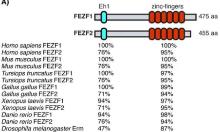
Figure 10: The Fez family of transcription factors. ... 49
Figure 11: Fezf1 and Fezf2 expression during embryogenesis. ... 50
Figure 12: Fezf1 and Fezf2 functions during forebrain neurogenesis. ... 52
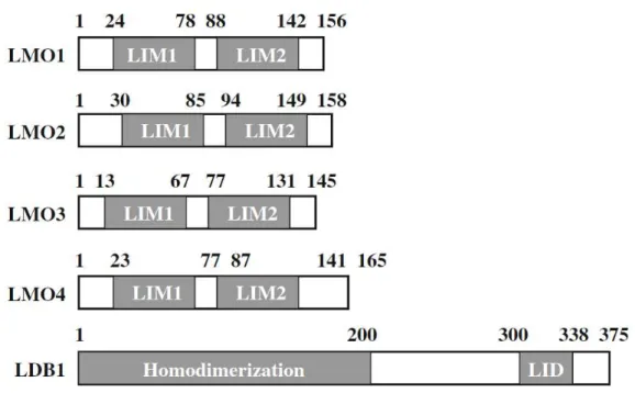
Figure 13: Schematic representation of the human LMOs and LDB1. ... 54
Figure 14: Schematic representation of LMOs in gene transcriptional regulation. ... 54
Figure 15: Schematic model of the putative mechanism by which Lmo4 de-represses Ctip2 expression ... 55
Figure 16: The Cook Islands model applied to neuronal reprogramming ... 71
Figure 17: Ectopic expression of Lmo4 induces lower layer markers at low efficiency in post-mitotic upper layer neurons ... 97
Figure 18: Ectopic expression of Lmo4 in UL neurons does not alter their axonal projection ... 99
Figure 19: Ectopic Expression of single Fezf2 or Fezf2 in combination with Lmo4 drives post-mitotic UL neurons to acquire molecular features characteristic of CFuPN. ... 102
Figure 20: cFezf2 & cLmo4 or single cFezf2 ectopic expression alter axonal projection of post-mitotic UL neurons towards corticofugal targets ... 105
Figure 21: Ectopic expression of cFezf2 & cLmo4 in UL neurons of the motor area induces higher levels of corticofugal identity markers CTIP2 and DARPP2 compared to somatosensory area. ... 107
Figure 22: Reprogrammed post-mitotic UL neurons maintain stable regarding their altered molecular identity... 112
Figure 23: A. Verification of iLmo4 and iFezf2 induction ... 114
Figure 24: Postnatal induction at P3 of single Fezf2 or Fezf2 & Lmo4 alters molecular identity of UL neurons ... 115
Figure 25: Postnatal induction of single iFezf2 or iFezf2 & iLmo4 combined with iCtip2 or iPBX1 does not increase reprogramming efficiency ... 117
Figure 26: Postnatal induction of single iFezf2 or iFezf2 & iLmo4 drives UL neurons to change their axonal projections towards corticofugal targets ... 119
Figure 27: Postnatal induction at P7 of iFezf2 & iLmo4 still drives UL neurons to express lower layer marker PCP4 ... 121
Figure 28: UL neurons receiving expression of single iFezf2 or iFezf2 & Lmo4 at P7 show aberrant projections to corticofugal targets ... 123
Figure 29: Ectopic expression of single iFezf2 or iFezf2 & Lmo4 in increasingly differentiating UL neurons show corticofugal projections ... 125
Figure 30: RNA extraction of UL neurons electroporated with cGFP, cFezf2 or cFezf2 and cLmo4 to screen for downstream targets via RNA-sequencing ... 127
11
List of Abbreviations
A1: Primary auditory AC: Anterior commissure
Ascl1: Achaete-Scute Family BHLH Transcription Factor 1
bHLH: basic Helix-Loop-Helix
Bhlhb5: Basic Helix-Loop-Helix Family Member E22
BRCA1: Breast Cancer 1
C. Elegans: Caenorhabditis Elegans
Cas9: CRISPR associated protein 9 CC: Corpus callosum
Cdh10: Cadherin 10
CFuPN: Corticofugal projection neuron CGE: Caudal ganglionic eminence cKO: conditional Knock-out Clim1: Ldb2
Clim2: Ldb1
CNS: Central nervous system
Coup-TFI: Chicken ovalbumin upstream promoter transcription factor 1
CP: Cortical plate CPD: Cerebral Peduncle
CPN: Commissural projection neurons Crb: Cerebellum
CRISPR: Clustered Regularly Interspaced Short Palindromic Repeats
Crym: Crystallin Mu
CSMN: Corticospinal motor neuron CST: Corticospinal Tract
CStr: Corticostriatal
CTA: Corticothalamic Axon
CThPN: Corticothalamic projection neuron Ctip2: B-cell lymphoma/leukemia 11B Cux1: Cut Like Homeobox 1
Cux2: Cut Like Homeobox 2
Darpp32: Protein Phosphatase 1 Regulatory Inhibitor Subunit 1B
DCC: DCC Netrin 1 Receptor
dLGN: dorsal lateral geniculate nucleus DNA: Deoxyribonucleic acid
Dnmt1: DNA methyltransferase 1 E: Embryonic day
Egf: Epidermal growth factor Eh1: Engrailed homology 1 Emx2: Empty spiracles homolog 2 Ep: Electroporation
Er81: ETV1
ERK: Extracellular Signal-regulated Kinase Erm: Earmuff
ERα: Estrogen Receptor alpha ESC: Embryonic Stem Cell
FACS: Fluorescence-activated cell sorting
Fezf2: FEZ Family Zinc Finger 2
FGF17: Fibroblast growth factor 17 FGF8: Fibroblast growth factor 8 Fog2: ZFPM2
FoxP2: Forkhead box protein P2 GABA: c-aminobutyric acid
12 GFP: Green Fluorescent Protein
GLIS1: Glis Family Zinc Finger 1 GW: Glial Wedge
HDAC: Histone deacetylase
Hes5: Hes Family BHLH Transcription Factor 5 IC: Internal Capsule
IGG: Indusium Griseum Glia iN: induced Neuron
IN: Interneuron
IP: Intermediate progenitor IPC: Intermediate precursor cell iPSC: induced Pluripotent Stem Cell IQ: Amino acids Isoleucine and Glutamine ISH: In Situ Hybridization
IT: Intratelencephalic kDa: kilo Dalton
Klf4: Kruppel-like factor 4 KO: Knock-out
L: Layer
Ldb1: LIM domain binding protein 1 Ldb2: LIM domain binding protein 2
Lmo4: LIM domain only 4
Lmx1a: LIM Homeobox Transcription Factor 1 Alpha
M1: Primary motor
MAPK: Mitogen-activated Protein Kinase Mbd3: Methyl-CpG Binding Domain Protein 3 MCc: caudal motor cortex
MCr: rostral motor cortex
MGE: Medial ganglionic eminence MGv: Medial geniculate nucleus
miRNA: micro RNA
MOE: Main Olfactory Epithelium mRNA: messenger RNA
MSN: Medium spiny neuron MTA1: Metastase Associated 1
MyoD: Myoblast Determination Protein MZ: Marginal zone
MZG: Midline Zipper Glia
NCAM: Neural Cell Adhesion Molecule NeuN: Fox-3
NeuroD1: Neurogenic Differentiation 1 Nkx-2: Nk2 Homeobox 2
NSC: Neural Stem Cells
Nurr1: Nuclear receptor related protein 1 OB: Olfactory bulb
Oct3/4: Octamer-binding transcription factor¾ oRG: outer Radial glial cell
OSKM: Oct3/4, Sox2, Klf4 and c-Myc OT: Optic tectum
P: Postnatal day Pax6: Paired box 6
Pbx1: Pre-B-cell Leukemia Homeodomain 1 PCP4: Purkinje-cell protein 4
PN: Projection neuron PO: Pons
POA: Preoptic area PP: Preplate
PSPB: Pallial/Subpallial Boundary PT: Pyramidal tract
RGC: Radial glial cell RNA: Ribonucleic acid
13 Robo: Roundabout Guidance Receptor
Rorβ: RAR-related orphan receptor beta RTN: Reticular nucleus
S1: Primary somatosensory SAP: Subapical progenitor
Satb2: Special AT-rich Sequence Binding Protein 2
SC: Spinal cord
SCPN: Subcerebral projection neuron SCS: Subcallosal Sling
SEMA3A: Semaphorin 3A SEMA3C: Semaphorin 3C
Sey: small eye hypomorphic mutant SFRP2: Secreted frizzled-related protein 2 Slit: Slit Guidance Ligand
Sox2: SRY-Box 2 Sox5: SRY-Box 5 SP: Subplate
Sp8: Trans-acting transcription factor 8 STN: Subthalamic nucleus
SVZ: Subventriuclar zone Tbr1: T-Box Brain Protein 1 TCA: Thalamocortical afferents TF: Transcription factor Th: Thalamus
Tle4: Transducin Like Enhancer Of Split 4 UNC5H3: Unc-5 Netrin Receptor C UV: Ultraviolet light
V1: Primary visual VL: Ventrolateral nucleus VNO: Vomeronasal Organ
VP: Ventroposterior nucleus
VPM: Ventral posteromedial nucleus VZ: Ventricular zone
Wnt: Vertebrate orthologs of drosophila wingless/integrated
14
15 During evolution, one part of the brain experienced a drastic development in complexity and size and is one of the major reasons for the evolutionary success of mammals: the cerebral cortex. It covers the outer portion of the cerebrum and is anatomically comprised of the archicortex, the paleocortex and the neocortex – its largest component (O’Leary, Chou and Sahara, 2007). The neocortex is the evolutionary newest part of the cerebral cortex and its cytoarchitecture is a distinguishing feature of mammals; in humans, it covers up to 80% of the total volume (Rakic, 2009; Kaas, 2011). The neocortex represents the major and critical organ for sensory input processing, movement, memory and language and is additionally responsible for higher cognitive functions, such as abstract thinking and self-awareness (Kaas, 2011). Due to these intrinsic features it is a major organ of interest for fundamental and medical research, but still represents a major challenge because of its high complexity. Therefore, gaining a better understanding on the molecular, cellular and systemic level is crucial to unravel its mystery and thus, this work on the mouse neocortex is intended to contribute to it.
I. The Mammalian Neocortex: Cytoarchitecture and Development
A. Cytoarchitecture of the neocortex
1. Cellular components
The neocortex is composed of neuronal cells and glia. Glutamatergic projection neurons (PNs) make up almost 80% of the total neuronal population in the neocortex, they are generating the neocortical output and are responsible for the connection of the neocortex to subcortical, subcerebral or intracortical targets. The other major population is composed of interneurons (INs), which make up around 20% of the neuronal population (Lodato and Arlotta, 2015). They are largely inhibitory and modulate the activity of neuronal circuits by release of c-aminobutyric acid (GABA). INs share a common feature of connecting within the neocortex via short-range projections even though they appear to be a very heterogenous class with diverse morphology, connectivity, biochemistry and physiological properties (Lim et al., 2018). Non-neuronal cells are the third category of cells that are present in the neocortex and are
16 named glia. Their role besides supporting neuronal circuits is very various. Three main subcategories of glial cells exist: Microglia, Oligodendrocytes and Astrocytes. Microglia are known as the ‘immunocompetent’ cells with phagocytic abilities. They cover a huge part of the brain parenchyma and not only survey the brain tissue for any damage, but are also implicated in the maintenance of the diverse cell populations that comprise the CNS (Schafer and Stevens, 2015). Oligodendrocytes are the myelin-producing cells insulating axons to allow fast axon potential propagation. Some oligodendrocytes are not producing myelin and may have other functions, as they are present in the grey matter of the cerebral cortex (Kang et
al., 2013). Astrocytes are the most abundant glial cell population in the neocortex, their
function in the neuronal circuit is various and ranges from water and ion homeostasis, participation in synapse signalling or maintenance of the blood brain barrier (Jäkel and Dimou, 2017).
The investigated cell type in this thesis is the excitatory projection neuron and therefore the following part of the introduction will be focused on this cell type.
2. Tangential organisation of the neocortex
The neocortex is tangentially organised into functional areas, each having a unique cytoarchitecture, distinct connections and specific functions. For the neuroscientist Brodmann, “each area represents an organ” of the adult brain (Brodmann, 1909), implying that each area is well distinguished from another one by rather distinct cytoarchitecture borders and has a unique function within the brain. Four major areas can be distinguished in all eutherian mammals (Kaas, 2011) and are defined as primary areas due to their function as first “receiver” of sensory inputs – auditory area A1, visual area V1 and somatosensory area S1 – or as first “executor”: the motor area M . The sensory areas are processing information from the cochlea (A1), the retina (V1) and the body (S1), while M1 controls voluntary movement of different body parts (O’Leary, Chou and Sahara, ).
3. Radial organisation of the neocortex
Tangentially, the neocortex is subdivided into functional areas, whereas in its radial dimension the neocortex is organised into six layers (see Fig. 1). Each layer is composed of a different
17 pool of neurons and, each layer varies in its cellular composition, its projections and its thickness depending on their areal identity (Lodato and Arlotta, 2015).
Layer (L)1 represents the first sheet just underneath the pial surface (the inner most membrane that envelopes the cortex) and mainly consists of axons, dendrites and sparse cells – named Cajal-Retzius cells (Kaas, 2011). The following layers are grossly classified by their projections within the central nervous system (CNS, see Fig. 1): L2-3 are mainly composed of neurons which extend their axons to the contralateral cortical hemisphere, named callosal projection neurons (CPNs) and associative projection neurons, projecting within the neocortex. Most of the CPN axons cross the midline through the corpus callosum (CC), which represents the major “bridge” between the two cortical hemispheres. Some CPNs send their axons through the anterior commissure (AC), which represents a second “bridge” with fewer cortical axonal projections, connecting both hemispheres (Greig et al., 2013). In fewer numbers, CPNs are also residing in L5 and L6. Neurons in L4 are the main recipient of thalamocortical afferents and the majority of L4 neurons project within one cortical hemisphere to L2/3 (Lodato and Arlotta, 2015).
PNs in L5 and L6 project away from the cortex to distinct subcortical or subcerebral structures. These projection neurons are named corticofugal projection neurons (CFuPNs) and can be further subdivided into subcerebral projection neurons (SCPNs) - residing in L5 – and, corticothalamic projection neurons (CThPNs), in L6. SCPNs are projecting towards distinct subcerebral targets in function of their areal location: from V1 to the superior colliculus (corticotectal PNs), from S1 to the brainstem (mostly pons, cortico-pontine PNs) and from M1 to the spinal cord (Corticospinal motor neurons, CSMNs). CThPNs project to specific thalamic nuclei, in a topographic manner, meaning that CThPNs from distinct areas project to specific nuclei in the thalamus (Molyneaux et al., 2007; Greig et al., 2013; Lodato and Arlotta, 2015). This is a rough classification and gives only a general idea of neuronal population differences within layers. For example, associative projection neurons are present in each layer and extend their axons within a single cortical hemisphere (Molyneaux et al., 2007). Additionally, some neurons send projections to multiple targets and therefore can be classified in several classes. Among them are CPNs, projecting to both the contralateral hemisphere and ipsilaterally within the cortex, whereas intrathelencephalic corticostriatal PNs, extend callosal axons to the ipsilateral but also to the contralateral striatum (Shepherd, 2013). SCPN often
18 project to multiple targets, as they send collaterals to diverse corticofugal targets (Shepherd, 2014). Also, some SCPN extend backward projections, sending axons to subcerebral targets and within the cortex (Molyneaux et al., 2007; Greig et al., 2013; Lodato and Arlotta, 2015).
19
Figure 1: Projection neuron diversity in the
cerebral cortex
Schematic presentation showing origin of PNs and their respective axonal projections. A. Commissural projection neurons project to
the contralateral cortical hemisphere. Most cross the midline through the corpus callosum (callosal projection neurons, CPN), while a smaller population crosses through the anterior commissure. CPN reside primarily in upper layer 2 - 3, with fewer in lower layers 5 and 6 and extend axons to mirror-image locations in the same functional area of the contralateral hemisphere.
B. Associative projection neurons are present
in all layers of the neocortex, projecting within a single cortical hemisphere. This populations includes short-distance intrahemispheric projection neurons, which extend axons within a single cortical column or nearby cortical columns and long-distance intrahemispheric projection neurons, which extend axons to adjacent or distant cortical areas (such as forward and backward projection neurons).
C. Corticofugal projection neurons (CFuPNs)
project outside the cortex to subcortical targets including corticothalamic projection neurons (CThPNs), which reside in layer 6, and subcerebral projection neurons (SCPNs), which reside in layer 5.
D. Neurons that send projection to multiple
targets can sometimes be classified into more than one category. Examples include CPN with frontal projections, which extend axons to the contralateral hemisphere and to ipsilateral frontal cortex. SCPN with backward projections, which extent axons to subcerebral targets and to ipsilateral caudal cortex. Intratelenecpahilic corticostriatal projection neurons, which extend projections to contralateral hemisphere and to ipsilateral striatum.
CC Corpus Callosum, Crb cerebellum, LGN lateral geniculate nucleus of thalamus, OB olfactory bulb, OT optic tectum, PO pons, SC spinal cord, Th thalamus, VL ventral lateral nucleus of thalamus, VP ventral posterior nucleus of thalamus. Taken from Greig et al. (2013).
20 4. Connections of the neocortex
Axonal trajectories are important messenger propagation routes to ensure communication between higher cognitive upper layer neurons and executive lower layer neurons. PNs can be characterized by their axonal projections, revealing their functional role in the cortex. For simplification, associative PNs and callosal PNs are grouped together as intratelencephalic (IT) PNs, as they project to the contralateral hemisphere or within the telencephalon (cortex and striatum) or both (bilateral IT neurons). Names of SCPNs and CThPNs are not changed.
a. Cortico-cortical projections
Inputs on cortical neurons originate mainly from other cortical neurons of the same hemisphere (associative projection neurons) or from neurons projecting from the contralateral hemisphere. The axonal pathway from one hemisphere to its contralateral region through the CC is a relatively recent feature in cortical evolution and is unique to placental mammals (Aboitiz, Morales and Montiel, 2003). Other interhemispheric commissures exist and are more ancient, the hippocampal commissure and the AC, but the CC is by far the largest and the only one which serves solely the communication between the two neocortical hemispheres (Aboitiz, Morales and Montiel, 2003). Axons in the CC arise mainly from CPNs which are abundantly located in L2 – 3 (around 80% in rodents), in L5 (around 20%) and some in L6 (Fame, MacDonald and Macklis, 2011). All CPNs extend an axon to the contralateral hemisphere, but they can also innervate local cortical neurons within the hemisphere and/or the striatum. In fact, mainly lower layer CPNs possess dual projections to the contralateral side and ipsilaterally within the forebrain, while upper layer CPNs project to cortical neurons in L2 – 3, to stellate cells in L4 and they also send collaterals to L5 and L6 cortical neurons (See Fig. 2, Petreanu et al., 2007). Thus, CPN function is mainly related to integration and connection of neuronal circuits within the neocortex.
b. Corticofugal projections
The sub-classification of CFuPNs is complex regarding their connectivity and is not yet completely understood. The complexity arrives from the fact that many CFuPNs target several
21 structures via branches along their trajectory. This is especially the case for L5 SCPNs, which can project to the midbrain, striatum, thalamus, and subthalamic nuclei, en route to their dorsal-most destinations in the brainstem and the spinal cord (Shepherd, 2013). Additionally, SCPNs also form ipsilateral connections within the cortex, but do not cross through commissures to the contralateral hemisphere (Shepherd, 2013). Less complex projections are found of L6 CThPNs, which are projecting to the thalamus only. Here, the major trajectories of CFuPNs will be presented.
c. The corticospinal tract (CST)
The control of voluntary movements in mammals is based on sophisticated motor and sensory communication in the CNS. Axonal trajectory of the CST enables fast and controlled motor execution that has been carried out by corticospinal motor neurons (CSMNs) in the neocortex. The functions of the CST comprise control of afferent inputs, spinal reflexes and motor neuron activity (Lemon and Griffiths, 2005). Axons in the CST originate from L5 SCPNs in the primary motor and somatosensory cortex and in less extent from parietal, cingulate, visual and prefrontal regions (Miller, 1987; Akintunde and Buxton, 1992; Tennant et al., 2011; Kamiyama
et al., 2015). The canonical path of the CST in mammals starts from axonal projections of L5
SCPNs in the neocortex, which then travel into the internal capsule (IC), further on to the cerebral peduncle (CP), into the brainstem and some reach the spinal cord (see Fig. 2). The axonal bundles of the CST remain at the ventral position until reaching the medulla in the brainstem. In rodents, 80 – 95% CST axons cross the midline at the junction between the brainstem and spinal cord, and pass from a ventral to a dorsal position, forming the pyramidal decussation (Welniarz, Dusart and Roze, 2017; Armand, 1982; Schreyer and Jones, 1982; Rouiller et al., 1991; Joosten et al., 1992). Therefore, the majority of the axon trajectory continues in the contralateral part of the SC and terminates in the ventral part of the dorsal funiculus, located between the dorsal horn and the midline (Kuypers, 1964). The remaining 5 – 20% uncrossed CST project in the ventral funiculus. However, both crossed CST and uncrossed CST originate from the same cortical regions (Galea and Darian-Smith, 1994; Brösamle and Schwab, 1997; Lacroix et al., 2004), but control different muscles: the crossed CST is involved in fine movements of distal extremities, whereas the uncrossed CST targets
22 proximal or axial musculature (Welniarz, Dusart and Roze, 2017). The terminations of crossed CST are located in the dorsal and intermediate horns (grey matter) of the contralateral spinal cord, uncrossed CST terminations on the ipsilateral side show the same pattern (Welniarz, Dusart and Roze, 2017). Interestingly, CSMNs of adult rodents do not form direct connections with motor neurons in the spinal cord (Alstermark and Ogawa, 2004), whereas higher primates have monosynaptic connections between CSMNs and motor neurons, allowing fine hand dexterity control (Lemon, 2008). However, this monosynaptic connection is present in juvenile mice and is eliminated by signalling molecules and is therefore lost in the adult mouse (Gu et
al., 2017). Thus, the CST connection to motor neurons in the spinal cord might be formed by
polysynaptic transmission via interneurons and propriospinal neurons (Lemon, 2008). Overall, L5 SCPNs extend their axons within the CST and project to several corticofugal targets via branches or collaterals of their axons, en route to their dorsal-most locations, the brainstem and the spinal cord.
d. Cortico-striatal connections
The striatum of the mammalian cerebrum is a critical component of motor and reward systems, including motor and action planning, motivation and reward perception (Yager et al., 2015). It receives glutamatergic input from the cortex and dopaminergic input from basal ganglia structures. Connectivity between the cortex and the striatum is directional, meaning that cortical projections form mono-synaptic input, whereas neurons in the striatum communicate indirectly with the cortex via polysynaptic downstream circuits (Shepherd, 2013). The cortical neurons innervating the striatum originate from L5 IT or SCPNs and L6 IT neurons. Moreover, cortico-striatal projections can either originate from L5 IT neurons or SCPNs, but not from both (Shepherd, 2013). L5/6 IT neurons projecting to the striatum have the unique attribute of being both corticofugal and callosal, because they project to the striatum and they additionally project to the contralateral hemisphere (Macklis et al., 2012). Interestingly, the striatum is special in receiving both L5/6 IT (bilateral) and SCPN (ipsilateral) inputs, differently from other cerebral areas. In the striatum, L5/6 IT neurons and SCPNs make synaptic contacts primarily with spines of striatal projection neurons, also called medium spiny neurons (MSNs, Reiner, 2010), which is by far the most abundant neuron type in the striatum
23 (Yager et al., 2015). MSNs themselves project to basal ganglia structures such as globus pallidus and substantia negra pars reticulate. Taken together, the cortical input the striatum receives originates from several types of PNs, which includes L5 and L6 IT neurons and SCPNs.
e. Connections between cortex and thalamus
The thalamus is a prominent structure in the diencephalon and is the major receiver and relay of sensory inputs, which propagates in a topographic manner to the cortex. The thalamus and the cortex are strongly interconnected and both are important for proper development of one another and represent an integrated processing unit, which regulates thalamic transmission of peripherally derived data for cortical processing (Leyva-Díaz and López-Bendito, 2013). The cortex innervates the thalamus, by corticothalamic axonal (CTA) projection and the thalamus projects to targets in the cortex, by thalamocortical axonal (TCA) projection.
The cortical innervation of the thalamus originates from CThPNs of L6 and to a smaller extent of SCPNs in L5. But, the projection patterns of L6 CThPNs differ from L5 SCPNs. For instance, L6 CThPNs project to all thalamic nuclei (including both first-order and higher-order relays), whereas SCNPs only project to higher-order thalamic nuclei (Hoerder-Suabedissen et al., 2018). First-order relays get their driving input from subcortical sources and form a reciprocal feedback pattern with CThPNs. Moreover, CTA of CThPNs innervate thalamic nuclei, which in turn send TCA to the same cortical region in L6 from where the input originated, depending on their area identity (Briggs and Usrey, 2008). These projections from CThPNs modulate how sensory information is relayed to the cortex (Olsen et al., 2012; Lam and Sherman, 2013; Crandall, Cruikshank and Connors, 2015). Most of TCA terminate in L4, thereby innervating local circuit neurons, named stellate cells. In addition, also L1 and L2 – 3 receive TCA input (Caviness and Frost, 1980). Thus, the neocortical primary areas are connected via CThPNs with the thalamic nuclei in the following way: M1 is connected with the ventrolateral (VL) nucleus, S1 with the ventroposterior nucleus (VP), V1 with the dorsal lateral geniculate nucleus (dLGN) and A1 with the ventral part of the medial geniculate nucleus (MGv, Guillery, 1967; Hoogland, Welker and Van der Loos, ; O’Leary, Chou and Sahara, ).
24 In contrast, higher-order relays get their driving input from SCPNs (Usrey and Sherman, 2018). SCPNs from S1 innervate posterior medial nucleus, V1 SCPNs innervate pulvinar and A1 SCPNs project to dorsal division of medial geniculate nucleus (Usrey and Sherman, 2018). These projection patterns imply that L6 CThPNs provide a more modulatory input to the thalamus and SCPNs provide driving inputs (Usrey and Sherman, 2018). Thus, thalamic nuclei receive input from L6 CThPNs and L5 SCPNs. CThPNs project to the thalamus and receive reciprocal thalamic afferents. SCNPs project solely to higher-order thalamic nuclei, providing cortical input. Overall, PNs of the neocortex can be classified by their axonal projections, which in turn
indicates their functional role. Upper layer IT neurons project within the cortex while some lower layer IT neurons also project to the striatum. Moreover, lower layer SCPNs project to multiple subcerebral targets and CThPNs solely innervate thalamic nuclei (see Fig. 2).
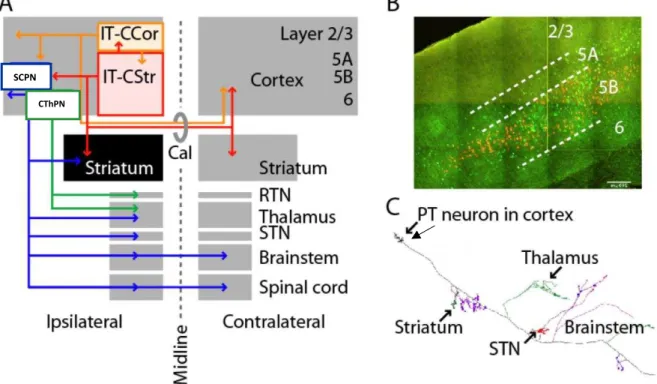
Figure 2: Long-range axonal projections define two classes of corticostriatal projection neuron
A. SCPNs (blue) project to ipsilateral striatum, thalamus, subthalamic nucleus (STN), and many brainstem and spinal cord regions. Intratelencephalic (IT) neurons project ipsi- or bilaterally (via corpus callosum, Cal), within the cerebral hemispheres to cortex (IT-CCol, orange), and many of these also to striatum (IT-CStr, red). The ipsilateral striatum (black) is unique in receiving Cstr input from both IT neurons and SCPNs. Layer 6 corticothalamic neurons (CT, green) project only to thalamus and its reticular nucles (RTN). B. Retrogradely labelled corticospinal CSMNs and callosally projecting IT-CStr neurons in mouse motor cortex. IT neurons (green) and SCPNs (orange) are intermingled in layer 5B, but not double-labeled. C. A single SCPN’s axon is multiprojectional, sending branches to many subcortical areas. Taken and modified from Shepherd (2013).
SCPN
25
B. Development of the neocortex
1. Corticogenesis
Early in development, the telencephalic wall is composed of neuroepithelial cells. They divide symmetrically, thereby generating two more neuroepithelial cells, intended for expansion purposes. These epithelial cells are multipotent and represent the first neuronal population in the neocortex (Haubensak et al., 2004; Greig et al., 2013; Tan and Shi, 2013). Starting at E9.5, neuroepithelial cells give rise to radial glial cells (RGCs). While RGCs start to proliferate symmetrically (for self-renewal purposes), some expand onto the lateral ventricle, establishing the germinative or ventricular zone (VZ). At later stages, RGCs undergo a final asymmetric division and give rise to some excitatory PNs, some RGCs in turn, give rise to outer RGCs (very few in rodents) and intermediate progenitors (IPs), forming the subventricular zone (SVZ). IPs act as transit-amplifying cells, undergoing asymmetric division to self-renew, limited proliferative divisions, and more often dividing symmetrically to produce two neurons (Haubensak et al., 2004; Noctor et al., 2004; Wu et al., 2005; Sessa et al., 2008). A fourth class of progenitors, the short neural precursors, reside in the VZ, producing post-mitotic neurons directly within the VZ (Gal, 2006; Stancik et al., 2010). Neocortical progenitors begin to produce excitatory PNs around E10.5 (Angevine and Sidman, 1961; Rakic, 1974). Earliest-born cortical neurons migrate past the VZ and form the preplate (PP), which is then split into two structures: the marginal zone (MZ) and the subplate (SP). The marginal zone will give rise to L1 neurons. Between these two structures, the cortical plate (CP) will develop and give rise to the other layers of the neocortex. The first newborn neurons migrate into the CP via somal-translocation, whereas later newborn neurons move along a glial scaffold via glia-guided neuronal migration. Throughout the rest of corticogenesis, newly born neurons migrate into the CP, organizing themselves in an “inside-out” fashion (see Fig. 3). This implies that early-born neurons form lower layers 6 and 5, and late-early-born neurons populate upper layers 4 – 2 (Greig et al., 2013). After neurogenesis is complete, neural progenitors transit to a gliogenic mode, generating astrocytes and oligodendrocytes.
26
Figure 3: Neocortical projections neurons are generated in an “inside-out” fashion by diverse progenitor types in the VZ and SVZ
This schematic depicts the sequential generation of neocortical projection neuron subtypes and their migration to appropriate layers over the course of mouse embryonic development
A. Radial glia (RG) cells in the VZ begin to produce projection neurons around E11.5. At the same time, RG generate intermediate progenitors (IP) and outer radial glia (oRG), which establish the SVZ and act as transit-amplifying cells to increase neuronal production. After neurogenesis is complete, neural progenitor transition to a gliogenic mode, generating astrocytes and oligodendrocytes. Caja-Retzius (CR) cells primarily migrate into neocortical layer I from non-cortical locations, while other PNs are born in the neocortical VZ/SVZ and migrate along radial glial process to reach their final laminar destinations.
B. Distinct PN sub-types are born in sequential waves over the course of neurogenesis. The peak birth of SP neurons occurs around E11.5 with the peak birth of CThPNs and SCPNs occurring at E12.5 and E13.5, respectively. Layer IV granular neurons (GN) are born around E14.5. Some CPNs are born starting at E12.5 and those CPN born concurrently with CThPNs and SCPNs also migrate to deep layers. Most CPNs are born between E14.5 and E16.5, and these late-born CPNs migrate to superficial cortical layers. Peak sizes are proportional to the approximate number of neurons of each sub-type born on each day. Ne, neuroepithelial cell, WM, white matter. Taken from Greig et al. (2013).
27 2. Development of the callosal projection neurons (CPNs)
The birthdate of CPNs is in line with their future location in the radial column of the neocortex, such as CPNs of L6 are born at E12.5 together with CThPNs, L5 CPNs at E13.5 and L2-3 CPNs from E15.5 to E17.5 (Molyneaux et al., 2007). As newborn CPNs are migrating to the CP, they already start to send axons towards premature zones of the developing CC. In ex vivo experiments, Semaphorin 3 has been shown to repel axons away from the cortical marginal zone (Polleux et al., 1998) and guide premature CPN axons towards the midline (Zhao et al., 2011). Parallel to the birth of CPNs, the two telencephalic hemispheres start to fuse with help of glial cells (Lindwall, Fothergill and Richards, 2007). A transient zone of a ‘bridge-like subcallosal sling’, which is formed by local neurons and glial cells, builds up a first window for CPNs axons to cross. Thanks to guidance cues, repelling or attracting from glial cells, first CPNs axons are crossing the midline (see Fig. 4). Since newborn CPNs in the cingulate cortex (most dorsomedial structure of the neocortex) are closest to the midline, they are referred to as pioneer axons crossing the hemisphere bridge (at E17, Rash and Richards, 2001) and they help guiding later-arriving CPN axons (approximatively 1 day later) in providing a structural framework (Norris and Kalil, 1991).
Due to the different birthdates, axons of lower layer CPNs cross the midline before axons of upper layer CPNs. When reaching the contralateral side, CPN axons turn dorsally and extend into the neocortex towards homotopic targets (Fame, MacDonald and Macklis, 2011). Important signalling molecules responsible for proper guidance of CPN axons are members of the Slit/Robo, Wnt, Netrin and Ephrin families. The specific mechanisms of the callosal axon guidance to their homotopic regions of the contralateral side are largely unknown. However, callosal fibers that fail to cross the midline due to wrong guidance cues or absence of molecular factors remain ipsilateral and form Probst bundles (Fame, MacDonald and Macklis, 2011).
28
Figure 4: CPN development and diversity
This schematic representation shows callosal projection development at embryonic stages and shows characteristic callosal connections in the adult mouse brain
A During development, callosal axons (red) turn toward the midline. Multiple glial populations (blue) and mixed neuronal/glial populations (purple) play critical roles in CPN axon guidance and midline crossing. Pioneering axons (brown) from neurons of the cingulate cortex begin the process of midline crossing. This schematic represents process that occur across multiple embryonic times during mouse CPN development. Abbreviations: CP, cortical plate; IZ, intermediate zone; SVZ, subventricular zone; VZ, ventricular zone; IGG, indusium griseum glia; GW, glial wedge; SCS, subcallosal sling; MZG, midline zipper glia.
B At least four major types of adult CPN can be classified based on projection patterns. These include: single projections to the contralateral cortex (red); dual projections to the contralateral cortex and ipsilateral or contralateral striatum (green); dual projections to the contralateral cortex and ipsilateral premotor cortex (blue); or dual projections to the contralateral cortex and ipsilateral sensorimotor cortex (purple). Taken from Fame, MacDonald and Macklis (2011).
29 3. Development of the CST
Axons of the CST originate from SCPNs, residing in lower layer 5B of the cortex. Axons of SCPNs, once generated, have to travel a long distance towards the spinal cord and do so by early outgrowth of the cortex. At E14, CST axons are already reaching the IC and at E17, CST axons are present in the brainstem. At P0, CST cross the midline after the pyramidal decussation and reach the spinal cord at postnatal day (P) 2 (see Fig. 5).
Initially, corticofugal projections, including axons from SCPNs and CThPNs, grow out of the cortex and make a lateral turn away from the midline, thus diverging from callosal projections (Welniarz, Dusart and Roze, 2017). This long trajectory implies proper guidance by extrinsic signals. Guidance cues accompany pioneer axonal projections and ensure they reach their targets. After CST axons have left the cortex, Semaphorin family members SEMA3A and SEMA3C together with NETRIN-1, attract CST axons away from the midline (Srivatsa et al., 2014). After CST axons have passed the pallial/subpallial boundary (PSPB) - a molecular and cellular border between pallial and subpallial structures - they make a medial turn to enter the IC, attracted by Slit guidance ligand 1 and 2 (SLIT1/2) and ROBO1/2 receptors (Bagri et al., 2002; Lopez-Bendito et al., 2007). Passing the IC, CST axons, with the help of NKX-2, enter the brainstem in the caudal medulla reaching the pyramidal decussation (Welniarz, Dusart and Roze, 2017). At the level of the pyramidal decussation, CST axons are starting to cross the midline. Several molecules are involved in the corticospinal midline crossing of CST. NETRIN-1, DCC and UNC5H3 are crucial factors, as loss of these molecules in animal models leads to a failure of most CST axons to cross the midline. The exact mechanisms are still not known and it is uncertain how the small proportion of CST axons are instructed not to cross the midline (Welniarz, Dusart and Roze, 2017). What is known is that once the CST cross the midline, they are prevented from re-crossing by repellent protein EPHRIN-B3, secreted from the midline (Kullander et al., 2001). However, a small fraction of CST axons does not cross the midline at the pyramidal decussation and instead project to the ipsilateral spinal cord (Welniarz, Dusart and Roze, 2017 see Fig. 6).
30
Figure 5: Mechanisms and time course of mouse corticospinal tract (CST) development
The left side of the figure shows the trajectory of the mouse CST from the cortex to the spinal cord. The genes involved in CST development are indicated at the corresponding level. The right side of the figure indicates the time course of CST development.
31
Figure 6: CST projections to the spinal cord in rodents
A The crossed CST (dark blue) is located in the most ventral part of the dorsal funiculus, and the uncrossed CST (red) runs in the ventral funiculus. Taken and modified from Welniarz, Dusart and Roze (2017).
4. Development of CTA
Newborn post-mitotic cortical neurons directly start to extend their axons before even leaving the IZ. Characteristic for corticofugal axons, they are attracted by Sema3C expressed in the IZ (Bagnard et al., 1998; Skaliora, 1998). Furthermore, the SEMA3 gradient within the IZ guides CTA towards the lateral side of the cortex (Bagnard et al., 1998). Then, CTA pass through the IZ and reach the lateral part of the IC at E13 – E15.5 (Jacobs et al., 2007). At E15.5, CTA cross the PSPB (Carney et al., 2009). At this point, ventrolateral oriented CTA tend to turn medially in order to reach the IC in the subpallium (Agmon et al., 1995). As it is the case in the development of the CC, also pioneer axons of the very first-born CThPNs in the subplate act as pioneer axons to guide later-arriving CTAs. Guidance cues of the Slit and Robo family ensure CTAs remain in the IC until they reach the diencephalon-telencephalon boundary (DTB, Leyva-Díaz and López-Bendito, 2013). At the DTB, CTA enter the premature thalamus and meet cells of the perireticular thalamic nucleus and the thalamic reticular nucleus (RTN), at E16 (Jacobs
et al., 2007). Then, CTA finally reach the thalamus (E18.5 – P2.5) and start to innervate their
specific nuclei, a process which can take several days (Jacobs et al., 2007). Therefore, the CTA form proper connections at early postnatal stages, for instance, the first CTA to reach the dLGN appear at P4.5 (Jacobs et al., 2007).
32 5. Development of TCA
The reciprocal connection between the cortex and the thalamus starts between E12 – E18 (Leyva-Díaz and López-Bendito, 2013). TCA follow a canonical pattern in order to reach the cortex. They run rostrally to the telencephalon, make a sharp turn at the DTB and follow through the striatum to reach the developing cortex (Leyva-Díaz and López-Bendito, 2013). TCA are already present in the IC at E13. Interestingly, they even reach the cortex before their appropriate target neurons are even born. Just before birth of the embryo, TCA grow into the CP and form branches and synapses in their appropriate layer (Leyva-Díaz and López-Bendito, 2013).
In summary, the development of the neocortex is organized in an “inside-outside” fashion, where PNs first populate lower layers and upper layers are populated at later embryonic stages. While PNs migrate to their destined locations, they already start to send axons to their appropriate targets and thus, establish first major connections at early postnatal stages.
C. Genetic regulation of PNs position and identity
1. Positional information in progenitors
The establishment of areas is driven by an interplay between intrinsic and extrinsic mechanisms throughout the development of the neocortex (see Fig. 7). First, broad patterning of areas is initiated by a complex interaction between morphogens and molecular signals secreted from opposing patterning centres of the neocortical primordium (Grove and Fukuchi-Shimogori, 2003; Alfano and Studer, 2013). Beginning at E9.5 in mice, fibroblast growth factors (FGF) 8 and 17 are secreted from the commissural plate, Wnt and Bmp signalling molecules are diffused from the cortical hem and SFRP2 and some Egf signalling molecules derive from the cortical antihem (Greig et al., 2013). Their spatial- and time-dependent secretion establishes a gradient expression of TFs in the VZ, which act in a complementary manner, thereby shaping boundaries in the developing telencephalon. Namely, transcription factors PAX6 (paired box 6), EMX2 (empty spiracles homolog 2), SP8 (trans-acting transcription factor 8) and COUP-TFI (chicken ovalbumin upstream promoter transcription factors 1) are expressed in progenitors of the VZ. (Bishop, Goudreau and O’Leary, ; Zhou, Tsai and Tsai,
33 2001; Sahara et al., 2007), the starting point of corticogenesis. While Pax6 is expressed in a high rostrolateral to low caudomedial manner, Emx2 is expressed in a low rostrolateral to high caudomedial manner. Moreover, Sp8 is expressed in a high rostromedial to low caudolateral manner, whereas Coup-TFI is expressed in a high caudolateral to low rostromedial manner. Progenitors located at different medio-lateral and rostro-caudal coordinates express distinct levels of these TFs, which establishes a fate map of cortical areas in the VZ. Loss-of-function and gain-of-function studies revealed the areal identity establishment driven by these TFs. Here, Fgf8 has been shown to function as key organizer of area identity. Upon overexpression of Fgf8, rostromedial areas of the cortex are expanded caudally (Fukuchi-Shimogori and Grove, 2001; Suzuki-Hirano et al., 2010). In contrast, reduced Fgf8 expression in hypomorphic mutants causes caudal areas of the cortex to expand rostrally (Garel, 2003).
Strong caudal expression of Emx2 and Coup-Tf1 promotes specification of sensory areas. In
Nestin-Emx2 transgenic mice, Emx2 is expressed more uniformly throughout the VZ, leading
to an increase in the size of visual cortex, and a concomitant size decrease and rostrolateral shift of somatosensory and motor areas. In contrast, upon absence of one allele of Emx2, motor areas expand, and sensory areas shift caudomedially (Hamasaki et al., 2004). Similarly, in Coup-tf1-conditional null mice, motor areas expand dramatically to occupy a large portion of cortex, while sensory areas are displaced to a narrow occipital band that contains compressed, but properly-configured, sensory representations (Armentano et al., 2007; Greig
et al., 2013). Rostrally, expression of Pax6 and Sp8 drives specification of motor identity. Both Sp8 and Pax6 conditional null mice, as well as Pax6sey/sey (“small eye”) hypomorphic
mutants, exhibit a drastic loss of motor areas (Bishop, Goudreau and O’Leary, ; Muzio et
al., 2002; Zembrzycki et al., 2007). Gain- and loss-of-function in utero electroporation
experiments, however, independently support a role for Sp8 in cortical area identity, both by cell-autonomous repression of Coup-tf1 in neocortical progenitors and indirectly by induction of Fgf8 (Sahara et al., 2007; Borello et al., 2014).
These complementary expression patterns allow coordination of areas in any tangential dimension and form a first rough area pattern, known as the ‘protomap’ (Rakic, 1988). As mentioned above, thalamocortical afferents reach the cortical primordium starting from E12. The developmental program of the thalamus and the neocortex is occurring temporally in
34 parallel and both structures are influencing each other during ontogeny (Moreno-Juan et al., 2017; Antón-Bolaños, Espinosa and López-Bendito, 2018). Their reciprocal interplay is crucial for proper development of both structures and disruption in the developmental program of one partner can have consequences for the other one. For example, mice with a reduced somatosensory cortex (artificially induced via miss-expression of Pax6 in cortical progenitors) have a miniaturized body map representation which can also be observed in its corresponding thalamic nuclei (Zembrzycki et al., 2013). On the contrary, ablation of TCA inputs to the developing V1 is leading to a loss of V1 identity, which is not anymore distinguishable from higher order visual areas (Chou et al., 2013). At postnatal stages, TCA innervations are responsible for refining the initial ‘protomap’, influencing the size and identity of specific cortical areas (Antón-Bolaños, Espinosa and López-Bendito, 2018).
Figure 7: Transcription factors in the VZ establish an area identity fate map
A. Arealization of the cerebral cortex is initiated by diffusible morphogens and signaling molecules secreted from opposing sides of the neocortical periphery (left panel). These signals induce expression of complementary and orthogonal transcription factor gradients such as Pax6/Emx2 and Sp8/Coup-TFI, seen in a schematized flatmount view of the ventricular zone (VZ)
35
B. Pax6 is expressed most highly rostrolaterally, in opposition to Emx2, which is expressed most highly caudomedially. Similarly, Sp8 is expressed most highly rostromedially, in opposition to Coup-TFI, which is expressed most highly caudolaterally. Gradients are shown in wholemount (left) and sagittal (right) views for each.
C. Progenitors located at different medio-lateral and rostro-caudal coordinates express specific levels of these transcription factors, which combinatorically establish a fate map of cortical areas in the ventricular zone. This fate map is later translated into a definitive area map in the cortical plate (CP), shown in flatmount view (left panel). Manipulation of morphogen signaling or VZ transcription factor expression results in dramatic changes in the size and position of cortical areas (right panel). Hatching indicates mixed area identity. A1, primary auditory cortex; Ep, electroporation; M1, primary motor cortex; S1, primary somatosensory cortex; sey/sey, small eye hypomorphic mutant; V1, primary visual cortex; YAC, yeast artificial chromosome. Taken from Greig et al. (2013).
2. Genetic regulation of PN diversity
In the beginning of corticogenesis, undifferentiated neurons share molecular sub-type identities. With proceeding developmental stages, neurons start to differentiate and sub-type identities become more distinct from each other. The sub-type identities of post-mitotic PNs are established by cross-repressive molecular controls. Molecular boundaries for example exist between CFuPN and CPN subpopulations. Furthermore, among CFuPNs, molecular controls diverge SCPN and CThPN subpopulations (see Fig. 8).
a. Molecular fate specification of CThPNs
Corticofugal projection neurons are among the first class of neurons to be generated during cortical neurogenesis (E12.5-E13.5) and are populating L6 and L5.
Tbr1
Important for the post-mitotic specification of CThPNs is the transcription factor T-Box Brain Protein 1 (TBR1). Its expression starts in the first post-mitotic neurons, the preplate neurons, followed by subplate neurons. Studies using genetic manipulations revealed that Tbr1 is necessary for the differentiation of L6 neurons. In Tbr1-/- mice, early-born neurons (E11.5) fail to express a subset of L6 marker such as Transducin Like Enhancer Of Split 4 (TLE4) and Forkhead box protein P2 (FOXP2). Instead these cells express markers associated with L5 neurons, such as B-cell lymphoma/leukemia 11B (CTIP2) and FEZ Family Zinc Finger 2 (FEZF2, McKenna et al., 2011). On the other hand, ectopic expression of Ctip2 and Fezf2 in L6 neurons