HAL Id: tel-02613739
https://tel.archives-ouvertes.fr/tel-02613739
Submitted on 20 May 2020HAL is a multi-disciplinary open access archive for the deposit and dissemination of sci-entific research documents, whether they are pub-lished or not. The documents may come from teaching and research institutions in France or abroad, or from public or private research centers.
L’archive ouverte pluridisciplinaire HAL, est destinée au dépôt et à la diffusion de documents scientifiques de niveau recherche, publiés ou non, émanant des établissements d’enseignement et de recherche français ou étrangers, des laboratoires publics ou privés.
human pathogen Candida glabrata
Antonin Thiebaut
To cite this version:
Antonin Thiebaut. Transcriptional networks of the stress responses in the human pathogen Can-dida glabrata. Biochemistry, Molecular Biology. Sorbonne Université, 2018. English. �NNT : 2018SORUS485�. �tel-02613739�
Institut de Biologie Paris-Seine - Team Structure of Genes Networks Doctoral school : Complexité du Vivant
Doctoral Thesis
Transcriptional networks of the stress responses in
the human pathogen Candida glabrata
Publicly defended on December 5th, 2018
Presented by :
Antonin THIÉBAUT
Under the supervision of :
Prof. Frédéric DEVAUX
Jury members :
Cécile NEUVÉGLISE
Sascha BRUNKE
Nuno Gonçalo Pereira MIRA
Christophe HENNEQUIN
Examiner Reporter Reporter President
“I would rather have questions that can’t be answered than answers that can’t be questioned.”
Acknowledgements
I would like to begin by thanking all my jury members : Sascha Brunke, Nuno Mira, Cécile Neu-véglise and Christophe Hennequin. Thank you for reading my work and giving me some of your time. I know it’s very valuable, given how hard it was to find a common date for my defense !
Fred, my PhD supervisor, thank you for all you have done for me, to begin with giving the opportunity to spend my PhD in your lab. I know how lucky I was to have this occasion and I don’t regret a single second taking the chance you offered me. I hope you feel the same. You taught me a lot, so, again, thank you for everything.
I would also like to thank the rest of the team : Thierry and his legendary humor and hunger, coupled with the best lab tricks and advices ; Mathilde, for all your precious advices ; Pierre-Louis, for the fascinating talks on iron and the mind visits of Italy ; Médine, for putting up with me even if bothering you was one of my favorite hobbies and Gaëlle, you are not in the team per se, but you too taught me so much. . . including bioinformatics ! I am here today thanks to you.
Thank you Aubin, Chloé and Sam, for the jokes, the escape rooms and the life at the lab. Antonio and Rossa, my favorite Italians. Nico and Stéphane, for the advices, the music and the loud discussions ! Marie-Laure and Fabienne, for all the lunches we spent together debating on so many topics.
My family. You deserve much more than a “Thank you” for always supporting me, helping me and caring for me. Nina, thank you for always being there and changing my life. I would not have made it that far without you.
And finally, thank you to all my friends Anne*, Antoine*, Brixme*, Célia*, Cookie*, Emeline*, Flo*, Gigi*, Jeanne*, Lancelot*, Machine, Paül*, Poulet*, Pygmée*, Thomas Linux*. (*alphabetical order, these great folks contributed equally to the friendship). Life would not be the same without you, your crazy ideas and our crazy parties !
Thank you all. This was a great adventure and it’s now time for me to open a new chapter. Or, as the great D.M. would say : "A tout le monde, à tous mes amis, je vous aime, je dois partir !".
Abstract
Candida glabratais simultaneously a commensal of human gut and a pathogenic yeast with an increasing prevalence. It is often associated with fatal bloodstream infections, notably because of its ability to resist azole treatments, evade the immune system and easily colonize the human host. Also, it displays incredible abilities to adapt and resist adverse growth conditions. However, little is known about C. glabratacapacities to adapt and the underlying regulations. This assessment led to the implementation of the Candihub project. It aims to describe the mechanisms that allow C. glabrata to survive and thrive as a pathogen and has a focus on the transcriptional regulatory networks promoting the yeast strong resistance to stress.
My PhD project was undertaken within the framework of Candihub. I tried to unravel the regulation networks associated to several transcription factors. These factors were chosen because of their key roles in controlling an array of stress responses : iron deprivation, iron excess, oxidative stress, osmotic stress... To achieve that goal, I performed high-throughput transcriptomic (microarrays) and genomic (ChIP -seq) analyses. This led to the construction of a wide network of interactions. Afterwards, I focused on smaller parts of this network.
The first part tackled the role of the CCAAT-Binding Complex in respiration and iron homeostasis. The CBC is very conserved across the fungi. In S. cerevisiae, it controls cellular respiration, while in pathogenic fungi such as C. albicans, it controls the iron homeostasis. We showed that the CBC has a dual role in C. glabrata : it interacts with the regulatory subunit Hap4 to control respiration and it collaborates with Yap5 to act on iron homeostasis.
The second part was based on the use of comparative transcriptomics to uncover unknown features of the iron starvation response of C. glabrata. We demonstrated the significance of Aft2 in response to iron starvation and we identified the regulatory network of Aft2. This revealed the involvement of genes responsible for ribosome rescue in the No GO Decay pathway, thus suggesting a link between iron homeostasis and the NGD.
Résumé
Candida glabrataest à la fois un commensal de l’homme et une levure pathogène dont la prévalence est en train d’exploser. Elle est souvent cause d’infections systémiques mortelles, notamment en raison de ses aptitudes à résister aux azoles, limiter sa détection par le système immunitaire et facilement coloniser l’hôte humain. Par ailleurs, elle possède une incroyable capacité à s’adapter et résister aux conditions de croissance défavorables. Cependant, on ne sait que très peu de choses sur les capacités d’adaptation de C. glabrataet les régulations qui les sous-tendent. Ce constat a mené à la création du projet Candihub, qui a pour but de décrire les mécanismes permettant à ce champignon de survivre et prospérer en tant que pathogène de l’homme. Candihub se focalise sur les réseaux de régulations transcriptionnelles favorisant la forte résistance au stress de Candida glabrata.
Mon projet de thèse s’est déroulé dans le cadre de Candihub. J’ai révélé les réseaux de régulation associés à plusieurs facteurs de transcription. Ces facteurs ont été spécialement choisis en raison de leurs rôles clés dans le contrôle de diverses réponses au stress, telles que la carence en fer, l’excès de fer, les stress oxydatif et osmotique... Dans ce but, j’ai réalisé des expériences haut-débit de transcriptomique (puces à ADN) et génomique (ChIP-seq). Cela a mené à la construction d’un vaste réseau d’interactions. Je me suis ensuite concentré sur des sous-ensembles de ce réseau.
La première partie a abordé le rôle du CCAAT-Binding Complex dans la respiration et l’homéostasie du fer. Le CBC est très conservé parmi les champignons. Dans S. cerevisiae, il contrôle la respiration cellulaire tandis que dans les champignons pathogènes tels que C. albicans, il gère l’homéostasie du fer. Nous avons montré que le CBC a un double rôle dans C. glabrata : il interagit avec la sous-unité régulatrice Hap4 pour contrôler la respiration et il collabore avec Yap5 pour réguler l’homéostasie du fer.
La deuxième partie est fondée sur l’utilisation de la transcriptomique comparative pour découvrir de nouvelles propriétés de la réponse à la carence en fer de C. glabrata. Nous avons démontré l’importance d’Aft2 dans la réponse à la carence en fer et identifié le réseau de régulation d’Aft2. Cela a révélé le rôle de gènes normalement impliqués dans le sauvetage des ribosomes dans la voie du No Go Decay, ce qui suggère l’existence d’un lien entre l’homéostasie du fer et le NGD.
Contents
Acknowledgements vii
Abstract ix
Résumé xi
Contents xiii
List of Figures xvii
List of Tables xix
List of Abbreviations xxi
Introduction 1
1 Candida glabrata, a yeast with many faces 3
1.1 A hectic phylogenetic classification . . . 3
1.2 Close to Saccharomyces spp, far from Candida spp ? . . . 4
1.3 An "emerging" opportunistic pathogen . . . 5
1.4 C. glabratagenome plasticity : a huge impact . . . 8
1.5 Breaking C. glabrata clichés . . . 10
1.5.1 A commensal yeast ? . . . 10
1.5.2 An asexual yeast. . . for now . . . 13
2 Candida glabrata and the stress responses 15 2.1 A general response : the Environmental Stress Response (ESR) . . . 15
2.2.1 Rox1, a mediator of response to hypoxia . . . 20
2.2.2 The oxidative stress response (OSR) . . . 21
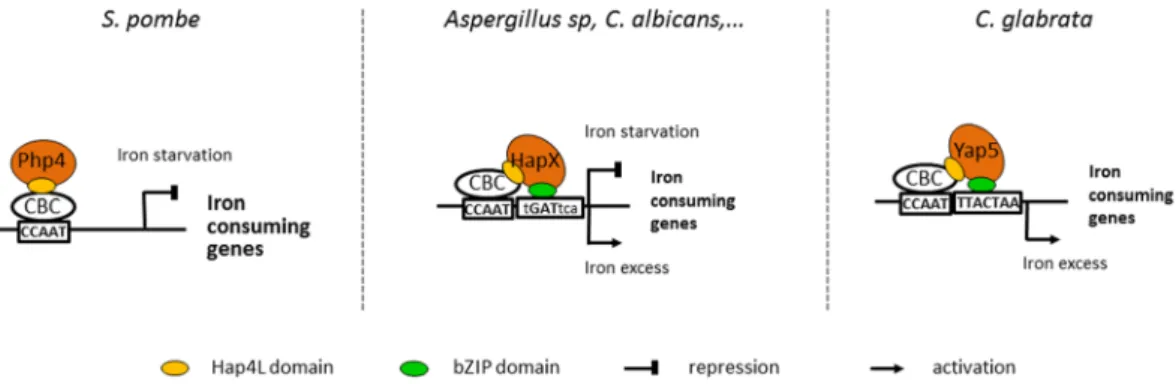
2.2.3 The CCAAT-Binding Complex as a link between oxidative stress and respiration 27 2.2.4 The iron homeostasis is linked with respiration and oxidation . . . 33
2.2.4.1 Response to iron-depleted media . . . 33
2.2.4.2 Response to iron-excess . . . 36
3 Candihub, the study of transcriptional networks of Candida glabrata stress responses 39 3.1 Networks : a model to represent connections between various elements . . . 39
3.2 The transcriptional regulatory networks . . . 41
3.3 The Candihub project : deciphering stress responses in Candida species using transcrip-tional regulatory networks . . . 42
3.3.1 Description and goals of the Candihub project . . . 43
3.3.2 Building the Candihub networks . . . 45
3.3.2.1 Types of C. glabrata strains used in this work . . . 45
3.3.2.1.1 ∆HTL strain . . . 45
3.3.2.1.2 TF deleted strains . . . 46
3.3.2.1.3 myc-tagged TF strains . . . 46
3.3.2.2 Approaches to build networks . . . 47
3.3.2.2.1 Mathematical inference of regulatory networks from experi-mental data . . . 47
3.3.2.2.2 Direct determination of regulatory networks . . . 49
3.3.3 Proof of concept of Candihub : the Yap network . . . 56
3.3.4 Goals of my PhD . . . 57
Results and discussions 59 4 Stress responses in Candida glabrata : a highly interconnected network 61 4.1 Introduction . . . 61
4.2 Selection of the ChIP conditions . . . 61
4.3 Sequencing of the immuno-precipitated DNA . . . 62
4.5 Representing the Candihub network . . . 65
5 The CBC Impacts Respiratory Genes and Iron Homeostasis in Candida Glabrata 67 5.1 Introduction . . . 67
5.2 Publication . . . 68
5.3 Supplementary results . . . 79
5.3.1 Introduction . . . 79
5.3.2 The YRE and the CCAAT motifs are required to activate the iron excess response 79 5.3.3 The YRE and the CCAAT motifs are differentially conserved in the Saccha-romycetales . . . 81
5.3.4 Hap4 might still interact with the CBC during iron excess response . . . 87
5.3.5 Conclusion . . . 88
6 Comparative Transcriptomics Reveals New Features of Iron Starvation in Candida glabrata 91 6.1 Introduction . . . 91
6.2 Publication . . . 93
6.3 Supplementary results . . . 112
6.3.1 Introduction . . . 112
6.3.2 Aft1 network in Candida glabrata . . . 112
6.3.3 C. glabrataAft1 has several functions shared with S. cerevisiae Aft1 . . . 114
6.3.4 C. glabrataAft1 and Aft2 roles are only partially redundant . . . 116
6.3.5 Relationship between Aft factors in S. cerevisiae share some features with C. glabrata. . . 117
6.3.6 Relationship between Aft factors in more distant species . . . 118
6.3.7 GRX3and GRX4 are involved in iron-deprivation response under different regu-lations . . . 119
Conclusion and perspectives 123
References 129
Appendices 169
List of Figures
1.1 Phylogenetic tree of the Saccharomycotina clade . . . 6
1.2 Infection strategies of C. glabrata and C. albicans . . . 9
1.3 Genomic processes responsible for the evolution of C. glabrata . . . 10
1.4 Phylogenetic structure of 33 Strains of C. glabrata . . . 11
1.5 Example of illegal repair events during switching in C. glabrata . . . 14
2.1 ESR genes in S. cerevisiae and C. glabrata genomes . . . 17
2.2 Pathways of the OSR in C. glabrata . . . 24
2.3 Representation of the four proteins of the HAP complex in S. cerevisiae . . . 29
2.4 Interaction of transcriptional regulators involved in the utilization of non-fermentable carbon sources in S. cerevisiae . . . 31
2.5 CBC known roles and associated transcription factors in S. cerevisiae . . . 32
2.6 Mechanisms of iron-deprivation response in C. glabrata . . . 36
2.7 Mechanisms of iron-excess response in C. glabrata . . . 38
3.1 Example of a network . . . 40
3.2 Construction of ∆HTL strain . . . 46
3.3 Workflow of GRN inference . . . 48
3.4 Microarray experiment worflow . . . 50
3.5 Chromatin Immuno-Precipitation protocol . . . 53
4.1 Network of interactions of C. glabrata stress responses . . . 66
5.1 Analysis of the impact of binding motifs presence on the activation of GRX4 promoter . 80 5.2 Analysis of the conservation of CCAAT-motif and YRE and their spacing in 30 species of the Saccharomycetales clade . . . 84
5.3 Representation of Hap4 and Hap5 networks . . . 88 5.4 Representation of the binding peaks of Hap4 in the promoters of 7 iron responsive genes
and 7 respiratory genes . . . 89 6.1 Visualization of Aft1 and Aft2 networks . . . 113 6.2 Visualization of the links between GRX genes and several transcription factors . . . 120 6.3 Analyses of the promoters of the GRX3/4 orthogroup in 28 Saccharomycetaceae species 121
List of Tables
1.1 Comparison of S. cerevisiae and C. glabrata genomes . . . 4
1.2 Comparison of C. glabrata, C. albicans and S. cerevisiae . . . 7
3.1 Number of high-throughput experiments performed in S. cerevisiae, C. albicans and C.glabrataup to 2013 . . . 43
4.1 Summary of the conditions used to perform ChIP-seq . . . 62
4.2 Summary of the reads number obtained after sequencing and processing . . . 63
4.3 Peaks, targets and GO enrichment of each transcription factor . . . 64
List of Abbreviations
aa amino-acid
BPS Batho Phenanthroline diSulfonate CBC CCAAT Binding Complex CGD CCandida Genome Database ChIP Chromatin Immuno-Precipitation ESR Environmental Stress Response
GO Gene Ontology
GRN Gene Regulatory Networks HAP Heme Activator Protein OSR Oxidative Stress Response SGD Saccharomyces Genome Database TF Transcription Factor
WGD Whole Genome Duplication
WT Wild Type
YAP Yeast Activator Protein
Candida glabrata, a yeast with many faces
Candida glabratais an interesting model organism. Despite its name, it is phylogenetically closer to Saccharomyces cerevisiae than to other Candida species. It gained interest a few years ago when its pathogenic potential was fully revealed. But even now, a lot is still unknown about this yeast. For example, we still don’t know whether it is asexual and if it really is commensal of the human host. All these questions will be tackled in the following part.
1.1
A hectic phylogenetic classification
The classification of the haploid hemiascomycete Candida glabrata has been quite vibrant for a long time. Due to the resemblance with some previously discovered fungi, it was first called Crypto-coccus glabratusafter its discovery on grapes (Berlese, 1894) and in human stools (Anderson,1917), a century ago. Its name switched to Torulopsis glabrata in 1938 (Lodder et al., 1938), after the authors noticed that the yeast wasn’t able to form pseudomycelium and had morphological and physiological characteristics resembling the ones of Torulopsis yeasts. Then, it changed to Candida glabrata in 1978 (Yarrow et al., 1978), following an important change in yeast classification and the disappearance of the Torulopsis genus. Finally, it was attributed to the Nakaseomyces clade by Kurtzman et al., 2003, after they performed a huge phylogenetic work thanks to the study of several genes, including rDNA genes, translation elongation factor, actin, RNA polymerase and mitochondrial genes. This clade also comprises three environmental species (N. bacilisporus, N. delphensis, C. castelli) added by Kurtzman et al., 2003, and two other pathogenic species (C. nivariensis and C. bracarensis) were added later to that clade (Alcoba-Flórez et al.,2005; Correia et al.,2006).
1.2
Close to Saccharomyces spp, far from Candida spp ?
The phylogenetic study of Kurtzman et al.,2003was soon followed by the publication of the whole genomic sequence of C. glabrata ATCC2001 strain by the Genolevures consortium (Dujon et al.,2004). They revealed that C. glabrata genome is composed of 13 chromosomes, totaling around 12.3 Mb. The genome has an average GC content of 38.8% and encodes for 5283 CDS. It resembles the overall structure of S. cerevisiae which is 12.1 Mb long, has an average GC content of 38.3% and encodes for 5807 CDS. Some other genomic similarities between the two species can be found in Table 1.1. This article highlighted the closeness of C. glabrata and S. cerevisiae. They both underwent a Whole Genome Duplication (WGD) event (Wolfe et al.,1997; Dietrich et al.,2004; Kellis et al.,2004) before their divergence as distinct species, as shown by the shared duplicated blocks of sister chromosomal regions (Lalo et al.,1993; Dujon et al.,2004). C. glabrata also displays exactly the same set of 42 tRNA encoding genes than S. cerevisiae. The two species even share an average of 65% of sequence identity between orthologous proteins ; this is translated by the fact that approximately 4800/5300 genes in C. glabratahave an homologue in S. cerevisiae (Gabaldón et al.,2016).
Species S. cerevisiae C. glabrata
Genome size (Mb) 12.1 12.3
Average GC content (%) 38.3 38.8
Total CDS 5,807 5,283
Total tRNA genes 274 207
Average gene density (%) 70.3 65.0
Average GC in CDS (%) 39.6 41.0
Average CDS size (codons) 485 493
Median CDS size (codons) 398 409
Maximum CDS size (codons) 4,911 4,881
TABLE 1.1. General characteristics of S. cerevisiae and C. glabrata genomes. This table is adapted from Dujon et al.,2004.
However, as close as they may be, this 35% discrepancy between these two yeasts is the same than the difference between human sequences and zebrafish (Gabaldón et al.,2013). In a general way, Nakaseomyces spp genomes are smaller and contain fewer genes than S. cerevisiae (Gabaldón et al., 2013). One explanation could be that C. glabrata has a much higher rate of loss of duplicated genes (Du-jon et al.,2004). According to this team, C. glabrata lost so much paralogues that it resulted in reductive evolution, associated with loss of functions and decrease genome redundancy. Even more striking, C. glabratagenome redundancy is equivalent to that of Kluyveromyces lactis, a pre-WGD species. Mainly, the genes were lost in galactose metabolism, phosphate metabolism, cell rescue, defence and virulence
and nitrogen and sulphur metabolism compared to S. cerevisiae and three other yeast species. Among other genomic differences, S. cerevisiae genome contains active transposon (Krastanova et al., 2005), while all Nakaseomyces species, including C. glabrata have transposon-free genomes (Gabaldón et al., 2013). Last but not least, C. glabrata is pathogenic while S. cerevisiae isn’t, which indicates some pro-found genomic differences in these yeast. As mentioned previously, C. glabrata has some specific genes with no homology to S. cerevisiae. Though one could have thought that these specific genes might be an explanation as to why C. glabrata is pathogenic, while S. cerevisiae is not, Gabaldón et al., 2016 indicated that most differences in gene content was not related to virulence.
Despite these differences and the fact that Candida albicans is also pathogenic, C. glabrata is even more distant from this yeast than it is from S. cerevisiae (Dujon et al.,2004; Fitzpatrick et al.,2006), as represented in Figure 1.1. Another major difference between C. glabrata and C. albicans is the change in C. albicansgenetic code : in this species, the codon CTG is translated as a serine, while in other yeasts, including S. cerevisiae and C. glabrata, it is translated as a leucine.
C. albicans and C. glabrata have a common ecological niche, they can both cause infections in human ranging from benign to fatal and they even share a common species name. However, despite these common points, they still are very distant species according to their genomic sequences as well as some of their phenotypic features. For example, C. glabrata cells are usually described as budding yeast or pseudohyphae (under certain specific conditions such as limiting nitrogen conditions (Csank et al., 2000)), while C. albicans cells can be shaped into yeast, pseudohyphae, hyphae or chlamydospores. A more complete comparison of several features in these two yeasts and S. cerevisiae can be found in Table 1.2. Nevertheless, these two Candida species are both pathogen, and this pathogenicity had a big impact in bringing them under the focus of research.
1.3
An "emerging" opportunistic pathogen
Candida glabratabegan to draw attention only long after its discovery, even though it was identified as early as the beginning of the 20th century. It only received significant consideration in the late 80’s, when it was noticed to be one of the main cause of fungal infections in immunocompromised individuals (Just et al.,1989). Following this statement, the increasing incidence of this yeast was acknowledged in the late 20th century when it was declared as an emerging pathogen (Hazen,1995). It was also noticed
FIGURE1.1. Phylogenetic tree of the Saccharomycotina clade. Schematic representation based of the phyloge-netic tree inferred by X.-X. Shen et al.,2016. The tree was inferred from the concatenation-based analysis of 1233 single-copy orthologues. The Whole Genome Duplication is indicated by a black star. The CUG-clade is depicted by red branches on the tree. C. glabrata, C. albicans and S. cerevisiae are highlighted in bold.
that its prevalence tends to increase with age, antibiotic treatment, length of stays in hospitals, diabetes mellitus. . . (Angoulvant et al.,2016).
Usually, Candida glabrata is a harmless fungi of the human gut microbiota. However, given some specific conditions, it can proliferate and trigger superficial benign infections such as oral or vaginal thrush, gastrointestinal tract infection or urinary bladder infection (Pfaller et al.,1998; Fidel et al.,1999), hence the adjective “opportunistic”. In the worst cases, it can breach the mucosal barriers, enter the bloodstream and disseminate throughout the body, causing systemic candidiasis infections with high mortality rates (40-60%), especially in immunocompromised individuals (cancer patients undergoing chemotherapy, transplanted patients, for example) and elder people (Angoulvant et al.,2016).
Feature/Species C. glabrata C. albicans S. cerevisiae
Ploidy Haploid Diploid Diploid
Virulence Opportunistic pathogen Opportunistic pathogen Non-pathogenic Major sites of infection Oral, vaginal, disseminated Oral, vaginal, disseminated Non-infectious
Mating genes Present Present Present
Sexual cycle Unknown Known (cryptic) Known
Clonal population structure Yes Yes No
Phenotypic switching Present Present Absent
True hyphae Absent Present Absent
Pseudohyphae Present Present Present
Biofilm formation Present Present Present
Major adhesins Lectins (EPA genes) Lectins Lectins (FLO genes) Hwp1 adhesin Sexual agglutinins
Als adhesins
Auxotrophy Niacin, thiamine, pyridoxine None None Azole resistance Innate resistance Susceptible Susceptible Mitochondrial function Petite positive Petite negative Petite positive TABLE 1.2. Comparison of C. glabrata, C. albicans and S. cerevisiae. ECM : extracellular matrix ; Hwp1 : hyphal cell wall protein. This table is adapted from Kaur et al.,2005.
et al.,2007; Azie et al.,2012) and Candida spp are in the top five causes of nosocomial bloodstream in-fections (Gudlaugsson et al.,2003; Pfaller et al.,2007). Most of the time, Candida albicans is the main cause of candidiasis (50-70%), followed by C. glabrata (20-30%) (Perlroth et al.,2007). However, the role of C. glabrata as cause of candidiasis is rapidly increasing, perhaps because of its natural resistance to the compounds usually administered as treatment of this disease, namely azoles (Jandric et al.,2011; Pfaller,2012). It can also easily acquire resistance to another class of antifungal, the echinocandins. It was shown that resistance increases in case of a pre-treatment. Additionally, a phenomenon of cross-resistance to other drugs can appear in strains already resistant to fluconazole (Komshian et al., 1989; Malani et al.,2005). One of the principal cause of the innate resistance of C. glabrata is the increased efflux of drugs driven out of the cells by pumps, usually regulated by PDR1. When this gene is over-expressed, the cells tend to bear an increased resistance and a increased virulence. Also, some changes in the cell wall have been reported to prevent drug diffusion (Parkinson et al., 1995; Clark et al.,1996; Vermitsky et al.,2004; Ferrari et al.,2011a). All these mechanisms are reviewed in Sanglard,2002.
C. glabrataand C. albicans can both easily infect humans, however their strategies are different on several points (reviewed in Brunke et al.,2013) :
Adhesion : C. albicans have Als and Hwp adhesins, while C. glabrata has Epa adhesins. Also, both species can form biofilms on host cells or medical devices (Iraqui et al.,2005; Nobile et al.,2006). Invasion : C. albicans mainly use its hyphae form to gain access to epithelial cells. The invasion mechanism in C. glabrata is not known yet, as this yeast doesn’t form true hyphae. However,
it was noticed that C. glabrata infections cause reduced inflammatory reaction from the host, contrary to C. albicans. It might suggest a smoother way for C. glabrata to enter its host.
Interaction with immune cells : The two species have very different approach : shortly after internaliza-tion in macrophages, C. albicans uses hyphae to burst them, while C. glabrata uses the phagosome as a safe haven to multiply before bursting it. Once again, this has the advantage to be more quiet against the immune system of the host.
Nutrient acquisition : C. albicans is much better equipped to face the host in nutrients acquisition. It has no known auxotrophy and owns several integration pathways for a lot of nutrients, including metallic ions. On the other hand, C. glabrata loss of genes after WGD caused the disappearance of several important pathways (mentioned in Section 1.2). This lack has to be compensated for the cells to survive and thrive.
All these mechanisms are summarised in Figure 1.2. Finally, given these dissimilarities and the phylogenetic disparity between C. glabrata and C. albicans, it is likely that their pathogenicity and their infection strategies came from different and independent evolutions.
1.4
C. glabrata genome plasticity : a huge impact
Over the years, it became clear that Candida glabrata possesses an impressive genome plasticity and can be rearranged easily. Several teams reported some interesting features and consequences to this very dynamic nature of the C. glabrata genome, which could go as far as considering the rearrangements as an adaptive mechanism. The first clue might be the high rate of loss of paralogues after the WGD, leading to a reductive evolution (Dujon et al.,2004) (Table 1.1). Shin et al.,2007reported that changes in karyotype can appear quickly in strains isolated at different time intervals in the same patient without antifungal therapy. They also showed that these strains were able to acquire azole resistance under treatment with azole compounds. Ahmad et al.,2013reported that the number of chromosomes of C. glabrata can vary among different isolates. Especially, they found extra chromosomes created by segmental duplication and translocation. They supposed that it was a way for C. glabrata to improve its fitness around new environments. Poláková et al.,2009also noticed some size variation in the chromosomes, but more im-portantly, they remarked that new supernumerary chromosomes carried duplicated genes among which
FIGURE1.2. Infection strategies of C. glabrata and C. albicans. These two yeasts have very different methods to infect the host. However, there is still a lot of unknown processes involved in C. glabrata pathogenicity. Adapted from Brunke et al.,2013.
the orthologues of S. cerevisiae family of ABC transporters, which plays a role in pleiotropic drug re-sistance and yeast-host interaction genes. Overall, these karyotype variations correlated with antifungal drug resistance.
C. glabratagenome contains the family of EPA genes (homologues of the FLO genes in S. cere-visiae), coding for cell-wall proteins, which are known to be responsible for the adherence to human epithelium (Cormack et al., 1999; Gabaldón et al.,2013). Coincidentally, Muller et al.,2009reported size variation in tandem arrays of repeated genes, which often encode the EPA genes, suggesting a role in adaptation to the host (Figure 1.3). It also suggests a role in virulence because these tandem repeats were also found to encode aspartyl proteases responsible for adherence to mammalian cells and survival in macrophages (Kaur et al.,2007).
However, this genomic changes also happen very quickly in lab strains : Bader et al.,2012noticed some karyotypic modifications that could be associated to phenotypic variations. It shows that genome
FIGURE1.3. Genomic processes responsible for the evolution of C. glabrata. EPA genes : family of GPI-anchored cell wall proteins that facilitate host recognition and C. glabrata adherence. Adapted from Moran et al.,
2011).
plasticity occurs not only during the harsh conditions of host infection, but also in laboratory conditions during a very short time and without strong selective pressures.
Hence, it seems that chromosome remodeling and gene duplications play an important role in the specialization to specific environments, virulence, and interaction with the host (Butler et al., 2009; Moran et al.,2011; Gabaldón et al.,2018).
1.5
Breaking C. glabrata clichés
1.5.1 A commensal yeast ?
Candida glabratais often considered as a human commensal, even if its prevalence can vary a lot between studies and is influenced by a range of factors, including the age, the previous use of antifungal treatments and the medical condition of a patient. C. glabrata has been detected in human flora (oropha-ryngeal, digestive, vaginal), on medical devices (Iraqui et al.,2005), phones (Kordecka et al.,2016) but it can also be found in non-human related niches such as fermenting coffee beans (de Melo Pereira et al., 2014) or cloaca of several bird species (Cafarchia et al.,2008; Francesca et al.,2014; Al-Yasiri et al., 2016). These are all diverse isolation sites, sometimes without any link between them (for example, mobile phone and bird cloaca or with human (for example, bird cloaca and human). It suggests that either C. glabrata is a commensal of several sources, or that it contaminated all these secondary sources from a primary source that is still unknown. This supposition is coherent with the fact that C. glabrata
is not always found in these niches. This type of behaviour has been shown for other yeasts, such as C. albicans(Bensasson et al.,2018) or S. cerevisiae (Goddard et al.,2015).
FIGURE1.4. Phylogenetic structure of 33 Strains of C. glabrata. This tree indicates the phylogenetic structure of the strains based on SNPs data analysis. Total number of SNPs isn’t displayed, but a range of 4.66 to 6.56 SNPs per kb per strain when compared to the reference strain (as sequenced by Dujon et al.,2004) is observed. Each colour designates a clade. Super-indices indicate pairs of strains isolated in the same patient, but on different body site or at a different date. Body site and country of the isolation are displayed, along with the mating type of the strains. Adapted from Carreté et al.,2018.
One strong argument in favour of the human commensality of C. glabrata is the existence of phylo-genetically distinct clades among the yeast population related to specific geographical origins. In other words, some distinct clades were defined according to genetic and phenotypic markers, and these clades were specific to a geographical zone (Dodgson et al.,2003; Dodgson et al.,2005; Brisse et al.,2009; Rolland et al.,2010; Schwarzmüller et al.,2014). However, these studies were only performed on a few markers, and consequently, the number of detected clades varied a lot. This problem was solved by the study of Carreté et al.,2018, who assessed genomic and phenotypic variations in 33 C. glabrata isolates coming from all over the world. They were able to sequence these strains and class them into tree with seven clades (Figure 1.4). On this tree, it appears quite clear that strains of the same clade clustered
together despite their geographical origin (for example, cluster I with USA and Belgium), which would go against a human commensality of C. glabrata : this undermines the idea of a genomic co-evolution between C. glabrata and human, as we could have expected if there was a commensal relationship be-tween them. It lets us think that human activities and transportations caused the crossing of strains that have been isolated for a long time, most likely in specific geographical areas.
However, C. glabrata displays several traits that let us think of it as a commensal of human. The optimal growth temperature of C. glabrata is close to 37°C, the normal internal temperature of the human body. This is a huge advantage as both a commensal and an opportunistic pathogen. C. glabrata has a higher stress resistance and an enhanced ability to sustain prolonged starvation. It can withstand very high concentrations of H2O2(also considered as oxidative stress), varying concentration of bio-available
iron, lack of oxygen, among others, and can also resist to shortage of nutrients caused by protective mechanisms of the human host. Especially, the adaptation to use alternative nutrients and survive long periods of starvation is essential for C. glabrata to survive macrophage engulfment (Roetzer et al.,2010) and use the macrophage as a “Trojan horse” to invade the whole body of the host. This type of adaptation reminds the bacteria surviving internalization in amoebae and the subsequent oxidative and osmotic stresses (Greub et al., 2004), as well as the likely selection for bacterial virulence and resistance traits (Tosetti et al.,2014; Hao et al.,2015). Additionally, earlier, C. glabrata can also resist to antifungal drugs and acquire new resistances, which is an useful mechanisms for a human pathogen. Also, C. glabrata genome has remodeled its cell-wall components resulting in a higher adherence. The ability to adhere to the host tissues is mediated by cell-wall-associated proteins called adhesins (reviewed in Groot et al., 2013). This translates into the presence of a high number of particular adhesins encoded by the EPA family of genes . This adhesins proved to be crucial in virulence and to form biofilms (Cormack et al., 1999; Roetzer et al.,2011a), an ability which increases resistance, virulence and persistence in the host. Finally, C. glabrata has lost several hundreds of genes, including genes in the pyridoxine, thiamine and nicotinic acid biosynthetic pathways (Dujon et al.,2004; Kaur et al.,2005). The fact that the human host is a really stable growth environment, because all its parameters (temperature, oxygen, nutrients. . . ) are regulated through homeostasis, could be an explanation to the loss of specific pathways in C. glabrata. It also means that this yeast has a higher dependence of the host, and rely on it to acquire nutriments. All these features tend to show an adaptation of C. glabrata to its human host.
Nonetheless, several of these features (growth at 37°C, loss of the nicotinic acid pathway and pres-ence of auxotrophies) are shared by all Nakaseomyces species, pathogenic and non-pathogenic, and the
expansion of the EPA family was linked to virulence in the pathogenic Nakaseomyces (C. glabrata, C. bracarensis, C. nivariensis). This means that not all these changes are specific to C. glabrata and its com-mensalism (or its pathogenicity). Hence, C. glabrata clearly displays commensal traits but the repercus-sions of this commensalism on C. glabrata genome are not clearly identified yet. This could be because C. glabratamight be a recent human commensal (which hasn’t left genomic traces yet) or/and because we still lack experimental data on the comparisons between C. glabrata and other Nakaseomyces.
1.5.2 An asexual yeast. . . for now
C. glabratais believed to be an asexual haploid fungus, which reproduces exclusively by budding. Though it is haploid and can bud, it might not be as asexual as we thought, even if its mating has never been actually observed. Yeast in the same clade than C. glabrata and S. cerevisiae often have two mating types in haploid cells. These haploid cells can merge, forming diploids, which can then undergo meiosis and sporulation (Muller et al.,2008). To increase their adaptive potential, haploid cells can also switch mating types through recombination with silent loci after an HO endonuclease cut (Haber,2012).
One of the reasons supporting a sexual reproduction of C. glabrata is the existence and the obser-vation of mating types a and α (Muller et al.,2008; Carreté et al.,2018). Even more striking, Brockert et al., 2003reported mating type switching during infection of a host and Butler et al., 2004reported mating type switching during laboratory growth. Interestingly, C. glabrata possesses a seemingly intact mating machinery in its genome (S. Wong et al., 2003), composed of orthologues of genes involved in mating in S. cerevisiae : MTL1, MTL2 and MTL3 genes were found in C. glabrata (Srikantha et al., 2003; Butler et al.,2004). MTL1 is the expression locus, while MTL2 and MTL3 encodes a and α mating informations. The genome also contains an homothallic (HO) endonuclease, which is known to be re-sponsible for gene conversion events that underlies mating type switching in S. cerevisiae. Additionally, Butler et al.,2004found a putative HO endonuclease recognition site in MTL1.
All these findings were further confirmed by Carreté et al.,2018, who furthermore demonstrated the existence of illegal repair events during switching (Figure 1.5). They also found traces of selective con-straints in mating genes. Even more interesting, they found the same levels of constraint in C. glabrata than in S. cerevisiae or C. albicans, which have functional sexual and parasexual cycles, respectively. This tends to show that sexual genes in C. glabrata are still under selective pressure, most likely because they are still functional. Also, switching is lethal to many C. glabrata cells in laboratories Boisnard et al.,
2015, which shows that the switch is tightly regulated. Besides, they discovered evidences of chimeric patterns in chromosomes, which is the result of sexual mating between yeasts of different clades.
FIGURE1.5. Example of illegal repair events during switching in C. glabrata. BS means Before Switching. AS means After Switching. Adapted from Carreté et al.,2018. Case 1 represents a normal conversion event at MTL1, which switched mating type from a to α. Case 2 shows the classic a-to-α switch at MTL1 accompanied by illegitimate conversion at MTL2, resulting in a triple-α strain. Case 3 displays an illegitimate MTL2 conversion without the MTL1 switch. Case 4 represents an illegitimate MTL3 conversion without the MTL1 switch, resulting in a triple-a strain.
To summarize, Candida glabrata keeps distinct a and α haploid mating types and distinct associated cellular identities (Muller et al.,2008) ; it shows evidences of mating type switches in populations issued of isolates and laboratory populations (Brockert et al.,2003; Butler et al.,2004) and sexual recombination (Dodgson et al.,2005; Carreté et al.,2018). All this lets us think that C. glabrata might still be mating but rarely and we just haven’t found yet the adequate conditions to observe it.
Candida glabrata and the stress responses
As mentioned in the previous part, one of the possible explanations of C. glabrata success as a human pathogen and a commensal of a lot of ecological niches could be its genome plasticity, among other things. This genome plasticity has been shown to allow the yeast to adapt to a range of environments, which can vary a lot in pH, temperature, oxygen and iron availability, oxidative stress. . . A brutal change in these conditions is perceived by the yeast as a stress and triggers several pathways that aim to adapt the cells to their new environment to improve their survival and growth. The phenomenon of adaptation to improve the cells fitness in a specific environment is called stress response. Besides genome plasticity, stress responses can also take the form of transcriptional adaptations inducing changes in gene expression and protein composition of the cell. We can distinguish two kinds of stress responses : a general stress response, which is activated as soon as the cells detect an environmental variation, regardless of the change, and stress responses specifically adapted to the triggers perceived by the cells.
2.1
A general response : the Environmental Stress Response (ESR)
The Environmental Stress Response (ESR) corresponds to the regulation of a core set of genes in response to sub-optimum growth conditions, regardless of the stress imposed. It means that the cells react to changes in the environment by up and down-regulating the same set of genes for every condition, whether it is temperature, pH or nutrients availability. . . On a more practical way, it also means that these genes will display close expression profiles during the stress response. The ESR was studied in Candida glabrataby Roetzer et al., 2008through the yeast transcriptional response to four conditions : glucose starvation, osmotic stress (addition of NaCl in the media), heat stress (growth at 42°C) and oxidative stress (addition of hydrogen peroxide in the media). They identified 782 genes as members of the ESR. Among those, 358 were induced and 424 were repressed. Interestingly, the ESR seems to be triggered not only by deeply detrimental growth conditions, but also by small environmental changes which do
not severely impact cell growth or viability. Nevertheless, the ESR is caused by the transition from an optimal environment to a sub-optimal environment and therefore is not activated when cells go back from a sub-optimal environment to an optimal growth environment. It is equally noteworthy that the ESR intensity is adapted to the severity of the environmental stress : the more severe the environmental changes are, the longer the ESR is and the more acute are the genes induction and repression.
The genes composing the ESR are enriched in several functional categories. More specifically, the stress response causes the general repression of cytosolic translation (ribosome biogenesis, rRNA and tRNA processing, translation initiation and elongation), nucleotide biosynthesis, cell growth and secre-tion. On the other hand, it causes the induction of HSP chaperone proteins (as well as protein folding and degradation), cellular redox reactions, DNA damage repair and genes involved in energy storage and car-bohydrate metabolism. In short, all these changes aim to protect DNA, save energy by repressing protein synthesis and cell growth and rearrange the proteome to keep the proteins essential for stress response and basic cell functioning. It gives rise to an interesting phenomenon : the simultaneous induction of both synthetic and catabolic enzymes, which aim to help produce the suitable proteins and destroy the useless ones. The ESR might also be the cause of cross-resistance to various stresses, in which cells exposed to a low dose of one stress become resistant to a low dose of a second stress : by protecting the pathways previously mentioned after a first stress, the cells are already in a resistance state which reduces the impact of a second stress.
Roetzer et al.,2008also showed that Msn2/Msn4 TF are partially responsible for the ESR. Deletion of Msn2/4 abolishes the induction/repression of a part of the ESR and render cells more sensitive to stress exposure. These factors bind on the STRE motif (AGGGG) (discovered by Martínez-Pastor et al., 1996 in S. cerevisiae), which happens to be one of the most enriched motifs in ESR genes. Msn factors expression and localisation are tightly regulated. When cells encounter stresses, these TFs are induced, and Msn2/4 proteins are relocated from cytoplasm to nucleus to induce several genes of the ESR. When cells return to a more growth-friendly environment, Msn2/4 are down-regulated and they are exported from the nucleus. Thus, the import/export of these TF is essential for their regulation abilities. The mechanisms involved in nuclear import and export are not fully understood yet, but it is clear that the Nuclear Export Signal (NES) (especially the conserved HD1 domain) and the Nuclear Localisation Signal (NLS) play an important role. The NLS drives nuclear import. Nuclear export requires HD1 domain presence, as well as exportin Msn5. Currently, it is supposed that HD1 is the target site of Msn5 exportin and that the Protein Kinase A (PKA) phosphorylation site within HD1 has a regulatory role.
Msn2 activity is likely regulated by PKA and TOR pathways, which phosphorylate the HD1 domain and the NLS, but these assumptions remain to be proven in C. glabrata.
These findings differ only slightly from the ESR study of S. cerevisiae (Gasch et al.,2000)). They used 13 different stress conditions and found that the core set of genes contains 868 genes, among which 283 are upregulated and 585 are downregulated. In both C. glabrata and S. cerevisiae, ESR genes represent 15% of the total number of genes in the genome. Several dozens of the ESR genes are common between the two species (Fig 2.1). As a consequence, there is an enrichment in the same GO categories in both species (see previous paragraphs). However, there are still a few differences in the targets : genes of sterol and ergosterol biosynthesis are deeply repressed upon stress in C. glabrata while they are not in S. cerevisiae. Some of these genes were showed to be linked with cell growth and drug resistance (Geber et al.,1995; Montañés et al.,2011), which might explain their down-regulation during the ESR. Additionally, some interesting genes are up-regulated in C. glabrata and down-regulated in S. cerevisiae : PHO84 (involved in heavy metal tolerance, Rosenfeld et al., 2010), VPH2 (involved in vacuolar pH and lifespan extension, Ruckenstuhl et al.,2014), TBF1 (TF involved in DNA protection, Ribaud et al., 2012). Interestingly, Wapinski et al.,2010showed that C. glabrata lost the down-regulation of ribosomal proteins in ESR, while it is conserved from S. cerevisiae to C. albicans.
FIGURE2.1. ESR genes in S. cerevisiae and C. glabrata genomes. The overlap between the ESR of the two species is depicted as a Venn diagram. C. glabrata and S. cerevisiae data are from Roetzer et al., 2008; Gasch et al.,2000, respectively.
The expression of ESR genes in S. cerevisiae is regulated by different TF, depending on the envi-ronmental condition encountered, and governed by several different upstream signaling pathways. Msn2 general role and localisation are especially conserved in S. cerevisiae (Görner et al.,1998; Görner et al., 2002). Other pathways are involved, and it seems that each stress condition activates specific pathways. Notably :
- PKA and PKC pathways trigger the ESR (especially repression of ribosomal proteins) in response to lack of nutrients and impaired secretion, respectively (Klein et al., 1994; Neuman-Silberberg et al.,1995; Nierras et al.,1999).
- ESR-induced genes during osmotic stress are regulated by the High Osmolarity Glycerol (HOG) pathway (Rep et al.,2000).
- ESR-triggering by DNA damage is controlled by DNA damage specific Mec1 pathway (Gasch et al.,2001).
Unfortunately, most of these transcription factors and pathways are poorly studied in C. glabrata. The PKA pathway seems to be involved in adaptation to nutrient deprivation and biofilm formation (Schwarzmüller et al.,2014; d’Enfert et al.,2016), but it wasn’t proven to be involved in ESR regulation. The PKC path-way plays a role in response to cell wall damaging agents, however it wasn’t linked to the ESR (Borah et al.,2011). Schwarzmüller et al.,2014noted that Mec1 isn’t essential in C. glabrata while it is in S. cerevisiae and that its deletion modifies the cells fitness which suggests that the Mec1 regulatory net-works functions differently between the two species, but once again, no link was made with the ESR. Nevertheless, Gregori et al.,2007and Roetzer et al.,2008showed that Hog1 function is conserved in C. glabrataand it retains its role in ESR, particularly during osmotic stress.
A part of the ESR genes is also conserved in other species such as L. kluyveri (Brion et al.,2016), S. pombe(D. Chen et al.,2003), Aspergillus spp (Kawasaki et al.,2002; Du et al.,2006). Usually, the target genes in these species have GO categories similar to those of S. cerevisiae and C. glabrata. The ESR existence is still unclear in C. albicans because of contradictory findings (Enjalbert et al.,2003; Harcus et al.,2004; Enjalbert et al.,2006; Roy et al.,2013), even if at least a small part of the regulations seems to be conserved.
Regarding the role of Msn2, there are contradictory signs in previously mentioned species. The Msn2 DNA binding domain is found in genes in other species, but this is usually the only conserved part
between these genes and C. glabrata Msn2. A team found STRE binding proteins in S. pombe (Kunitomo et al.,2000), however they didn’t seem to be involved in stress response. Nevertheless, the HD1 domain is conserved in K. lactis and A. gossypii, as well as the embedded PKA phosphorylation site and is enough to trigger nuclear export of Msn2 in these species. However, the HD1 region doesn’t appear to be conserved in C. albicans and its close species, which led Roetzer et al.,2008to hypothesise that the HD1 domain and the Msn2-mediated stress response appeared after the divergence of C. albicans from the evolutionary path leading to S. cerevisiae and C. glabrata. Enjalbert et al., 2006found no role for Msn2 in C. albicans ESR. Ramsdale et al.,2008showed that C. albicans Mnl1 (Msn2 correspondent in this yeast) is still binding to STRE motifs, however, they only reported its role in weak-acid response, and not in a larger environmental stress response. On the other hand, Alonso-Monge et al., 1999reported an important role for Hog1 in several types of stress in C. albicans (osmotic stress, weak acid stress, oxidative stress). Also, it was shown by D. Chen et al., 2003, that Sty1 (Hog1 S. cerevisiae orthologue in S. pombe) is required for ESR regulation in S. pombe. Hence, the ESR of these two species might be carried out by transcription factors that are different from Msn2 orthologues.
Besides the control of the ESR, these signaling systems have also been involved in regulating more specialized stress responses and the associated gene expression. These pathways simultaneously regulate both the ESR and specialized responses specifically adapted to the environmental conditions that activate the pathways.
2.2
Condition-specific stress responses
Like most unicellular organisms, Candida glabrata has a multitude of ways to deal with ever-changing growth conditions or unfavorable media and nutrients starvation. These situations are per-ceived by the cells as stresses and trigger stress responses specifically adapted to answer the changes in growth media. As a result, C. glabrata has a lot of different stress response pathways. In the following parts, a handful of them will be addressed, as well as the strong connections that exist between them : the absence of oxygen, the oxidative stress response, the respiratory pathway and iron homeostasis. We focused on these stresses because they are of interest to our team and because some of them (oxidative stress, changes in iron concentration) are important for C. glabrata virulence.
2.2.1 Rox1, a mediator of response to hypoxia
ROX1was extensively studied in S. cerevisiae since the early 80’s. It was first discovered as a repres-sor of ANB1, a translation elongation factor especially expressed during hypoxia (Lowry et al.,1986). Lowry et al.,1988confirmed the repressing effect of Rox1 on another gene involved in hypoxia, CYC7, and Ter Linde et al.,2002and others used genome-wide approaches to show that under aerobic media, Rox1 represses several tens of genes required for hypoxia transition and adaptation. This regulation is permitted by Rox1 binding on the ATTGTTCTC motif found in the promoters of its targets (Bala-subramanian et al., 1993). The binding is mediated by HMG (High Mobility Group) domain. Deckert et al., 1995also found that Rox1 ability to repress its targets was probably linked to its DNA bending capacities : when Rox1 is bound, it bends DNA at an angle of 90 degrees, preventing other activators or polymerases to bind and promote or start transcription. Currently, ROX1 is almost uncharacterised in C. glabrata. Schwarzmüller et al.,2014reported no impact of Rox1 deletion on C. glabrata fitness in the condition they tested (azole susceptibility and biofilm formation). However, Gupta et al., 2017showed that Rox1 has a role in biofilm formation and biofilm resistance to azole under hypoxic condition. Addi-tionally, they suggested that Rox1 is involved in resistance to hypoxia. This last role resembles a lot to the role of S. cerevisiae Rox1.
However, in S. cerevisiae, Rox1 activity is also mediated by its interactions with several other proteins (Kastaniotis et al., 2000; Sertil et al., 2003; Mennella et al., 2003; Klinkenberg et al., 2005). Rox1 interacts synergistically with Mot3 to repress the transcription of hypoxic genes, and that Mot3 activity is regulated by oxygen, like Rox1. It is also able to recruit the Tup1/Ssn6 complex, a known general transcriptional repressor, to repress its targets.
With all these data, the ability of Rox1 to regulate its targets was also rapidly linked to the presence of oxygen and heme : Lowry et al., 1988and L. Zhang et al., 1999showed that ROX1 is induced by heme, and this finding laid the bases of ROX1 regulatory network. Indeed, Keng, 1992demonstrated that Heme Activated Protein 1, Hap1, was required to induce ROX1 to regulate HEM13. Zitomer et al., 1997went further and found that the presence of heme induces Hap1 which in turns induces Rox1 which represses his targets. On the other way, absence of heme causes ROX1 repression by Hap1, thus lifting Rox1 repression on hypoxia genes. This makes sense, given that heme biosynthesis is strongly linked to oxygen availability. Also, according to Proft et al.,2005, Sko1 is another possible regulator of ROX1, as well as Ixr1 (Castro-Prego et al.,2009).
Besides its regulation by heme and oxygen and its impact on hypoxic genes, S. cerevisiae Rox1 was found to be involved in a variety of roles, such as lipid metabolism (Vasconcelles et al., 2001; Kwast et al.,2002), cell wall mannoproteins regulation (Abramova et al.,2001), ergosterol biosynthesis regulation with Hog1 during osmotic stress (Jensen-Pergakes et al., 2001; Montañés et al.,2011) and fermentation (Fujiwara et al.,1999). More interestingly, Rox1 was also linked to metal homeostasis and oxidative stress response, which is not so surprising given the relationships between oxygen and ROS production : C.-M. Wong et al., 2003showed that Rox1 regulates TSA2, a key gene of the OSR. Liu et al.,2013determined that oxidative stress lowers the occupancy of Rox1 on hypoxic gene promoters, thus inducing their de-repression. Finally, Caetano et al., 2015confirmed that during oxidative stress caused by cadmium, Yap1 activates ROX1 to repress FET4, providing a link between Rox1, OSR and metals. This link might be conserved in C. glabrata, because Merhej et al.,2016found that Yap1 binds and regulates ROX1 in response to selenite, which causes oxidative stress, among others. Still in C. glabrata, Rox1 has another role in metal homeostasis : Srikantha et al., 2005showed that it regulates CCC2, a copper transporter. This regulation is conserved in S. cerevisiae (Ter Linde et al.,2002). Also, C. glabrataRox1 was linked to mitochondrial dysfunction (Ferrari et al.,2011b). Nevertheless, the role of Rox1 doesn’t seem to be conserved in K. lactis (Fang et al.,2009) nor in C. albicans, where it controls filamentous growth (Khalaf et al.,2001; Kadosh et al.,2001).
2.2.2 The oxidative stress response (OSR)
Oxidative stress occurs when the concentration of Reactive Oxygen Species (ROS) present in or around the cell is higher than the detoxification abilities of the yeast or when damages resulting from ROS accumulation starts proving difficult to repair. ROS are highly reactive chemical species contain-ing oxygen, such as the superoxide anion (O2–), hydrogen peroxide (H2O2) and hydroxyl radical (HO ). These compounds can damage DNA, oxidize proteins and lipids and ultimately cause cell death. There are several sources of ROS (reviewed in D’Autréaux et al., 2007). First, they are by-products of nor-mal aerobic metabolism, especially cellular respiration : for example, they are formed during oxidative phosphorylation, which aims to produce ATP. Additionally, hydroxyl radicals and superoxide anions are produced during Fenton reactions, in which metallic ions (which are often iron and copper, see Stohs et al.,1995) in contact with hydrogen peroxide are oxidized, leading to the formation of the previously mentioned species. Heavy metals such as cadmium and lead can also cause oxidative stress by peroxi-dising lipids and oxiperoxi-dising glutathiones (Stohs et al.,1995; Z.-S. Li et al.,1997). The damages caused
by oxidation of proteins and lipids has a great cost in ATP and NADP(H) for the cells, because the re-duction processes of these compounds are very expensive in energy. Finally, pathogenic yeasts such as C. glabrata are likely to come into contact with ROS when they are engulfed by macrophages during the systemic invasion of a host. Phagocytes are the first line of host defense against fungal infections and when they encounter a pathogen, they quickly use the NADPH oxidase complex to produce ROS designed to kill it (Segal et al.,2012), such as, for example, hypochlorous acid, the main reactive compo-nent of bleach. Failing in NADPH oxidase functioning of the host often leads to increased susceptibility to fungal infections, which displays a strong link between oxidative stress and virulence (Roetzer et al., 2011b; Seider et al.,2014). This shows how important it is for C. glabrata to efficiently deal with ROS and maintain a proper redox homeostasis.
It was also shown that the Oxidative Stress Response (OSR) plays a key role in C. glabrata survival and replication after engulfment in macrophages (Kaur et al.,2007; Wellington et al.,2009; Roetzer et al., 2010; Roetzer et al.,2011b; Seider et al.,2011). The OSR is so efficient in C. glabrata that this yeast can withstand very high H2O2concentrations : while the H2O2concentration inside a macrophage is 0.4 mM, C. glabrata can resist up to 40 mM H2O2 given proper conditions (Roetzer et al.,2010), which might
partially explain its persistence in these immune cells. Studies showed that C. glabrata is particularly resistant to hydrogen peroxide, compared to S. cerevisiae and isolates of C. albicans (Cuéllar-Cruz et al., 2008), even if OSR is quite conserved between S. cerevisiae and C. glabrata (Roetzer et al.,2008; Salin et al., 2008; Saijo et al., 2010; Roetzer et al., 2011b; Gutiérrez-Escobedo et al., 2013). Cuéllar-Cruz et al.,2008also proved that C. glabrata ROS resistance is further increased in stationary phase.
The currently available data suggest that C. glabrata has a robust and most likely redundant antioxi-dant system. The OSR in C. glabrata is performed through the induction of enzymatic (catalases, SODs, and peroxidases) and non-enzymatic (glutathione) mechanisms. It will mainly use three pathways to deal with ROS and especially hydrogen peroxide.
- The catalase pathway (Green box in Figure 2.2) : catalases decompose H2O2to water and
oxy-gen. While S. cerevisiae has two catalases, CTT1 (cytoplasmic catalase) and CTA1 (peroxisomal catalase), C. glabrata has only one, CTA1. Interestingly, CgCTA1 combines features (induction conditions and intracellular localisation) of the two catalases of S. cerevisiae (Roetzer et al.,2010; Roetzer et al.,2011b). Even if CTA1 orthologue of C. albicans (CAT1) was proved to be important for virulence (Nakagawa et al.,2003), that is not the case in C. glabrata, despite being essential to hydrogen peroxide resistance (Cuéllar-Cruz et al.,2008; Roetzer et al.,2010).
- The glutathione pathway (Blue box in Figure 2.2) : it is constituted by glutathione (GSH), glutare-doxins and a glutathione reductase. This system uses a series of reactions that are expensive in energy and reduced materials. Glutathione (GSH) is an essential tripeptide composed of glycine, cysteine and glutamate synthesized by the action of GSH1 and GSH2 (Meister, 1974). In C. glabrata, GSH1 is essential (Yadav et al.,2011). GSH acts as an electron donor for conversion of H2O2into water. Accordingly, it produces an oxidised form of GSH, glutathione disulfide (GSSG) (Penninckx,2000). This reaction is catalysed by the glutathione peroxidase enzymes Gpx1 and Gpx2. GSH is reformed by reduction of GSSG by the glutathione reductase, Glr1, using NADPH as an electron donor. The glutaredoxins Grx1 and Grx2 may also be involved in the maintenance of a high GSH to GSSG ratio.
- The thioredoxin pathway (Red box in Figure 2.2) : this system also uses a series of complex and expensive reactions. Detection of ROS leads to the activation of TRR1, a reductase which consumes NADPH to reduce the thioredoxin Trx2. Trx2 then reduces the thioredoxin peroxidase Tsa1, which converts H2O2to H2O. In the same reaction, Tsa1 is also reduced back to its initial
form (Grant,2001). In S. cerevisiae, Tsa1 is expressed at high basal levels even in unstressed cells. This might be because it is an essential antioxidant of ROS (its deletion renders cells sensitive to a range of oxidative stressors) but also because Tsa1 was linked to genome stability and protection (Iraqui et al.,2009).
Furthermore, superoxides anions and hydroxyl radicals are converted to hydrogen peroxide by super-oxide dismutases (SOD) (Briones-Martin-Del-Campo et al.,2014). This allows the anions to enter one of the three recycling pathways mentioned above. SODs are metalloenzymes that catalyze the dismuta-tion of O2–into H2O2and oxygen. SODs always have a metal cofactor, which can be copper, zinc, iron,
manganese. . . C. glabrata has two SOD genes, SOD1 (Copper and Zinc SOD) and SOD2 (Manganese SOD). SOD1 and SOD2 are constitutively expressed, even in the absence of oxidative stress (Roetzer et al., 2011b), contrary to S. cerevisiae, and are also highly induced in glucose starvation. Unlike in C. albicans, sole deletion of SOD1 doesn’t impact survival in macrophages (Roetzer et al.,2011b).
Additionally, C. glabrata can reduce the impact of heavy metals with the use of chelator proteins called metallothioneins. Two of them are identified in this yeast : MT-I (CAGL0D01265g) and MT-II (CAGL0H04257g) (Mehra et al.,1989), however their functioning is still unclear. Mehra et al.,1994also demonstrated the use of phytochelatins (GSH derivatives) to chelate cadmium. Finally, pigments also
FIGURE2.2. Pathways of the OSR in C. glabrata. Drugs involve for example Menadione, a known cause of O2–production, or benomyl. Fenton processes are oxidation reactions between hydrogen peroxide and metals, such as iron. Heavy metals can be Cadmium ions, for example. ROS and ROS producers are in orange boxes and orange drawing respectively. The three main H2O2detoxification pathways are also boxed : catalase (green, in cytoplasm and peroxisomes), glutathione (blue) and thioredoxin (red). TFs are in violet. The genes identified as transcriptionally regulated by oxidative stress and Yap1 are written in red. The conformational change of Yap1 which prevents it from leaving the nucleus is represented by a black cross. Curved arrows are redox reactions. Green double-ended arrows imply a positive feedback. Dashed arrows represent putative pathways.
play a role against H2O2protection : Brunke et al.,2010showed that a tryptophan-derived pigment is a by-product of the Ehrlich pathway of tryptophan degradation and its production is mainly driven by the aromatic aminotransferase I (Aro8).
The OSR in C. glabrata is mainly governed by four transcription factors, which have redundant targets. These information are summarised in Figure 2.2. The first TFs are Msn2 and Msn4, which were previously mentioned for their importance in the ESR. The most striking gene under their control is CTA1. Additionally, Cuéllar-Cruz et al., 2008showed that Msn2 is important for resistance to ROS especially in stationary phase, while Msn4 is essential in both stationary phase and log-phase.
number suppressor of a strain defective in β -glucan synthesis : J.L. Brown et al.,1993revealed that Skn7 was controlling KRE9, a gene encoding for a glycoprotein involved in cell wall beta-glucan assembly. Several teams confirmed that result and found other target genes involved in the same pathways, namely osmotic stress response and cell wall regulation.
Afterwards, a huge focus was made to understand Skn7 activation upon osmotic stress. In particular, Ketela et al.,1998; Lu et al.,2003; Lu et al.,2004; Shankarnarayan et al.,2008deciphered the mechanism of Skn7 activation. It starts with the detection of osmotic stress by the sensor kinase module Sln1. This event provokes the auto-phosphorylation of Sln1 and the transfer or the phosphorylated group from Sln1 to Ypd1. This last protein is a shuttle that will go into the nucleus to interact with Skn7, which it activates by transfer of the phosphorylated group with the help of Mog1. After its activation, Skn7 will regulate its targets and contribute to the regulation of the HOG pathways, the main pathway to respond to osmotic stress. In parallel, Skn7 can also be activated by the plasma membrane-associated cell wall sensor MID2 (Ketela et al.,1999). Interestingly, MID2 is known to interact with genes required for cell wall construction and cell wall integrity signalling, and deletion of MID2 leads to resistance to calcofluor white, an agent attacking the fungal cell wall and triggering the osmotic response.
The osmotic stress function of Skn7 might be conserved in C. glabrata : Juárez-Cepeda et al.,2015 showed that the EPA2 adhesin encoding gene is responsive to stress and that its activation relies on Skn7, the EPA genes being well-known for their role in biofilm formation. However, it seems that this function is lost in C. albicans : Basso et al., 2017reported that in C. albicans, Skn7 lost its role in cell wall regulation and osmotic stress response, but acquired the ability to regulate filamentation and morphogenesis.
In parallel, Skn7 was also found to be involved in the oxidative stress response in S. cerevisiae (Morgan et al., 1997; Lee et al., 1999; Tsuzi et al., 2004). This role is conserved in C. glabrata : Skn7 is responsible for the induction of genes critical to the OSR such as GPX2, TRX2, TRR1, TRR2, TSA1, TSA2or CTA1 (Cuéllar-Cruz et al.,2008; Saijo et al.,2010). Interestingly, Skn7 is constitutively localised in the nucleus in C. glabrata (Roetzer et al., 2011b), so its activation during stress doesn’t rely on cellular localisation. Eventhough, it is still unclear how Skn7 is activated, Gómez-Pastor et al., 2013showed in S. cerevisiae that Trx2p regulates Skn7p phosphorylation and thus modulates Skn7p-dependent promoter activation during oxidative stress. This displays a clear link with the fact that Skn7 has to be phosphorylated to activate antioxidant genes (He et al.,2009). Given that both Skn7 and Trx2
are conserved in C. glabrata and that Skn7 still regulates TRX2, it is possible that phosphorylation may also play a role in Skn7 activation in this species. Additionally, Skn7 might also be activated by the Ras/PKA and MAPK pathways (Charizanis et al., 1999). However, these mechanisms weren’t studied in C. glabrata. The role of Skn7 as a regulator of oxidative stress is conserved across many species of fungi, including C. albicans (Homann et al., 2009; Basso et al., 2017), A. fumigatus (Lamarre et al., 2007) and C. neoformans (K.-W. Jung et al.,2015). The conservation of the Skn7 regulatory network across several species is reviewed in Pais et al.,2016.
Additionally, other roles were associated to Skn7 in S.cerevisiae : it is involved in recombinational repair of double-strand breaks in DNA by controlling Rad51 with the help of Yap1 (Yi et al., 2016). It might also be involved in heat shock response, given that it can interact with Hsf1 to activate heat shock genes (Raitt et al., 2000) and that cells deleted for Skn7 are more sensitive to heat Morano et al., 2012. Also, Skn7 is often considered as a Heat Shock Factor-like TF, because it shares structural homologies with Hsf1 (Raitt et al., 2000) and both Skn7 and Hsf1 bind the same DNA motif, namely TTCnnGAAnnTTC (Raitt et al., 2000and Amin et al., 1988). In C. glabrata, Skn7 is also linked to virulence : Saijo et al.,2010showed that cells deleted for Skn7 present an attenuated virulence in murine model. It implies that this factor or its targets are likely to be involved in virulence processes.
Finally, in C. glabrata, most of the genes mentioned here, including Skn7, are also co-regulated by Yap1 (Merhej et al., 2016), the last factor that will be mentioned here and the most important in term of transcriptional regulation. It has been shown that Yap1 and Skn7 have independent roles in regulating oxidative stress adaptation, but also co-operate to regulate many genes by co-binding the same promoters, and this is similar to S. cerevisiae (Roetzer et al., 2011b). Moreover, in S. cerevisiae, Skn7 has to be phosphorylated to physically interact with Yap1 (Mulford et al.,2011). In C. glabrata, contrary to Skn7, Yap1 is essential to the OSR, but not in virulence (Cuéllar-Cruz et al., 2008; K.-H. Chen et al.,2007). Upon oxidative stress, Yap1 is translocated from the cytoplasm to the nucleus and phosphorylated in S. cerevisiae : Gpx3 is a key sensor of oxidative stress, and when it is reduced, it changes Yap1 conformation and especially its NES, preventing its export of the nucleus by Crm1 (Yan et al., 1998), which leads to Yap1 accumulation in the nucleus. While there is no effective clue on Gpx3 role in OSR in C. glabrata, Yap1 functional domains (NLS, NES and Gpx3 interaction domain) are conserved, so this mechanism might also be conserved. Indeed, Roetzer et al., 2010showed that Yap1 is localised in the nucleus after oxidative treatment. Once in the nucleus, the bZIP TF Yap1 will regulate a wide range of targets, including genes in oxidative stress and redox homeostasis (previously