Tissue-specific expression of two genes for sucrose synthase in carrot
(Daucus carota L.)
Arnd Sturm
∗, Susanne Lienhard, Stephan Schatt and Markus Hardegger
Friedrich Miescher-Institut, Maulbeerstrasse 66, 4058 Basel, Switzerland (∗author for correspondence)
Received 26 March 1998; accepted in revised form 2 September 1998
Key words: sucrose synthase, Daucus carota, sucrose partitioning, starch accumulation
Abstract
Sucrose synthase, which cleaves sucrose in the presence of uridine diphosphate (UDP) into UDP-glucose and fructose, is thought to be a key determinant of sink strength of heterotrophic plant organs. To determine the roles of the enzyme in carrot, we characterized carrot sucrose synthase at the molecular level. Two genes (Susy∗Dc1 and Susy∗Dc2) were isolated. The deduced amino acid sequences are 87% identical. However, the sequences upstream of the translation initiation codons are markedly different, as are the expression patterns of the two genes. Susy∗Dc2 was exclusively expressed in flowers. Transcripts for Susy∗Dc1 were found in stems, in roots at different developmental stages, and in flower buds, flowers and maturing seeds, with the highest levels in strong utilization sinks for sucrose such as growing stems and tap root tips. Expression of Susy∗Dc1 was regulated by anaerobiosis but not by sugars or acetate. The carrot sucrose synthase protein is partly membrane-associated and this insoluble form may be directly involved in cellulose biosynthesis. Tap roots of the carrot cultivar used accumulated starch in the vicinity of the vascular bundles, which correlated with high sucrose synthase transcript levels. This finding suggests that soluble sucrose synthase in tap roots channels sucrose towards starch biosynthesis. Starch accumulation appears to be transient and may be involved in sucrose partitioning to developing tap roots.
Introduction
Heterotrophic plant organs are dependent on a con-stant supply of nutrients, including an energy-rich form of carbon. In most plants, carbon is transported as sucrose, which can be utilized only after cleav-age into hexoses by invertase (β-fructofuranosidase, EC 3.2.1.26) or sucrose synthase (UDP-glucose:D-fructose 2-α-D-glycosyl transferase, EC 2.4.1.13) [20, 40]. Hydrolysis by invertase does not preserve the en-ergy of the glycosidic bond between glucose and fruc-tose. In contrast, cleavage by sucrose synthase leads to the formation of the energy-rich hexose derivate UDP-glucose. In vitro, this conversion is reversible in a pH-dependent manner, but in vivo the enzyme is primarily involved in sucrose breakdown [14, 20].
The nucleotide sequence data reported will appear in the EMBL, GenBank and DDBJ Nucleotide Sequence Databases under the accession numbers Y16090 and Y16091.
One of the numerous functions of sucrose synthase is to channel sucrose toward the synthesis of starch [8, 41, 46, 47]. In this process, the UDP-glucose produced is converted by UDP-glucose pyrophosphorylase into glucose-1-phosphate for further production of ADP-glucose, the substrate for starch synthesis [44]. Su-crose synthase also supplies UDP-glucose for cell wall biosynthesis [11]. Recent studies on developing cot-ton fibers have shown that at least half of the cellular sucrose synthase is tightly associated with the plasma membrane, supporting a model in which the enzyme forms a complex with β-glucan synthetase and serves to channel carbon directly into cellulose [1]. Su-crose synthase may also provide UDP-glucose for the synthesis of callose [1, 28]. Furthermore, sucrose synthase appears to be involved in the catabolism of sucrose in companion cells. The ATP made is used to generate a proton gradient across the plasma mem-brane, which is required for the loading of sucrose into the phloem by a sucrose/proton cotransporter [9, 23,
28]. Finally, sucrose synthase appears to be involved in meeting the increased glycolytic demand during anaerobiosis and cold stress [13, 22, 33, 34, 43, 52].
In monocotyledonous species such as maize, su-crose synthase is encoded by two differentially ex-pressed, non-allelic genes, Sh1 (Shrunken; also termed Sus2) and Sus1 [6, 8]. Expression of Sh1 is restricted to the endosperm, whereas the second sucrose syn-thase isoform was found in most tissues, including endosperm, embryos, roots, and shoots [7]. Expres-sion of the two maize genes is modulated differentially by sugar levels [19]. In root tips, Sh1 is maximally expressed under conditions of limited carbon supply. In contrast, Sus1 transcripts are low or undetectable under sugar-depleted conditions and peak at 10-fold greater glucose or sucrose concentrations. The two maize genes also show a differential response to anaer-obiosis: Sh1 expression is induced by a reduced level of oxygen, whereas Sus1 is not affected [24, 39].
In dicotyledonous plants, the presence of only one sucrose synthase gene has been suggested [15, 47], but more detailed analyses of the genomes of potato [12] and Arabidopsis thaliana [5, 23] led to the identifica-tion of two genes. As in maize, the expression of the two potato genes is differentialy regulated [12]. One of the genes (Sus3) is highly expressed in stems and roots and appears to provide the vascular function of the enzyme. In contrast, Sus4 is expressed primarily in the storage and vascular tissue of tubers and appears to facilitate sink function. Detached leaves incubated in medium at increasing sucrose concentrations showed increasing levels of Sus4 transcripts. In contrast, Sus3 transcripts were not sucrose-inducible. In tubers, the level of sucrose synthase transcripts was markedly in-creased by anaerobiosis and drastically reduced upon wounding [35]. Of the two sucrose synthase genes in Arabidopsis, only expression of Asus1 was detected [23]. In transgenic Arabidopsis plants harboring a 1.5 kb fragment of the Asus1 promoter transcription-ally fused to the β-glucuronidase reporter gene, GUS activity was found in the vascular tissues of leaves and in all parts of the roots. Elevated GUS activity was also found in response to low sugar levels, anaerobiosis and cold treatment [23].
In a recently published study on sucrose synthase from carrot [37], only one type of cDNA was isolated from a root cDNA library, whereas analysis of carrot genomic DNA suggested the presence of two genes. Here, we report the structures of these two genes, their expression patterns in different organs at various developmental stages and in response to potential
reg-ulators. Finally, we discuss possible sucrose synthase functions in sucrose utilization and storage in carrot plants with developing tap roots.
Material and methods Plant material
Carrot plants (Daucus carota L. cv. Nantaise) were grown in a garden near Basel. After harvest, plant or-gans were directly frozen in liquid nitrogen and stored at−80◦C.
Cells of cv. Nantaise and of wild carrot (D. carota L. cv. Queen Annes’s Lace, W001C [42]) were grown in Murashige and Skoog medium (MS medium [27]) supplemented with 0.1 mg/l 2,4-dichlorophenoxyacetic acid (2,4-D) at 26 ◦C in the dark. The suspension cells were transferred at 1-week intervals into fresh MS medium.
Construction of a genomic library and screening High-molecular-weight DNA was isolated from carrot leaves according to the method of Dellaporta et al. [10]. The DNA was partially restricted with Sau3AI (Boehringer) and then size-fractionated by prepara-tive agarose-gel electrophoresis. Fragments of 9–20 kb were ligated into the lambda vector Embl-3 (Strata-gene) and packaged into phage particles (Gigapack II Gold, Stratagene). The library consisted of 120 000 independent recombinant phage clones, which on av-erage harbored inserts of 15 kb and, thus, represented 1.8× 10⁹bp. Since the carrot genome is about 1.4× 10⁹ bp [2], the library contained approximately one whole genome.
For the isolation of genes for sucrose syn-thase, the amplified library was screened with a
32P-labelled KpnI/SacI fragment (1150 bp) of the
carrot sucrose synthase cDNA [37]. The miss-ing 30 end of Susy∗Dc1 was amplified from EcoRI-restricted genomic DNA using PCR with the oligonucleotides CATCACCAGCACATTCC (nu-cleotides 1572–1788 of the Susy-1 cDNA) and AG-CAAAGACTGAAATTG (reverse and complement of nucleotides 2850–2866 of the Susy-1 cDNA).
Analysis of DNA and protein sequences
Inserts of genomic clones were subcloned into the pBluescript II KS (+/−) vector (Stratagene) and
both strands were automatically sequenced by the dideoxynucleotide chain-termination method [25].
Computer-assisted analysis of DNA and protein sequences was performed using the Wisconsin Pack-age Version 9.0, Genetics Computer Group (GCG), Madison, WI.
Regulation of sucrose synthase gene expression by sugars
Segments (1 cm long) of young carrot petioles were isolated from the upper parts of leaves. Segments (about 2 g per Petri dish) were covered with 0.5× MS medium [27] lacking hormones, vitamins, and sugars, or supplemented with 50, 100, 150, 200, or 250 mM sucrose, glucose, fructose, or mannitol, and incubated for 24 h at room temperature under natural light on a shaker at 30 rpm.
Slices of carrot tap roots were incubated in sugar solutions of different concentrations under similar conditions. For this purpose, tap roots were washed, peeled, and cut into 2 mm thick slices. Slices (about 15 g per Petri dish) were incubated for 24 h in the dark in 30 ml of medium as described above.
Callus cultures of wild carrot or of the cultivar Nantaise were grown for several weeks on solidified MS medium, containing 2% glucose, fructose, or su-crose. Cells were harvested for RNA isolation 1 week after the final subculture.
Regulation of sucrose synthase gene expression by acetate, 2,6-dichlorobenzonitrile (DCB), or anaerobiosis
A solution of 1 M potassium acetate, pH 5.8, was slowly added to suspension cultures (50 ml) of wild carrot or cv. Nantaise in logarithmic growth phase. The final concentrations were 0, 1, 2.5, 5, 10, or 20 mM acetate. Cells were harvested after 24 h of constant shaking at 140 rpm at 27◦C in the dark.
For the inhibition of cellulose biosynthesis [26], a solution (50 µl) of 10 mM 2,6-dichlorobenzonitrile in dimethylsulfoxide was added to suspension cultures of carrots (50 ml) in logarithmic growth phase. Cells were harvested immediately, or 1, 3, 6, or 24 h af-ter treatment. Control cultures were only treated with DMSO.
To induce anaerobiosis, tap roots of plants grown in pots were submerged in water for 1–3 days. Leaves were separated from roots and both organs used for RNA isolation.
Analysis of RNA and DNA
Total RNA was prepared by the method described by Prescott and Martin [32] modified by adding 20 mg of Polyclar AT (Serva) per gram of tissue before grinding in liquid nitrogen. For RNA gel blot analysis, total RNA (10 µg/lane) was separated on a 1.2% agarose gel, containing 6% formaldehyde [36]. The blots were carefully calibrated by loading equal amounts of rRNA and were repeated at least twice. For DNA gel blot analysis, restricted DNA was separated on a 0.8% agarose gel (10 µg/lane). RNA and DNA were trans-ferred to nylon membranes (Hybond-N, Amersham). Gene-specific DNA fragments from the 30-non-coding regions of the Susy-1 cDNA (XmnI/EcoRI, 330 bp) and the Susy∗Dc2 gene (XmnI/HindIII, 200 bp), and a fragment of the Susy-1 cDNA recognizing both genes (C-terminal probe, SacI/XmnI, 235 bp) were labeled with32P by random priming [36]. The blots were pre-hybridized at 65◦C for 5 h in 6× SSC, 5× Denhardt’s solution, 100 mg/ml denatured calf thymus DNA and 0.5% SDS [36]. Hybridization was carried out in the same buffer overnight at 65◦C. Blots were washed at 65◦C with 1× SSC and 0.1% SDS, and 0.1× SSC and 0.1% SDS for 30 min each.
Isolation and analysis of soluble and membrane proteins
About 2 g of cells of carrot cells in logarithmic growth phase were suspended in 14 ml protoplast buffer (400 mM mannitol, 0.1 mM CaCl2, 0.5 mM DTT,
0.05% BSA, and 10 mM Tris/MES pH 5.7) containing 1% cellulase Onozuka 10 and 0.4% Macerozyme R-10 (Yakult Honsha). The mixture was kept at 30◦C under constant shaking at 100 rpm. After 4 h, the pro-toplasts were filtered through a 100 µm mash into a 15 ml Falcon Tube. The turbid filtrate was underlay-ered with 1 ml of 20% (w/v) Ficoll in Ficoll buffer (400 mM mannitol, 0.1 mM CaCl2, 1 mM DTT,
10 mM Tris/MES pH 5.7) and protoplasts harvested by centrifugation at 3000 rpm for 3 min. The brown-ish supernatant was removed and 2 ml of 20% Ficoll was added to the protoplasts on the Ficoll cushion. Cells were thoroughly mixed with the Ficoll and the mixture was overlayed with 8 ml of 4% Ficoll and 4 ml Ficoll buffer. After centrifugation at 2500 rpm for 30 min, protoplasts were collected from the upper interface. The protoplasts (ca. 1 ml) were diluted with 10 ml Ficoll buffer and harvested by centrifugation at 1500 rpm for 3 min. The pellet was resuspended
in 3 ml lysis buffer (50 mM Tris/MES pH 5.7, con-taining 0.5 mM DTT, 1 mM EDTA, 1 mM EGTA, 1 mM PMSF). After careful homogenization with a Polytron homogenizer (Kinematika, Kriens/Luzern, Switzerland) for 3× 15 s at full speed, membranes were separated from soluble proteins by centrifugation in a swing-out rotor (SW60, Beckman Instruments) at 35 000 rpm for 120 min. The supernatant was collected, proteins precipitated with 7% TCA and re-dissolved in 500 µl of 100 mM Tris/HCl pH 7.5, containing 2% SDS. The membrane pellet was sus-pended in 1 ml of 100 mM Tris-HCl pH 7.5, and proteins precipitated with acetone (final concentration 80% v/v). After 2 h at−20 ◦C, the precipitate was dissolved in 500 µl of 100 mM Tris-HCl pH 7.5, containing 2% SDS.
Proteins (20 µg/lane) were separated on an 18% SDS-polyacrylamide gel [21] and either stained with Coomassie blue or transferred onto nitrocellulose membranes (Hybond-ECL, Amersham Life Science). The free polypeptide-binding sites on the nitrocellu-lose membrane were blocked for 30 min with 5% non-fat milk powder (Blocker, BioRad) in phosphate-buffered saline (20 mM sodium phosphate pH 7.5, 500 mM NaCl) containing 0.1% Tween 20 (TPBS). Immunodetection of proteins on nitrocellulose mem-branes was done by ECL western blotting (Amersham Life Science) with an antibody against maize sucrose synthase [7] at a dilution of 1:2000.
Analysis of chlorophyll and starch
Chlorophyll was determined in ethanol extracts of leaves by measuring the absorbance at 652 nm [45]. Briefly, leaf lamina were homogenized in liquid nitro-gen and 0.5 g extracted for 1 h at room temperature in 90% ethanol.
Starch was histochemically detected in ca. 1 mm thick hand sections of carrot tap roots by incubating them for a few minutes in a small volume of Lu-golscher solution ([29]; 0.4 g potassium iodide and 0.2 g iodine in 60 ml of water). The sections were immediately photographed.
Results
Characterization of genes for two isoforms of carrot sucrose synthase
An amplified library of genomic DNA from cv. Nan-taise was screened with a cDNA for carrot sucrose
synthase (Susy-1; [37]). Several clones were obtained. Southern blot analysis of purified lambda DNA al-lowed the division of the clones into two classes corre-sponding to the Susy-1 cDNA or coding for a second sucrose synthase isoform. The two sucrose synthase genes were termed Susy∗Dc1 and Susy∗Dc2.
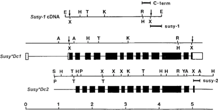
A comparison of the sequence of the Susy-1 cDNA with the sequence of Susy∗Dc1 revealed 14 exons (Figure 1). Like the genes for sucrose synthase from maize [50], Susy∗Dc1 has a long intron in the 50 -non-coding leader sequence (1.2 kb). The exons of Susy∗Dc2 were also deduced from a comparison with the Susy-1 cDNA (Figure 1). Because the 50 -non-coding sequences of Susy-1 and Susy∗Dc2 share no homologies, it is not clear whether the gene for the second carrot sucrose synthase isoform also has a leader intron. The overall gene structures of Susy∗Dc1 and Susy∗Dc2 are very similar (Figure 1). The DNA sequences share about 85% and 45% identity between their exon and intron regions, respectively. In both carrot genes and the Asus1 gene of Arabidopsis [23], the introns 6 and 13 of the Sh1 gene of maize [23] and rice [49] are absent. The deduced amino acid se-quences of the two carrot sucrose synthase isoforms are 87% identical. Their N-termini contain no sig-nal peptide-like sequences but the phosphorylation site Ser-15 and some of the neighboring amino acids of the Sus1 polypeptide of maize are conserved [18].
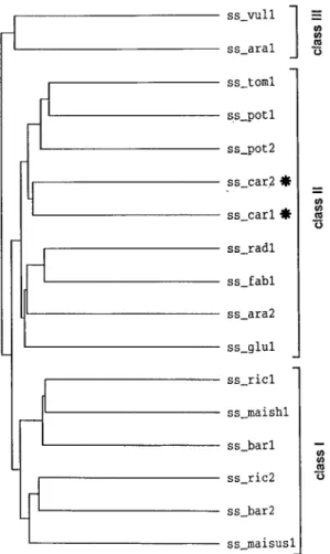
A phylogenetic tree generated from the amino acid sequences of sucrose synthases from carrot and other plant species shows their division into three main classes (Figure 2). Class I consists of enzymes from monocotyledonous plants, which can be subdivided into proteins with high homology to either the prod-ucts of the Sh1 (ss−maish1) or the Sus1 (ss−maisus1) gene from maize [6, 7, 8]. Classes II and III com-prise enzymes from dicotyledonous plants. Class II has numerous members with high sequence identity of about 85%, whereas the sequences belonging to class III diverged more from the sequences of the other two classes; for example the sequences of isoforms I from carrot (ss−car1) and Arabidopsis (ss−ara1) share only 68% identity.
The genome of carrot may code for additional sucrose synthase isoforms with low homology to the cloned genes
To determine whether the carrot genome contains additional sucrose synthase genes, a general probe for sucrose synthase genes and specific probes for
Figure 1. Restriction and structural maps of the genomic subclones for Susy∗Dc1 and Susy∗Dc2. The bottom line represents the scale in kilobases, the upper line the restriction map of the cDNA for Susy∗Dc1. Signs on the restriction maps are as follows: A, HindIII; E, EcoRI; H, HindII; K, KpnI; P, PstI; R, SacI; S, SalI; T, SphI; X, XmnI; Y, XbaI; arrows indicate beginning and end of open reading frames (a cDNA clone for Susy∗Dc2 was not isolated and, therefore, the exons of the gene could only be deduced from the Susy-1 cDNA). On the structural maps of the two sucrose synthase genes, the coding regions are represented by filled boxes with introns shown as lines. The open boxes on the structural map of Susy∗Dc1 mark the 50and 30non-coding regions of the corresponding Susy-1 cDNA. The sequences used as probes are marked by bars above and below the structural maps (C-term, general sucrose synthase probe; susy-1 and susy-2, specific probes for Susy∗Dc1 and Susy∗Dc2, respectively).
Susy∗Dc1 and Susy∗Dc2 were designed. A compari-son of known amino acid sequences of sucrose syn-thases from different plant species identified the C-terminus as a highly homologous region (more than 85% identity). Thus, a 235 bp long fragment of the Susy-1 cDNA (HindII/XmnI) was isolated from the 30 end of the open reading frame (probe C-term, see Figure 1). A comparison of the 30-non-coding regions of the Susy-1 cDNA with the respective re-gion of Susy∗Dc2 revealed only 35% identity and, therefore, a 330 bp long fragment of the Susy-1 cDNA (XmnI/EcoRI) and a 200 bp long fragment of Susy∗Dc2 (XmnI/HindIII) were isolated (probes susy-1 and susy-2, see Figure 1).
Genomic DNA from the carrot cultivar Nantaise was restricted with HindII (Figure 3, lane a), HindIII (lane b), or PstI (lane c), all of which generated fragments harboring both the common sequence (C-term) and the gene-specific sequences (susy-1 and susy-2) on the same fragments. The DNA gel blot hybridized with the common probe (Figure 3, blot C-term) showed one strong signal per lane, corre-sponding to the respective fragments of Susy∗Dc1 (see blot susy-1). A few weaker signals were also visible on blot C-term. By probing an identical blot with the gene-specific probe for Susy∗Dc2 (Figure 3, blot susy-2), most of the weaker signals could be assigned to Susy∗Dc2. Two weak signals in the HindII digest, one weak signal in the HindIII digest, and one weak sig-nal in the PstI digest (Figure 3, blot C-term, bands
marked at their left side with asterisks) could not be assigned to either of the two genes. These hybridiza-tion signals may indicate the presence of one or more additional sucrose synthase gene with low homology to the cloned genes, such as a gene of the sucrose synthase sequence class III (Figure 2).
The two cloned genes for carrot sucrose synthase differ markedly in their expression patterns
In order to identify where and when in the carrot plant the two cloned sucrose synthase isoforms are ex-pressed, gene-specific steady-state mRNA levels were determined in different organs at different develop-mental stages (Figure 4). Transcripts of Susy∗Dc1 were found in young leaves and roots of all devel-opmental stages, as well as in flower buds, flowers, and developing seeds (main form of carrot sucrose synthase). In contrast, mRNA of Susy∗Dc2 was only found in flowers (reproductive form of carrot sucrose synthase).
The main form of sucrose synthase is highly expressed in sucrose utilization sinks
Source leaves, which had not yet reached their final size, were dissected into lamina and six consecutive stem segments (Figure 5, upper panel). Highest lev-els of Susy∗Dc1 transcripts were found in the growing stem (Figure 5, lower panel), indicating a function of sucrose synthase in sucrose utilization for growth.
Figure 2. Comparison of amino acid sequences of plant sucrose synthases. The dendrogram was generated by comparison of the known sequences of plant sucrose synthases by the PileUp pro-gram of the Genetics Computer Group Sequence Analysis Software Package. ss−vul1, Beta vulgaris (X81974); ss−ara1, Arabidopsis thaliana (X60987); ss−tom1, tomato (L19762); ss−pot1, potato (M18745); ss−pot2, potato (U24088); ss−car2 (polypeptide en-coded by Susy∗Dc2), carrot (Y16091); ss−car1 (polypeptide en-coded by Susy∗Dc1), carrot (X75332 and Y16090); ss−rad1, Vi-gna radiata (D10266); ss−ara2, Arabidopsis thaliana (X70990); ss−fab1, Vicia faba (X69773); ss−glu1, Alnus glutinosa (X92378); ss−ric1, rice (Z15028); ss−maish1, maize (X02382); ss−bar1, bar-ley (X65871); ss−ric2, rice (X59046); ss−bar2, barley (X69931); ss−maisus1, maize (L22296). The two carrot sequences for sucrose synthase are marked by asterisks.
Only very low mRNA levels were found in the leaf lamina.
Developing tap roots were also divided into zones with different biological activities. At the tip (Fig-ure 6A, segment 6), the root elongates and thickens (utilization sink for sucrose) and in the upper part (seg-ments 1–5), storage of sugars occurs (storage sink for sucrose). These upper segments also grow in diameter (secondary root growth) but their increase is small in
Figure 3. DNA gel blot analysis of sucrose synthase sequences in the carrot genome. Genomic DNA (10 µg/lane) from cv. Nantaise was digested with HindII (lane a), HindIII (lane b), or SphI (lane c). The fragments were separated by agarose gel electrophoresis and blotted before hybridization with a32P-labelled general probe for sucrose synthase (C-term, left panel), or the gene-specific probes for Susy∗Dc1 (susy-1, middle panel) or Susy∗Dc2 (susy-2, right panel). The hybridization signals obtained with the general sucrose syn-thase probe (C-term), which are indicated by asterisks (left panel), do not correspond to the maps of Susy∗Dc1 or Susy∗Dc2.
comparison with the rate of root elongation at the root tip. Transcripts for the main form of sucrose synthase were found in all root sections, with higher levels in the root tip. Again, these findings suggest a function of the enzyme in sucrose utilization for growth.
Figure 4. Steady-state mRNA levels of Susy∗Dc1 and Susy∗Dc2 in leaves and roots of three different developmental stages and in reproductive organs of carrot plants. Upper panel: northern blot with total RNA (10 µg/lane) from 4-, 10-, and 16-week old leaves, 4-, 10-, and 16-week old roots, and flower buds (B), flowers (F), small developing seeds (Gs), large developing seeds (Gl), and mature
seeds (S). The blot was hybridized with the32P-labelled probe for Susy∗Dc1 (susy-1). Lower panel: an identical blot was hybridized with a32P-labelled probe for Susy∗Dc2 (susy-2).
Figure 5. Steady-state mRNA levels of Susy∗Dc1 in leaf lamina and sections of the stem. A (top). Leaves of about 12-week old plants which had not yet reached their mature size were dissected into lamina and 6 consecutive stem sections. B (bottom). Northern blot with total RNA (10 µg/lane) from leaf lamina (L) and stem sections (1–6; section 6 is a highly branched stem segment, con-necting the numerous leaf lamina). The blot was hybridized with the32P-labelled probe for Susy∗Dc1 (susy-1).
Figure 6. Steady-state mRNA levels of Susy∗Dc1 in sections of de-veloping tap roots. Twelve-week old carrot plants grown in soil in a field were harvested in the middle of a sunny, warm day. The tap roots were washed and divided into 6 equal and consecutive sections (A, left top). B (bottom). Northern blot with total RNA (10 µg/lane) from the six root sections (1–6). The blot was hybridized with the
32P-labelled probe for Susy∗Dc1 (susy-1).
The main form of sucrose synthase is partially membrane-associated, indicating a function of the enzyme in cellulose biosynthesis
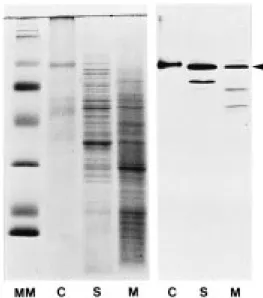
Protoplast from rapidly growing carrot cells were sep-arated into soluble (S) and membrane proteins (M). After the separation of these proteins by SDS-PAGE, the amount of sucrose synthase in the two fractions was analyzed on immunoblots with a polyclonal an-tibody against sucrose synthase from maize, which cross-reacts well with sucrose synthase from carrot [37]. In several independent experiments, 25–50% of the sucrose synthase polypeptide was found associated with the membrane fraction (Figure 7).
Figure 7. Analysis of sucrose synthase in soluble proteins (S) and membrane proteins (M) from carrot protoplasts by immunoblotting. Protoplasts isolated from a carrot suspension culture were homog-enized and membranes harvested by ultra-centrifugation. Soluble proteins (S) from the supernatant (20 µg/lane), membrane proteins (M) from the pellet (20 µg/lane), and soluble proteins (C, con-trol) extracted from a maize leaf (4 µg/lane) were separated on a SDS-polyacrylamide gel (18%) and stained with Coomassie blue (left panel). Proteins from an identical gel were blotted onto ni-trocellulose and carrot sucrose synthase detected with a polyclonal antibody against sucrose synthase from maize (right panel, sucrose synthase is marked by an arrowhead; additional lower molecular weight bands in lanes S and M most likely are due to break-down of the sucrose synthase protein). Prestained marker proteins (Gibco-BRL) were loaded on the left lane (MM).
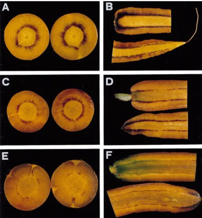
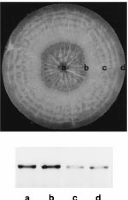
Developing tap roots transiently accumulate starch in the vicinity of their phloem strands, suggesting a function of sucrose synthase in starch biosynthesis Despite the current belief that carrot tap roots are free of starch [31], we detected the β(1,4)glucan by staining hand sections (Figure 8A–F) with iodine. The roots analyzed were from developing plants, harvested on a sunny and warm day and not from mature plants already harvested for consumption. In cross sections, starch was mainly found in a star-like pattern, origi-nating at the cambial ring and extending towards the periderm (Figure 8A). This pattern coincides with the localization of active phloem bundles [3]. In longitu-dinal sections, starch was found all along the cambium from the crown to the tip (Figure 8B).
To test whether starch accumulation in tap roots is transient, developing plants growing in a field were covered with large opaque cardboard boxes for up to three days. During this time, the chlorophyll content of the leaf lamina dropped 25% (data not shown), and
Figure 8. Detection of starch in cross- and longitudinal sections of developing tap roots with iodine. Twelve-week old tap roots were harvested in the middle of a sunny and warm day. The roots were hand-sectioned into 1 mm thick slices (panel A, cross sections from the central region of the root, halfway between crown and tip; panel B, longitudinal section) and immediately stained with Lugolsche solution [29], containing 0.2 g idodine and 0.4 g potassium iodide in 60 ml of water. Plants from the same field were covered with large opaque cardboard boxes and kept in the dark for up to 3 days. At least 10 roots per time point were sectioned and stained. Representative sections of tap roots kept in the dark for 1 night (panels C and D) and 1 night and 2 additional days (panels E and F) are shown.
Figure 9. Steady-state mRNA levels of Susy∗Dc1 in different tis-sues of developing tap roots (top). The middle third of tap roots of 12-week old plants (excluding crown and tip) were dissected into xylem (a), cambium plus a few millimeters of adjacent phloem (b), the remaining phloem (c), and periderm (d). B (bottom). Northern blot with total RNA (10 µg/lane) from the four root tissues. The blot was hybridized with the32P-labelled probe for Susy∗Dc1 (susy-1).
the starch content of the majority of the roots clearly decreased (Figure 8C–F). These results indicate that a constant supply of photoassimilates is required for starch accumulation in the roots, and that the starch pool is rapidly turned over.
To understand whether the tissue, in which starch preferentially accumulates, correlates with high ex-pression of Susy∗Dc1, developing tap roots were di-sected into xylem tissue, cambium plus a few milime-ters of phloem tissue (the starch-accumulating tissue), the remaining phloem tissue, and periderm (Figure 9, upper panel). High transcript levels were found in the starch-accumulating tissue (Figure 9, lower panel). Because sucrose synthase is a key enzyme for starch synthesis in the starch-accumulating storage organs of crop plants such as the endosperm of maize kernels and the tubers of potatoes, the enzyme is most likely also involved in the biosynthesis of the polysaccharide in carrot tap roots.
Figure 10. Response of Susy∗Dc1 to anaerobiosis. Tap roots of plants grown in pots (lane C, control plants,) were submerged in water for 1–3 days (lanes 1–3). Top: northern blot with total RNA (10 µg/lane) from leaves. Bottom: northern blot with total RNA (10 µg/lane) from roots. The blots were hybridized with a
32P-labelled probe for Susy∗Dc1 (susy-1).
The expression of the main form of sucrose synthase is regulated by anaerobiosis
The effect of anaerobiosis on sucrose synthase gene expression was studied by submerging tap roots of developing carrot plants in water for up to 3 days. The steady-state levels of Susy∗Dc1 mRNA increased markedly in response to reduced oxygen levels in tap roots (Figure 10, lower panel) but were only slightly affected in the leaves which were still exposed to air (Figure 10, upper panel). The marked increase of Susy∗Dc1 transcripts in organs with reduced oxygen supply supports the suggested function of the enzyme in providing the stressed tissues with fermentable sugars [13, 22, 33, 34, 43, 52].
The expression of the main form of sucrose synthase is not affected by sugars, inhibition of cellulose biosynthesis, or acetate
Different tissues (young petioles and slices of develop-ing tap roots) and cells (calli and suspension cultures) of carrot were exposed to up to 250 mM of glu-cose, fructose, or sucrose, or to the non-metabolizable sugar mannitol to only increase the osmolarity of the medium for up to 24 h. No alterations of the steady-state mRNA levels for Susy∗Dc1 were found (data not shown).
A negative result was also obtained when carrot cells were treated for up to 24 h with up to 20 µM 2,6-dichlorobenzonitrile, a strong inhibitor of cellu-lose biosynthesis [26]. Furthermore, expression of
Susy∗Dc1 was not altered when carrot cells were ex-posed to up to 20 mM of potassium acetate for up to 24 h (data not shown).
Discussion
Two genes for sucrose synthase (Susy∗Dc1 and Susy∗Dc2) were isolated from carrot. Their structures are very similar and the proteins encoded have highly homologous amino acid sequences. The marked dif-ferences in the sequences upstream of the translation initiation codons coincided with different organ- and development-specific expression patterns. Transcripts of Susy∗Dc2 were only found in flowers (reproductive form of carrot sucrose synthase), whereas mRNA for Susy∗Dc1 was detected in stems, in roots of differ-ent developmdiffer-ental stages, as well as in flower buds, flowers and maturing seeds (main form of carrot su-crose synthase). Thus, the expression patterns of the two carrot sucrose synthase genes are similar to those of the two sucrose synthase genes of maize (Sh1 is endosperm-specific, and Sus1 expressed in leaves, roots, and developing seeds [7]). In contrast, they are clearly different to the expression patterns of the two sucrose synthase genes from potato (3 and Susy-4), expressed either in vascular tissues of leaves and roots (Susy-3), or in the vascular and storage tissue of tubers (Susy-4) [12].
Southern blot analysis with a general probe for sucrose synthase and specific probes for Susy∗Dc1 and Susy∗Dc2 identified a few weak hybridization sig-nals which could not be assigned to the two isolated genes. Thus it is possible that carrot, like some dicote-lydonous species contains genes coding for distantly related forms of sucrose synthase (class III genes, Figure 2).
In developing leaves, the main form of sucrose synthase was only weakly expressed in the lamina. Low transcript levels were also detected in leaves of tomato [48], potato [12] and sugar-beet [16]. This is in contrast to high β-glucurosidase (GUS) activity in phloem bundles of leaves of transgenic plants ex-pressing the GUS gene driven by the sucrose synthase promoter [12], which may be due to the high stabil-ity of the β-glucuronidase polypepetide. Expression of sucrose synthase in companion cells of leaves was interpreted as evidence for a function of the enzyme in the supply of energy for phloem loading in source tissues [23, 28], and may also explain the function of the enzyme in carrot leaf lamina.
Strong expression of Susy∗Dc1 was found in the growing stems with highest transcript levels towards the stem base. This is also where the stem grows most extensively (Arnd Sturm, unpublished observation). High transcript levels were also found in developing tap roots with highest levels in root tips. Here, the root develops and elongates, suggesting again a function of the enzyme in sucrose utilization for growth.
Expression of sucrose synthase from maize [19], bean [15], Arabidopsis [23] and potato [12] is regu-lated by sugars such as sucrose. In carrot as well as in sugarbeet [15] and tomato fruit [30], sugar regu-lation of sucrose synthase gene expression was not detected. In contrast to plants such as maize and potato, the storage organs of carrot, sugar-beet and tomato accumulate high concentrations of sugars and short-term physiological changes will only lead to mi-nor sugar concentration changes, not large enough to efficiently alter the regulation of gene expression. Thus, it is likely that the genes for sucrose synthase from sugar-storing plants are regulated by a different, so far unknown mechanism. Along this line, we tested 2,6-dichlorobenzonitrile, a strong inhibitor of cellu-lose biosynthesis [26] which most likely leads to an accumulation of UDP-glucose, but no effect on carrot sucrose synthase gene expression was found. Like-wise, acetate up to 20 mM did not alter transcript levels, although it is a potent inducer of metabolic repression of photosynthetic genes [38].
Carrot sucrose synthase most likely has sev-eral functions. In growing tissues, the membrane-associated form of the enzyme may feed sucrose di-rectly into cellulose biosynthesis. Originally, a partial association of sucrose synthase with the plasma mem-brane was identified in cotton fibers [1], where sucrose is converted at high rates to cellulose, and in cells of maize endosperm [4]. A model was put forward in which sucrose synthase is bound to the catalytic subunit of cellulose synthase, feeding UDP-glucose directly into the synthesis of the β-glucan [11].
The soluble form of the enzyme may feed su-crose directly into plant metabolism, for example via glucose-1-phosphate, made by pyrophosphorolysis of UDP-glucose [41]. Susy∗Dc1 was strongly induced in tap roots in response to reduced oxygen levels. Thus, bridging sucrose anabolism with catabolism via py-rophosphorolysis seems to operate most efficiently at times of high glycolytic demand, such as anaerobiosis [13, 22, 33, 34, 43, 52].
In plants with starch-accumulating storage organs, sucrose synthase feeds sucrose into the biosynthesis
of α-glucans. The finding that developing tap roots also accumulate starch, suggests a function of sol-uble carrot sucrose synthase in starch biosynthesis. Accumulation of starch in the vicinity of the phloem strands may have two functions. First, starch could be a store for photoassimilates when carbon dioxide assimilation exceeds storage and utilization capaci-ties. Second, biosynthesis of an insoluble polymer directly after unloading from the phloem may gen-erate a steep sucrose concentration gradient requried for rapid transport of sucrose from source leaves into sink organs [17]. This latter hypothesis is strongly supported by preliminary experiments indicating that starch is stored only transiently.
The hypothesis is also supported by our finding that the sucrose proton/cotransporter for the loading of sucrose into the phloem of carrot leaves is regulated in a diurnal fashion (Roshani Shakya and Arnd Sturm, unpublished results). Thus, it may be possible that the bulk of sucrose is transported into the tap roots only during the day and there to a large extent converted into starch. During the night, starch may become de-graded and sucrose resynthesized, which is strongly supported by the transient nature of the starch and a high sucrose phosphate synthase activity in tap roots ([51], Heidi Petersen and Arnd Sturm, unpublished results).
Acknowledgements
We are grateful to Prem Chourey (University of Florida, Gainesville) for the antiserum against sucrose synthase from maize and Inge Obergföll (FMI) for photography. We also thank Tang Guo-Qing, Manfred Heinlein and Patrick King for critical reading of the manuscript.
References
1. Amor Y, Haigler CH, Johnson S, Wainscott M, Delmer DP: A membrane-associated form of sucrose synthase and its po-tential role in synthesis of cellulose and callose in plants. Proc Natl Acad Sci USA 92: 9353–9357 (1995).
2. Bennett MD, Smith JB: Nuclear DNA amounts in an-giosperms. Phil Trans R Soc Lond 274: 227–274 (1976). 3. Caplin SM: Translocation of14C metabolites in carrot root.
Am J Bot 60: 703–707 (1973).
4. Carlson SJ, Chourey PS: Evidence of plasma membrane-associated forms of sucrose synthase in maize. Mol Gen Genet 252: 303–310 (1996).
5. Chopra S, Del-favero J, Dolferus R, Jacobs M: Sucrose syn-thase of Arabidopsis: genomic cloning and sequence charac-terization. Plant Mol Biol 18: 131–134 (1992).
6. Chourey PS: Genetic control of sucrose synthetase in maize endosperm. Mol Gen Genet 184, 372–376 (1981).
7. Chourey PS, Latham MD, Still PE: Expression of two sucrose synthetase genes in endosperm and seedling cells of maize: evidence of tissue specific polymerization of protomers. Mol Gen Genet 203: 251–255 (1986).
8. Chourey PS, Nelson OE: The enzymatic dificiency condi-tioned by the shrunken-1 mutations in maize. Biochem Genet 14: 1041–1055 (1976).
9. Daie J: Phloem loading of sucrose: update and opportunities in molecular biology. Plant Mol Biol Rep 7: 106–115 (1989). 10. Dellaporta SJ, Wood J, Hicks JB: A plant DNA
miniprepara-tion: version II. Plant Mol Biol Rep 1: 19–21 (1983). 11. Delmer DP, Amor Y: Cellulose biosynthesis. Plant Cell 7:
987–1000 (1995).
12. Fu H, Park WD: Sink and vascular-associated sucrose syn-thase functions are encoded by different gene classes in potato. Plant Cell 7: 1369–1385 (1995).
13. Guglielminetti L, Perata P, Alpi A: Effect of anoxia on car-bohydrate metabolism in rice seedlings. Plant Physiol 108: 735–741 (1995).
14. Hawker JS: Sucrose. In: Dey PM, Dixon RA (eds) Biochem-istry of Storage Carbohydrates in Green Plants, pp. 1–51. Academic Press, London (1985).
15. Heim U, Weber H, Baumlein H, Wobus U: A sucrose-synthase gene of Vicia faba L.: expression pattern in developing seeds in relation to starch synthesis and metabolic regulation. Planta 191: 394–401 (1993).
16. Hesse H, Willmitzer L: Expression analysis of a sucrose syn-thase gene from sugar beet (Beta vulgaris L.). Plant Mol Biol 30: 863–872 (1996).
17. Ho LC: Metabolismus and compartmentation of imported sug-ars in sink organs in relation to sink strength. Annu Rev Plant Physiol Plant Mol Biol 39: 355–378 (1988).
18. Huber SC, Huber JL, Liao P-C, Gage DA, McMichael RW Jr, Chourey PS, Hannah LC, Koch K: Phosphorylation of serine-15 of maize leaf sucrose synthase. Plant Physiol 112: 793–802 (1996).
19. Koch KE, Nolte KD, Duke ER, McCarty DR, Avigne WT: Sugar levels modulate differential expression of maize sucrose synthase genes. Plant Cell 4: 59–69 (1992).
20. Kruger JN: Carbohydrate synthesis and degradation. In: Den-nis DT, Turpin DH (eds) Plant Physiology, Biochemistry, and Molecular Biology, pp. 59–76. Longman Scientific & Technical Publishers, Harlow, UK (1990).
21. Laemmli UK: Cleavage of structural proteins during assembly of the head of bacteriophage T4. Nature 227: 680–685 (1970). 22. Maraña C, García-Olmedo F, Carbonero P: Differential ex-pression of two types of sucrose-synthase-coding genes in wheat in response to anaerobiosis, cold shock and light. Gene 88: 167–172 (1990).
23. Martin T, Frommer WB, Salanoubat M, Willmitzer L: Expres-sion of an Arabidopsis sucrose synthase gene indicates a role in metabolization of sucrose both during phloem loading and in sink organs. Plant J 4: 367–377 (1993).
24. McCarty DR, Shaw JR, Hannah LC: The cloning, genetic mapping, and expression of the constitutive sucrose synthase locus of maize. Proc Natl Acad Sci USA 83: 9099–9103 (1986).
25. Messing J: New M13 vectors for cloning. Meth Enzymol 101: 20–78 (1983).
26. Montezinos D, Delmer DP: Characterization of inhibitors of cellulose synthesis in cotton fibers. Planta 148: 305–311 (1980).
27. Murashige T, Skoog F: A revised medium for rapid growth and bioassays with tobacco tissue culture. Physiol Plant 15: 473–497 (1962).
28. Nolte KD, Koch KE: Companion-cell specific localization of sucrose synthase in zones of phloem loading and unloading. Plant Physiol 101: 899–905 (1993).
29. Nultsch W: Mikroskopisch-Botanisches Praktikum, pp. 64ff. Georg Thieme Verlag, Stuttgart/New York (1995).
30. Ohyama A, Ito H, Sato T, Nishimura S, Imai T, Hirai M: Supression of acid invertase activity by antisense RNA modi-fied sugar composition in tomato fruit. Plant Cell Physiol 36: 369–376 (1995).
31. Peterson CE, Simon PW: Carrot Breeding. In: Bassett MJ (ed) Breeding Vegetable Crops, pp. 321–356. AVI Publishing Company, West Port, CT (1986).
32. Prescott A, Martin C: A rapid method for the quantitative as-sessment of levels of specific mRNAs in plants. Plant Mol Biol Rep 4: 219–224 (1987).
33. Ricard B, Rivoal J, Spiteri A, Pradet A: Anaerobic stress in-duces the transcription and translation of sucrose synthase in rice. Plant Physiol 95: 669–674 (1991).
34. Ricard B, VanToai T, Chourey P, Saglio P: Evidence for the critical role of sucrose synthase for anoxic tolerance of maize roots using a double mutant. Plant Physiol 116: 1323–1331 (1998).
35. Salanoubat M, Belliard G: The steady-state level of potato sucrose synthase mRNA is dependent on wounding, anaero-biosis and sucrose concentration. Gene 84: 181–185 (1989). 36. Sambrook J, Fritsch EF, Maniatis T: Molecular Cloning: A
Laboratory Manual, 2nd ed. Cold Spring Harbor Laboratory, Cold Spring Harbor, NY (1989).
37. Šebkova V, Unger C, Hardegger M, Sturm A: Biochemical, physiological and molecular characterization of sucrose syn-thase from Daucus carota. Plant Physiol 108: 75–83 (1995). 38. Sheen J: Metabolic repression of transcription in higher plants.
Plant Cell 2: 1027–1038 (1990).
39. Springer B, Werr W, Starlinger P, Bennett DC, Zokolica M, Freeling M: The shrunken gene on chromosome 9 of Zea mays L. is expressed in various plant tissues and encodes an anaerobic protein. Mol Gen Genet 205: 461–468 (1986). 40. Sturm A: Molecular characterization and functional analysis
of sucrose-cleaving enzymes in carrot (Daucus carota L.). J Exp Bot 47: 1187–1192 (1996).
41. Sung SJS, Xu DP, Black CC: Identification of actively filling sucrose sinks. Plant Physiol 89: 1117–1121 (1989).
42. Sung ZR: Turbidimetric measurements of plant cell culture growth. Plant Physiol 57: 460–462 (1976).
43. Taliercio EW, Chourey PS: Post-transcriptional control of su-crose synthase expression in anaerobic seedlings of maize. Plant Physiol 90: 1359–1364 (1989).
44. Visser RGF, Jacobsen E: Towards modifying plants for altered starch content and composition. Trends Biotechnol 11: 63–68 (1993).
45. von Schaewen A, Stitt M, Schmidt R, Sonnewald U, Willmitzer L: Expression of a yeast-derived invertase in the cell wall of tobacco and Arabidopsis plants leads to accu-mulation of carbohydrate and inhibition of photosynthesis and strongly influences growth and phenotype pf transgenic tobacco plants. EMBO J 9: 3033–3044 (1990).
46. Wang F, Sanz A, Brenner ML, Smith A: Sucrose synthase, starch accumulation, and tomato fruit sink strength. Plant Physiol 101: 321–327 (1993).
47. Wang F, Smith AG, Brenner ML: Isolation and sequencing of tomato fruit sucrose synthase cDNA. Plant Physiol 103: 1463– 1464 (1993).
48. Wang F, Smith AG, Brenner ML: Temporal and spatial expres-sion pattern of sucrose synthase during tomato fruit develop-ment. Plant Physiol 104: 535–540 (1994).
49. Wang M-B, Boulter D, Gatehouse JA. A complete sequence of the rice sucrose synthase-1 (RSs1) gene. Plant Mol Biol 19: 881–885 (1992).
50. Werr W, Frommer WB, Maas C, Starlinger P: Structure of the sucrose synthase gene on chromosome 9 of Zea mays L. EMBO J 4: 1373–1380 (1985).
51. Zamski E, Barnea A: Characterization of sucrose phosphate synthase, sucrose synthase and invertase in roots of two nearly isogenic carrot lines that differ in their capacity to accumulate sucrose. J Plant Physiol 149: 301–306 (1996).
52. Zeng Y, Wu Y, Avigne WT, Koch KE: Differential regulation of sugar-sensitive sucrose synthases by hypoxia and anoxia in-cicate complementary transcriptional and posttranscriptional responses. Plant Physiol 116: 1573–1583 (1998).