HAL Id: hal-01451895
https://hal.archives-ouvertes.fr/hal-01451895
Submitted on 1 Feb 2017
HAL is a multi-disciplinary open access
archive for the deposit and dissemination of
sci-entific research documents, whether they are
pub-lished or not. The documents may come from
teaching and research institutions in France or
abroad, or from public or private research centers.
L’archive ouverte pluridisciplinaire HAL, est
destinée au dépôt et à la diffusion de documents
scientifiques de niveau recherche, publiés ou non,
émanant des établissements d’enseignement et de
recherche français ou étrangers, des laboratoires
publics ou privés.
Adsorption of nucleotides on biomimetic apatite: the
case of cytidine 5’ monophosphate (CMP)
Maela Choimet, Audrey Tourrette, Christophe Drouet
To cite this version:
Maela Choimet, Audrey Tourrette, Christophe Drouet. Adsorption of nucleotides on biomimetic
apatite: the case of cytidine 5’ monophosphate (CMP). Journal of Colloid and Interface Science,
Elsevier, 2015, vol. 456, pp. 132-137. �10.1016/j.jcis.2015.06.021�. �hal-01451895�
To link to this article:
DOI:10.1016/j.jcis.2015.06.021
http://dx.doi.org/10.1016/j.jcis.2015.06.021
This is an author-deposited version published in:
http://oatao.univ-toulouse.fr/
Eprints ID: 16530
To cite this version:
Choimet, Maela and Tourrette, Audrey and Drouet, Christophe Adsorption
of nucleotides on biomimetic apatite: the case of cytidine 5′
monophosphate (CMP). (2015) Journal of Colloid and Interface Science,
vol. 456. pp. 132-137. ISSN 0021-9797
O
pen
A
rchive
T
oulouse
A
rchive
O
uverte (
OATAO
)
OATAO is an open access repository that collects the work of Toulouse researchers and
makes it freely available over the web where possible.
Any correspondence concerning this service should be sent to the repository
administrator:
staff-oatao@listes-diff.inp-toulouse.fr
Adsorption of nucleotides on biomimetic apatite: The case of cytidine 5'
monophosphate (CMP)
Maëla Choimet
a.b.Audrey Tourrette
b'Christophe Drouet
a.*'CJRIMAT Carnot /nstitute, UMR CNRS/INPT/ UPS 5085, University of Toulouse, Ensiacet, 4 allée E. Manso. 31030 Toulouse cedex 4, France
bctRJMAT Carnot / nstitute, UMR CNRS/INPT/UPS 5085, University of Toulouse, Faculté des Sciences Pharmaceutiques, 35 Chemin des Maraîchers, 31400 Toulouse, France
G R A P H I C A L A B S T R A C T A B S T R A C T Keywords: DNA Adsorption Biomimetic apatite Cytidine monophosphate Paleogenetics
Hydroxyapatite
The chemical interaction between DNA macromolecules and hard tissues in vertebrate is of foremost
importance in paleogenetics, as bones and teeth represent a major substrate for the genetic material after
cell death. Recently, the empirical hypothesi s of DNA "protection" over tirne thanks to its adsorption on hard tissues was revisited from a physico-chemical viewpoint. In particular. the existence of a strong
interaction between phosphate groups of DNA backbone and the surface of apatite nanocrystals (mimick-
ing bone/ dentin minerai) was evidenced on an Dexperimental basis. In the field of nanomedicine, DNA
or RNA can be used for gene transport into cells.and apatite nanocarriers then appear promi sing. ln order to shed some more light on interactions between DNA molecules and apatite, the present study focuses on the adsorption of a "mode!" nucleotide, cytidine 5' monophosphate (CMP), on a carbonated biomimetic apatite sample.The follow-up of CMP kinetics of adsorption pointed out the rapidity of interaction with stabilization reached within few minutes.The adsorption isothenn could be realistically fitted to the Sips mode! (Langmuir-Freundlich) suggesting the influence of surface heterogeneities and adsorption cooper- ativity in the adsorption process. The desorption study pointed out the reversible character of CMP adsorption on biomim etic apatite.This contribution is intended to prove helpful in view of better appre- hending the molecular interaction of DNA fragments and apatite compounds, independently of the appli- cation domain, such as bone diagenesis or nanomedicine. This study may also appear informative for researchers interested in the origins of life on Earth and the occurrence and behavior of primitive biomolecules.
*
Corresponding author.E-mail address: christophe.drouet@ensiacet.fr (C. Drouet).
"· j Cytidine monophosphate (CMP) nucleotide
1. Introduction
After the death of an organism, its constitutive cells rapidly break up and release their contents, including DNA (deoxyribonu- cleic acid). The analysis of DNA extracts from body remains has led to much progress in domains such as paleogenetics, paleo-microbiology, animal and vegetal adaptations and even forensic sciences. Among the genetic markers studied are the uni- parental markers (e.g. located on mitochondrial DNA inherited from the mother, or on the Y chromosome inherited from the father), and the biparental DNA markers located on the autosomes. Ali these markers are very important for following evolutionary histories of populations [1] or revealing family relationships within funeral areas burials [2]. Another area of interest related to the analysis of DNA residues is paleo-microbiology. The analysis of lice (head bugs) found on soldiers from the Vilnius battle by Napoleon in 1812 is an illustration of this point application [3].
The development of molecular biology techniques has permit- ted to extract information from mineralized tissues (bone and teeth remains) that are the predominant vertebrates' vestiges found in archeological searches [4]. The study of DNA extracted from hard tissues then permitted to gain information on more and more ancient periods of time. Indeed, DNA seems to be better preserved in hard tissues than in soft tissues [5]. Today, DNA sequences dating of more than 1700000 years can be extracted from skeletal remains [6]. The preservation of DNA in such ancient specimens was at first surprising, because experimental data [7]
only allowed one to expect a priori a much faster DNA degradation. From the better conservation of DNA associated to bone and teeth arose the hypothesis of a possible role played by a specific interaction between DNA and the apatite contained in mineralized tissues. This hypothesis has long been used by paleo-geneticists
(7], but without being actually demonstrated. Recently, a physico-chemical study [8] investigated the interaction between DNA fragments and biomimetic apatite analogous to bone min- eral/dentin. By exploring DNA adsorption and desorption onto apa- tite nanocrystals, the existence of strong chemical links was evidenced: the existence of a clear interaction between phosphate groups of DNA backbone and the surface of apatite nanocrystals was indeed evidenced on an experimental basis [8]. The absence of desorption upon simple dilution was observed (while it was favored upon adding phosphate ions in the medium). The implica- tion of DNA phosphate backbone in adsorption processes was also reported in other studies involving different substrates and for dis- tinct applications [9-12].
In another domain, the adsorption of plasmid DNA or RNA on nanosystems such as colloidal nanoparticles can prove helpful for gene transfection/gene silencing applications [13-17]: since apatite-based nanoparticles have been shown to have a great potential in nanomedicine, the use of DNA- or RNA-loaded apatite nanoparticles for the transportation of extemal genes not naturally expressed by cells appeared indeed as an encouraging route.
DNA, however, is a complex macromolecule with altemating nucleotides organized in double-stranded helicoidal structure, where a nucleotide is the result of the association of a nucleic base (typically Cytosine, Adenine, Guanine or Thymine) with a pentose (deoxyribose) and a phosphate group. With the aim to inspect fur- ther the type of interaction that may occur between DNA and apa- tite, and independently of the application (paleogenetics, nanomedicine, . ..), the present contribution investigates the adsorptive behavior of a "model" nucleotide, cytidine 5'
monophosphate (CMP), onto a biomimetic apatite substrate as pre- viously used (see for example [8]). After summarizing the main characteristics of the apatitic substrate used in this work, the CMP adsorption kinetics and isotherm were investigated, at room
temperature, and desorption was followed in the presence or in the absence of phosphate ions. These results could serve as a com- plementary instructive dataset in future DNA/apatite-related works, independently of the application field.
2. Materials and methods
2.1.Synthesis of the biomimetic apatite substrate
The synthetic sample of carbonated biomimetic apatite used in this work was obtained by precipitation of a calcium nitrate Ca(N03)z-4H20 solution (52.2 g in 750 ml of deionized water) with an ammonium hydrogenphosphate (NH4)zHP04 solution and a sodium bicarbonate NaHC03 solution (90 g of each in 1500 ml of
deionized water), at room temperature (20 °C). In the second solu- tion, an excess of phosphate was used to buffer the pH of the solu- tion at 7.2, close to the physiological values (7.4). The precipitate was then left to mature at room temperature for 1 week (7 days), filtered on Büchner funnel, and thoroughly washed with deionized water. The sample was then freeze-dried for 3 days, sieved in dif- ferent size fractions, and stored at -18°C until further use. In this study, we worked with granulometry fraction below 100 µ m. In the rest of the text, this 1-week matured sample will be referred to as "hac-lw".
2.2. Physico-chemical characterization of the apatite substrate
The physico-chemical characteristics of the hac-1w apatite were detailed in a previous study using several techniques [8]. Briefly, the total calcium and phosphate contents in the apatite phase were determined by complexometry and by spectrophotom- etry (Shimadzu UV 1800, Â. = 460 nm) respectively, while the car- bonate titration was carried out by coulometric method (UIC, !ne. CM 5014 coulometer with CM 5130 acidification unit). The nature of the crystalline phase was determined by powder X-ray diffrac- tion using an INEL diffractometer CPS 120 and the monochromatic Co Ka. radiation (,1. = 1.78892 A). Fourier transform infrared (Ff!R) spectroscopy analyses were achieved on a Thermo-Nicolet 5700 spectrometer with a resolution of 4 cm-1, using the KBr pellet method. A Microtrac BELSORP mini II apparatus (BET method based on nitrogen adsorption ) was used to determine the specific surface area Sw. Zeta potential (determined from electrophoretic mobility measurements) was determined at 25 °C using a Malvern Zetasizer Nano Analyzer apparatus. The apatitic substrate was studied after dispersion of the powder in water set at different pH values in the range 6-9.5 and filtration on micropore filter for avoiding the sedimentation of large aggregates (>1 µm) during the analyses.
2.3. Cytidine 5' monophosphate ( CMP) adsorption and desorption
Cytidine 5' monophosphate, in the form of di-sodium sait, was purchased from Sigma Aldrich; its chemical formula is given in
Fig. 1. For ail experiments of adsorption or desorption, 20 mg of apatite powder (0 < 100 µm) was dispersed in 5 ml of CMP solu- tions at room temperature. The CMP concentration present in the supernatant was evaluated using spectrophotometry (Shimadzu UV 1800, Â. = 272 nm). At any time, the adsorbed amount could be determined by difference of the initial quantity introduced in the medium and the residual amount in solution.
Adsorption kinetics was investigated by following the amount of adsorbed CMP versus time. For these experiments, a CMP solu- tion of 300 mg/l in I<Cl solution (0.01 M) was used, at room tem- perature and physiological pH. Different contact times ranging from 5 min to 3 h were followed so as to evaluate the adsorption
v,(Pq) 0 (a) 25000 ::l .l!l c: 20000 15000 ::l 10000 ü 5000 0 20 (b) 30 40 50 2 theta (°) 60 70
Fig. 1. Chemical formula of cytidine 5' monophosphate (CMP) di-sodium sait. 0.6 v v,(CO,) +>iHPC{)
equilibrium of CMP on the apatite substrate.After the selected con- tact time, the mixture was centrifuged for 2 min at 5000 rpm and the supernatant was withdrawn and analyzed by spectrophotome-
try as indicated above. The CMP adsorption isotherm was then
built by following (after 1h of contact time) the adsorbed amount
versus the equilibrium concentration in solution, varying from 0 up to 1200 mg/!.
When mentioned in the text. the amount of phosphate and cal-
cium ions present in the supernatant after adsorption were respec-
tively evaluated by spectrophotometry and atomic absorption
(Analytik jena ContrAA300, N20/acetylene flame, .1. = 422.67 nm).
The eventual desorption of CMP was investigated for some dat-
apoints corresponding to the isotherm (250, 600 and 1000 mg/L).
After 1 h of adsorption, a known amount of supernatant was with-
drawn and replaced by KCI solution (0.01 M). After the same
immersion time, the solution was analyzed by spectrophotometry.
The effect of an addition of phosphate ions on the release of CMP
was also followed by the addition of KH2P04 at a final concentra-
tion of 18 mM.
3.Results and discussion
3.1.Physico-chemical characterization of apatite substrate
As mentioned above, the biomimetic apatite substrate, hac-1w,
used in this work was previously characterized (see [8] for details);
for the sake of completeness however its main features are sum- marized hereafter.
The apatitic nature of the sample was confirmed by powder
X-ray diffraction (XRD) analysis, complemented by
Fourier-Transform lnfrared (FTIR) spectroscopy. ln particular, the XRD
pattern showed the absence of detectable secondary phases (Fig.
2a), and the biomimetic character of this sample can be assessed
by the similarity of crystalline features to those of bone specimens (see Supporting Information, Fig. SI 1). The broadness of the diffraction peaks can be attributed bath to the existence of
microstrains in the apatitic network and to nanosized constitutive
crystals. The application of Scherrer's formula to diffraction lines
(002) and (310) suggested, in a first approximation, a mean crys-
tallite length of around 16 nm and a mean width/depth close to
5 nm, underlining the nanometer dimensions of this hac-1w sam-
ple, similarly to those of bone minerai [18]. The specific surface
area of the granulometry fraction selected in this work reached
122 m2/g. For additional information on the physic-chemical char-
acteristics of the apatitic substrate used in this work, its zeta
potential was also measured, in a wide pH range from 6 to 9.5. It
was found to be negative throughout this pH range, and close to -30 ± 2 mV. Such negative values are consistent for such
Wavenumber (cni1)
Fig. 2. (a) XRD pattern (2 theta between 20° and 70°) of the biornirnetic carbonated
apatite sample hac-lw (maturation of 1 week) used in this work, with ( hk l)
indexation after JCPDS file #09-432 relative to hydroxyapatite, and (b) FTIR spectrum for hac-1w with phosphate, carbonate and water band attributions.
nonstoichiometric nanocrystalline apatites as previously reported [19]. Note however, that this measurement only gives a global value of the surface charge and does not imply that only negatively charged surface sites are present as bath calcium and phosphate (protonated) ions coexist.
The carbonation of this sample was confirmed by the presence
of carbonate bands observable by FTIR (Fig. 2b), in the ranges
1350-1550 cm-1 (v3(C03) domain) and 840-910 cm-1 (v2(C03)
domain). Bath direct coulometric measurements and FTIR carbon-
ate titration using a recently reported method (20] revealed a car-
bonate amount of 3.6 wt.%. The Ca/( P + C) overall molar ratio
determined by complementary chemical titrations led to 1.36.
This value, noticeably Iower than 1.67 (characteristic of stoichio- metric hydroxyapatite), points to the nonstoichiometry of this
compound, as for bone samples (18], confirming again the biomi-
metic nature of this apatite compound.
The above results thus substantiate the choice of this apatite
sample for the present study, aiming at inspecting the interaction of the CMP nucleotide with a substrate close to bone minerai. 3.2.Investigation of CMP adsorption and desorption
CMP adsorption onto hac-1w was investigated here in two
ways. First, the sorption kinetics was followed by varying the
CMP/apatite contact time, so as to determine the conditions of
achievement of thermodynamic equilibrium. Then, for a selected contact time, the adsorbed amount was measured for increasing CMP concentrations in solution, in order to define the adsorption
isotherm (at room temperature ).
3.2.1.Adsorption kinetics
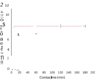
Fig. 3 shows the adsorption kinetics of CMP on hac-lw (for an
initial concentration of 300 mg/l), by plotting the amount of CMP
adsorbed as a function of contact time. The thermodynamic equi-
librium was reached very quickly after Jess than 10 min.
In the present
ê
-0 1+
'
2
12 a. Cil 10 CJ) Ôl Q.ds = B ·Ln(A)+
B ·Ln(Ceq ) Temkin (3)where KL. Nm.Kf,n,A and B are the related constants, at a given tem- perature. The application of these models to the experimental data led to the following correlation parameters (adjusted R2): 0.9916,
.
o.s
..8 ,--- --- 1--- -t 0.9637 and 0.9453. These findings suggest that pure Freundlich or
ô
1 "'a
6 ë :::i 4 Cil1l
2 0 (/)Temkin adsorptive behaviors cannot adequately explain the exper- imental variation observed here. Instead, an improved fit was found with the Langmuir equation (with = 32.33 ± 2.41 mg/g (0.82 ± 0.06 µmol/m2 ) and KL = 0.0017 ± 0.0002 1/mg). A recent study on the adsorption of tetracycline on biomimetic apatite [23) however pointed out the interest to test also the adequacy of another mode!, known as Sips equation [24,25) (also known as "L
o ,._
0 20 40 60 80 100 120 140 160 180 200
Contactlime (min)
Fig. 3. Kinetics of adsorption of CMP onto biomimetic apatite hac-1w (room
angmuir-Freundlich" isotherm) which appears as a modified ver-sion of the Langmuir mode! by incorporating the exponent "m" as follows:
Ks ·
c
:q
temperature). Q.cts,e = Qm · K
cm
Sips isothermS · eq
(4)
corresponding to an adsorbed amount of ca. 7.5 mg/g of apatite (0.19 µ mol/m2 ).
Taking into account the rapidity of this adsorption process, probably painting to a high affinity of CMP molecules for the sur- face of biomimetic apatite, it was not possible to draw advanced conclusions relatively to the type of kinetic mode!.
3.2.2.Adsorption isotherm
The adsorption isotherm of CMP on apatite is shown in Fig. 4. The shape of the plot indicates a progressive rise of the adsorbed amount (Q,ctsl for increasing equilibrium concentrations (Ceq). ln order to explore further the adsorption mechanism implied in the CMP/biomimetic apatite system, the experimental datapoints were then tentatively fitted to various mathematical models often encountered in adsorption studies, including with apatite sub- strates (e.g. [8,21,22]).
The models of Langmuir, Freundlich and Temkin were the first to be tested; being respectively described by the following equations:
The application of this equation to our experimental points led to the adjusted R2 correlation parameter 0.9933, indicating that
this mode! is the most relevant , among the four tested, to describe adequately the obtained datapoints. The corresponding Sips parameters are m = 1.20 ± 0.11, = 26.1± 2.6 mg/g (0.66 ± 0.07 µ mol/m2 ), and Ks = 0.00052 ± 0.00027. In particular, the value of "m", noticeably greater than unity, confirms that the isotherm should not be considered as fully Langmuirian.The max- imal adsorbed amount (monolayer coverage in our experimen- tal conditions) thus reaches about 26.1 mg/g (0.66 µ mol/m2 ), which is equivalent, in micromoles per gram, to 80.8 µ mol/g. This value is roughly of the same order of magnitude as the one found for tetracycline on a 1-day matured [23) (the concept of maturation of nanocrystalline apatites being explained elsewhere [26]) carbonated apatite sample ( 82 µ mol/g). On the other hand, it is difficult to directly compare this maximal adsorbed amount to that found for much larger macromolecules such as DNA (of the order of0.49 µ mol/g [8]), due to very different molec- ular sizes and dynamics. Nonetheless, the adsorption of CMP mole- cules on apatite probably involves at least an interaction via the exposed phosphate end-group which is known to have a high affin-Langmuir
Qads = KF · C!" Freundlich
Concentration of CMP at equilibrium (mmol/I)
(1)
(2)
ity for apatitic surfaces, as was also suggested by other studies (e.g. bisphosphonates [27,28) or DNA [8)).
The value of "m" in Sips equation is also informative on the type of interaction existing in the adsorbent/adsorbate system, as was discussed previously [25): it was in particular related to the exis- tence of lateral interactions between adsorbed molecules (which is not considered in the Langmuir theory). For m > 1, "positive"
24
0.5 1.0 1.5 2.0 2.5 3.0 3.5 4.0 4.5
20
2
---1cooperativity between adjacent molecules is hypothesized, while m < 1 would correspond to "negative" cooperativity. In the present case, m 1.20 could thus suggest a favoring role of adsorbed mole- cules on the adsorption process. The fact that Sips isotherm fits
a. 16 Cil o... ,,, 12 , ü CJ) /,j'
---·
better the experimental data as compared to Langmuir may also highlight the heterogeneous nature of the surface of apatites.
The Sips constant Ks also carries some thermodynamic meaning as it was proposed to be linked to the change in standard Gibbs free
"
,
.
.s
8 /a""'
•
Qads, experimentalenergy of adsorption through the following equations [24) (with K5 recalculated with Ceq expressed in mol/!):
4
•
,f Fit with Sips adsorption model (5)0
0 200 400 600 800 1000 1200 1400
Concentration of CMP at equilibrium (mg/1)
Fig. 4. CMP adsorption isotherm (-20 °C) on hac-1w biomimetic apatite (concen- trations given in mmol/l and in mg/l).
case, this leads to the negative value
Gacts0 -19 kj/mol (uncertainty estimated to ±1 kj/mol ), indicat-
ing a favorable adsorption of CMP on biomimeti c apatite in stan- dard conditions. For information, this value is of the same order as the one (-22 kj/mol) found for tetracycline adsorption [23).
•
•
•
a,...
In order to inspect further the adsorption mechanism, the
amounts of calcium and phosphate ions potentially released in
the medium after adsorption were measured. lndeed, previous
studies have shown that the adsorption of some molecules, such
as bisphosphonates [21,29], were accompanied by the concomitant
release of ions from apatite nanocrystals surfaces, leading to a physical "anchoring" of the molecule on the substrate. ln the pre-
sent case, the titration of Ca and P in the supernatants showed however their presence only in small quantities after adsorption.
After subtraction of the amounts of calcium and phosphate ions naturally released due to the partial dissolution of the apatite sub- strate in the medium (as any ionic compound ), it is then possible to examine whether any additional release is observed due to the adsorption process itself. For <lacts varying from 0 to 24 mg/g (see isotherm Fig. 4; corresponding in moles to the range 0-75 µ mol/g), the amount of phosphate released remained low and comprised
between 0 and 6 µ mol/g, while calcium was found to vary around
a mean position of 1OO µ mol/g. No clear trend was observed in function of the adsorbed amount. Thus, these findings suggest that
the adsorption of CMP on nanocrystalline apatite is not accompa- nied by a significant simultaneous release of surface ions.
3.2.3. Desorption study
In continuation with the adsorption study, the possible desorp- tion of adsorbed CMP molecules was followed by dilution of the medium (see dilution details in experimental section). Severa( des-
orption points were examined, corresponding to various degrees of
surface coverage, as shown in Fig. 5. On this figure, the "hypothet- ical" points calculated by supposing the absence of desorption upon dilution are indicated in parentheses , for information. For each point studied, dilution of the medium led, in contrast, to par- tial CMP desorption as marked by the curved arrows. ln the
assumption that this adsorption process would be fully reversible, desorption should occur in a way that the point would follow the adsorption isotherm in the opposite way. ln the case of nanocrys- talline apatites, however, the adsorption of some molecules was found to be irreversible upon dilution; and this observation was
related to a concomitant ion release from the surface of the nanocrystals: for example, the adsorption of bisphosphonates cited above was shown to occur simultaneously to the release of surface phosphate ions, with no clear release of calcium, thus leading to an
"anchoring" of the molecule on the nanocrystal surface, in a "sur- face phosphate" position.
ln the present case, the experimental desorption of CMP,
another phosphate-bearing molecule, (see square "D" datapoints in Fig. 5) are found to clearly approach the isotherm (although
without being strictly superimposed to it); therefore one can
conclude that the CMP/apatite adsorption process is for the most part "reversible" upon simple dilution. This conclusion seems to
be corroborated by the fact that the addition of phosphate ions
in the diluted medium (in the form of dissolved KH2P04 sait, see
star
"
*
"
datapoints in Fig. 5) does not promote desorption. Also, this is in good agreement with our above results (see previous sub- section) indicating that the adsorption process was not accompa- nied by a significant phosphate release in the supernatant.3.3. Concluding remarks
In this contribution, the adsorption of a nucleotide, CMP, onto a nanocrystalline apatite substrate mimicking bone minerai was investigated in detail. The adsorption isotherm, established for a contact time permitting to reach equilibrium, was found to follow a Sips (Langmuir-Freundlich) mode!, and data analysis suggested a cooperative adsorption of CMP molecules. The study of the desorp- tion upon dilution and the analysis of supernatants allowed us to insinuate that CMP adsorption on nanocrystalline (biomimetic) apatite was highly reversible and not accompanied by a concomi- tant ion release from the apatite substrate (despite the presence of a terminal phosphate group on the molecule). This type of analysis, on a single nucleotide - forming one "building black" of DNA or RNA macromolecules - will have to be extended to polynucleotide
subunits and compared to data recently obtained on the adsorp-
tion of DNA on biomimetic apatite [8]. Nonetheless, it already pro-
vides some additional data in view of better comprehending the interaction between apatite compounds and DNA, e.g. in post- mortem settings (study of bone diagenesis) or for nanomedicine applications.
By extrapolation, this work may also appear informative for
researchers interested in the origins of life on Earth, where the
occurrence and preservation of primitive biomol ecules such as nucleotides is of prime relevance. Indeed, as in the case of DNA (see Ref. [8]), the adsorption of nucleotides onto minerai substrates
constitutive of the Earth's geochemistry such as apatites (e.g. pre- sent in metamorphic rocks [30] of pelitic, carbonate, basaltic, and ultramafic composition) may provide protection against premature degradation by environmental factors. Also, in the adsorbed state,
nucleosides/nucleotides may then undergo surface reactions (pos-
sibly catalytic) to promote chemical modifications and/or their combination into polynucleotides of biological relevance [31].
Therefore, better apprehending adsorption processes involving
nucleotides and minerais like apatites could participate to better understanding how "primitive" biomolecules succeeded to evolve toward more elaborate systems.
Acknowledgment 20 18
2
16 ro (o)-- -- -- -- -- ---=--=--=- -•.,
.
, ,
;
/ /
The authors wish to thank the CIRIMAT carnot Institute for
internai funding of Maela Choimet's PhD thesis.
Appendix A. Supplementary material
o.. 14
0) 12
0::
10
ü
---Supplementary data associated with this article can be found, in the online version, at http://dx.doi.org/ 10.1016/j.jcis .2015.06.021.
0) 8
,./
---experirœntal.§., 6
"""'
o ,.;; -·---- Fit with Sips adsorption model References
a
"' 2•
/
,.
•
.,
o Experimental desorption point
<*) Hypothetical point v.ithout desorption, v.ithKH,PO, [1] C. Keyser, C. Bouakaze, E. Crubezy, V.G. Nikolaev, D. Montagnan, T. Reis, B.
*
Experimental desolption point,v.ith KH,PO, 00 100 200 300 400 500 600 700 800
CMP concentration alequilibrium (mg/I)
Fig.5. Desorption study upon dilution, and effect of the addition of phosphate ions in the medium (KH2P04).
Ludes, Hum. Genet. (2009) 126.
[2] C. Keyser-Tra cqui, E. Crubezy, B. Ludes, Am. j. Hum. Genet. (2003) 73. [3] D. Raoult, O. Dutour, L. Houhamdi, R.jankauskas, P.E. Fournier, Y.Ardagna, M.
Drancourt, M. Signoli, V.D. La, Y. Macia, R. Aboudharam, j. Infect. Dis. (2006) 193.
[4] E. Hagelberg, B. Sykes, R. Hedges, Nature ( 1989) 342.
[5] P. Gill, P.L. Ivanov, C. Kimpton, R. Piercy, N. Benson, G. Tully, 1. Evett, E.
Hagelberg, K. Sullivan, Nat. Genet. 6 (1994).
,•
(o)- --·--;--·• (o) Hypothetical point without desorption 4
(6] L. Orlando, A. Ginolhac, G. Zhang, D. Froese, A. Albrechtsen, M. Stiller, M. Schubert, E. Cappellini, B. Petersen, 1. Moltke, P.L.F. Johnson, M. Fumagalli, j.T. Vilstrup, M. Raghavan, T. Korneliussen, A.-S. Malaspinas,]. Vogt, D. Szklarczyk, C.D. Kelstrup, J. Vinther, A. Dolocan, J. Stenderup, A.M.V. Velazquez, j. Cahill, M. Rasmussen, X. Wang, j. Min, G.D. Zazula, A. Seguin-Orlando, C. Mortensen, K. Magnussen, J.F. Thompson, J. Weinstock, K. Gregersen, K.H. Roed, V. Eisenmann, Cj. Rubin, D.C. Miller, D.F. Antczak, M.F. Bertelsen, S. Brunak, K.A.S. Al-Rasheid, O.Ryder, L. Andersson , j. Mundy, A. Krogh, M.T.P. Gilbert, K. Kjaer, T. Sicheritz-Ponten, L.j. Jensen, j.V. Olsen, M. Hofreiter, R. Nielsen , B. Shapiro, J. Wang, E. Willerslev, Nature 499 (2013) 74-78.
[7] T. Lindahl, Nature 362 (1993).
[8] A. Grunenwald, C. Keyser, A.M. Sautereau, E.Crubezy, B. Ludes, C. Drouet, Appl. Surf. Sei. 292 (2014) 867-875.
(9) B. Liu,]. Liu, Langmuir 31 (2015) 371-377.
(10] X. Zhang, F. Wang, B. Liu, E.Y. Kelly. M.R. Servas, ]. Liu, Langmuir 30 (2014)
839-845.
(11] B. Liu,]. Liu, Chem. Commun. 50 (2014) 8568-8570.
(12] R. Pautler, E.Y. Kelly, P.-j.J. Huang, J. Cao, B. Liu, J. Liu, ACS Appl. Mater.
Interfaces 5 (2013) 6820-6825.
(13] T. Welzel, 1. Radtke, W. Meyer-Zaika, R. Heumann, M. Epple, j. Mater. Chem. 14
(2004) 2213-2217.
[14] S. Hossain, A. Stanislaus, M.J. Chua, S. Tada, Y.-1. Tagawa, E.H. Chowdhury, T.
Akaike, j. Contrai. Release 147 (2010) 101-108.
(15] S. Hossain, S. Tada, T. Akaike, E.H.Chowdhury, Anal. Biochem.397 (2010) 156- 161.
(16] E.H. Chowdhury, T. Akaike, Curr. Anal. Chem. 1 (2005) 187-192.
[17] E.H. Chowdhury, Expert Opin. Drug Deliv. 4 (2007) 193-196.
(18] A. Grunenwald, C. Keyser, A.M. Sautereau, E.Crubezy, B. Ludes, C. Drouet, Anal. Bioanal. Chem. 406 (2014) 4691-4704 .
(19] 1. Rodriguez-Ruiz, ]. Manuel Delgado-Lapez, M.A. Duran-Olivencia , M. Iafisco,
A. Tampieri, D. Colangelo, M. Prat, J. Gomez-Morales, Langmuir 29 (2013) 8213-8221.
(20] A. Grunenwald, C. Keyser, A.M. Sautereau, E. Crubézy, B. Ludes, C. Drouet, ]. Archaeol. Sei. 49 (2014) 134-141.
(21] P. Pascaud, P. Gras, Y. Coppel, C. Rey, S. Sarda, Langmuir 29 (2013) 2224-2232.
(22] L. Benaziz, A. Barroug, A. Legrouri, C. Rey, A. Lebugle, ]. Colloid Interface Sei. 238 (2001 ) 48-53.
(23] S. Cazalbou, G. Bertrand, C. Drouet,]. Phys. Chem. B 119 (7)(2015) 3014-3024.
(24] Y. Liu, Sep. Purif. Technol. 61 (2008) 229-242.
[25] LI<. Koopal, W.H. Vanriemsdijk,j.C.M. Dewit, M.F. Benedetti,].Colloid Interface Sei. 166 ( 1994) 51-60.
(26] N. Vandecandelaere, C. Rey, C. Drouet, J. Mater. Sci.-Mater. Med. 23 (2012) 2593-2606.
[27] P. Pascaud, F. Errassifi, F. Brouillet, S. Sarda, A. Barroug, A. Legrouri, C. Rey,]. Mater. Sci.-Mater. Med. 25 (2014) 2373-2381.
[28] F. Errassifi, S. Sarda, A. Barroug, A. Legrouri, H. Sfihi, C. Rey, J. Colloid Interface
Sei. 420 (2014) 101-111.
(29] A. Al-Kattan, F. Errassifi, A. Sautereau, S. Sarda, P. Dufour, A. Barroug, 1. Dos
Santos, C. Combes, D. Grossin, C. Rey, C. Drouet, Adv. Eng. Mater. 12 (2010) B224-B233.
[30] F.S.Spear,j.M. Pyle, Apatite, monazite, and xenotime in metamorphic rocks, in:
MJ. Kahn, ]. Rakovan, j.M . Hughes (Eds.), Phosphates: Geochemical, Geobiological, and Materials Importance, vol. 48, Reviews in Mineralogy and Geochemistry, Mineralogical Society of America, Washington, D.C., 2002, pp.
293-335.