HAL Id: tel-02414560
https://tel.archives-ouvertes.fr/tel-02414560
Submitted on 16 Dec 2019HAL is a multi-disciplinary open access archive for the deposit and dissemination of sci-entific research documents, whether they are pub-lished or not. The documents may come from teaching and research institutions in France or abroad, or from public or private research centers.
L’archive ouverte pluridisciplinaire HAL, est destinée au dépôt et à la diffusion de documents scientifiques de niveau recherche, publiés ou non, émanant des établissements d’enseignement et de recherche français ou étrangers, des laboratoires publics ou privés.
Sen1-mediated RNAPIII transcription termination
controls the positioning of condensin on mitotic
chromosomes
Julieta Rivosecchi
To cite this version:
Julieta Rivosecchi. Sen1-mediated RNAPIII transcription termination controls the positioning of condensin on mitotic chromosomes. Cellular Biology. Université de Lyon, 2019. English. �NNT : 2019LYSEN041�. �tel-02414560�
3
Acknowledgements
Firstly, I would like to express my gratitude to my supervisor Vincent Vanoosthuyse for his dedicated and patient guidance, and also the knowledge and scientific rigour he transmitted to me throughout these four years of work.
I would like to extend my sincere thanks to past and present members of the Lab: Pascal Bernard, Esther Toselli, Léonard Colin, Xavier Robellet and Clémence Hocquet for their support, enriching discussions and personal advice.
I would like to thank my thesis committee: Cyril Bourgeois and Gaëlle Legube for their helpful comments and encouragement during our two meetings. I also thank the directors of the BMIC doctoral school: Françoise Monéger and Mathias Faure for their assistance.
I acknowledge Marc Larochelle, Frédéric Grenier and François Bachand for contributing with valuable genome-wide data to our shared publication. I thank also Amélie Malapert and Camille Teste for their participation in the publication. I would like to give special thanks to all members of the Emiliano Ricci Lab for their constant and insightful suggestions.
I am also pleased to thank the PhD jury members: Stéphane Marcand, Kiran Padmanabhan, Francesca Palladino, and Damien Hermand and Armelle Lengronne for taking the time to revise this thesis.
I am truly grateful to all those who gave me their humanity and helped me keep things in perspective.
5
Table of contents
Acknowledgements
... 3Preface
... 9Introduction
... 13I. Genome architecture changes throughout the cell cycle ... 13
A) Genome organization in interphase ... 13
B) Genome reorganization in mitosis ... 17
i. Chromosomes are individualized and compacted in mitosis ... 17
ii. Mitotic chromosomes are organized into arrays of consecutive loops ... 17
C) Chromatin loops are different between interphase and mitosis ... 21
II. The SMC complexes organize the chromosomes in the nuclear space ... 23
A) Discovery of SMC ... 23
B) Structure of SMC complexes ... 25
C) Prokaryote SMC ... 27
D) Eukaryote SMC ... 28
i. The SMC5/6 complex ... 29
ii. The cohesin complex ... 29
iii. The condensin complex ... 30
a) The interaction of condensin with DNA ... 32
b) The activities of condensin in vitro ... 32
E) The loop extrusion model to explain SMC-driven chromatin loop formation ... 34
F) How could cohesin and condensin in interphase and in mitosis form different types of chromatin loops? ... 38
G) Condensin function in the context of chromatin ... 41
i. Genomic distribution of condensin on chromosomes ... 41
ii. Loading of condensin on chromosomes ... 45
iii. Translocation of condensin on chromosomes ... 46
iv. How does gene transcription contribute to position condensin on mitotic chromosomes?48 III. Genetic experiments indicate that the transcription-associated factors Sen1 and Swd22 act as negative regulators of condensin ... 50
6
Materials and Methods
... 55I. Fission yeast strains and culture ... 55
II. Genetic methods ... 55
A) Crosses ... 55
B) Tetrads dissection ... 55
III. Mutagenesis ... 56
A) SPCTRNAARG.10 and SPCTRNATHR.10 mutants ... 56
B) sen1-G1534D and rpc37-V189D mutants ... 56
IV. Chromatin Immunoprecipitation (ChIP) ... 57
V. Immunofluorescence (IF) ... 57
VI. Western Blot ... 59
VII. Strand specific and random hexamers RT-qPCR ... 59
VIII. Northern blot ... 59
IX. Mapping of the 3’ end of read-through transcripts at SPATRNAPRO.02. ... 60
X. Transcription termination assay ... 60
Chapter 1: The RNAPIII-associated factor Sen1 regulates the accumulation of
condensin in the vicinity of RNAPIII-transcribed genes.
... 671.1. Introduction ... 67
1.1.1. Identification of negative regulators of condensin that function in transcription termination ... 67
1.1.2. The concomitant deletions of Sen1 (sen1∆) and Swd22 (swd22∆) result in the increased accumulation of condensin around some RNAPIII-transcribed genes ... 68
1.1.3. The accepted model to explain the accumulation of condensin at RNAPIII-transcribed genes: direct recruitment by the transcription factors TFIIIC and Tbp1. ... 68
1.1.4. An alternative model: condensin recognizes one or more transcription-associated chromatin feature(s) ... 71
1.2. Results ... 73
1.2.1. Synchronization methods ... 73
1.2.2. Sen1 antagonizes the accumulation of condensin specifically at RNAPIII-transcribed genes in mitosis. ... 75
7
1.2.3. Lack of other proteins related to Sen1 function does not impact the distribution of
condensin ... 77
1.2.4. Condensin accumulation at RNAPIII-transcribed genes cannot be explained by increased TFIIIC or Tbp1-dependent recruitment ... 79
1.2.5. Sen1 acts in –cis at RNAPIII-transcribed genes to regulate the positioning of condensin .. 81
1.2.6. Topological stress accumulates around RNAPIII-transcribed genes in the absence of Sen1 in G2 and in mitosis ... 83
1.3. Discussion ... 85
1.3.1. Is the regulation of condensin by Sen1 dependent on RNAPIII transcription? ... 85
1.3.2. Could the accumulation of condensin specifically at RNAPIII-transcribed genes in the absence of Sen1 explain the suppressor effect? ... 86
1.3.3. Condensin does not accumulate in WT cells at RNAPIII-transcribed genes in fission yeast 87
Chapter 2: Sen1 is required for robust RNAPIII transcription termination
... 892.1. Introduction ... 89
2.1.1. Function of Senataxin/Sen1 in RNAPII transcription termination in human and in S. cerevisiae ... 89
2.1.2. Function of Sen1 in RNAPIII transcription termination in S. pombe ... 92
2.2. Results ... 93
2.2.1. Publication: Senataxin homologue Sen1 is required for efficient termination of RNA polymerase III transcription ... 93
2.2.2. The helicase activity of Sen1 is required for efficient RNAPIII transcription termination .. 95
2.2.3. Sen1 dissociates RNAPIII molecules that override the primary terminator ... 97
2.2.4. Investigating the transcription termination defects in the RNAPIII mutant rpc37-V189D . 97 2.3. Discussion ... 99
2.3.1. The role of Sen1 in transcription termination is conserved ... 99
2.3.2. Does Sen1 act on both RNAPIII termination and RNAPIII elongation? ... 101
8
Chapter 3: The control of RNAPIII transcription termination by Sen1 determines
condensin positioning in the vicinity of RNAPIII-transcribed genes
... 1053.1. Introduction ... 105
3.1.1. Transcription as a positioning factor for cohesin ... 105
3.1.2. Transcription interferes with translocation of bacterial SMC ... 106
3.1.3. Which feature of RNAPIII transcription controlled by Sen1 could impact condensin? ... 106
3.2. Results ... 107
3.2.1. RNAPIII accumulates on its target genes in the absence of Sen1 in mitosis ... 107
3.2.2. The accumulation site of condensin coincides with the peak of the RNAPIII domain in sen1∆ ... 107
3.2.3. RNAPIII that readthrough the primary terminator in the absence of Sen1 are responsible for positioning condensin 3’ of RNAPIII-transcribed genes... 109
3.2.4. Readthrough transcription at RNAPIII-transcribed genes is not sufficient to impact condensin ... 111
3.2.5. Cohesin accumulates at RNAPIII-transcribed genes in the absence of Sen1 ... 113
3.2.6. Introducing 10 Reb1 sites downstream of a tRNA gene is not sufficient to impact condensin ... 113
3.3. Discussion ... 115
3.3.1. Speculative model: High RNAPIII density over tRNA loci in the absence of Sen1 creates a barrier for condensin ... 116
3.3.2. Nucleosomes as barriers for SMC loading ... 118
3.3.3. CTCF as directional barriers to cohesin ... 119
3.3.4. The telomere-binding factor Rap1 as a condensin stalling factor ... 120
3.3.5. The permeable moving barrier model from bacterial SMC to explain RNAPIII/condensin interactions in the Sen1 mutant ... 121
3.3.6. Can we gain insights into condensin function from condensin positioning? ... 124
Conclusion
... 1259
Preface
The three-dimensional arrangement of the genome in the nucleus plays a fundamental role in regulating its activities (Fraser and Bickmore, 2007). Disruption of genome architecture is associated with genome instability such as mutations, chromosomal rearrangements and aneuploidy (Fudenberg et al., 2011). These abnormalities can ultimately lead to cancer (Corces and Corces, 2016). The study of genome architecture is a very dynamic and active field of research aiming to understand the relationship between nuclear organization and genome function.
At each cell division, the genome undergoes a dramatic reorganization to form highly compacted and individualized chromosomes, as seen classically as X-shaped chromosomes in metaphase (Figure 1). This process, called chromosome condensation, is essential for the proper transmission of the genome to daughter cells (Hirano, 2016). It was discovered 25 years ago already that the highly conserved condensin complex is a key driver of mitotic chromosome condensation (Hirano and Mitchison, 1994; Saka et al., 1994).
In the last few years, Chromosome Conformation Capture assays in different organisms have revealed that condensin organizes mitotic chromosomes into consecutive loops of chromatin organized around a central axis (Gibcus et al., 2018; Kakui et al., 2017; Naumova et al., 2013; Tanizawa et al., 2017). Very recently, in vitro data show how condensin can translocate on naked DNA and extrude DNA to form these loops (Ganji et al., 2018; Kong et al., 2019; Terakawa et al., 2017). However, how condensin behaves in vivo on chromatin, in the face of numerous obstacles such as nucleosomes, tightly DNA-bound proteins or the transcription machinery, is still unknown.
The association of condensin with chromatin is a prerequisite for chromosome condensation. How condensin is recruited to chromosomes is still unclear. Studies in various organisms have shown that condensin covers the whole genome but is specifically enriched near highly expressed genes in a transcription-dependent manner (D’Ambrosio et al., 2008; Dowen et al., 2013; Gruber and Errington, 2009; Kim et al., 2013, 2016; Kranz et al., 2013; Nakazawa et al., 2015; Sutani et al., 2015). This suggests that transcription can impinge on the distribution of condensin along chromosomes. However, it is not clear whether these high-occupancy condensin sites represent loading sites or sites where condensin accumulates in the face of obstacles. Condensin peaks could represent sites where condensin is initially recruited. Alternatively, they could represent sites where condensin accumulates after sliding from its loading sites. Techniques used to assess genome-wide localization of condensin (based on Chromatin Immunoprecipitation) do not allow to distinguish between these two possibilities.
In vertebrates at least, the link between transcription and condensin is paradoxical, as condensin associates with chromatin mostly in mitosis, when transcription by all three RNA
10
polymerases is largely repressed (Gottesfeld and Forbes, 1997). How does transcription contribute to establish condensin-positioning sites in mitosis, when transcription is inactive? Recent works have demonstrated that transcription might be maintained at low levels in mitosis (Palozola et al., 2017) and that bursts of transcription at the onset of mitosis precede the full extinction of transcription (Liang et al., 2015), suggesting that transcription and condensin loading on chromosomes could temporally coexist. Some observations show that transcription factors could directly recruit condensin to chromatin at highly transcribed genes (D’Ambrosio et al., 2008; Iwasaki et al., 2010, 2015; Kim et al., 2016). Conversely, it has been suggested that a by-product generated during transcription such as nucleosome free regions at promoters of genes (Toselli‐Mollereau et al., 2016), transcription-associated topological stress (Legros et al., 2014), ssDNA or RNA (Nakazawa et al., 2019; Sutani et al., 2015) could impact the loading/positioning of condensin in mitosis. Taken together, these observations suggest that transcription must play a crucial and conserved role in the recruitment and/or the positioning of condensin complexes along chromosomes. However, what transcription-associated features impact the loading/positioning of condensin remains poorly understood (Bernard and Vanoosthuyse, 2015; Robellet et al., 2016).
At the beginning of my PhD project, the literature was quite controversial on this topic. Some publications claimed a positive role for transcription in determining condensin positioning, but other works stated the opposite. During my project I focused on this intriguing interplay between transcription and condensin, using tRNA genes as models.
Prior to my arrival, the lab had used genetic screens in Schizosaccharomyces pombe to identify factors associated with the transcription machinery that impact the function of condensin (Vanoosthuyse et al., 2014). When I joined the lab, I focused on one of those transcription-associated regulators of condensin, a conserved DNA/RNA helicase called Sen1. The lab had shown previously that the deletion of Sen1 was able to partially suppress the growth defects of a condensin mutant (Legros et al., 2014).
During my 4 years in the lab (M2 + PhD), I characterised Sen1 as a factor preventing the accumulation of condensin specifically in the vicinity of RNA polymerase III (RNAPIII)-transcribed genes (Chapter 1). I demonstrated that Sen1 is a cofactor of RNAPIII, required for efficient transcription termination (Chapter 2). Finally, I described how the function of Sen1 in RNAPIII transcription termination underlies its role in the positioning of condensin at RNAPIII-transcribed genes (Chapter 3).
11 Two main contributions emerged from my work:
1) The identification of Sen1 as the first accessory factor required for RNAPIII transcription termination, challenging the current model that RNAPIII does so autonomously. This work is published in EMBO Journal.
2) The demonstration that the quality control of RNAPIII transcription contributes to the positioning of condensin in mitosis.
13
Introduction
I. Genome architecture changes throughout the cell cycle
Eukaryotic genomes must be carefully folded and packaged to fit inside the small space of the cell nucleus. The length of total DNA in a single human cell, if stretched out, is nearly 2 meters long, but it is folded inside a nucleus 5-10 μm in diameter. Numerous cellular processes share the same DNA template, including for example transcription and DNA replication, and require the genome to remain accessible. It is therefore crucial that chromosomes are folded in a way that is compatible with these essential cellular processes and also with processes that require a more compact and rigid genome structure, i.e. the segregation of chromosomes towards the two daughter cells in mitosis (Fraser et al., 2015).
As cells progress through the cell cycle, chromosomes undergo dramatic morphological changes. Decondensed and loose chromosomes in interphase (Figure 1A) are rapidly and efficiently packaged into highly compact mitotic chromosomes (Figure 1B). Thus, chromosome condensation at the onset of mitosis occurs in a short period of time and must therefore be highly efficient. This massive reorganization of genome architecture at each cell division requires the action of dedicated machineries. One of these protein machines, known as condensin, belongs to the Structural Maintenance of Chromosomes (SMC) family of proteins. The conserved SMC complexes, including cohesin and condensin, associate with DNA and influence a large variety of DNA-based processes, including sister chromatid cohesion, chromosome condensation, transcription and replication (reviewed in Hirano, 2016). The role of condensins in shaping mitotic chromosomes is the focus of my research.
In this general introduction, I will first present an overview of genome organization through the cell cycle. I will then introduce the different SMC complexes and their roles in genome organization. Finally, I will focus on the SMC condensin complex and present the currently available data describing the possible links between gene transcription and condensin function.
A) Genome organization in interphase
The folding of interphase chromosomes follows a hierarchical organization. In recent years, microscopic techniques such as Fluorescence In Situ Hybridization (FISH) and molecular approaches including Chromosome Conformation Capture (3C) technologies allowed scientists to interrogate chromatin organization at multiple resolutions (reviewed in Fraser et al., 2015). In particular, the high-throughput version of the 3C method, called Hi-C, enables the detection of chromatin contacts
14
Figure 1: Chromosome compaction changes during the cell cycle. Fluorescent micrographs of mitotic newt lung cells. Microtubules are stained with Anti-β-Tubulin. Chromosomes are counterstained with Hoechst 33342. (A) In interphase, chromatin is in its least condensed state and appears loosely distributed throughout the nucleus. (B) Reorganization of chromosomes in mitosis. Stages of mitosis: A-B: prophase. Chromosome compaction begins during prophase. C: prometaphase. Chromosomes are highly compacted. D-E: Metaphase. Alignment of chromosomes at the metaphase plate. F: anaphase. Chromosomes are segregated into daughter nuclei. G: telophase. In late anaphase and telophase the mitotic chromatin decondenses to re-establish its interphase structure. H: cytokinesis. From Rieder & Khodjakov, 2003.
A
15
at a whole-genome scale (Lieberman-Aiden et al., 2009). The progressive improvement in the resolution of Hi-C techniques in the last decade uncovered general principles of chromosome folding (Figure 2) (Szalaj and Plewczynski, 2018).
At the level of the whole nucleus, individual chromosomes occupy separated territories that are irregular in shape but typically about 1–2 μm in diameter (Figure 2A, B and F). In higher eukaryotes, every interphase chromosome has its preferred location and preferred neighbouring chromosomes within the nucleus. These chromosome territories constitute the largest feature of nuclear architecture (Cremer and Cremer, 2010).
At the megabase level, chromosomal arms segregate into regions of preferential long-range interactions that form two mutually excluded types of chromatin, referred to as “A” and “B” compartments (∼5 Mb in size) (Figure 2C and G). “A” compartments correlate with gene density, transcriptional activity, chromatin accessibility, and activating chromatin marks. “B” compartments are mostly enriched in repressive chromatin. Each compartment is characterized by extensive contacts with multiple domains of the same type (A or B) (Nagano et al., 2017; Rao et al., 2014).
At the scale of several hundreds of kilobases (kb), chromosomes fold into smaller domains. Chromatin interactions are much favoured within a domain compared to inter-domain interactions with neighbouring chromatin domains on the same chromosome. These contacts (∼1 Mb in size) are referred to as “topologically-associating domains” (TADs) and have been described in many species, indicating that they may represent a conserved feature of genome organization (Figure 2D and G) (Dekker and Heard, 2015; Lieberman-Aiden et al., 2009).
At lower scales (10-100 kb), chromatin is able to form long-range interactions in which two distant DNA segments are brought close to each other forming a loop (Figure 2E and H). These loops exhibit a great variability in their length and stability. Different types of loops may be involved in various cellular mechanisms. One of the best-characterized function of loops is to bring together distant enhancers and promoters (Szalaj and Plewczynski, 2018).
Disruption of this hierarchical organization of the genome in interphase has been linked to changes in gene transcription (Fraser and Bickmore, 2007). Illustrating this functional link, tethering of chromosomes to the nuclear periphery has been shown to influence gene expression (Finlan et al., 2008) and deletion of the sequence at a TAD boundary has been shown to produce transcription misregulation (Nora et al., 2012). Numerous studies have pointed out that chromatin looping plays a role in gene regulation by promoting or preventing contacts between gene promoters and regulatory elements. Studies investigating the impact of DNA looping on gene expression have looked at several loci such as the prolactin gene (Cullen et al., 1993), the β-globin locus (Tolhuis et al., 2002), the H19/Igf2 locus (Murrell et al., 2004) and the Shh locus (Amano et al., 2009).
16
Figure 2: Hierarchical genome organization in interphase. Hi-C heatmaps at different scales: (A) Whole genome (chromosomes occupy separate chromosomal territories and rarely interact with each other). (B) Whole chromosome. (C) Megabase (checkered pattern corresponding to compartments A and B) (D) Megabase (clear square formations along the diagonal are indicative of topological domains). (E) Hundred kilobases (individual peaks corresponding to chromatin loops ). (F-H) Model of genome folding at these scales. Adapted from Szalaj and Plewczynski, 2018.
17
In conclusion, chromatin loops in interphase constitute the structural unit of the hierarchical organization of the genome. The loops are subjected to a complex and tight regulation, aiming at controlling cellular processes such as transcription.
B) Genome reorganization in mitosis
The fidelity of mitosis is essential for life, and successful completion of this process relies on drastic and rapid changes to the organization of chromosomes (Hirano, 2016).
i. Chromosomes are individualized and compacted in mitosis
The dramatic reorganization of the nucleus during cell division has fascinated microscopists for more than a century. The German anatomist Walther Flemming described cell division in 1882 as we know it today. The substance in the nucleus was termed “chromatin” because of its affinity for dyes. Flemming proposed the term “mitosis” to characterize the formation of paired threads (greek = mitos) during division of the cell nucleus. At the time, Flemming proposed that chromatin network in resting nucleus (interphase) transforms into threads (mitotic chromosomes), thereby representing continuity of the nuclear material (Figure 3, compare to Figure 1B) (Flemming, 1882).
Mitotic chromosome condensation at the onset of mitosis is responsible for the individualization and compaction of chromosomes (Hirano, 2016). As DNA replication results in the entanglement of the two DNA molecules (Sundin and Varshavsky, 1980), the resolution of such intertwines (catenanes) upon mitotic entry is crucial for efficient and faithful chromosome segregation. Therefore, the formation of mitotic chromosomes requires both the resolution of replicated sister chromatids and the compaction of chromatin. Additionally, condensation of chromatin into sturdy chromosomes is also necessary to confer the physical properties required for their segregation. Chromosomes must be stiff, resilient, and elastic enough to withstand forces coming from pulling microtubules during anaphase to prevent damage and breaks caused by external tensions (Piskadlo and Oliveira, 2016).
How are chromatin fibres organized within mitotic chromosomes?
ii. Mitotic chromosomes are organized into arrays of consecutive loops
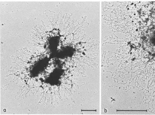
In the late 1970s, Laemmli and colleagues proposed that mitotic chromosome structure arises from a set of non-histone proteins that fold the chromatin fibres into loops (Figure 4). To test their hypothesis, Laemmli and colleagues isolated histone-depleted chromosomes after treating mitotic chromosomes from Hela cells with an excess of polyanions dextran sulphate and
18
Figure 3: Drawing of mitosis in newt cells by Walther Flemming. Sequence of chromosome movements during mitosis. (A to C) During prophase the chromosomes form within the nucleus from a substance termed “chromatin” because of its affinity for dyes. (D) After nuclear envelope breakdown, the chromosomes interact with the two separating “centrosomes” to form a spindle-shaped structure. (E) Prometaphase. (F) Metaphase. After the chromosomes attach to the spindle, they become positioned on its equator, halfway between the two poles. (G) Anaphase. The two chromatids move toward the opposing poles. (H) Telophase. During the final stages of mitosis, neighbouring chromosomes within the two groups fuse to form the daughter nuclei. (I) Cytokinesis. (J) Microtubules pulling apart sister chromatids to opposite poles in anaphase. Compare to Figure 1B. Adapted from Flemming, 1882.
19
heparin (Paulson and Laemmli, 1977). They found that DNA remained highly organized by a core of non-histone proteins whose structure retained the size and shape of the original chromosomes. The authors referred to these proteins as the “chromosome scaffold” and observed that loops of DNA extended outward from the scaffold (Paulson and Laemmli, 1977). Laemmli and colleagues demonstrated later that in intact mitotic chromosomes swollen in a low-salt buffer, chromatin loops emanate from the chromosome scaffold (Figure 4) (Earnshaw and Laemmli, 1983; Lewis and Laemmli, 1982; Maeshima et al., 2005).
Recent studies, using chromosome conformation capture techniques confirmed such organisation in chromatin loops observed in electron micrographs (Gibcus et al., 2018; Kakui et al., 2017; Naumova et al., 2013; Tanizawa et al., 2017). Naumova et al. performed chromosome conformation capture experiments in different human cell types in interphase and in mitosis. These experiments revealed that mitotic genome organization strongly differs from the compartmentalized and cell type-specific organization of the genome in interphase (Naumova et al., 2013). In metaphase, chromosomes are made of consecutive and homogenous arrays of chromatin loops. This folding of chromatin within mitotic chromosomes is consistent among cell types and mitotic chromatin loops are formed by loci located 2-12 Mb apart (Naumova et al., 2013). Consistent with this work, in fission yeast, Hi-C studies have shown that short range (<100 kb) interactions in interphase are replaced by long range (∼1 Mb) interactions in mitosis (Kakui et al., 2017; Tanizawa et al., 2017).
Gibcus et al. have described a pathway for mitotic chromosome formation using DT40 chicken cells. The authors collected synchronous cells at different time points during mitotic progression and analysed chromosome organization by microscopy and Hi-C. Their study revealed that upon mitotic entry, interphase organization is lost and chromatin fibres are converted into arrays of consecutive loops (Gibcus et al., 2018), consistent with previous studies (Naumova et al., 2013). The authors proposed a model in which, in prophase an inner chromosome scaffold forms with a radial arrangement of loops from 30 to 40 kb up to 60 kb in size around the scaffold (Figure 5). As cells progress through mitosis, the distance between interacting loci increases. During prometaphase, the central scaffold acquires a helical arrangement with loops rotating around the scaffold as steps in a spiral staircase. As prometaphase progresses, outer loops grow, and they are subdivided into smaller loops producing a nested arrangement of loops. The number of kilobases per turn continues to grow and results in the shortening of chromosomes to form the mature mitotic chromosome (Figure 5) (Gibcus et al., 2018).
In conclusion, mitotic chromosomes show a highly uniform organization in which large chromatin loops constitute the structural unit and are consecutively arranged.
20
Figure 4: Architecture of metaphase chromosomes. (A) Electron micrographs of a metaphase chromosome swollen in a low-salt buffer showing radial loops emanating from points all along the chromatid arms. (B) Higher magnification view showing the nucleosomal arrangement of the chromatin in the loops. Adapted from Earnshaw & Laemmli, 1983.
Figure 5: Model for mitotic chromosome formation. In prophase, chromosomes are compacted into arrays of consecutive loops around a chromosome scaffold. During prometaphase, the central scaffold acquires a helical arrangement with loops rotating around the scaffold as steps in a spiral staircase. As prometaphase progresses, outer loops grow, they split into smaller loops, the number of loops per turn increases, resulting in the shortening of chromosomes to form the mature mitotic chromosome. Adapted from Gibcus et al, 2018.
21
C) Chromatin loops are different between interphase and mitosis
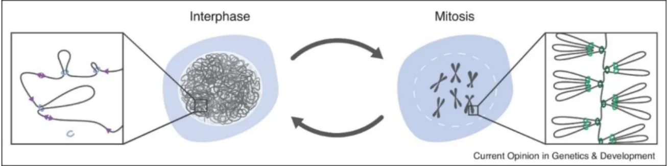
As described above, the genome is extremely structured in the cell nucleus and its organization undergoes drastic changes throughout the cell cycle. Interphase chromatin is hierarchically organized but remains, however, accessible to transcription and DNA replication machineries. In addition, the loose conformation of chromatin allows the control of interactions between genomic loci, through chromatin loops. On the other hand, mitotic chromosomes are organized in helically arranged loop arrays and chromatin acquires a compact structure. Chromatin loops represent, therefore, the structural units of chromosomes during all the cell cycle (Figure 6).
A variety of microscopy-based and sequencing-based techniques have shown, however, that the nature of interphase and mitotic chromatin loops is different (Figure 6). Interphase loops are smaller (∼10-100 kb), heterogeneous, they are not arranged around a central axis and they control gene expression. On the contrary, mitotic loops are larger (∼1-10 Mb), they form all over mitotic chromosomes, they are arranged in homogenous arrays of loops, they are formed at the onset of mitosis in a very short period of time, and their purpose is to compact chromosomes (Figure 6). These differences between interphase and mitotic loops could be explained either by a unique machinery responsible for the formation of the loops that is differently regulated between interphase and mitosis, or alternatively, two different machines that form different loops.
Biochemical and genetic experiments led to the discovery of Structural Maintenance of Chromosomes (SMC) complexes as major components of chromosomes. Numerous studies demonstrated that the SMC complex condensin is central for chromatin looping during mitosis while the SMC complex cohesin underlies the formation of chromatin loops in interphase (Gibcus et al., 2018; Naumova et al., 2013; Rao et al., 2014, 2017).
In the next sections, I will describe the discovery, structure and function of SMC complexes, with particular focus on cohesin and condensin. I will then describe the in vitro data supporting the model in which cohesin and condensin fold the genome by extruding loops of DNA. I will discuss whether structural differences between cohesin and condensin could underlie the formation of different loops. Finally, I will focus on condensin and its function in the context of chromatin.
22
Figure 6 : Chromatin loops in interphase and mitosis. The interphase genome is organized at a low scale by irregular loops. Mitotic chromosomes by contrast are compact rod-like structures containing arrays of homogenous nested loops. Adapted from Sedeño Cacciatore and Rowland, 2019.
23
II. The SMC complexes organize the chromosomes in the nuclear space
SMC complexes are key players in the spatiotemporal organization and maintenance of chromosomes from bacteria to humans.
A) Discovery of SMC
SMC proteins were discovered independently by both biochemical and genetic approaches. As mentioned previously, Laemmli and colleagues used electron microscopy to analyse the structure of mitotic chromosomes. The authors observed that a chromosome scaffold forms the backbone of mitotic chromosomes with chromatin loops attached to this central axis (Figure 4) (Earnshaw and Laemmli, 1983; Lewis and Laemmli, 1982; Paulson and Laemmli, 1977). It was shown that the scaffold consists of a subset of non-histone proteins that includes two major proteins, ScI and ScII (Lewis and Laemmli, 1982). ScI was identified as topoisomerase II (Earnshaw et al., 1985). Topoisomerase II is an evolutionary conserved protein that can untangle DNA and relax the intertwined supercoils in a DNA molecule by passing one DNA molecule through a transient double-stranded break in another (Wang, 1996). The requirement of Topoisomerase II for chromosome condensation was also shown in fission yeast using genetic approaches, as Topoisomerase II mutants fail to segregate chromosomes (Uemura et al., 1987).
Another biochemical approach used mitotic chromosomes assembled in vitro. When incubated in Xenopus egg extracts, sperm chromatin is converted into condensed mitotic chromosomes (Hirano and Mitchison, 1994). Hirano and Mitchison identified two chromosome-associated proteins (CAPs) present in Xenopus egg extracts, XCAP-C and XCAP-E (Hirano and Mitchison, 1994).
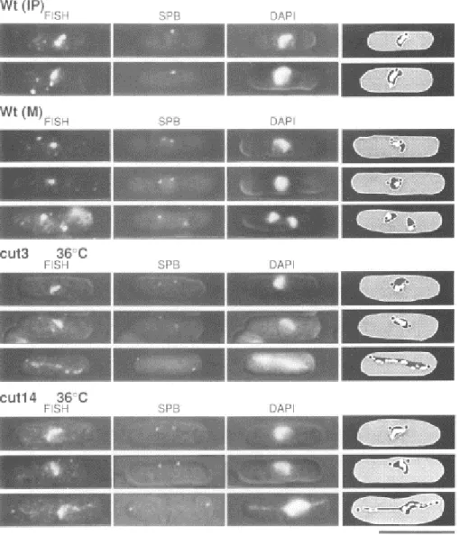
Independent genetic studies led to the discovery of two gene products required for chromosome segregation in yeast. Cloning of a budding yeast gene responsible for the 'Stability of Mini Chromosomes' led to the identification of Smc1 (Strunnikov et al., 1993). Yanagida's group demonstrated that the proteins mutated in the cut3 and cut14 fission yeast mutants showing the “cell untimely torn” (cut) phenotype, were homologues of the SMCs involved in chromosome condensation and segregation (Figure 7) (Saka et al., 1994). Almost simultaneously, Saitoh et al. showed that ScII, one of the major components of the chromosome scaffold, is a chicken homologue of an SMC protein (Saitoh et al., 1994).
Hirano and his colleagues later characterised the CAP proteins and discovered a pentameric complex, that they named condensin, including a heterodimer of XCAP-C (SMC4) and XCAP-E (SMC2) and three non-SMC subunits XCAP-D2, XCAP-G, and XCAP-H (Hirano et al., 1997). The authors showed that immunodepletion of condensin results in chromatin decompaction, suggesting that the
24
Figure 7 : Thermosensitive mutants of fission yeast condensin lead to chromosome condensation and sister chromatid separation defects . Painting of the chromosome arm in cut3-477 and cut14-208 by FISH. Wild type, cut3-477 and cut14-208 cells were cultured at 36°C and fixed. The probes for FISH consisted of mixed DNAs derived from 11 cosmids in the left arm of chromosome 2. The spindle pole bodies (SPB) (functionally equivalent to the centrosome) were visualized using anti-sad1 antibodies. DAPI-staining of nuclear chromatin is shown in the third column. Superimposed images of FISH, SPB and DAPI are depicted on the right. Note in cut3-477 and cut14-208 cells the formation of chromosome bridges in anaphase. Wt IP: wild type interphase. Wt M: wild type mitosis. The bar indicates 10 µm. Adapted from Saka et al, 1994.
25
complex has a key role in chromosome condensation (Hirano et al., 1997). In 1997 also the laboratory of Kim Nasmyth discovered the cohesin complex in budding yeast (Michaelis et al., 1997).
In 1995, Smc2 was identified in budding yeast and it was shown to be required for chromosome segregation (Strunnikov et al., 1995).
The identification of a second condensin complex in vertebrate cells was later reported (Ono et al., 2003). The condensin complex initially discovered was named condensin I, while the newly identified condensin complex was named condensin II. Whereas the two SMC subunits are identical in both complexes, the non-SMC subunits are different (Ono et al., 2003). Both condensins I and II are almost ubiquitous in eukaryotes, and only a limited number of organisms have condensin I only (such as S. cerevisiae and S. pombe).
The results from many diverse technical approaches have converged in the discovery of a large superfamily of SMC. SMC have been found in all eukaryotes examined, and numerous prokaryotes as well, and play crucial roles in chromatid cohesion, chromosome condensation, and DNA repair. Although SMC complexes have diverse functions, they share a conserved architecture.
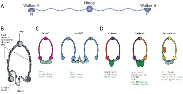
B) Structure of SMC complexes
SMC complexes are characterized by their ring-like shape structure (Figure 8). The ring is made up of three proteins along its circumference: two SMC subunits (homo or heterodimers) complemented by a kleisin subunit (Figure 8B). The SMC family of proteins regroups large ATPases with an unusual domain organization. Each SMC subunit is 1,000–1,500 amino acids in length and has a central hinge domain flanked by two long coiled-coils. The N-and C-terminal domains of the coiled coils contain Walker A and Walker B motifs, crucial for ATP binding (Figure 8A). The SMC folds back on itself through antiparallel coiled-coil interactions, creating an ATP-binding head domain.
Within a complex, two SMC proteins interact at the hinge region, forming long-armed V-shaped dimers (Figure 8B). The length of each arm is ∼50 nm, equivalent to the length of 150 bp of dsDNA.
The ATPase domain is structurally similar to an ATP-binding cassette (ABC) domain and their ATP-binding and hydrolysis cycle modulates engagement and disengagement of the two head domains (Hirano, 2016). In the presence of ATP, the two ATPase heads engage and create a compartment between the hinge and the engaged heads (Chapard et al., 2019; Vazquez Nunez et al., 2019).
The activities of the SMC ring are regulated by peripheral subunits, many of which are either composed of α-helical HEAT (Huntingtin, Elongation factor 3, protein phosphatase 2A, TOR1 domain) repeats or Tandem Winged-Helix Domains (WHD) and associate via the kleisin subunit (Haering and Gruber, 2016a; Uhlmann, 2016). Non-SMC subunits are responsible for differences in localization,
26
Figure 8: Structure of SMC. (A) Each SMC protein has a central globular hinge domain. This domain is flanked by two extended coiled-coils, both of which end with a globular domain that contains a Walker A or a Walker B motif (amino acid consensus sequences that are present in NTP-binding proteins). The protein is folded at the hinge, leading to anti-parallel interaction between the coiled domains. This brings the amino and carboxyl termini together to form a functional ATPase domain, which is structurally similar to an ATP-binding cassette (ABC) domain. Adapted from Jeppsson et al., 2014. (B) Within a complex, two SMC proteins interact at the hinge region, forming long-armed V-shaped dimers, which in turn are associated with complex-specific non-SMC subunits. A kleisin subunit completes the ring. The activities of the core complexes are regulated by diverse peripheral subunits, many of which are either composed of α-helical HEAT-repeats or Tandem Winged-Helix Domains (WHD) and associate via the kleisin protein. (C) Prokaryote SMC. (D) Eukaryote SMC. Adapted from Eeftens and Dekker, 2017.
A
27
dynamic and function of SMC complexes (Andrews et al., 2005; Fujimoto et al., 2005; Green et al., 2012; Ono et al., 2003).
The ring-shaped structure and the ATPase activity of SMC lead to the proposal that they can act as machines that manipulate chromosomal DNA within their compartments, in order to shape chromosomes. SMC could embrace DNA inside the ring or inside the compartment created when SMC heads engaged through ATP. It has been shown that SMC can encircle DNA (Cuylen et al., 2011; Haering et al., 2008; Ivanov and Nasmyth, 2005; Kanno et al., 2015; Wilhelm et al., 2015) and more recently, a better description of DNA entrapment inside the SMC compartments has been reported (Chapard et al., 2019; Kong et al., 2019; Vazquez Nunez et al., 2019). Interestingly, disrupting the integrity of the condensin ring, results in segregation defects in budding yeast (Cuylen et al., 2011, 2013), suggesting that topological entrapment of DNA within the condensin ring might be required for chromosome condensation. However, it is not yet clearly established how the ring-like structure of SMC contribute to their functions.
Prokaryote and eukaryote SMC share functional properties in chromosome organization. Eukaryotes have at least six SMC proteins that interact with different partners to form the SMC complexes. SMC complexes have a conserved structure but play different roles in chromosome architecture. Many efforts are focused on understanding what features of eukaryote SMC specify their functions. Bacteria have a single SMC, and even if it has not be formally demonstrated that bacterial SMC is the functional counterpart of the eukaryotic condensin complex, the analysis of this bacterial protein would contribute to our understanding of the more sophisticated eukaryotic complexes (Hirano, 2005).
C) Prokaryote SMC
Prokaryotes have only a single type of SMC that forms a homodimer. Bacillus Subtilis and Caulobacter Crescentus contain a homodimer of Smc and the kleisin protein ScpA. The additional regulatory protein ScpB, which contains a WHD domain, associates with ScpA (Figure 8C). On the other hand, the structure of the Escherichia Coli SMC-like protein comprises the MukB homodimer, and its accessory proteins, MukE and MukF. MukE contains tandem WHDs and MukF is the kleisin subunit (Figure 8C). The structure of MukBEF differs slightly from those of the other SMC complexes in the fact that the MukF kleisin domain forms dimers that allows the formation of multimers of SMC complexes (Hirano, 2016), at least in vitro (Matoba et al., 2005).
Disruption of Smc and MukB results in increased anucleate cells (Moriya et al., 1998; Yamazoe et al., 1999). These anucleate cells form after abnormal cell division at the ends of elongated cells that
28
result from defective segregation. These studies suggest that bacterial SMC play a role in chromosome partitioning.
The roles of Smc and MukB in chromosome organization have been investigated at the molecular level. Recent experiments have shown that upon chromosomal loading at parS, Smc-ScpAB moves onto flanking DNA aligning the two chromosomal arms progressively from ori to ter. Hi-C maps of thermosensitive Smc mutants in B. Subtilis at the non-permissive temperature showed the loss of the interactions between the arms, suggesting that Smc promotes the colinearity of chromosomal arms (Wang et al., 2017). C. Crescentus SMC is not essential for cell survival, suggesting that other mechanisms act to ensure proper chromosome segregation. Consistent with data in B. Subtilis, Hi-C maps of Δsmc, ΔscpA, and ΔscpB cells in C. Crescentus revealed a decrease in inter-arm interactions (Tran et al., 2017).
In E.Coli, the function of MukBEF differs from Smc-ScpAB. Chromosome conformation capture experiments indicate that MukBEF may generate a series of DNA loops covering most parts of the chromosome instead of co-aligning the two chromosome arms. This process would therefore be more similar to what happens in eukaryotes. A contact map of cells depleted for mukB showed a reduction in long-range interactions, suggesting that MukBEF is needed to establish chromosomal contacts between distant DNA regions (Lioy et al., 2018).
Despite differences in loading and action, both bacterial SMC complexes play an essential role in chromosome organization. Smc-ScpAB seems to act as a translocation factor moving through chromosome arms. On the other hand, MukBEF appears to be required for the formation and maintenance of loops.
Through this thesis I will mention several works in B. subtilis and C. crescentus regarding SMC loading and translocation on chromosomes that contribute to my study on condensin function.
D) Eukaryote SMC
Eukaryote SMC complexes, contrary to prokaryote, are built from heterodimers of different SMC subunits. There are three different SMC complexes in eukaryotes, cohesin, condensin and SMC5/6 complexes (Figure 8D). Despite their structural homology, each of these SMC complexes performs specific, non-redundant functions in the maintenance of chromosomes. Current models predict that this is because they associate with different types of regulatory subunits and exhibit different localization pattern on chromosomes (Baxter et al., 2019).
29
i. The SMC5/6 complex
SMC5-6 complex was initially identified as a complex involved in DNA repair (Lehmann et al., 1995; Nasim and Smith, 1975). However, several studies now show that SMC5/6 could play a role in regulating gene transcription during plant development and in inhibiting transcription of viral genomes (reviewed in Aragón, 2018).
SMC5/6 is composed of a SMC5–SMC6 heterodimer and at least four additional non-SMC elements (Nse proteins). In addition to the ATPase activity (Bermúdez-López et al., 2015; Kanno et al., 2015), the SMC5/6 complex possess the Nse2 subunit, a SUMO E3 ligase that facilitates the SUMOylation of substrates by the SMC5/6 complex and also self-SUMOylation (Andrews et al., 2005). Nse4 is the kleisin subunit that bridges the SMC5/6 heads. Nse1 and Nse3 contain tandem winged-helix domains (WHD) (Figure 8D). Two additional HEAT-containing repeats, Nse5 and Nse6 form a subcomplex and associate with SMC5/6 (not depicted in Figure 8D) (Aragón, 2018).
A study in budding yeast shows the role of Smc5/6 in reducing topological stress accumulated downstream of replication forks (Kegel et al., 2011). This work indicates that SMC5/6 via its interplay with topology can impact chromosome dynamics, however a role in chromosome organization has not yet been reported. In order to pursue this study in SMC-dependent genome organization, I will stay focused on cohesin and condensin.
ii. The cohesin complex
Cohesin was first discovered for its vital role in holding sister chromatids together from the time of DNA replication until anaphase onset to ensure proper chromosome segregation in anaphase (Michaelis et al., 1997).
Cohesin complexes consist of typical SMC ring structures (Figure 8D) that interact with several accessory subunits. Mammalian cohesin is composed of SMC1, SMC3 and two non-SMC subunits, the kleisin subunit SCC1 (Scc1 in budding yeast and Rad21 in fission yeast) and a HEAT-repeat subunit, SA (stromal antigen) (Scc3 in budding yeast and Psc3 in fission yeast) (Figure 8D). The hinge domains of SMC1 and SMC3 bind tightly to each other, whereas the ATPase heads of both proteins are physically connected by SCC1. The ATPase activity of cohesin is required for its loading (Arumugam et al., 2003) and function (Hu et al., 2011; Petela et al., 2018). The loader NIPBL-MAU2 (Scc2-Scc4 in budding yeast) recruits cohesin to chromosomes (Ciosk et al., 2000; Watrin et al., 2006). PDS5 (a HEAT repeat-containing subunit) and WAPL are two other proteins that associate with the four canonical cohesin subunits and act to dissociate cohesin from chromosomes (Haering and Gruber, 2016a; Uhlmann, 2016).
30
Apart from its prominent role in sister chromatid cohesion, cohesin takes part in many other chromosomal processes, including organization of the genome into chromatin loops (reviewed in van Ruiten and Rowland, 2018).
Cohesin and the insulator protein CTCF were found to colocalize in mammalian cells on chromatin (Wendt et al., 2008), at the anchors of loops (Rao et al., 2014) and at boundaries of TADs (Lieberman-Aiden et al., 2009; Nora et al., 2012; Rao et al., 2014).
CTCF is an essential protein that is highly conserved from fly to human but absent in yeast, C. elegans and plants. CTCF binds genomic DNA through a central 11-zinc-finger DNA binding domain and its binding sites contain a directional sequence consensus of 11 to 15 bp (Ong and Corces, 2014). It was suggested that DNA loops correlate with the presence of pairs of CTCF sites arranged in a convergent orientation (Rao et al., 2014). This was supported by a study in mammals, showing that inversion of CTCF-binding sites alters looping and expression of a locus (Guo et al., 2015).
In yeast, (CTCF is absent) cohesin is enriched at sites of convergent transcription that represent the boundaries of the domains (Gullerova and Proudfoot, 2008; Lengronne et al., 2004; Mizuguchi et al., 2014).
Consistent with a role for CTCF and cohesin in genome organization, depletion of CTCF results in fewer intra-TAD contacts (Zuin et al., 2014). Loss of cohesin function causes a complete loss of interphase loops in mouse liver cells (Schwarzer et al., 2017) and human cell lines (Rao et al., 2017). In fission yeast, cohesin is required for maintaining interacting domains ∼50-100 kb in size, since a loss of function mutation of cohesin causes the disruption of local contacts (Mizuguchi et al., 2014).
It has been proposed that CTCF defines contact points for cohesin-mediated chromosomal interactions. A current model suggests that, once loaded onto DNA, cohesin extrudes chromatin loops until it encounters convergent CTCF sites or convergent genes (Busslinger et al., 2017; Fudenberg et al., 2016; Wutz et al., 2017). This “loop extrusion” model will be presented in detail in the section II.E.
Cohesin remains the most studied SMC complex. During this thesis I will bring elements regarding cohesin loading, translocation and loop formation that contribute to my study on the role of condensin in mitotic chromosome organization.
iii. The condensin complex
Condensin is essential for chromosome assembly and segregation during mitosis. Inactivation of condensin in organisms from bacteria to human cells leads to failure in chromosomes condensation. As a consequence, chromosomes remain entangled leading to the formation of anaphase
31
chromosome bridges (Figure 7) (Cuylen et al., 2013; Hirano et al., 1997; Saka et al., 1994; Woodward et al., 2016).
The five subunits of condensin form the ring and contrary to cohesin, no other accessory proteins have been found. The condensin complex contains a SMC2–SMC4 heterodimer, a kleisin subunit and two HEAT repeats subunits (Figure 8D). Mammals possess two different condensin complexes: condensin I and II, that share the SMC subunits but have different non-SMC subunits. Condensin I contains HEAT subunits called CAP-D2 and CAP-G, and the CAP-H kleisin subunit. Condensin II has CAP-D3, CAP-G2 (HEAT-repeat subunits) and the CAP-H2 kleisin subunit (Ono et al., 2003).
Without condensin, mitotic chromosomes retain their interphase structure with local interactions. Two studies in fission yeast analysed the contribution of condensin to large mitotic loops, using different tools to inhibit condensin. Kakui et al. arrested cells in metaphase by repressing the expression of the Slp1 activator of the anaphase-promoting complex. Condensin was depleted by repressing the expression of Cut14 and promoting its degradation vie an auxin-inducible degron. Kakui et al. observed by Hi-C that under these conditions, long-range interactions were lost, suggesting that condensin is responsible for contact interactions in mitosis (Kakui et al., 2017). Tanizawa et al. performed Hi-C in thermosensitive mutants of condensin cut14-208 and cut3-477 at 36°C and found that large domains interactions were reduced (Tanizawa et al., 2017). These studies suggest that condensin mediates the formation of long-range contacts during mitosis and predict that this organization could explain chromosome compaction.
Gibcus et al. investigated the differing contributions of condensin I and II to mitotic chromosome formation in vertebrates. They depleted condensin I or II in G2-arrested cells and Hi-C was performed as cells progressed through mitosis. They showed that either condensin can mediate the formation of arrays of chromatin loops, however, they play distinct roles at different structural levels in mitotic chromosome formation. Condensin II, is nuclear in interphase and associates with chromosomes as early as prophase; it was shown to be centrally located on chromosomes and to compact chromosomes into arrays of consecutive loops. On the other hand, condensin I only gains access to chromosomes after nuclear envelope breakdown and it was shown to subdivide large condensin II-mediated loops into smaller loops (see Figure 5, condensin I is shown in red and condensin II in blue ) (Gibcus et al., 2018).
It has been proposed that condensin, like cohesin, can extrude DNA to form mitotic loops (Ganji et al., 2018; Kong et al., 2019). This “loop extrusion” model, and the evidences supporting it, will be explained in the section II.E.
To further understand condensin function, I detail in the next sections what is currently known of in vitro condensin-DNA interactions and activities of condensin.
32 a) The interaction of condensin with DNA
A prerequisite for chromosome condensation is the association of condensin with chromatin. Thermosensitive mutants of fission yeast condensin, in which condensin association with chromosomes is reduced fail to properly segregate chromosomes in anaphase (Nakazawa et al., 2015; Sutani et al., 2015; Tada et al., 2011).
To study the association of condensin with chromatin, I will address two main points: the nature of condensin-DNA interactions, and the cell-cycle regulated association of condensin with chromosomes.
How does condensin interact with DNA? Three types of condensin-DNA interactions have been described in vitro: topological entrapment of DNA (Cuylen et al., 2011), binding of SMC subunits to ssDNA (Akai et al., 2011; Piazza et al., 2014) and binding of HEAT-repeat subunits to dsDNA (Kschonsak et al., 2017; Piazza et al., 2014).
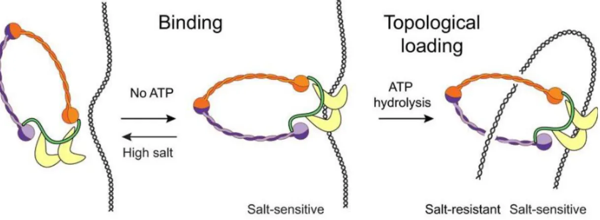
An attractive multistep binding of condensin to DNA has been recently described (Figure 9). Using magnetic tweezers, it has been shown that the association of condensin with DNA can take place in the absence of ATP. This ATP‐independent interaction is able to survive washing steps with physiological salt concentrations, but it does not survive in buffer conditions of high ionic strength, which indicates that the ATP‐independent interaction of condensin with DNA may be electrostatic in nature. When condensin is added to DNA in the presence of ATP, it is, however, able to survive high‐ salt conditions. This suggests that the ATP‐dependent mode of DNA binding must be exceptionally stable. These evidences suggest a model in which condensin first interacts electrostatically through the HEAT-repeat subunits to then topologically entrap DNA in an ATP-dependent manner (Figure 9) (Eeftens et al., 2017).
When does condensin associate with chromosomes? In higher eukaryotes, condensin II is located within the nucleus during interphase and associates with chromosomes in prophase. Condensin I only contact chromosomes after nuclear envelope breakdown in prometaphase (Haering and Gruber, 2016b; Hirota et al., 2004; Ono et al., 2003). In organisms with a closed mitosis like fission yeast, condensin has to be imported into the nucleus before mitotic chromatin compaction. In the case of fission yeast, condensin requires the CDK-dependent phosphorylation of Smc4/Cut3 for nuclear localization (Thadani et al., 2012).
b) The activities of condensin in vitro
SMC2 and SMC4 subunits provide the ATPase activity to the condensin complex, required for chromosomes condensation. However, how the energy of ATP hydrolysis promotes chromosome compaction is still unclear.
33
Figure 9: Multistep binding mechanism of condensin with DNA. Condensin first binds to DNA electrostatically, presumably through the HEAT‐repeat subunits. Upon ATP hydrolysis, condensin embraces the DNA topologically. High salt or high force can disrupt the electrostatic interactions in vitro. Adapted from Eeftens et al, 2017.
34
In vitro studies have uncovered several activities of condensin, namely, the ability to introduce positive supercoiling into DNA, ssDNA reannealing, DNA translocation and DNA loop extrusion (Akai et al., 2011; Ganji et al., 2018; Kim et al., 2019; Kimura and Hirano, 1997; Kong et al., 2019; Sutani and Yanagida, 1997; Sutani et al., 2015; Terakawa et al., 2017).
In vitro studies revealed that condensins are able to introduce ATP-dependent positive supercoils on circular DNA in the presence of topoisomerase I (Kimura and Hirano, 1997). Yet it is not clear if condensin supercoiling activity is required for in vivo chromosome condensation.
Using magnetic-tweezers, a study with Xenopus condensin showed that condensin can bind to DNA in the absence of ATP, but it only compacts the DNA in the presence of hydrolysable ATP (Strick et al., 2004). Similar results were obtained in another magnetic-tweezers study on the S. cerevisiae complex that examined how the rate of compaction depends on protein concentration, ATP concentration, and the force applied on the DNA (Eeftens et al., 2017). Interestingly, both magnetic-tweezers studies on eukaryotic condensin failed to detect a supercoiling activity for condensin, which disagree with the observations of Hirano (Kimura and Hirano, 1997).
It has been shown in vitro that SMC subunits of condensin promote DNA renaturation reactions (the rewinding of single-strand DNA into double helical DNA) (Akai et al., 2011; Sutani and Yanagida, 1997). In vivo studies in fission yeast demonstrated, that an RPA (Replication protein A, the major protein that binds to ssDNA) mutant with reduced affinity to ssDNA restores the growth of condensin mutants (Akai et al., 2011). This observation suggests that ssDNA stabilized by RPA could hinder chromosome segregation. Additionally, it was shown in fission yeast that condensin accumulation sites are sensitive to nuclease P1 (which digests ssDNA or ssRNA), suggesting that these sites contain ssDNA (Sutani et al., 2015). The authors of this study proposed that unwound DNA produced by transcription, detrimental for chromosomes condensation, is recognized and rewound by condensin (Sutani et al., 2015).
To what extent these biochemical activities of condensin contribute to chromosome condensation in vivo is unclear.
Very recently, DNA translocation and DNA extrusion activities of condensin have been demonstrated in vitro (Ganji et al., 2018; Terakawa et al., 2017). These evidences, together with in vitro evidences for cohesin translocation on DNA (Davidson et al., 2016; Stigler et al., 2016), support the proposed “loop extrusion model”.
E) The loop extrusion model to explain SMC-driven chromatin loop formation
How do cohesin and condensin function to organize chromosomes into chromatin loops? Currently, two models have been proposed to explain how SMC complexes bring together distal
35
elements (Figure 10). The stochastic crosslinking model proposes that SMC complexes entrap DNA elements that by chance are in close proximity and thereby stabilize stochastic interactions (Figure 10A) (Cuylen et al., 2013; Thadani et al., 2012). In this model, the topological entrapment of two DNA strands within condensin would explain the long-range interactions mediated by condensin. Alternatively or additionally (Sakai et al., 2018), another model has recently gained a lot of attention: the loop extrusion model (Figure 10B). An increasing amount of evidence supports the notion that the common molecular mechanism that underlies the action of SMC complexes is their ability to create and progressively enlarge loops of DNA (Alipour and Marko, 2012; Goloborodko et al., 2016; Nasmyth, 2001). In this model, an SMC initially binds DNA. Then DNA then bends to form an initial loop. The SMC ring reels DNA generating a loop (Figure 10B). Cohesin extrusion would be blocked by convergent CTCF sites in human, and convergent genes in yeast (Rowley and Corces, 2018). There is no evidence yet that condensin extrusion could be blocked by DNA-bound proteins or transcription. Condensin II does not accumulate at CTCF sites in interphase cells (Dowen et al., 2013), suggesting that CTCF can not block condensin II.
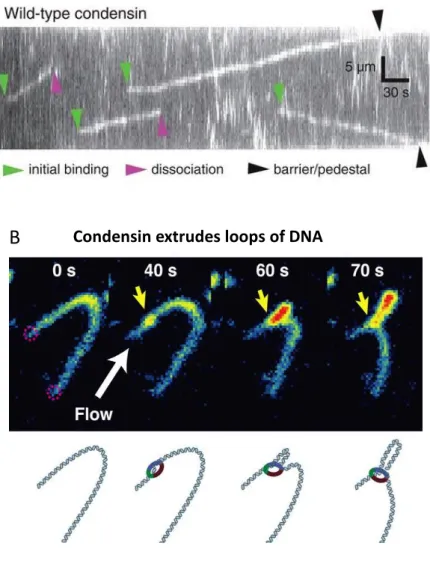
Several in vitro experiments performed on purified condensin support the loop extrusion model. An elegant DNA-curtain study showed that the S. cerevisiae condensin complex is a mechanochemical molecular motor that translocates on naked DNA (Figure 11A) (Terakawa et al., 2017). Translocation was ATP dependent, persisted for very long distances (>10 kb), and showed an average velocity of ∼60 base pairs per second (Figure 11A) (Terakawa et al., 2017). Subsequent single-molecule experiments further demonstrated that the linear translocation observed for condensin could be converted into DNA loop extrusion. Addition of purified budding yeast condensin to DNA molecules, which had been tethered under low tension, allowed the real-time visualization of ATP-dependent formation and gradual expansion of DNA loops of several kbp in size. Condensin-dependent loop extrusion was strictly asymmetric, suggesting that condensin may anchor onto DNA and reels it in from only one side (Figure 11B) (Ganji et al., 2018). The same group has recently published that under the same conditions, higher concentrations of budding yeast condensin result in multiple loop formation on the same DNA molecule (Kim et al., 2019). Surprisingly, they observed a structure containing three dsDNA stretches, connected in parallel. The in-depth study of these structures led them suggest that condensin complexes are able to traverse each other (Kim et al., 2019). These observations challenge the posited model of single looping by condensin and open a new perspective in the mechanisms of chromosome organization. Condensin might create complex loops in which more than a single condensin molecule is implicated. Another very recent work using DNA-curtains and U-shaped DNA molecules (low tension) demonstrated that both human condensin I and II can translocate and extrude loops of DNA in an ATP-dependent manner, as yeast condensin (Kong et al., 2019). In contrast, human condensins DNA loops seem to form symmetrically. The
36
Figure 10: Models of SMC function. (A) The random cross-linking model. An SMC complex links DNA together by trapping two DNA strands inside its ring. Looping can be accomplished either by a single SMC complex or by two interacting SMC complexes. Adapted from van Ruiten & Rowland, 2018. (B) The extrusion process involves SMC loading, DNA bending to form the initial loop, and extrusion. Extrusion could be blocked by DNA-bound proteins (CTCF likely blocks cohesin sliding) or other SMC. A final step could involve release of SMC.. Adapted from Rowley & Corces, 2018.
37
Figure 11: In vitro condensin activities that support the loop extrusion model. (A) ATP–dependent translocation of condensin on dsDNA. DNA curtain assay in which dsDNA is tightly tethered at both ends. Adapted from Terakawa et al, 2017. (B) Condensin extrudes loops of DNA asymmetrically. Double-tethered DNA molecule in which the ends are attached at a distance much shorter than its contour length, in order to keep it loose. Application of a flow allows the visualization of the loop. Adapted from Ganji et al, 2018.
A
Condensin translocates on DNA38
authors also showed that both condensins proceed without stopping upon encountering nucleosomes on single-tethered DNA curtains, and compact DNA (Kong et al., 2019). However, they were only able to assemble 3-4 nucleosomes on a 48,5 kb DNA molecule, which is a much lower density than in vivo chromatin.
Data about cohesin movement on naked DNA seem to be more controversial. Studies using DNA-curtains demonstrated diffusion of fission yeast and human cohesin on DNA but found no evidence for ATP-dependent translocation (Davidson et al., 2016; Stigler et al., 2016). Mobility of fission yeast cohesin can be restricted by nucleosomes, nucleosomes arrays, and DNA-bound proteins (Stigler et al., 2016). Human cohesin can pass over some DNA‐bound proteins and nucleosomes but is constrained in its movement by transcription and CTCF (Davidson et al., 2016). A third study probed the dynamics of Xenopus cohesin on flow-stretched DNA. In contrast to the above reports, these authors claim that cohesin diffusion is dependent on both ATP and the cohesin-loading complex Scc2–Scc4 (Kanke et al., 2016).
In vitro data are very useful and informative to decipher the mechanism of function of SMC and to test the loop extrusion model, but they are performed on naked DNA, so they do not reflect the situation in vivo. Several questions remain to be addressed: How do SMC first contact DNA and associate with chromatin? Is DNA entrapment required for translocation? How do SMC catch the first loop of DNA? How do SMC behave in vivo when they encounter DNA bound proteins that could act as obstacles/roadblocks?
F) How could cohesin and condensin in interphase and in mitosis form different types of
chromatin loops?
Chromatin loops are the structural unit of genome organization in interphase and in mitosis, however, their nature is different (see below section I.C) (Naumova et al., 2013). Short-range interphase loops depend on cohesin and long-range mitotic loops are dependent on condensin (Tanizawa et al., 2017). It is proposed that both machineries drive chromatin looping by extruding DNA (Fudenberg et al., 2016; Ganji et al., 2018). How can we explain the differences in loop sizes between interphase and mitosis?
Cohesin and condensin share similar structures, however they have specific interactors that could differentially regulate their function in loop formation. Non-SMC regulatory proteins could provide specific DNA contacts that would influence SMC translocation. Differences in processivity (speed of loop extrusion and residence time on chromatin) of SMC complexes could be linked to the size of loops generated. For example, condensin I and II have different residence times, with condensin II being more stably bound to chromatin and condensin I more dynamic (Walther et al.,