HAL Id: hal-02991995
https://hal.archives-ouvertes.fr/hal-02991995
Submitted on 20 Nov 2020
HAL is a multi-disciplinary open access
archive for the deposit and dissemination of
sci-entific research documents, whether they are
pub-lished or not. The documents may come from
teaching and research institutions in France or
abroad, or from public or private research centers.
L’archive ouverte pluridisciplinaire HAL, est
destinée au dépôt et à la diffusion de documents
scientifiques de niveau recherche, publiés ou non,
émanant des établissements d’enseignement et de
recherche français ou étrangers, des laboratoires
publics ou privés.
Synthesis and Intracellular Uptake of
Rhodamine–Nucleolipid Conjugates into a
Nanoemulsion Vehicle
Anthony Cunha, Geoffrey Prévot, Yannick Mousli, Philippe Barthélémy,
Sylvie Crauste-Manciet, Benjamin Dehay, Valérie Desvergnes
To cite this version:
Anthony Cunha, Geoffrey Prévot, Yannick Mousli, Philippe Barthélémy, Sylvie Crauste-Manciet, et
al.. Synthesis and Intracellular Uptake of Rhodamine–Nucleolipid Conjugates into a Nanoemulsion
Vehicle. ACS Omega, ACS Publications, 2020, 5 (11), pp.5815-5823. �10.1021/acsomega.9b03992�.
�hal-02991995�
1
Synthesis and Intracellular uptake of Rhodamine-nucleolipid
conju-gates into a nanoemulsion vehicle
Anthony Cunha
a,b,c‡, Geoffrey Prévot
a‡, Yannick Mousli
a, Philippe Barthélémy
a, Sylvie
Crauste-Manciet
a, Benjamin Dehay
b,c#*and Valérie Desvergnes
a#*.
aUniversité de Bordeaux, INSERM, U1212, CNRS UMR 5320, ARNA, ARN : Régulations Naturelle et Artificielle,
ChemBio-Pharm, 146 rue Léo Saignat, 33076 Bordeaux CEDEX, France.
bUniversité de Bordeaux, Institut des Maladies Neurodégénératives, UMR 5293, F-33076 Bordeaux, France.
cCNRS, Institut des Maladies Neurodégénératives, UMR 5293, Centre Broca Nouvelle-Aquitaine, 146 rue Léo Saignat,
33076 Bordeaux CEDEX France
‡ A. Cunha and G. Prévot contributed equally to this work.
#* V. Desvergnes and B. Dehay are co-last corresponding authors. benjamin.dehay@u-bordeaux.fr, valerie.desvergnes@u-bordeaux.fr.
ABSTRACT: Neurodegenerative diseases represent some of the greatest challenges for both basic science and clinical medicine. Due to their prevalence and lack of known biochemical-based treatments, these complex pathologies represent an increasing societal cost. Increasing genetic and neuropathological evidence point that lysosomal impairment may be a common factor linking these diseases, laying the development of therapeutic strategies aimed at restoring lysosomal function. Here, we propose the design and synthesis of a nucleolipid conjugate as a non-viral chemical nanovector to target specifically neuronal cells and intracellular organelles. Herein, a thymidine appropriately substi-tuted to increase its lipophilicity, was used as a model nucleoside and a fluorophore moiety, covalently bound to the nucleoside, allowed to monitor the nucleolipid’s internalization in vitro. In order to improve nucleolipid protection and cellular uptake these conjugates were formulated in nanoemulsions. In vitro biological assays demonstrated cell uptake and internalization associated colocalization with lysosomal markers. Overall, this nucleolipid-nanoemulsion based formulation represents a promising drug delivery tool to target the central nervous system, able to bring drugs to restore lysosomal impaired function.
Introduction
The selective filter of the blood brain barrier (BBB) is one of the major obstacle to achieve efficient drug delivery1,2 and therapeutic effect to the brain3,4which hampers further treat-ments for brain-related diseases. Nowadays, innovation in the field of nanosystems allows a better crossing of biologi-cal membranes, thus paving the way for new therapeutic approaches1,3,5. Nucleolipids (NLs) are bifunctional hybrid molecules in which a lipid moiety and a nucleic acid moiety (nucleoside, nucleotide, nucleobase or oligonucleotide) are
covalently linked6. Exhibiting a wide structural variety, these molecules can be natural such as Algelasine F and Tunica-mycin7,8 or made of synthetic origin like the DOTAU9. NLs, either natural or synthetic, are interesting for their potential biological activities, including antimicrobial, antifungal, antiviral and antitumor properties10–12, but also for their re-markable ability to self-assemble. Indeed, amphiphilic mole-cules, such as the DOTAU, can form supramolecular objects like micelles or liposomes, which can be used to deliver DNA, antisense oligonucleotides or siRNA directly into the cells9. Because of its similarity to the lipid bilayer of cell membranes, these molecules are expected to cross the plasma membrane without the need of membrane transporters. Con-sidering the benefit of nanoemulsions (NEs) as vehicles for therapeutic agents to target the brain13–15, their combination with NLs, as potential absorption promoter, seemed to be a tantalizing approach to improve the passage of the BBB. Oil-in-water (O/W) NEs, made of submicrometric oily drop-lets stabilized by a corona of amphiphilic surfactants, present enhanced stability compared to other nanosystems and high loading capacity of hydrophobic drugs or imaging probes13,16–18. It has also been very recently reported that some NLs, such as a NL radiotracer, was successfully able to permeate the BBB19, suggesting that NLs could be promising absorption promoters. In this work, original NEs associated with nucleolipids were developed to enhance the membrane crossing properties for therapeutic purposes and particularly in the context of neurodegenerative diseases. Indeed, previ-ous works have shown that both nanoparticles or NEs loaded with an acidic cargo made of poly(DL-lactide-co-glycolide) (PLGA) were able to induce neuroprotective effects, and following systemic injection, NEs were able to cross the
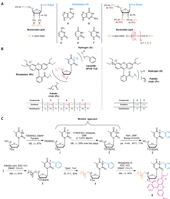
Figure 1. (A) General structure of nucleolipids. (B) Chemical structure of the different compounds synthetized for this study. (C)
Synthet-ic sequence leading to NL-A.
Herein, we report the design and synthesis of original NLs and their formulation into NEs to cross the plasma membrane. We show the contribution role of NEs in the uptake and internaliza-tion of NLs into neuronal cells in-vitro, and colocalizainternaliza-tion of NLs with lysosome was observed. These results suggest that lysosomes can be efficiently targeted by the use of these nanotechnology-based systems for drug delivery to brain diseases, in particular of interest in neurodegenerative diseases where lysosomal impair-ment occurs among others22,23.
Results and discussion
Design and synthesis of nucleolipidic platforms
Six compounds, among which three original NLs (Figure 1A), were then designed, synthetized and biologically evaluated. Commercially available thymidine was used as a nucleosidic
platform and key positions were conveniently functionalized step by step (Figure 1B). A benzyl group was introduced at N-3 posi-tion of thymidine to enhance the lipophilicity of the compound and allow the solubilization in oily phase to provide NE formula-tion later on. The first NL, compound A, was substituted with a palmitic chain at 5’ position and a Rhodamine B fluoroprobe at position 3'. In NL compound B the lipid chain and the Rhodamine B positions were switched. Thereby, these two lipophilic NLs were formulated into NEs, to carry out in vitro biological assays and evaluate the role of NLs, as well as the position of the differ-ent substitudiffer-ents and their influence on the uptake into cells. The third NL (compound C) has been similarly designed by removing the benzyl group to make it water soluble and investigate the role of NE itself. The three last compounds were used as control. Compound D was Rhodamine B, one formulated in a NE and one solubilized in water, in order to determine the role of the NEs, and compound E was a Rhodamine-lipid conjugate (Figure 1B).
B
C A
Compounds A B C
Positions N-3 O-3’ O-5’ N-3 O-3’ O-5’ N-3 O-3’ O-5’ Substituents Lg Rh Pc Lg Pc Rh H Rh Pc
Compounds D E Positions COO- COO-Substituents H Pc
Compound E was designed to compare its internalization into neuronal cells with that of compound A and investigate the bene-fits of using a NL rather than a simple lipid chain.
Nucleolipids A and B were prepared in 7 and 5 steps-sequences respectively. The first steps were selective protections of the nucleoside in order to functionalize, thereafter, either position 3' or 5'. The benzyl group was added on the thymidine N-3 position by a simple alkylation following a procedure using microwave activation previously developed in the laboratory24. An esterifica-tion reacesterifica-tion between the palmitic acid and the thymidine was performed at the free hydroxyl positions, (position 5’ for com-pound A and position 3’ for comcom-pound B). The protective group was then cleaved before subsequent esterification step with the Rhodamine B moiety (Figure 1C). Compound C was obtained through a six-step sequence while compound E required a unique esterification step.
Nanoemulsions preparation and characterization
Lipidic nature and plasticity of O/W NEs make them suitable intravenous (I.V.) systems for drug delivery. NE exhibit a good encapsulation ability for hydrophobic drugs and improve effi-ciently their stability, I.V. tolerance and bioavailability25. Five nanoemulsions were formulated with Medium Chain Triglycer-ides oil Miglyol® 812N as the oily phase and two surfactants Polysorbate 80 (Tween® 80) and egg phospholipids (Lecithin® E80) (Table 1). NE-A, NE-B, NE-D and NE-E were loaded re-spectively with nucleolipids A and B, Rhodamine (D) and Rhodamine-lipid conjugate (E). An unloaded NE named NE-O was used as a control.
NEs
composition NE-O NE-A NE-B NE-D NE-E
Miglyol® 812 20% 20% 20% 20% 20% Lecithin® E80 1.2% 1.2% 1.2% 1.2% 1.2% Tween® 80 2.5% 2.5% 2.5% 2.5% 2.5% Compounds mass (mg) 0 10 10 5 7,5 Quantity of compounds (mmol) 0 9.7×10⁻³ 9.7×10⁻³ 1.0×10⁻² 1.1×10⁻² Water q.s 100 100 100 100 100
Table 1. Composition of loaded NEs with the compound A, B, D
or E and the unloaded NE (NE-O). (The amounts of Miglyol® , Lecithin® and Tween® is expressed in % (w/w)).
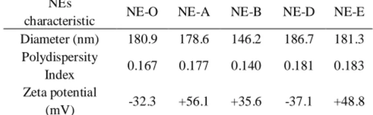
Physico-chemical analysis of the formulations by DLS showed that all five formulations have submicrometric size range (Table 2). All formulations displayed a monodispersed distribution with a dispersity below 0.2. Zetameter measurement showed a negative zeta potential for the control formulation (NE-O). Indeed, this negative charge is linked with the lecithin E80 surfactant (mixture of phospholipids including negatively charged phospholipids). Interestingly NE-A and NE-B turned out to exhibit positive zeta potential. The same phenomenon was observed with NE-E, the Rhodamine-lipid-based NE, which is also naturally positively charged. Contrariwise, a negative zeta potential was spotted with the Rhodamine B-based formulation (NE-D), because non-lipid conjugated Rhodamine was solubilized in oil phase at the final concentration of 25 mg/ml, suggesting that in this case Rhodamine B was placed inside the oily droplet and not at its surface (Figure 2).
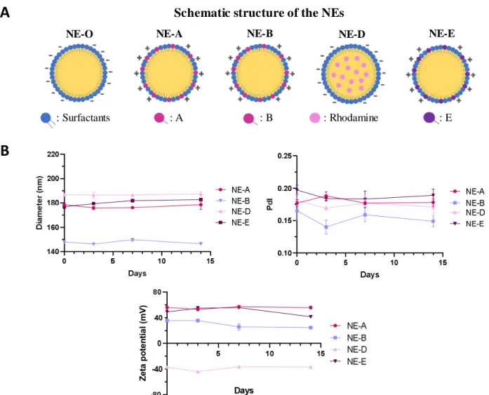
Finally, the colloidal stability of three of these NEs was monitored in Dynamic Light Scattering (DLS) to assess the size of the glob-ules and the index of polydispersity26. Moreover, Zeta potential was measured using a zetameter (Figure 2B). Satisfactory, all formulations proved to remain monodispersed with a mean di-ameter under 200 nm for at least 14 days, and their zeta potential was consistent over the same time. Moreover NE-A, NE-D NE-E assessed for longer time were found to be stable over 30 days.
NEs
characteristic NE-O NE-A NE-B NE-D NE-E Diameter (nm) 180.9 178.6 146.2 186.7 181.3 Polydispersity
Index 0.167 0.177 0.140 0.181 0.183 Zeta potential
(mV) -32.3 +56.1 +35.6 -37.1 +48.8
Table 2. Physico-chemical characteristics of unloaded and loaded
Figure 2. Physico-chemical characteristics of unloaded and loaded-NEs. (A) Schematic structure of the different NEs. (B) Evolution of the
diameter, polydispersity index (PdI) and zeta potential of NE-A, NE-D and NE-E over time.
In vitro cytotoxicity evaluation of nucleolipids-loaded nanoemulsions
Six formulations have been used in vitro to evaluate their respec-tive cytotoxicity onto human neuroblastoma cell lines (BE (2)-M17 cells) during either 24h or 48h. They are the four NEs pre-pared (NE-A, NE-B, NE-D and NE-E) and two aqueous formula-tions of compounds C and D (C: NL without the benzyl group and D: Rhodamine B). After 24 or 48 hours of exposure, no sig-nificant toxicity was observed except for the formulations Aq-D and NE-E (in a range of 20% to 36%) (Figure 3).
Figure 3. Cell viability evolution over time. BE (2)-M17 cells
treated with NE-A, NE-B, NE-D and NE-E and compounds C and D solubilized in water compared to control condition (UT) after 24 h and 48h. * p < 0.05 compared with untreated cells.
Role of NLs loaded NEs for internalization into lysosomes in vitro
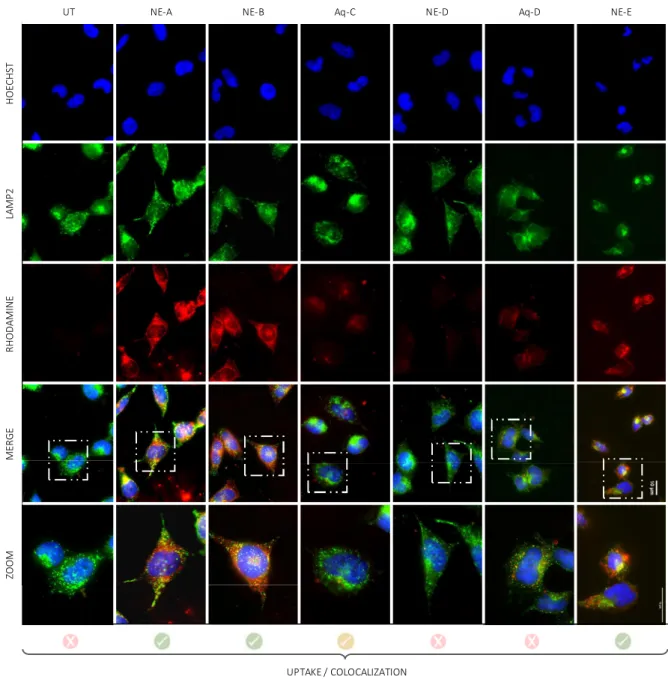
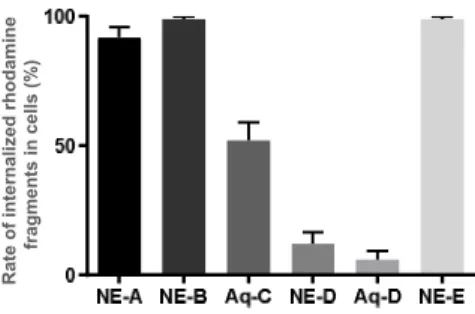
As previously mentioned, lysosomal impairment is a common factor in neurodegenerative diseases. Lysosomes are intracellular acidic compartments that contain hydrolytic enzymes involved in the degradation of intracellular components through several deg-radation pathways, including endocytosis, phagocytosis, and autophagy26,27. Therefore, to determine if these NE-NL nanovectors can be used as a drug delivery system for neuronal cells, cellular uptake and lysosomal colocalization of the different compounds, labeled with Rhodamine B, were investigated (Figure 4). Foremost, it has been observed that compounds/formulations
NE-A, NE-B, NE-E and Aq-C were well internalized into cells,
while solely 12% and 6% of cells uptake for formulations NE-D and Aq-D. One possible explanation regarding the low rate of internalization of NE-D and Aq-D, might be explained by the leakage of free Rhodamine outside the oily droplet of the NE, preventing the NE from fully playing its carrier function. Never-theless, an uptake of 100% was obtained for the two NLs and the Rhodamine-lipid conjugate loaded into NEs (A, B or E), and it has been found out that the rate of NL internalized into cells was greatly enhanced with the use of NE. Indeed, we observed that only 50% of Aq-C, solubilized in water, get through plasma membranes (Figure 4 and 5). Furthermore, each of these com-pounds labeled with rhodamine (red) colocalized into lysosomes (LAMP2, green). Z e ta p o te n ti a l (m V )
Schematic structure of the NEs
NE-O
NE-A
NE-B
NE-D
NE-E
A
B
: A
: B
: Rhodamine
: E
: Surfactants
Figure 4. In vitro biological assessment. Epifluorescence microscopy pictures showing uptake and colocalization evaluation into BE
(2)-M17 cells treated with compounds A, B, D and E formulated into NEs and compounds C and D solubilized in water. Nuclei were stained with Hoechst (blue), lysosomes with LAMP2 (green) and compounds A to E are highlighted by the presence of rhodamine B (red). Scale bar: 10 µm. HOECHST LAMP2 RHODAMINE MERGE ZOOM
UT NE-A NE-B Aq-C NE-D Aq-D NE-E
Consequently, combination of nanoemulsions with properly sub-stituted nucleolipids A and B provided efficient and non-toxic nanovectors for neuronal cells internalization and subsequent lysosome colocalization.
Figure 5. Quantification of internalized rhodamine in treated-cells
to evaluate the uptake into BE (2)-M17 cells of compounds A, B,
D and E formulated into NEs and compounds C and D solubilized
in water.
Conclusions
We developed the synthesis of original thymidine-derived NLs bearing the fluorophore rhodamine B to study their biocompatibil-ity and their role as absorption promoter to cross biological mem-branes. First, we designed lipophilic NLs to afford solubilization into the oily phase of O/W NE. Then, the lipidic chain on sugar moiety and the benzyl group were substituted on the thymine amino group, allowing the formulation of NLs into NEs. The colloidal stability of such NL-NE system was monitored over time by DLS and Zeta potential analysis before carrying out in vitro experiments. Cytotoxicity evaluation of NL-NE complexes on human neuronal cells showed that NLs are fully biocompatible after 24h and 48h of exposure, while other amphiphilic com-pound, such as Rhodamine-lipid conjugates display cellular tox-icity. Uptake evaluations into cells indicate that NL-NE are suc-cessfully internalized, highlighting the contribution of NEs as drug delivery vehicles. More importantly, these original NLs bearing a fluorophore moiety are colocalized with lysosomes suggesting that these nanotechnology-based systems can also be efficient tools to target lysosomes, where its impairment has been involved in neurodegenerative diseases. Therefore, these results were promising and pave the way to test in future studies and improve these new NL-NE systems for drug delivery in vivo, offering the possibilities for specific therapeutic solutions target-ing pathologies associated with lysosomal impairment.
EXPERIMENTAL SECTION General information
All reactions were carried out under an argon atmosphere. Yields refer to chromatographically and spectroscopically (1H-NMR) homogeneous materials, unless otherwise stated. All reagent-grade chemicals were obtained from commercial suppliers and were used as received, unless otherwise stated.
1
H-NMR and 13C-NMR were recorded on a Bruker Avance 300 (1H: 300 MHz, 13C: 75.46 MHz) spectrometer using residual CHCl3 as internal reference (7.26 ppm) and at 293K unless other-wise indicated. The chemical shifts (δ) and coupling constants (J) are expressed in ppm and Hz respectively. The following abbrevi-ations were used to explain the multiplicities: s = singlet, d =
doublet, t = triplet, q = quartet, m = multiplet, b = broad. FT-IR spectra were recorded on a Perkin-Elmer FT spectrometer Spec-trum two (UATR two). For the ESI HRMS analyses, a Waters Micromass ZQ instrument equipped with an Electrospray source was used in the positive and/or negative mode. MALDI TOF mass spectrometric analyses were performed on a PerSeptive Biosystems Voyager-De Pro MALDI mass spectrometer in the Linear mode using 3,4-dihydroxybenzoic acid as the matrix. Analytical thin layer chromatography was performed using silica gel 60 F254 pre-coated plates (Merck) with visualization by ultra-violet light, potassium permanganate or sulfuric acid. Flash chro-matography was performed on silica gel (0.043-0.063 mm).
Procedure for compound A synthesis
Synthesis of compound 1. To a solution of thymidine (1 eq., 6 g,
24.77 mmol) in pyridine (118 mL) and under argon are sequen-tially added the TBDMSCl (1.2 eq., 4.48 g, 29.72 mmol) and DMAP (spatula tip). The mixture is stirred overnight at room temperature to completion. The reaction is then quenched by addition of NaHCO3 (10 mL, aq.) and diluted with water (10 mL). The aqueous phase is extracted three times with DCM (3x30 mL), and the combined organic phases are dried over Na2SO4 before concentration to dryness under vacuum. The resulting crude is purified by flash chromatography on silica gel (Pen-tane/EtOAc, 70/30) in order to obtain the expected compound 1 as a white powder (7.68 g, 21.55 mmol, 87%). NMR data are consis-tent with literature. Rf : 0.24 (Pentane/EtOAc, 70/30). IR (ATR) ν (cm-1) : 3549, 3167, 2929, 2855, 1696, 1471, 1257, 1120, 832, 779.
Synthesis of compound 2. 1) To a solution of 1 (1 eq., 14.7 g,
41.21 mmol) in DMF (88 mL) and under argon are sequentially added the imidazole (1.2 eq., 3.37 g, 49.54 mmol) followed by TBDPSCl (1.2 eq., 12.88 mL, 49.54 mmol). The mixture is stirred overnight at room temperature to completion. The mixture is then diluted with toluene (100 mL) and DMF was coevaporated under vacuum to obtain a yellow oil. The oil is diluted in diethyl ether (10 mL) and the white precipitate formed is filtered, and washed with an aqueous solution of NaCl. The organic phase is dried over Na2SO4 before concentration to dryness under vacuo. 1’ was obtained as a yellow oil.2) To a solution of 1’ in methanol (80 mL) and under argon is added para-toluenesulfonic acid monohydrate (0.471 g, 2.477 mmol). The mixture is stirred over-night at room temperature to completion. The methanol is then evaporated under vacuum to obtain a yellow oil. The crude prod-uct diluted with EtOAc (30 mL) and the organic phase is washed three times with NaHCO3 (10 mL, aq.) and brine (3x20 mL). The organic phase is dried over Na2SO4 before concentration to dry-ness under vacuo. The resulting crude is purified by flash chroma-tography on silica gel (Petroleum Ether/EtOAc, 50/50) in order to obtain the expected compound (2) as a white powder (6.90 g, 14.36 mmol, 58% over two steps). Rf : 0.29 (Petroleum ether/EtOAc, 50/50). 1H NMR (300MHz, CDCl3) δ ppm : 9.18 (bs, 1H, NH), 7.59-7.69 (m, 4H, H Ar), 7.34-7.50 (m, 6H, H Ar), 7.30 (d, J= 1.2Hz 1H, H6), 6.26 (dd, J= 6.0Hz, 7.8Hz, 1H, H1’), 4.41-4.48 (m, 1H, H3’), 3.94-4.00 (m, 1H, H4’), 3.62 (dd, J= 2.4Hz, 12.0Hz, 1H, H5b’), 3.24 (dd, J= 3.0Hz, 12.3Hz, 1H, H5a’), 2.43 (bs, 1H, OH), 2.26 (ddd, J= 3.0Hz, 6.0Hz, 13.2Hz, 1H, H2b’), 2.04-2.22 (m, 1H, H2a’), 1.82 (d, J= 1.2Hz, 3H, H7), 1.08 (s, 9H, t Bu). 13C NMR (75.46MHz, CDCl3) δ ppm : 164.0 (C4), 150.5 (C2), 136.9 (C6), 135.8 (CH Ar), 133.4 (Cq Ar), 133.3 (Cq Ar), 130.2 (CH Ar), 130.2 (CH Ar), 128.0 (CH Ar), 111.0 (C5), 87.8 (C4’), 86.6 (C1’), 73.1 (C3’), 62.1 (C5’), 40.4 (C2’), 27.0 (CH3 tBu), 19.1 (Cq tBu), 12.5 (C7). IR (ATR) ν (cm-1) : 2932, 1682, 1471, 1274, 1104, 1031, 740, 702.
Rate of internalized rhodamine
Synthesis of compound 3. To a solution of 2 (1 eq., 1 g, 2.08
mmol) in anhydrous DMF (10 mL) and under argon atmosphere, NaH (1.2 eq., 0.135 g, 3.37 mmol) is added in a vial before activation in a microwave oven (2 min, 40°C, 200W). Then Benzyl bromide (1.2 eq., 0.4 mL, 3.37 mmol) is added and the reaction mixture is placed in a microwave oven using a temperature control mode (4 min, 40°C, 200W). DMF is then co-evaporated with toluene and the resulting crude is purified by flash chromatography on silica gel (Petroleum Ether/AcOEt, 70/30) to obtain a white powder 3 (0.846 g, 1.48 mmol, 71%). Rf : 0.26 (Petroleum Ether/EtOAc, 70/30). 1H NMR (300MHz, CDCl3) δ ppm : 7.61-7.69 (m, 4H, H Ar), 7.36-7.51 (m, 8H, H Ar), 7.21-7.35 (m, 4H, H6 and H Ar), 6.28 (appearing t, J= 6.6Hz, 5.2Hz, 1H, H1’), 5.11 (m, 2H, H8), 4.41-4.49 (m, 1H, H3’), 3.94-4.00 (m, 1H, H4’), 3.62 (dd, J= 2.1Hz, 11.7Hz, 1H, H5b’), 3.23 (dd, J= 2.9Hz, 12.0Hz, 1H, H5a’), 2.29 (ddd, J= 2.3Hz, 4.9Hz, 13.1Hz, 1H, H2b’), 2.08-2.33 (m, 1H, H2a’), 1.89 (d, J= 0.9Hz, 3H, H7), 1.48-1.70 (bs, 1H, OH), 1.10 (s, 9H, tBu). 13C NMR (75.46MHz, CDCl3) δ ppm : 163.5 (C4), 151.1 (C2), 136.9 (C6), 135.9 (CH Ar), 134.9 (CH Ar), 133.4 (CH Ar), 133.2 (CH Ar), 130.2 (CH Ar), 129.3 (CH Ar), 128.5 (CH Ar), 128.0 (CH Ar), 127.7 (CH Ar), 110.5 (C5), 87.7 (C1’ orC4’), 87.5 (C1’ or C4’), 73.1 (C3’), 62.3 (C5’), 44.6 (C8), 40.4 (C2’), 27.0 (CH3 tBu), 19.2 (Cq t
Bu), 13.4 (C7). IR (ATR) ν (cm-1) : 3456, 3071, 2932, 2859, 1666, 1634, 1428, 1240, 1103, 1024, 741, 700.
Synthesis of compound 4. To a solution of 3 (1 eq., 0.160 g,
2.80x10⁻¹ mmol) in DCM (1.6 mL) and under argon are sequen-tially added the palmitic acid (1.2 eq., 0.086 g, 3.36x10⁻¹ mmol), the EDC.HCl (1 eq., 0.052 g, 2.71x10⁻¹ mmol) and the DMAP (0.3 eq., 0.010 g, 8.40x10⁻² mmol). The mixture is stirred 48h at room temperature to completion. The reaction is then quenched by addition of NH4Cl (5 mL, aq.) and diluted with water (10 mL). The aqueous phase is extracted three times with DCM (3x10 mL), and the combined organic phases are dried over Na2SO4 before concentration to dryness under vacuum. The resulting crude is purified by flash chromatography on silica gel (Pentane/AcOEt, 90/10) in order to obtain compound 4 (0.138 g, 1.70x10⁻¹ mmol, 63%) as a white foam. Rf : 0.20 (Pentane/EtOAc, 90/10). 1H NMR (300MHz, CDCl3) δ ppm : 7.62-7.75 (m, 4H, H Ar), 7.23-7.54 (m, 11H, H Ar), 7.19 (d, J= 0.9Hz, 1H, H6), 6.50 (dd, J= 6.0Hz, 7.5Hz, 1H, H1’), 5.16 (q, J= 13.8Hz, 2H, H8), 4.24-4.33 (m, 1H, H3’), 4.08-4.15 (m, 1H, H4’), 3.94 (dd, J= 3.0Hz, 12.0Hz, 1H, H5b’), 3.80 (dd, J= 4.2Hz, 12.0Hz, 1H, H5a’), 2.43 (ddd, J= 2.5Hz, 5.5Hz, 13.2Hz, 1H, H2b’), 2.23 (t, J= 7.5Hz, 2H, O=C-CH2 lipid), 1.94 (s, 3H, H7), 1.77-1.90 (m, 1H, H2a’), 1.48-1.61 (m, 2H, CH2 lipid), 1.26-1.28 (m, 24H, CH2 lipid), 1.14 (s, 9H, tBu), 0.93 (m, 3H, CH3 lipid). 13C NMR (75.46MHz, CDCl3) δ ppm : 172.9 (C=O), 163.2 (C2), 150.8 (C4), 136.9 (Cq Ar), 135.7 (CH Ar), 135.7 (CH Ar), 133.0 (CH Ar), 132.9 (CH Ar), 132.8 (C6), 130.2 (Cq Ar), 130.2 (CH Ar), 129.1 (CH Ar), 128.3 (CH Ar), 128.0 (CH Ar), 127.9 (CH Ar), 127.5 (Cq Ar), 110.3 (C5), 85.6 (C1’), 84.7 (C4’), 73.0 (C3’), 63.2 (C5’), 44.5 (C8), 40.8 (C2’), 34.0 (CH2 lipid), 31.9 (CH2 lipid), 29.7 (CH2 lipid), 29.7 (CH2 lipid), 29.6 (CH2 lipid), 29.6 (CH2 lipid), 29.4 (CH2 lipid), 29.4 (CH2 lipid), 29.2 (CH2 lipid), 29.1 (CH2 lipid), 26.9 (CH3 tBu), 24.7 (CH2 lipid), 22.7 (CH2 lipid), 19.0 (Cq tBu), 14.1(CH3 lipid), 13.4 (C7). IR (ATR) ν (cm-1) : 3333, 2925, 2854, 1743, 1704, 1668, 1648, 1463, 1271, 1106, 1026, 700, 613, 506. HRMS (ESI) [M+H+] : calcd = 809.49194, found = 809,49210.
Synthesis of compound 5. To a solution of 4 (1 eq., 0.326 g,
4.03x10⁻¹ mmol) in THF (4.54 mL) and under argon is added the TBAF (1M in THF, 0.403 mL) at 0°C. The mixture is stirred 2 hours at 0°C. The reaction is then quenched by addition of water (5 mL), and the mixture is warmed to room temperature before addition of DCM (10 mL). The aqueous phase is extracted three times with DCM (3x10 mL), and the combined organic phases are
dried over Na2SO4 before concentration to dryness under vacuum. The resulting crude is purified by flash chromatography on silica gel (Pentane/AcOEt, gradient 90/10 to 70/30) in order to obtain the expected compound 5 (0.190 g, 3.33x10⁻¹ mmol, 83%). Rf : 0.31 (Pentane/EtOAc, 92/8). 1H NMR (300MHz, CDCl3) δ ppm : 7.39-7.48 (m, 2H, H Ar), 7.18-7.35 (m, 4H, H Ar and H6), 6.32 (appearing t, J= 6.3Hz, 1H, H1’), 5.11 (s, 2H, H8), 4.21-4.41 (m, 3H, H3’, H5b’ and H5a’), 4.13 (bs, 1H, H4’), 3.56-3.81 (bs, 1H, OH), 2.29-2.46 (m, 3H, O=C-CH2 lipid and H2b’), 2.00-2.13 (m, 1H, H2a’), 1.96 (s, 3H, H7), 1.56-1.71 (m, 2H, CH2 lipid), 1.27 (m, 24H, CH2 lipid), 0.85-0.93 (m, 3H, CH3 lipid). 13C NMR (75.46MHz, CDCl3) δ ppm : 173.6 (C=O), 163.4 (C2), 150.9 (C4), 136.7 (Cq Ar), 133.4 (C6), 128.8 (CH Ar), 128.4(CH Ar), 127.6 (CH Ar), 110.3 (C5), 85.8 (C1’), 84.3 (C4’), 71.3 (C3’), 63.7 (C5’), 44.5 (C8), 40.5 (C2’), 34.2 (CH2 lipid), 31.9 (CH2 lipid), 29.7 (CH2 lipid), 29.6 (CH2 lipid), 29.5 (CH2 lipid), 29.4 (CH2 lipid), 29.2 (CH2 lipid), 29.1 (CH2 lipid), 24.9 (CH2 lipid), 22.7 (CH2 lipid), 14.1 (CH2 lipid), 13.4 (CH3 lipid). IR (ATR) ν (cm-1) : 3363, 2918, 2850, 1697, 1621, 1470, 1178, 1092, 711. HRMS (ESI) [M+H+] : calcd = 571.37416, found = 571.37405.
Synthesis of compound A. To a solution of 5 (1 eq., 0.190 g,
3.33x10⁻¹ mmol) in DCM (1.9 mL) and under argon are sequentially added the Rhodamine B (1.2 eq., 0.191 g, 3.99x10⁻¹ mmol), EDC.HCl (1 eq., 0.062 g, 3.23x10⁻¹ mmol) and DMAP (0.3 eq., 0.012 g, 9.99x10⁻² mmol). The mixture is stirred 48h at room temperature. The reaction is then quenched by addition of NH4Cl (5 mL, aq.) and diluted with water (10 mL). The aqueous phase is extracted three times with DCM (3x10 mL), and the combined organic phases are dried over Na2SO4 before concentration to dryness under vacuum. The resulting crude is purified by flash chromatog-raphy on silica gel (DCM/MeOH, 95/5) in order to obtain the expected compound A as a purple powder (0.132 g, 1.28x10 -1
mmol, 40%). Rf : 0.24 (DCM/MeOH, 95/5). 1H NMR (300MHz, CDCl3) δ ppm : 8.24-8.34 (d, J= 30.9Hz, 1H, H Ar), 7.80 (dt, J= 7.5Hz, 14.7Hz, 30.3Hz 2H, H Ar), 7.31-7.41 (m, 2H, H Ar), 7.13-7.30 (m, 5H, H6 and H Ar), 7.09-6.86 (m, 4H, H Ar), 6.74 (bs, 2H, H Ar), 5.08-6.08 (m, 1H, H1’), 5.09-5.15 (m, 1H, H3’), 5.01-5.02 (d, J= 3.0, 2H, H8), 4.08-4.24 (m, 2H, H5’), 3.90-3.98 (m, 1H, H4’), 3.50-3.66 (m, 8H, CH2-CH3 Rhodamine), 2.04-2.28 (m, 4H, H2’ and O=C-CH2 lipid), 1.86 (s, 3H, H7), 1.40-1.52 (m, 2H, CH2 lipid), 1.07-1.32 (m, 36H, CH2 lipid and CH2-CH3 Rhodamine), 0.79 (t, J= 6.3Hz, 3H, CH3 lipid). 13C NMR (75.46MHz, CDCl3) δ ppm :172.8 (C=O), 164.1 (CH Ar), 163.0 (C2), 157.5 (Cq Ar or CH Ar), 155.4 (Cq Ar or CH Ar), 155.5 (CH Ar), 155.5 (CH Ar), 150.7 (C4), 136.6 (C6), 133.6 (CH Ar), 133.4 (CH Ar), 131.4 (CH Ar), 130.9 (CH Ar), 130.9 (CH Ar), 130.4 (CH Ar), 130.3 (CH Ar), 128.7 (CH Ar), 128.2 (CH Ar), 127.4 (CH Ar), 114.5 (Cq Ar), 113.2 (C5), 110.5 (Cq Ar), 96.3 (CH Ar), 85.7 (C1’), 81.8 (C4’), 75.5 (C3’), 63.5 (C5’), 46.1 (CH2-CH3 Rhodamine), 44.3 (CH2 lipid), 31.7 (C2’), 29.5 (CH2 lipid), 29.5 (CH2 lipid), 29.4 (CH2 lipid), 29.2 (CH2 lipid), 29.2 (CH2 lipid), 29.1 (CH2 lipid), 28.9 (CH2 lipid), 24.6 (CH2-CH3 Rhodamine), 22.5 (CH2 lipid), 13.9 (CH2 lipid), 13.2 (CH2 lipid), 12.6 (C7). IR (ATR) ν (cm-1) : 3333, 2923, 2852, 1716, 1704, 1647, 1586, 1465, 1412, 1336, 1272, 1246, 1178, 1131, 1072, 923, 682. HRMS (ESI) [M+] : calcd = 995.58924, found = 995.58830.
General procedure for NEs preparation
Lipoid® E80 (Lipoid GmbH, Ludwigshafen, Germany) was dispersed in heated (i.e. 70°C) oil phase Miglyol® 812N (IOI Oleo GmbH, Hamburg, Germany) using ultrasound delivered by a sonication bath. Once the dispersion was complete, 1x10⁻⁵ mol of the compound (A, B, D or F) were solubilized
in oil and surfactant mixture. Hydrophilic surfactant Tween® 80 (SEPPIC, Paris, France) was dispersed in heated (i.e. 70°) water phase (Milli-Q water). The emulsification was ob-tained by adding the water phase to the oil phase thanks to phase inversion method. Homogenization was performed using a sonication probe (Sonic Vibra Cell - VC 250) for 10 min to obtain submicron size range oil droplets. Prior to in vitro experiments, formulation osmolality was adjusted with the addition of Glycerol (Coopération pharmaceutique française) at 2% and pH was adjusted close to physiological value using sodium hydroxide 0.1 N.
NEs characterization and stability
Granulometric profile of NE were characterized by Dynamic Light Scattering (DLS), using Malvern Instruments (Zetasizer Nano ZS). NE were diluted at 1:1000 (v/v) and the average size and the polydispersity index were determined on 3 independent measurements performed at 25 °C. To analyze the Zeta potential, NEs were diluted at 1:1000 (v/v) and measurements were performed using Zetasizer Nano ZS coupled with Folded Capillary Cell (DTS1060) from Mal-vern Instruments. Short term stability assessment of the NE was performed during 1 month by checking the lack of visual creaming or phase separation and by checking the granulometric profiles and zeta potential.
Cell culture and cell viability assay
Cell lines BE (2)-M17 (human neuroblastoma) were obtained from ATCC (CRL-2267) and grown in OPTIMEM (Life Technologies, 31985-047) adding 10% fetal bovine serum (Sigma-Aldrich) and 1% penicillin/streptomycin. For all experiments, NE solutions were used as freshly prepared and cells were grown at 70% to 80% confluence. Cells have been treated for 24h or 48h with 0,5 µl of loaded NE. Each exper-iment was reproduced at least three times. Cell viability was estimated by MTS assay (ATCC/LGC Promochem) follow-ing manufacturer instructions.
Immunostaining and imaging
For all experiments, and NE solutions were used as freshly prepared and. For loaded NE internalization and colocalization imaging assays, cells were trypsinized and replated in 6-well plates (NUNC) with coverslips. BE (2)-M17 cells were grown at 70% to 80% confluence per well. Once cells were attached, they were exposed 24h at 37°C to the different loaded nanoemulsions. All nanoemulsions were added at a final concentration of ~50 µg/ml. Cells were then fixed with 4% paraformaldehyde 20 min at 4°C then washed with PBS. Cells permeabilization and blocking steps are realized by addition of a mixture composed of 5 mL of PBS, 450 µL of normal goat serum and 225 µL of triton. Lyso-some are marked with the LAMP2 (Mx, H4B4) antibody overnight at 4°C and staining was revealed with appropriate secondary antibody conjugated with GAM 488 (Life Tech-nologies). For cytoplasm and nuclei staining, the cells were incubated for 8 min at room temperature with 8µM Hoechst dye (ThermoFisher Scientific, #3342) prior to mounting. Slices were air-dried and mounted on #1.5 coverslips with Dako fluorescent mounting medium and left to dry overnight in darkness.
Images stacks (pixel size ~100 nm, z-step 0.3 µm) were acquired on a Leica TCS SP8 microscope laser scanning confocal microscope (Leica Microsystems) using a 63X Plan Apo CS oil immersion objective. Rhodamine B was detected by using excitation wavelength of 568 nm (DPSS laser) and
with a detection window between 570 and 590 nm. Lyso-some were detected by using excitation wavelength of 488 nm (argon laser) and with a detection window between 545 and 605 nm. Cells without loaded-nanoemulsion treatments were used in parallel as autofluorescence controls using the corresponding excitation and detection wavelengths. Images were analyzed with LAS AF v2.6 acquisition software equipped with HCS-A module (Leica Microsystems). Rhodamine B intensity relative to cell surface was quantified with Definiens XD Developer v2.5 software (Definiens). Colocalization, fluorescence profiles, orthogonal projections and maximal intensity projections were obtained with the ImageJ (NIH) distribution Fiji.
Statistical analysis
Statistical analysis was performed in GraphPad Prism 6 software. For functional assays, statistical significance of the data was evaluated after the calculation of a one-way anal-yses of variance (ANOVA) followed by Tukey’ s multiple comparison test. The level of significance was set at p < 0.05.
Supporting Information
Synthetic procedures and characterization of compounds 1, 2, 3, 4, 5, A, 6, 7, 8, B, 9, 10, C, and E.
Author contributions
A.C., G.P. and Y.M. carried out experiments. P.B., S.C-M., V.D. and B.D. designed the experiments, analyzed the data and wrote the manuscript. P.B. provided critical tool and reagents. P.B. and B.D. secured the funding. All authors read and approved the manuscript.
Acknowledgements
This work was supported by Fondation de France Grant number 00066525 and an IDEX Emergence Grant number OPE-2018-410 (B.D.). A.C. is a recipient of a MSER ship (France). G.P. has been supported by a MESR fellow-ship and the Fondation de France. The LABEX Brain, the University of Bordeaux and the Centre National de la Re-cherche Scientifique provided infrastructural support.
Conflicts of interest
There are no conflicts to declare.
References
(1) Furtado, D.; Björnmalm, M.; Ayton, S.; Bush, A. I.; Kempe, K.; Caruso, F. Overcoming the Blood–Brain Barrier: The Role of Nanomaterials in Treating Neurological Diseases. Adv. Mater. 2018, 30 (46). https://doi.org/10.1002/adma.201801362.
(2) Teleanu, D. M.; Negut, I.; Grumezescu, V.; Grumezescu, A. M.; Teleanu, R. I. Nanomaterials for Drug Delivery to the Central Nervous System. Nanomaterials 2019, 9 (3), 1–18. https://doi.org/10.3390/nano9030371.
(3) Cho, C. F.; Wolfe, J. M.; Fadzen, C. M.; Calligaris, D.; Hornburg, K.; Chiocca, E. A.; Agar, N. Y. R.; Pentelute, B. L.; Lawler, S. E. Blood-Brain-Barrier Spheroids as an in Vitro Screening Platform for Brain-Penetrating Agents. Nat. Commun. 2017, 8, 1–14. https://doi.org/10.1038/ncomms15623.
Teleanu, R. I. Neuronanomedicine: An up-to-Date Overview. Pharmaceutics 2019, 11 (3), 1–23. https://doi.org/10.3390/pharmaceutics11030101. (5) Tang, W.; Fan, W.; Lau, J.; Deng, L.; Shen, Z.; Chen, X.
Emerging Blood-Brain-Barrier-Crossing
Nanotechnology for Brain Cancer Theranostics. Chem.
Soc. Rev. 2019, 48 (11), 2967–3014.
https://doi.org/10.1039/c8cs00805a.
(6) Baillet, J.; Desvergnes, V.; Hamoud, A.; Latxague, L.; Barthélémy, P. Lipid and Nucleic Acid Chemistries: Combining the Best of Both Worlds to Construct Advanced Biomaterials. Adv. Mater. 2018, 30 (11), 1–24. https://doi.org/10.1002/adma.201705078.
(7) Takatsuki, A.; Tamura, G. Tunicamycin, a New Antibiotic. II. Some Biological Properties of the Antiviral Activity of Tunicamycin. J. Antibiot. (Tokyo).
1971, 24 (4), 224–231. https://doi.org/10.7164/antibiotics.24.224.
(8) Awasthi, N.; Zhang, C.; Schwarz, A. M.; Hinz, S.; Wang, C.; Williams, N. S.; Schwarz, M. A.; Schwarz, R. E. Comparative Benefits of Nab-Paclitaxel over Gemcitabine or Polysorbate-Based Docetaxel in Experimental Pancreatic Cancer. Carcinogenesis 2013,
34 (10), 2361–2369.
https://doi.org/10.1093/carcin/bgt227.
(9) Chabaud, P.; Camplo, M.; Payet, D.; Serin, G.; Moreau, L.; Barthélémy, P.; Grinstaff, M. W. Cationic Nucleoside Lipids for Gene Delivery. Bioconjug. Chem. 2006, 17 (2), 466–472. https://doi.org/10.1021/bc050162q. (10) van Tiel, F. H.; Boere, W. A.; Harmsen, T.; Kraaijeveld,
C. A.; Snippe, H. Determination of Inhibitory Concentrations of Antiviral Agents in Cell Culture by Use of an Enzyme Immunoassay with Virus-Specific, Peroxidase-Labeled Monoclonal Antibodies. Antimicrob.
Agents Chemother. 1985, 27 (5), 802–805.
https://doi.org/10.1128/aac.27.5.802.
(11) Riccardi, C.; Musumeci, D.; Irace, C.; Paduano, L.; Montesarchio, D. Ru III Complexes for Anticancer Therapy: The Importance of Being Nucleolipidic.
European J. Org. Chem. 2017, 2017 (7), 1100–1119.
https://doi.org/10.1002/ejoc.201600943.
(12) Simeone, L.; Mangiapia, G.; Irace, C.; Di Pascale, A.; Colonna, A.; Ortona, O.; De Napoli, L.; Montesarchio, D.; Paduano, L. Nucleolipid Nanovectors as Molecular Carriers for Potential Applications in Drug Delivery.
Mol. Biosyst. 2011, 7 (11), 3075–3086.
https://doi.org/10.1039/c1mb05143a.
(13) Prévot, G.; Kauss, T.; Lorenzato, C.; Gaubert, A.; Larivière, M.; Baillet, J.; Laroche-Traineau, J.; Jacobin-Valat, M. J.; Adumeau, L.; Mornet, S.; et al. Iron Oxide Core Oil-in-Water Nanoemulsion as Tracer for Atherosclerosis MPI and MRI Imaging. Int. J. Pharm.
2017, 532 (2), 669–676. https://doi.org/10.1016/j.ijpharm.2017.09.010.
(14) Prokhorov, D. I.; Sariev, A. K.; Abramov, D. A.; Voronina, T. A.; Kapitsa, I. G.; Zhuravko, A. S.; Shiriaeva, M. V.; Kaplun, A. P.; Seifulla, R. D. Experimental Investigation of Pharmacodynamics and Pharmacokinetics of Carbamazepine Nanoemulsion.
Bull. Exp. Biol. Med. 2014, 157 (6), 742–746.
https://doi.org/10.1007/s10517-014-2657-z.
(15) Kumar, S.; Ali, J.; Baboota, S. Design Expert ® Supported Optimization and Predictive Analysis of Selegiline Nanoemulsion via the Olfactory Region with Enhanced Behavioural Performance in Parkinson’s Disease. Nanotechnology 2016, 27 (43), 435101. https://doi.org/10.1088/0957-4484/27/43/435101. (16) Jaiswal, M.; Dudhe, R.; Sharma, P. K. Nanoemulsion:
An Advanced Mode of Drug Delivery System. 3 Biotech
2015, 5 (2), 123–127.
https://doi.org/10.1007/s13205-014-0214-0.
(17) Gonzales, J.; Kossatz, S.; Roberts, S.; Pirovano, G.; Brand, C.; Pérez-Medina, C.; Donabedian, P.; de la Cruz, M. J.; Mulder, W. J. M.; Reiner, T. Nanoemulsion-Based Delivery of Fluorescent PARP Inhibitors in Mouse Models of Small Cell Lung Cancer. Bioconjug. Chem.
2018, 29 (11), 3776–3782. https://doi.org/10.1021/acs.bioconjchem.8b00640. (18) Gianella, A.; Jarzyna, P. A.; Mani, V.; Ramachandran,
S.; Calcagno, C.; Tang, J.; Kann, B.; Dijk, W. J. R.; Thijssen, V. L.; Griffioen, A. W.; et al. Multifunctional Nanoemulsion Platform for Imaging Guided Therapy Evaluated in Experimental Cancer. ACS Nano 2011, 5 (6), 4422–4433. https://doi.org/10.1021/nn103336a. (19) Swastika; Chaturvedi, S.; Kaul, A.; Hazari, P. P.; Jha, P.;
Pal, S.; Lal, S.; Singh, B.; Barthélémy, P.; Mishra, A. K. Evaluation of BBB Permeable Nucleolipid (NLDPU): A Di-C15-Ketalised Palmitone Appended Uridine as Neuro-Tracer for SPECT. Int. J. Pharm. 2019, 565
(December 2018), 269–282.
https://doi.org/10.1016/j.ijpharm.2019.04.074.
(20) Prévot, G.; Soria, F. N.; Thiolat, M. L.; Daniel, J.; Verlhac, J. B.; Blanchard-Desce, M.; Bezard, E.; Barthélémy, P.; Crauste-Manciet, S.; Dehay, B. Harnessing Lysosomal PH through PLGA Nanoemulsion as a Treatment of Lysosomal-Related Neurodegenerative Diseases. Bioconjug. Chem. 2018, 29 (12), 4083–4089. https://doi.org/10.1021/acs.bioconjchem.8b00697. (21) Bourdenx, M.; Daniel, J.; Genin, E.; Soria, F. N.;
Blanchard-Desce, M.; Bezard, E.; Dehay, B. Nanoparticles Restore Lysosomal Acidification Defects: Implications for Parkinson and Other Lysosomal-Related Diseases. Autophagy 2016, 12 (3), 472–483. https://doi.org/10.1080/15548627.2015.1136769. (22) Dehay, B.; Martinez-Vicente, M.; Caldwell, G. A.;
Caldwell, K. A.; Yue, Z.; Cookson, M. R.; Klein, C.; Vila, M.; Bezard, E. Lysosomal Impairment in Parkinson’s Disease. Movement Disorders. NIH Public
Access June 2013, pp 725–732.
https://doi.org/10.1002/mds.25462.
(23) Bonam, S. R.; Wang, F.; Muller, S. Lysosomes as a Therapeutic Target. Nat. Rev. Drug Discov. 2019, 1–26. https://doi.org/10.1038/s41573-019-0036-1.
(24) Hamoud, A.; Barthélémy, P.; Desvergnes, V. An Organocatalyzed Stetter Reaction as a Bio-Inspired Tool for the Synthesis of Nucleic Acid-Based Bioconjugates.
Org. Biomol. Chem. 2018, 16 (10), 1760–1769.
(25) Piorkowski, D. T.; McClements, D. J. Beverage Emulsions: Recent Developments in Formulation, Production, and Applications. Food Hydrocoll. 2014, 42, 5–41. https://doi.org/10.1016/j.foodhyd.2013.07.009. (26) Luzio, J. P.; Pryor, P. R.; Bright, N. A. Lysosomes:
Fusion and Function. Nat. Rev. Mol. Cell Biol. 2007, 8 (8), 622–632. https://doi.org/10.1038/nrm2217.
(27) Lübke, T.; Lobel, P.; Sleat, D. E. Proteomics of the Lysosome. Biochim. Biophys. Acta - Mol. Cell Res. 2009,
1793 (4), 625–635.