HAL Id: hal-01221362
https://hal.archives-ouvertes.fr/hal-01221362
Submitted on 27 May 2020
HAL is a multi-disciplinary open access
archive for the deposit and dissemination of
sci-entific research documents, whether they are
pub-lished or not. The documents may come from
teaching and research institutions in France or
abroad, or from public or private research centers.
L’archive ouverte pluridisciplinaire HAL, est
destinée au dépôt et à la diffusion de documents
scientifiques de niveau recherche, publiés ou non,
émanant des établissements d’enseignement et de
recherche français ou étrangers, des laboratoires
publics ou privés.
β-amyloid induces a dying-back process and remote
trans-synaptic alterations in a microfluidic-based
reconstructed neuronal network
Bérangère Deleglise, Sébastien Magnifico, Eric Duplus, Pauline Vaur, Vanessa
Soubeyre, Morgane Belle, Maeva Vignes, Jean-Louis Viovy, Etienne Jacotot,
Jean-Michel Peyrin, et al.
To cite this version:
Bérangère Deleglise, Sébastien Magnifico, Eric Duplus, Pauline Vaur, Vanessa Soubeyre, et al..
β-amyloid induces a dying-back process and remote trans-synaptic alterations in a microfluidic-based
reconstructed neuronal network. Acta Neuropathologica Communications, BioMed Central part of
Springer Science, 2014, 2, pp.145. �10.1186/s40478-014-0145-3�. �hal-01221362�
R E S E A R C H
Open Access
β-amyloid induces a dying-back process and
remote trans-synaptic alterations in a
microfluidic-based reconstructed neuronal
network
Bérangère Deleglise
1,2,3,4†, Sebastien Magnifico
3,4†, Eric Duplus
1,2,3,4, Pauline Vaur
1,2,4, Vanessa Soubeyre
3,4,
Morgane Belle
3,4, Maeva Vignes
3,4,5,6, Jean-Louis Viovy
5,6, Etienne Jacotot
1,2,4,7, Jean-Michel Peyrin
1,2,3,4and Bernard Brugg
1,2,3,4*Abstract
Introduction: Recent histopathological studies have shown that neurodegenerative processes in Alzheimer’s and Parkinson’s Disease develop along neuronal networks and that hallmarks could propagate trans-synaptically through neuronal pathways. The underlying molecular mechanisms are still unknown, and investigations have been impeded by the complexity of brain connectivity and the need for experimental models allowing a fine manipulation of the local microenvironment at the subcellular level.
Results: In this study, we have grown primary cortical mouse neurons in microfluidic (μFD) devices to separate soma from axonal projections in fluidically isolated microenvironments, and appliedβ-amyloid (Aβ) peptides locally to the different cellular compartments. We observed that Aβ application to the somato-dendritic compartment triggers a “dying-back” process, involving caspase and NAD+
signalling pathways, whereas exposure of the axonal/distal compartment to Aβ deposits did not induce axonal degeneration. In contrast, co-treatment with somatic sub-toxic glutamate and axonal Aβ peptide triggered axonal degeneration. To study the consequences of such subcellular/local Aβ stress at the network level we developed new μFD multi-chamber devices containing funnel-shaped micro-channels which force unidirectional axon growth and used them to recreate in vitro an oriented cortico-hippocampal pathway. Aβ application to the cortical somato-dendritic chamber leads to a rapid cortical pre-synaptic loss. This happens concomitantly with a post-synaptic hippocampal tau-phosphorylation which could be prevented by the NMDA-receptor antagonist, MK-801, before any sign of axonal and somato-dendritic cortical alteration.
Conclusion: Thanks toμFD-based reconstructed neuronal networks we evaluated the distant effects of local Aβ stress on neuronal subcompartments and networks. Our data indicates that distant neurotransmission modifications actively take part in the early steps of the abnormal mechanisms leading to pathology progression independently of local Aβ production. This offers new tools to decipher mechanisms underlying Braak's staging. Our data suggests that local Aβ can play a role in remote tauopathy by distant disturbance of neurotransmission, providing a putative mechanism underlying the spatiotemporal appearance of pretangles.
Keywords: Microfluidics, Neuronal network, Axonal degeneration, Tau, Synapse loss
* Correspondence:[email protected]
†Equal contributors
1Institut de Biologie Paris Seine (IBPS), 75005 Paris, France 2
CNRS UMR 8256, Biological Adaptation and Ageing, Team 5: Degenerative Processes in Neurons and Networks, F-75005 Paris, France
Full list of author information is available at the end of the article
© 2014 Deleglise et al.; licensee BioMed Central Ltd. This is an Open Access article distributed under the terms of the Creative Commons Attribution License (http://creativecommons.org/licenses/by/4.0), which permits unrestricted use, distribution, and reproduction in any medium, provided the original work is properly credited. The Creative Commons Public Domain Dedication waiver (http://creativecommons.org/publicdomain/zero/1.0/) applies to the data made available in this article, unless otherwise stated.
Introduction
The two main hallmarks of Alzheimer’s disease (AD) are
extracellular deposits composed of β-amyloid peptide
(senile plaques) and intracellular filamentous aggregates composed of self-assembled hyperphosphorylated Tau proteins (neurofibrillary tangles, NFTs). Histopathological studies show that these hallmarks spread, each in their own stereotyped fashion, within specific regions of the brain during disease evolution [1,2]. This progression follows neuro-anatomical pathways and could be the sign of nexopathy-related processes [3]. A large body of evidence indicates that neurons affected in AD follow a dying-back pattern of degeneration, where abnormalities in synaptic function and axonal integrity long precede somatic cell death [4,5]. Since neurons are highly polarized, this raises the question whether local Aβ and Tau protein abnormalities in the vicinity of different neuronal subparts (somatic, dendritic, axonal) lead to local degenerative pro-cesses or could initiate distant dysfunction within neurons or even within neuronal networks, through (trans-) synap-tic alterations. However, though various mechanisms initi-ating local primary dysfunctions have been described, the molecular or structural reasons underlying distant break-down of specific networks and the relationship between amyloid and Tau pathology are still unclear.
Recent advances in the development of microfluidic devices (μFD) facilitate microenvironment development adapted to neuronal structures and subdomains with easy access and control [6,7]. We have previously designed and
developed a μFD system to polarize neuronal networks
using funnel-shaped micro-channels, also called axonal diodes, allowing the reconstruction of an orientated functional cortico-striatal neuronal network in vitro [8]. In
the present study, we used a similarμFD-based approach
to study the molecular mechanism of neurodegenerative processes in compartmentalized neurons and neuronal networks. By compartmentalizing axon terminals from cell bodies of cortical neurons, we demonstrate that axons are partially resistant to axonal Aβ-peptide exposure, while somatic treatments trigger an anterograde degeneration signal in axons, inducing a dying-back pattern. We then reconstructed an oriented cortico-hippocampal network
inμFD, and showed that somato-dendritic deposits of Aβ
on cortical neurons trigger a rapid cortical presynaptic disconnection concomitant with a glutamate-dependent postsynaptic hippocampal tau-phosphorylation before any axonal and somatic cortical degeneration.
Results
Somato-dendritic exposure of cortical neurons to Aβ-peptide induces an axonal dying back pattern
We wondered whether localized β-amyloid deposition
on different subcellular compartments (somato-dendritic or axonal) lead to local degenerative signals or to a global
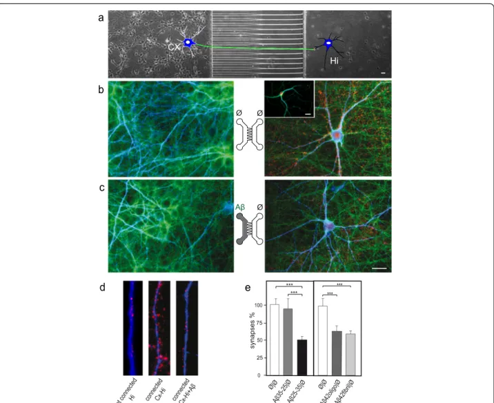
neuronal degeneration. Axons were compartmentalized from the somato-dendritic compartment by seeding
cortical neurons in μFD devices comprising two
cham-bers connected through asymmetrical micro-channels (2C-μFD diodes). When seeded in the left chamber, cortical neurons projected their axons from the somato-dendritic compartment to the distal compartment through filter-ing micro-channels (Figure 1a-a’,b). Analysis of den-drites (MAP2, red) and nuclear chromatin visualization (DAPI, blue) indicated that the seeded cortical neurons were healthy after two weeks in culture (Figure 1b, Additional file 1: Figure S1a). Somato-dendritic application of fibrillar Aβ25-35, mimicking amyloid plaques, induced axonal degeneration process (Figure 1c, g). While somato-dentritic application of aggregated Aβ did not affect somatic and dendritic viability (MAP2, red; DAPI, blue; Figure 1c) some local synaptic damage was evidenced in the som-atic chamber (MAP2, blue; Vglut1, red; Additional file 1:
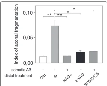
Figure S1b). Addition of NAD+, pharmacological inhibitors
of c-Jun N-terminal kinases (JNK, SP600125), or caspases inhibitor (z-VAD-fmk), to the axonal compartment signifi-cantly reduced Aβ-induced axonal degeneration (Figure 2). In contrast, distal axonal application of aggregated Aβ failed to induce any significant axonal degeneration in our paradigm (Figure 1d-g). Interestingly, while low dose of somato-dendritic glutamate treatment did not trigger axonal degeneration per se (Figure 1e-g, tubulin, grey), the combination of distal Aβ application with this sub-toxic dose of glutamate to the somato-dendritic compartment triggered a massive axonal fragmentation (Figure 1f-g). This suggests that axonal resistance toward local Aβ deposition is tightly controlled by the somato-dendritic behavior and that upstream neurotransmitter dysfunction may precipi-tate neuronal network collapse.
Cortical somato-dendriticβ-amyloid peptide exposure induces a rapid disconnection of cortico-hippocampal synapses, which precedes axonal degeneration
To decipher whether local Aβ might induce remote toxic effect on synapses we used 2C-μFD “axonal diodes” chips that allows reconstructing oriented neuronal networks [8], to create a unidirectional cortico-hippocampal network. When primary cortical neurons (Cx) were cultured at high density in the left chamber of the device and hippocampal neurons (Hi) were seeded at low density in the right chamber (Figure 3a), cortical neurons projected axons
(Figure 3b-c; α-tubulin, green) through the funnel-shaped
micro-channels and established synapses with hippocampal neurons in the right chamber (Figure 3b-d; MAP2, blue; α-synuclein, red). Thanks to the funnel-shaped μ-channels, hippocampal neurons did not project axons backwards. While hippocampal neurons cultured alone showed few presynaptic cluster along their dendritic shaft (insert in Figure 3b and d), Hippocampal neurons grown in contact
Deleglise et al. Acta Neuropathologica Communications 2014, 2:145 Page 2 of 9
anti-MAP2/Dapi Dapi/Thioflavine anti-β-Tubulin/Thioflavine
b
0,10 0,05 0,00**
index of axonal fragmentation
Ø|Ø
c
Glut Ø Aβ Ø Ø Glut|Ø Glut| Aβ 25-35**
***
d
e
f
g
Ø Aβ Ø Glut Aβa
a
’
a’ Aβ 35-25|Ø Aβ 25-35|Ø Ø|A β25-35Figure 1 Somatic applications of Aβ peptides induce a dying-back pattern. a. Schematic representation of a two-chamber microfluidic device (2-CμFD) with two cell culture chambers interconnected by funnel-shaped micro-channels [8] (a’: cross-section of the microfluidic device. A neuron is represented. Note that only axons can reach the right chamber). b-f. Fluorescence microscopy analysis of axonal degeneration vs somatic status after addition of Aβ peptide in the left (soma) vs right (distal part of axons) chambers. Cortical neurons were grown for 12 days in two-compartment chips and each chamber was treated for 48 h with Aβ peptides as indicated. The two left micrographs show the somato-dendritic chamber after staining with anti-MAP2 (red), DAPI (blue) and Thioflavine S (green). The right rectangular micrographs show the distal chamber after immunostaining with anti-α-tubulin and labeling with Thioflavine S. Each central scheme represents the device with indicated localization of treatment (left chamber vs right chamber): b) control cultures (Ø/Ø), c) somato-dendritic treatment with Aβ 25–35 peptide (Aβ/Ø, 30 μM), d) axonal treatment with Aβ 25–35 peptide (Ø/Aβ, 30 μM), e) somato-dendritic treatment with glutamate (Glut/Ø, 10 μM), f) somato-dendritic treatment with glutamate combined with axonal treatment Aβ 25–35 (Glut/Aβ, 10 μM/30 μM). Scale bar: 20 μm. g) Quantification of axonal fragmentation. Aβ25-35, glutamate (Glut), and control Aβ35-25 were added, or not (Ø), to the chamber as indicated. Axonal fragmention index was calculated as described in Methods section (n = 3; **P < 0.01; ***P < 0.001, ANOVA).
with cortical fibers showed high density presynaptic clusters
as evidenced by denseα-synuclein (Figure 3d) or VGLUT1
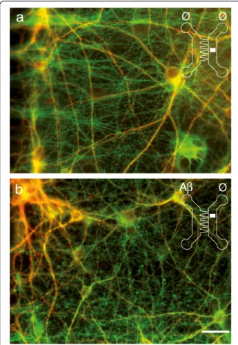
(not shown) staining along the hippocampal dendrites. Application of Aβ25-35 peptide to the Cx compart-ment induced cortico-hippocampal synapse loss in the Hi compartment within 24 h (Figure 3c-e), whereas cortical axons and soma showed no obvious sign of de-generation (Figure 3c). Similar results were obtained with nanomolar doses of oligomeric or fibrillar Aβ1-42 (Figure 3e) suggesting that this process does not rely on the aggregation state of the peptides. While cortical fibers were still intact 24 h after Aβ (peptide or oligomer) appli-cation, at 48 h after treatment, connected axons started to degenerate (Figure 4). We thus observed that the first structural alteration following somato-dendritic Aβ deposits is a distant synapse loss that is followed by delayed axonal degeneration, reminiscent of a dying back process.
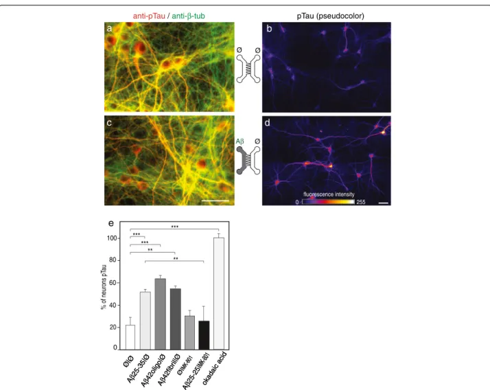
Selective cortical Aβ peptide exposure induces Tau Thr231 phosporylation in a distant fluidically isolated hippocampal neuron through synaptic NMDA receptor-dependent neurotransmission
It is known that a direct exposure of hippocampal neurons to extracellular soluble Aβ oligomers induce in vivo and
in vitro intracellular Tau hyperphosphorylation (tauHP+)
and trigger decreased microtubule stability and NFT formation [9]. We consequently examined whether Aβ deposits (oligomeric or fibrillar Aβ1-42, or Aβ25-35) in the Cx chamber could induce distant post-synaptic Tau phosphorylation in the Hi chamber. The antibody
recognizing the phospho-threonine 231 epitope detects one of the earliest phosphorylation changes observed in AD patients [10]. After 24 h of Aβ treatment in the Cx chamber we found that phosphorylation levels of Tau Thr231 increased in post-synaptic hippocampal neurons (Figure 5; pTau T231, pseudo-colors). Tau Thr231 phosphorylation was particularly concentrated in the cell bodies and proximal dendrites of hippocampal neurons, rather than in distal dendrites and axons. This effect appeared to be glutamate dependent as phosphoryl-ation was prevented by hippocampal pre-treatment with the NMDA receptor antagonist, MK801 (Figure 5e). Hence, Aβ-mediated disturbance of glutamatergic neurotrans-mission and concomitant synapse loss can induce Tau phosphorylation in connected hippocampal neurons and subsequently axonal degeneration of cortical fibers, and therefore initiates progressive neuronal network collapse (Figure 6).
Discussion
A large body of evidence indicates that neurons affected in AD follow a dying-back pattern of degeneration, where such loss of axonal integrity precede somatic cell death and has a profound effect on neuronal network function. However, the molecular mechanism underlying dying-back of neurons and its consequences on the neuronal network in AD remain elusive and difficult to
study in vivo. Using a new μFD system [8], we modeled
for the first time the perforant pathway, known to be affected early in AD [1,2]. Somato-dendritic Aβ cortical application within cortico-hippocampal network leads to a rapid presynaptic collapse before cortical axonal or somatic loss [4,5]. Since these synapses were not exposed to Aβ, this suggests that local somato-dendritic Aβ deposits have fast remote toxicity on the unchallenged synapses. This could be due either to a self-propagation [11] and to rapid distribution of Aβ through axonal transport [12] or to the induction of a signal in the soma, which is trans-mitted through the neuron. We recently described similar distant synapto-toxicity following axotomy [13]. Although no short-term morphological alteration of postsynaptic hippocampal neurons was observed, the Aβ-induced remote loss of cortical presynapses was concomitant to Tau Thr231 phosphorylation in the in-terconnected postsynaptic hippocampal neurons, and occurred well before axonal and somatic degeneration of cortical neurons. Thus local Aβ deposits generate fast propagation of degenerative signals across networks leading to early dysfunctions in remote areas. Our results show that local somato-dendritic (but not axonal) Aβ triggers distal-to-proximal axonal degeneration before any somato-dendritic abnormalities, a process reminiscent of dying-back pattern observed in various neurodegenera-tive syndromes [14]. Thus Aβ toxicity depends not only Figure 2 Axonal administration of NAD+, broad-spectrum
caspase inhibitor, and JNK inhibitor reduces Aβ-induced axonal degeneration. Cortical (Cx) neurons were cultured for 12 days in μFD chambers as in Figure 1. Somatic Aβ peptide treatment (48 h) induced a strong axonal fragmentation, which could be reversed by pre-treatment of the axonal compartment with 5 mM NAD+, 50
μM z-VAD-fmk or 50μM SP600125 (n = 5; *P < 0.01; **P < 0.001, ANOVA).
Deleglise et al. Acta Neuropathologica Communications 2014, 2:145 Page 4 of 9
on direct contact but also on the location of its subcellular deposition. Axons are relatively resistant to direct Aβ exposure, which is in line with our recent observation showing that axonal endings are resistant to direct pro-apoptotic insults [6]. Our observation with JNK and cas-pase pharmacological inhibitors suggest that both enzyme families are implicated locally (within axon) in the process of axonal degeneration, as already observed with other
neuronal death inducers [6]. We also shows, for the first
time, that axonal addition of NAD+ is protective against
Aβ-peptide induced axonal degeneration (Figure 2). Since
NAD+ is a well-established axo-protectant in the
con-text of Wallerian degeneration, this raises the question
whether there might be a NAD+ controlled molecular
pathway implicated in both Aβ-induced axon degeneration
and Wallerian degeneration. Therefore NAD+ related
Figure 3 Cortical Aβ-peptide exposures induce early synaptic loss in a reconstructed cortical-hippocampal network. a. Phase contrast picture of the microfluidic device comprising funnel-shaped micro-channels allowing unidirectional axonal growth. Cortical neurons (Cx) were seeded in the left chamber, hippocampal neurons (Hi) in the right chamber, and were cultured for 14 days to reconstruct a cortical-hippocampal network. A cartoon representing one cortical neuron connected to one hippocampal neuron is inserted for clarity. b-e. Effect of Aβ42 oligomers and Aβ25-35 peptides on synaptic connections. Cortical and hippocampal neurons were cultured in μFD chambers as shown in a. b,c) Representative fluorescence micrographs from somato-dendritic compartment of Cx neurons (left panels) and from Hi neurons in the distal chamber receiving cortical fibers (right panels). Cx chambers were treated with sham (b, Ø/ Ø) or Aβ25-35 (c, Aβ /Ø). Neurons, axons and synapses were immunodetected using anti-MAP2 (blue), anti-α-tubulin (green), and anti-α-synuclein (red) respectively. Scale bar: 20 μm. Similar presynaptic patterns were observed with anti-synaptophysin labeling (not shown). d) Representative higher magnification showing presynaptic clusters (anti-α-synuclein, red) affixed to hippocampal dendrites (anti-MAP2, blue) in hippocampal neurons cultured alone (Hi) connected as in b (Hi-Cx) and connected plus treated with Aβ as in c (Hi-Cx + Aβ). e) Quantification of presynaptic structures affixed to hippocampal dendrites after cortical exposure to oligomeric Aβ1-42 (10 nM), fibrillar Aβ1-42 (10 nM), Aβ25-35 peptides (10 μM), and Aβ35-25 peptides (10 μM). (n = 3; **P < 0.01; ***P < 0.001, ANOVA).
pathways might offer interesting targets to slow down dying back induced processes.
In some AD patients, increased Aβ is associated with
complex disturbances of neuronal activity (e.g. epileptic activity) [15] and neurotransmission dysfunctions have been described in early phases of AD models [16,17]. Such local circuit disturbance might potentially lead to a broader network disruption in remote areas through neuronal projections. Interestingly, we observed that a mild somatic glutamatergic stress exacerbates
dis-tant axonal toxicity of Aβ. This suggests that
cumula-tive and multi-focal stresses might play an important role in disease progression, by switching from local (and probably recoverable) minor dysfunctions to ex-tended neuronal alterations like permanent synaptic, axonal or even cell bodies loss. Several non-exclusive
mechanisms may account for AD-related spatiotemporal progression in the brain. This may involve neurotrophic factors withdrawal, aberrant neurotransmission, synaptic loss, excitotoxicity and prion-like spread of pathogenic misfolded proteins like trans-synaptic spread of Aβ [3]. Interestingly, in our paradigm, blocking NMDA receptors on hippocampal neurons during cortical somato-dendritic Aβ treatment prevented Tau Thr231 phosphorylation. These results are consistent with studies reporting that Aβ could potentiate potassium-evoked glutamate release from neurons [18,19].
Conclusion
While brain complexity, with its interconnected neuronal loops, complicates the in vivo analysis of pathophysiological initiation and spreading mechanisms, we were able for the first time to evaluate the distant effects of local
cor-ticalβ-amyloid deposits on neuronal subcompartments and
networks inμFD-based reconstructed cortico-hippocampal
networks. We show that a strictly local somato-dendritic amyloid trigger is sufficient to recapitulate a dying-back process, and to initiate an oriented neuron-to-neuron pro-gression of pathological events. Aβ peptide accumulation in the somato-dendritic compartment of cortical neurons leads to a fast anterograde propagation of degenerative signals toward endings, resulting in presynaptic collapse. This fast loss of cortical presynapses is associated with early trans-synaptic dysfunction such as NMDAR-dependent tau phosphorylation in postsynaptic hippocampal neurons.
Hence, reconstructed cortico-hippocampal μFD networks
offer a new tool to decipher mechanisms that could under-lie dying back and Braak’s staging.
Methods
Primary culture in microfluidic chips
Microfluidic chips were realized as described in [8]. The design used for network reconstruction comprises two culture chambers each connected to two reservoirs and
separated by a series of 500 μm-long asymmetrical
micro-channels (3μm high, tapering from 15 μm to 3 μm)
[8]. E16 embryos (Swiss mice, Janvier, France) were
micro-dissected in GBSS (without CaCl2and MgCl2) + 0.1%
glucose (Merck), digested with papain (20U/mL) and mechanically dissociated in DMEM containing DNAse.
120.103 cortical cells and 45.103hippocampal cells were
seeded in the chamber on contact with the wide and narrow sides of micro-channels, respectively. Cells were cultured in DMEM glutamax (Invitrogen) supple-mented with 10% FBS (PAA), N2, streptomycin/penicillin and B27 (Invitrogen). The culture medium was renewed every 3–5 days. Cortical axon entered the micro-channels and reached the hippocampi-containing chamber in 4–5 days. The cortico-hippocampal oriented network was main-tained routinely up to 2–3 weeks in vitro.
a
b
Ø Ø
Ø
A
β
Figure 4 Exposure of cortical somata to Aβ-peptide induces an axonal loss in a reconstructed cortical-hippocampal network. Cortical (Cx) and hippocampal (Hi) neurons were cultured for 14 days inμFD chambers as in Figure 2. Hippocampal neurons and projecting cortical axons were immunodetected using anti-MAP2 (red), anti-α-tubulin (green). Cx chambers were treated with sham (a, Ø/ Ø) or 10μM Aβ42 oligomers (b, Aβ /Ø) for 48 hours. Representative fluorescence micrographs from the distal hippocampal chamber receiving cortical fibers. Scale bar: 20μm.
Deleglise et al. Acta Neuropathologica Communications 2014, 2:145 Page 6 of 9
Oligomeric and fibrillar Aβ peptide preparations
Oligomeric and fibrillar forms of Aβ1-42 (Tocris Bioscience, MN, USA), were produced according to [20] and controlled by electron microscopy (Additional file 2: Figure S2). Briefly, lyophilized peptides were solubilized at 1 mM in 1, 1, 1, 3, 3, 3,-hexafluoro-2-propanol (HFIP, Sigma Aldrich). After 30 min of incubation at RT, HFIP was evaporated overnight and peptides were dried (Speed Vac, 1 h 4°C). Then, Aβ peptide stock solution were obtained byresolubilizationet 5 mM in dimethylsulfoxide (DMSO, Sigma Aldrich). To obtain oligomers, Aβ stock solution was diluted at 100 μM
in phenol-free DMEM-F12 medium (Life technologies), incubated 24 h at 4°C and centrifuged at 20 000 g (10 min; 4°C) before supernatant (soluble Aβ fraction) collection. To obtain fibrils, Aβ stock solution was diluted at 100 μM in HCl 10 mM and then incubated at 4°C for 24 h.
Electron microscopy
Aβ sample aliquots (10 μM) were allowed to adsorb onto carbon-coated 200-mesh copper grids (EMS, PA, USA) for 2 minutes before blotting off. Then, grids were incu-bated for 45 seconds in uranyl acetate 2.5% (w/v) to anti-pTau / anti-β-tub pTau (pseudocolor)
a c 0 255 fluorescence intensity b d Ø|Ø 80 40 20 *** % of neurons pT au Aβ 25-25| MK-801 60 0 ** Ø|MK-801 Aβ 25-35|Ø Aβ 42oligo|Ø Aβ42fibr il|Ø okadaic acid Ø|Ø Aβ 25-25| MK-801 Ø|MK-801 Aβ 25-35|Ø Aβ 42oligo|Ø Aβ42fibr il|Ø okadaic acid e 100 *** ** Ø Ø Ø ***
Figure 5 Cortical Aβ peptide deposition induces glutamate-dependent hippocampal Tau phosphorylation. Cortical (Cx) and hippocampal (Hi) neurons were cultured inμFD chambers as in Figure 2a. a,c. Representative fluorescence micrograph ofcortical neurons (left μFD chamber) after immunostaining of phosphorylated Tau (pTau Thr231, red;β-tubulin, green). Effect of Aβ42 oligomers and Aβ25-35 peptides on synaptic connections. Cortical and hippocampal neurons were cultured inμFD chambers as shown in a. b,c) Representative fluorescence micrographs from somato-dendritic compartment of Cx neurons (left panels) and from Hi neurons in the distal chamber receiving cortical fibers (right panels). b,d. Representative fluorescence micrograph of hippocampal neurons receiving cortical axons after immunostaining of pTau (Thr231; pseudo-colors). b) Control conditions (Ø/Ø), d) cortical treatment with Aβ25-35 (10 nM) for 24 h. Scale bar: 20 μm. e. Quantification of pTau-positive hippocampal neurons after 24 h cortical exposure to fibrillar Aβ25-35, oligomeric Aβ42 or fibrillar Aβ42 (10 nM, each) in the presence or absence of the NMDAR antagonist MK801 (10μM). The results were compared to pTau-positive neurons induced by exposure of Hi neurons to okadaic acid (1 nM, 24 h) (n = 3 **P < 0.01; ***P < 0.001, ANOVA).
produce a negatively stained protein loaded grid. Images were recorded on a Zeiss 912 omega electron microscope (Carl Zeiss Group, Oberkochen, Germany).
Treatments
The following compounds were used: Aβ1-42 peptide (1428, Tocris), Aβ25-35 peptide (H-1192, Bachem), control Aβ35-25 peptide (H2964, Bachem), glutamate
(G8415, Sigma), MK-801 (0924, Tocris),5 mM NAD+
(Sigma), 50μM z-VAD-fmk (R&D Systems, Minneapolis,
MN, USA) and 50μM SP 600125 (Sigma). Oligomeric and
fibrillar forms of Aβ1-42 were produced according to [20] and controlled by electron microscopy. To ensure fluidic isolation between compartments, a hydrostatic pressure difference was generated by over-pressurizing the non-treated chamber as described in [21].
Immunofluorescence detection
Cultures were fixed (4% PFA, 20 min RT) and immuno-stained as described in [13]. Primary antibodies included anti-α-tubulin-FITC (F2168, sigma), anti-MAP2 (M4403, Sigma), anti-Synaptophysin (S5768, Sigma), anti-Vglut1, anti-α-synuclein (4179, cell signaling), anti-pTau Thr231 (44746G, Invitrogen). Species-specific secondary antibodies (coupled to Alexa 350, 488, 555) were used (Invitrogen). Images were acquired with an Axio-observer Z1 (Zeiss) fitted with a cooled CCD camera (CoolsnapHQ2, Ropert Scientific) and images were analyzed using ImageJ.
Quantification of axonal fragmentation
For axonal fragmentation analysis and quantification we used fluorescence microscopy (immuno-detection of tubulin), phase contrast, and picture analysis according to a previously described protocol [22]. Briefly, we used a macro developed in NIH ImageJ software that utilizes the Otsu thresholding
algorithm, and a particle analyzer algorithm of ImageJ. The total area of axonal regions with circularity greater than 0.9 was determined and normalized by the total axonal area, which was measured from the thresholded image. This ratio, termed fragmentation index, is an indicator of the average axonal fragmentation level and is used in statistical comparisons. Indices of 0.005, 0.083, and 0.157 correspond to <5%, 50%, and >95% fragmenta-tion, respectively.
Quantification of synapse loss
Synaptic disconnection was assessed through fluorescence
microscopy by counting α-synuclein clusters affixed to
MAP2 positive hippocampal dendrites. Images were all ob-tained with the same acquisition parameters. The images were similarly processed with ImageJ software before being used for quantification: the brightness/contrast of all control images was optimized manually to eliminate the background and to maximize the signal. The means of the minimum and maximum intensities were then calculated in the con-trol condition, after which these settings were applied to
all images. The images of the threeα-synuclein/α-Tubulin/
MAP2 stainings were merged and the resulting image was used to define the zone where hippocampal dendrites were
sufficiently innervated by cortical fibres.α-synuclein/MAP2
merges were then used for quantification.
Quantification of Tau phosphorylation
Tau phosphorylation was assessed by counting the number of neurons presenting pTau levels above a fixed threshold. Images were all obtained using the same acquisition param-eters. The images were similarly processed with ImageJ soft-ware before being used for quantification: the brightness/ contrast of all control images was optimized manually to eliminate the background and to maximize the signal. The means of the minimum and maximum intensities were then calculated in the control condition, after which these set-tings were applied to all images. All pTau images were then
processed with the“Lookup Tables, fire” plugin to visualize
the intensity of pTau staining with pseudo-colours. The number of neurons above a fixed colour threshold was then counted and normalized by the total number of neurons to have the percentage of hyperphosphorylated tau neurons.
Statistical analysis
Differences were assessed by ANOVA, followed, when appropriate, by a post-hoc Bonferoni test. For all analysis * p-value < 0.05; ** p-value < 0.01; *** p–value < 0.001.
Additional files
Additional file 1: Figure S1. Dendritic morphology of cortical neurons before and after Aβ peptide treatment. Cortical (Cx) neurons were cultured for 14 days inμFD chambers as in Figure 1. Dendrites and Figure 6 Hypothetical model of synaptic, post-synaptic and
axonal pathological events induced by Aβ in a cortico-hippocampal neuronal network.β-amyloid application to the somato-dendritic compartment triggers a so-called“dying-back” leading to a rapid cortical presynaptic loss concomitant with a post-synaptic hippocampal Tau-phosphorylation which could be pre-vented by the NMDA-receptor antagonist, MK-801, before any sign of axonal and somato-dendritic cortical alteration.
Deleglise et al. Acta Neuropathologica Communications 2014, 2:145 Page 8 of 9
pre-synaptic clusters of cortical neurons were immuno-detected using anti-MAP2 (blue), anti-VGLUT1 (red). Cx chambers were treated with sham (a, Ø/ Ø) or 10μM Aβ42 oligomers (b, Aβ /Ø) for 48 hours. Representative fluorescence micrographs of the cortical chamber are shown. Scale bar: 20μm.
Additional file 2: Figure S2. Aβ morphology analysed by electron microscopy. Negative stain of TEM images of either Aβ1-42 oligomers (a) or fibrils (b) and Aβ25-35 aggregates (c).
Competing interests
The authors declare that they have no competing interests. Acknowledgments
This work was supported by“Foundation plan Alzheimer 2010-13”, ANR “PrionSensiTNF: Blan-1312-02” 2011–14 and ANR “Neuroscreen: 2011-RPIB-008-01” 2012–16. B.D. was funded by an MNRT fellowship. Author details
1Institut de Biologie Paris Seine (IBPS), 75005 Paris, France.2CNRS UMR 8256,
Biological Adaptation and Ageing, Team 5: Degenerative Processes in Neurons and Networks, F-75005 Paris, France.3CNRS UMR 7102,
Neurobiologie des Processus Adaptatifs, F-75005 Paris, France.4Sorbonne Universités, UPMC Université Paris 6, F-75005 Paris, France.5Institut Curie,
Laboratory of Macromolecules and Microsystems in Biology and Medicine, F-75005 Paris, France.6CNRS UMR 168, F-75005 Paris, France.7Institute of
Reproductive and Developmental Biology, Imperial College London, Hammersmith Hospital, London W12 0NN, UK.
Received: 16 August 2014 Accepted: 14 September 2014
References
1. Braak H, Alafuzoff I, Arzberger T, Kretzschmar H, Del Tredici K (2006) Staging of Alzheimer disease-associated neurofibrillary pathology using paraffin sections and immunocytochemistry. Acta Neuropathol 112(4):389–404 2. Thal DR, Rüb U, Orantes M, Braak H (2002) Phases of A beta-deposition in
the human brain and its relevance for the development of AD. Neurology 58(12):1791–1800
3. Warren JD, Rohrer JD, Schott JM, Fox NC, Hardy J, Rossor MN (2013) Molecular nexopathies: a new paradigm of neurodegenerative disease. Trends Neurosci 36(10):561–569
4. Terry RD (2000) Cell death or synaptic loss in Alzheimer disease. J Neuropathol Exp Neurol 59(12):1118–1119
5. Scheff SW, Price DA, Schmitt FA, DeKosky ST, Mufson EJ (2007) Synaptic alterations in CA1 in mild Alzheimer disease and mild cognitive impairment. Neurology 68(18):1501–1508
6. Magnifico S, Saias L, Deleglise B, Duplus E, Kilinc D, Miquel MC, Viovy JL, Brugg B, Peyrin JM (2013) NAD + acts on mitochondrial SirT3 to prevent axonal caspase activation and axonal degeneration. FASEB J 27(12):4712–4722 7. Millet LJ, Gillette MU (2013) New perspectives on neuronal development via
microfluidic environments. Trends Neurosci 35(12):752–761
8. Peyrin JM, Deleglise B, Saias L, Vignes M, Gougis P, Magnifico S, Betuing S, Pietri M, Caboche J, Vanhoutte P, Viovy JL, Brugg B (2011) Axon diodes for the reconstruction of oriented neuronal networks in microfluidic chambers. Lab Chi 11(21):3663–3673
9. Mairet-Coello G, Courchet J, Pieraut S, Courchet V, Maximov A, Polleux F (2013) The CAMKK2-AMPK kinase pathway mediates the synaptotoxic effects of Abeta oligomers through Tau phosphorylation. Neuron 78(1):94–108 10. Luna-Munoz J, Chávez-Macías L, García-Sierra F, Mena R (2007) Earliest
stages of tau conformational changes are related to the appearance of a sequence of specific phospho-dependent tau epitopes in Alzheimer's disease. J Alzheimers Dis 12(4):365–375
11. Jucker M, Walker LC (2013) Self-propagation of pathogenic protein aggregates in neurodegenerative diseases. Nature 501(7465):45–51 12. Song HL, Shim S, Kim DH, Won SH, Joo S, Kim S, Jeon NL, Yoon SY (2014)
beta-amyloid is transmitted via neuronal connections along axonal membranes. Ann Neurol 75(1):88–97
13. Deleglise B, Lassus B, Soubeyre V, Alleaume-Butaux A, Hjorth JJ, Vignes M, Schneider B, Brugg B, Viovy JL, Peyrin JM (2013) Synapto-protective drugs evaluation in reconstructed neuronal network. PLoS One 8(8):e71103
14. Adalbert R, Coleman MP (2013) Axon pathology in age-related neurodegenerative disorders. Neuropathol Appl Neurobiol 39(2):90–108 15. Palop JJ, Mucke L (2009) Epilepsy and cognitive impairments in Alzheimer
disease. Arch Neurol 66(4):435–440
16. Palop JJ, Chin J, Roberson ED, Wang J, Thwin MT, Bien-Ly N, Yoo J, Ho KO, Yu GQ, Kreitzer A, Finkbeiner S, Noebels JL, Mucke L (2007) Aberrant excitatory neuronal activity and compensatory remodeling of inhibitory hippocampal circuits in mouse models of Alzheimer's disease. Neuron 55(5):697–711 17. Palop JJ, Mucke L (2010) Amyloid-beta-induced neuronal dysfunction in
Alzheimer’s disease: from synapses toward neural networks. Nat Neurosci 13(7):812–818
18. Kabogo D, Rauw G, Amritraj A, Baker G, Kar S (2008) ss-amyloid-related peptides potentiate K +−evoked glutamate release from adult rat hippocampal slices. Neurobiol Aging 31(7):1164–1172
19. Liang Z, Liu F, Iqbal K, Grundke-Iqbal I, Gong CX (2009) Dysregulation of tau phosphorylation in mouse brain during excitotoxic damage. J Alzheimers Dis 17(3):531–539
20. Stine WB, Dahlgren KN, Krafft GA, LaDu MJ (2003) In vitro characterization of conditions for amyloid-beta peptide oligomerization and fibrillogenesis. J Biol Chem 278(13):11612–11622
21. Taylor AM, Blurton-Jones M, Rhee SW, Cribbs DH, Cotman CW, Jeon NL (2005) A microfluidic culture platform for CNS axonal injury, regeneration and transport. Nat Methods 2(8):599–605
22. Kilinc D, Peyrin JM, Soubeyre V, Magnifico S, Saias L, Viovy JL, Brugg B (2011) Wallerian-like degeneration of central neurons after synchronized and geometrically registered mass axotomy in a three-compartmental microfluidic chi. Neurotox Res 19(1):149–161
doi:10.1186/s40478-014-0145-3
Cite this article as: Deleglise et al.:β-amyloid induces a dying-back process and remote trans-synaptic alterations in a microfluidic-based reconstructed neuronal network. Acta Neuropathologica Communications 2014 2:145.
Submit your next manuscript to BioMed Central and take full advantage of:
• Convenient online submission
• Thorough peer review
• No space constraints or color figure charges
• Immediate publication on acceptance
• Inclusion in PubMed, CAS, Scopus and Google Scholar
• Research which is freely available for redistribution
Submit your manuscript at www.biomedcentral.com/submit
![Figure 1 Somatic applications of A β peptides induce a dying-back pattern. a. Schematic representation of a two-chamber microfluidic device (2-C μ FD) with two cell culture chambers interconnected by funnel-shaped micro-channels [8] (a ’ : cross-section of](https://thumb-eu.123doks.com/thumbv2/123doknet/14628793.547841/4.892.86.807.125.841/somatic-applications-peptides-schematic-representation-microfluidic-chambers-interconnected.webp)