Assessment of In Vitro Engineered Microvascular Networks and
their Application in the Treatment of Chronic Wounds
by
Connie Hong-Yee King
S.B. Materials Science and Engineering Massachusetts Institute of Technology, 2006
Submitted to the Department of Materials Science and Engineering in Partial Fulfillment of the Requirements for the Degree of Master of Engineering in Materials Science and Engineering
at the
Massachusetts Institute of Technology September 2007
C Connie Hong-Yee King. All rights reserved.
The author hereby grants to MIT permission to reproduce and to distribute publicly paper and electronic copies of this thesis document in whole or in part
in any medium now known or hereafter created.
Signature of Author
Department o~?aterials Science and Engineering
/ August 7, 2007
Certified by
Certified by
Accepted by
Mark Keegan Senior Member of the Technical Staff, The Charles Stark Draper Laboratory
. A Thesis Supervisor
- •. IjV ,
- .(/ ' V " I Darrell J. Irvine
Associate Professor of Materials Science & Engineering and Biological Engineering
I, Thesis Supervisor
/ LI -Samuel M. Allen
POSCO Professor of Physical Metallurgy
Chair, Departmental Committee on Graduate Students
Assessment of In Vitro Engineered Microvascular Networks and
their Application in the Treatment of Chronic Wounds
by
Connie Hong-Yee King
Submitted to the Department of Materials Science and Engineering on August 7, 2007 in partial fulfillment of the
Requirements for the degree of Master of Engineering in Materials Science and Engineering
Abstract
As the number of individuals suffering from tissue loss and end-stage organ failure continues to grow, researchers are turning to tissue engineering to provide better methods of treatment. The field, however, still faces many technical challenges that are limiting its applications. One challenge faced in engineering more complex tissues and organs is the need for inherent microvasculature to supply the tissue with nutrients and oxygen. Researchers at The Charles
Stark Draper Laboratory have developed a method for engineering microvascular networks in vitro using various microfabrication techniques. This paper discusses the current state of the research and technical challenges to overcome before commercializing the technology. The feasibility of using the networks in the nearer term application of treating chronic wounds will also be assessed, and a potential business strategy will be laid out.
Thesis Supervisor: Mark Keegan
Title: Senior Member of the Technical Staff, The Charles Stark Draper Laboratory Thesis Supervisor: Darrell Irvine
Acknowledgements
I would like to thank my advisor Mark Keegan for all of his guidance and support throughout this past year. This thesis would not have been possible without our numerous meetings at
Starbucks and e-mail exchanges. Darrell Irvine, my DMSE advisor, also deserves many thanks for helping me through the entire thesis process. I would also like to acknowledge the M.Eng director and DMSE for their administrative assistance.
I am grateful for the continual support of my family and friends. Thank you especially to Hannah and Stephen for encouraging me every step of the way. Finally, thank you God for everything.
Table of Contents
Table of Figures ... 7 Table of Tables ... 8 1 Introduction ... 2 Background ... 10 2.1 Tissue Engineering ... .. ... 102.1.1 Tissue Engineering of Skin... ... ... 11
2.1.1.1 Cutaneous W ounds ... 11
2.1.1.2 Physiology of W ound H ealing... ... .. ... 12
2.1.1.3 Current State of Research ... ... 15
2.1.2 Tissue Engineering of M icrovasculature... 16
2.2 M icrofabrication Technologies... ... ... ... ... ... 17
2.2.1 Photolithography ... ... 18
2.2.2 Plasm a Etching... ... ... ... 19
2.2.3 Replica M olding... ... 19
2.3 D raper's Technology ... ... ... . ... 20
2.3.1 Fabrication of M aster M olds ... ... 21
2.3.2 Fabricating Polym er Tem plates ... ... 22
2.3.3 Cell Seeding of N etworks ... ... 23
3 Technical Challenges & Considerations ... ... 25
3.1 M aterial Selection... ... 25 3.1.1 A liphatic Polyesters ... ... 27 3.1.2 Polyurethanes ... ... 29 3.1.3 Poly(ortho esters) ... 31 3.1.4 Polyanhydrides...31 3.1.5 Poly(glycerol sebacate) ... ... 32
3.1.6 N atural Polym ers... ... 33
3.2 Network Geom etry... ... ... ... 34
3.3 Porosity... 35
3.4 Coatings and Surface M odifications... ... 36
3.5 Cell Seeding... ... 38
3.6 Perfusion M edia... .... ... 39
4 Chronic W ound H ealing Applications ... ... 46
4.1 Chronic Wounds ... ... ... ... 46
4.1.1 Burns ... 46
4.1.2 V enous Stasis U lcers... ... 47
4.1.3 Diabetic U lcers... 47
4.1.4 Pressure U lcers... 48
4.2 Market Analysis ... 48
4.3 Competitive Technologies... ... 51
4.3.1 Grafts...51
4.3.3 O ther Com m ercialized Technologies ... 56
4.3.4 Technologies in D evelopm ent... 57
4.4 D raper's Potential Product ... ... 58
4.5 Intellectual Property ... 60 4.6 Business Plan... ... 61 4.6.1 Cost A nalysis ... ... 61 4.6.2 Business M odel ... ... 62 4.6.3 FD A R egulatory Procedures ... 64 5 C onclusion ... 66 R eferences ... ... 67
Table of Figures
Figure 1. Anatomy of skin...in ... 11
Figure 2. Acute vs. impaired chronic wound healing ... 13
Figure 3. Photolithography processing steps ... 19
Figure 4. Basic steps in fabricating Draper's microvascular networks ... 20
Figure 5. 2D microfluidic network model designed with a shear-based algorithm ... 21
Figure 6. Scanning electron micrograph of a vascular network etched on a silicon wafer ... 22
Figure 7. Microfabricated PDMS network...23
Figure 8. Schematic of culture system used with PDMS networks ... ... 24
Figure 9. (a) Confluent layer of HMEC-1 cells in a PDMS network after approximately 1 week of culturing (b) HMEC-1 cells after 2 weeks of culturing ... 24
Figure 10. Cross-section of a bonded PGS capillary... 34
Figure 11. 2004 U.S. medical device market revenues by medical specialty... ... 49
Figure 12. Compound Annual Growth Rate (CAGR) of U.S. medical device industry segments (2004-11) ....49
Figure 13. Wound care and management market: segmentation and corresponding revenues in 2004...50
Figure 14. (a) Graphic of AlloDerm® acellular matrix components and (b) application of GRAFTJACKETr to a w ound...52
Figure 15. Apligraf (a) in storage dish and (b) applied to a foot ulcer... ... 55
Figure 16. Schematic of potential wound healing product based on Draper's technology ... 59
Table of Tables
Table 1. Synthetic biodegradable polymers for tissue engineering applications...26
Table 2. Angiogenic growth factors... 40
Table 3. Wound healing growth factors... 41
Table 4. Major participants in the interactive wound care market ... 50
Table 5. Skin substitutes ... 53
Table 6. List of Draper's current patent applications ... 61
1 Introduction
In the past few decades, the field of tissue engineering has emerged to address the growing issue of tissue loss and organ failure among individuals. Currently, more than half of U.S. health care expenditures, or over $600 billion, are spent treating these conditions.1 Transplants, autografts, and prostheses have been used with some success thus far, but these methods are accompanied by problems of limited supply and lack of long term biocompatibility. While tissue engineering seeks to provide a superior alternative to current methods of treatment, many challenges must be overcome before its applications can be made widely available for clinical use. One major challenge faced in engineering complex organs is the necessity of microvasculature to deliver nutrients and oxygen and remove waste. Researchers at The Charles
Stark Draper Laboratory are attempting to address this issue by fabricating three dimensional microfluidic networks to serve as in vitro engineered microvasculature in tissue engineering applications. This paper serves to assess the current state of the technology and the feasibility of its nearer term application in the treatment of chronic wounds.
2 Background
2.1 Tissue Engineering
The field of tissue engineering draws on knowledge from engineering and science disciplines to accomplish two main purposes: to achieve a better understanding of tissue
structure-function relationships and to use this understanding to develop biological substitutes to regenerate and repair tissue.2 As research continues, new approaches to restoring tissue loss and organ failure will be introduced. Current approaches such as transplants, autografts, and
prostheses have saved many lives and improved the quality of life for many others. However, these approaches are far from sufficient. With transplants, the demand far exceeds the supply. In 2002, there were 79,512 patients waiting for transplants in the U.S. alone. Less than a third received organs, and over 6,000 died waiting on the list.3 Currently, there are 96,957 individuals awaiting transplants. With only 9,218 transplants performed in the first four months of this year, there is an obvious need for additional therapies.4 Even after receiving a transplant, there is
always the concern of organ rejection. Other conditions such as burns or bone and vascular injuries may be treated with autografts. By using tissue grafts from the patients themselves, concerns of rejection are eliminated; however, availability is even more limited. In some cases, the physical function of injured organs can be maintained through the use of prostheses;
examples include artificial hips and pacemakers. While prostheses are considered biocompatible, they are still accompanied by long-term complications.5 Tissue engineering shows great
potential for serving as an approach that addresses all of these problems.
The goal of developing biological substitutes has been approached through a variety of in vivo and in vitro methods. Several strategies deal only with the isolation and expansion of cell
biomolecules that induce tissue regeneration. Lastly, much research has concentrated on the development of matrices to aid in the regeneration of tissue.6 These matrices, designed to
possess properties similar to those of the tissue to be regenerated, also often incorporate cells and/or biomolecules. They can be implanted immediately at the site of injury or allowed to synthesize tissue in vitro prior to implantation.5
2.1.1 Tissue Engineering of Skin
2.1.1.1 Cutaneous Wounds
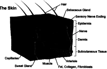
The skin (Figure 1) consists of two layers, the epidermis and dermis, which are separated by a basement membrane. Although it is only approximately 100 pm thick, the epidermis serves as the body's primary protective barrier against infection and fluid loss. The dermis is a thicker layer of vascular connective tissue that contains skin appendages such as sweat glands, hair follicles, and sebaceous glands. It provides mechanical and metabolic support for the thinner, avascular epidermis.
The
Skin\
\
Sebaceous GlandSensory Nerve Ending -Epidermie -Nerve -Dermis -Subcutaneous Tissue Ca S Gland/_ YaMusc l \
Sweat GlandFat, Collagen, Fibroblasts
Figure 1. Anatomy of skin7
When only the epidermis and basement membrane sustain injury, the body is able to spontaneously regenerate the lost tissue. Wounds that extend into the dermis, on the other hand, are healed through a repair process. The tissue resulting from this process differs structurally from that of the original tissue.5 Deeper wounds are described as partial or full thickness wounds when a portion or all of the dermis is injured in addition to the epidermis. Partial thickness wounds can be further categorized as superficial or deep to describe the degree of damage to the dermis more precisely.8
2.1.1.2 Physiology of Wound Healing
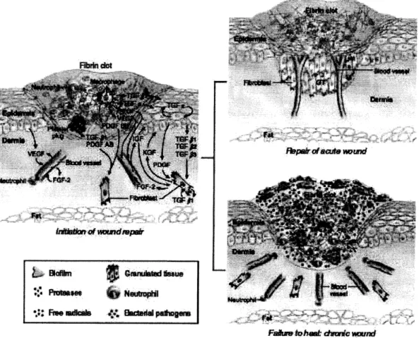
The repair process (Figure 2) of a typical wound consists of three phases: inflammation, proliferation, and remodeling. Two main responses occur during the inflammation phase: a vasomotor-vasopermeability response and leukocyte infiltration. Immediately after injury, hemostasis is reestablished through vasoconstriction and the formation of a fibrin clot via platelet adhesion. Subsequent vasodilation and increased capillary permeability allow protein-rich serum and cell populations to infiltrate the wound. 9 These cells are attracted by the chemical signals released as the wound healing process is initiated. Neutrophils begin the process of cleansing the wound and preventing infection. Monocytes also migrate to the wound and develop into
macrophages. These cells continue to cleanse the wound through phagocytosis and also release growth factors necessary for the continuation of the repair process.'1
i1f o UWadtvp
PepaiiOftii-o mul
Figure 2. Acute vs. impaired chronic wound healing. Reprinted by permission from Macmillan Publishers Ltd: Journal of Investigative Dermatology Vol. 127, 1018-1029, copyright 2007.12
TGF - transforming growth factor; FGF - fibroblast growth factor; VEGF - vascular endothelial growth factor; PDGF - platelet-derived growth factor; IGF - insulin-like growth factor; KGF - keratinocyte growth factor; GT - granulation tissue
The proliferation phase begins two to three days after injury and is dominated by fibroblast activity. Fibroblasts proliferate, migrate to the wound, and begin synthesizing
extracellular matrix (ECM) components, including collagen and glycosaminoglycans (GAGs), to replace lost connective tissue. Angiogenesis also occurs during this phase to support the
metabolic needs of the fibroblasts.9 As a result, the newly synthesized tissue is highly vascularized, giving it a granular appearance and thus its name - granulation tissue.10 If angiogenesis fails to occur, fibroblast migration is halted, and wound healing is inhibited.9 Reepithelialization begins hours after injury with the migration of keratinocytes from skin
_I
appendages and wound edges and continues throughout the proliferation phase. In the second week after injury, myofibroblasts also begin compacting connective tissue and contracting the wound to aid in wound closure.10
The final phase of wound healing begins after homeostasis between collagen synthesis and degradation has been achieved. This occurs approximately three weeks after the wound is created, but remodeling can continue for up to two years. As remodeling occurs, many of the blood vessels and fibroblasts in the newly synthesized connective tissue undergo apoptosis, transforming the granulation tissue into a scar. Collagen fibers are also reorganized to strengthen the new tissue.'0 The wound strength gradually increases from approximately 20% (at three weeks after injury) until it plateaus at 70-80% of the strength of undamaged tissue.9'10
The final morphology of the repaired skin differs from normal skin in several ways. Normal dermal tissue consists of randomly aligned collagen fibers, and the dermo-epidermal junction contains rete ridges. In scar tissue produced through repair, collagen fibers are mainly aligned in the plane of the epidermis, and the dermo-epidermal junction lacks the characteristic rete ridges. Additional differences include the composition of the connective tissue and lack of
skin appendages that were damaged during injury.3
In some cases, the wound healing process does not complete properly, resulting in a chronic wound (Figure 2). Chronic wounds are characterized by a lack of epidermal migration and granulation tissue growth; this often results from difficulty in moving past the inflammation phase."1,12 A variety of issues can lead to the inhibition of healing. Vascular problems that may
contribute to the problem range from arterial insufficiency to venous hypertension to the failure of angiogenesis at the wound site. Without functioning vasculature to maintain metabolic activities, fibroblasts are unable to continue migrating and synthesizing new tissue. In other
cases, sufficient amounts of growth factors may be lacking due either to undersecretion or rapid metabolism. Other factors including infection and edema, the accumulation of fluid in tissues, may also slow healing.9 Some chronic wounds will eventually heal with increased amounts of
granulation tissue and fibrosis, which lead to significant scarring and reduction of functionality.9
2.1.1.3 Current State ofResearch
Research regarding the tissue engineering of skin has resulted in the development of many commercialized skin substitutes for use in cutaneous wound healing. Scientists are now capable of isolating and expanding keratinocyte cultures by 10,000 fold in 2-3 weeks. This has allowed for the development of autologous and allogeneic epidermal replacements using cultures from patients and neonatal foreskin, respectively.12,13 Two main types of dermal substitutes have also been developed. Cadaveric allografts have been treated to remove immunogenic elements such as cells and in some cases, the epidermis. Other dermal substitutes have been prepared by constructing scaffolds from ECM components. In some cases, these matrices are subsequently seeded with allogeneic fibroblasts. Bilayered skin substitutes are the most recent advances to be commercialized. These substitutes provide both an epidermal component with keratinocytes and a dermal component with fibroblasts.'2 While both skin components are supplied, they are limited in thickness and do not always take well. This is due to the fact that they lack vascularization to supply nutrients to the cells within the scaffolds. The cells must rely on imbibition, or the diffusion of nutrients, until neovascularization occurs.14
2.1.2 Tissue Engineering of Microvasculature
While thinner and simpler tissues have been successfully engineered, the challenge of engineering vascularized tissue has prevented the engineering of thicker and more complex tissues. While the former is capable of relying initially on diffusion for oxygen and nutrients, the latter cannot.14 An inherent vasculature would enable the engineering of tissues with greater metabolic needs and thicknesses greater than the 100-200 gtm nutrient diffusion limit.3
To date, success in engineering vasculature has been primarily confined to individual, large diameter (> 5 mm) blood vessels. Bypass grafts have been constructed using Dacron and expanded polytetrafluoroethylene (ePTFE). These materials, however, have proven insufficient for smaller diameter blood vessel substitutes due to their thrombogenicity. 5 Clotting and scarring of the blood vessels often leads to stricturing or even complete occlusion. To counteract this effect, many devices are coated with anti-thrombogenic coatings, such as heparin-based coatings. Researchers have also begun conducting studies on lining materials with endothelial cells to prevent thrombosis.
Although researchers are currently investigating a wide range of methods for engineering blood vessels with a high patency, the gold standard for microvascular grafts (internal diameter <
1 mm) is still the autologous vein graft. Autografts, however, cannot be used to provide
microvasculature for tissue-engineered organs due to the fact that a complete and intact capillary bed cannot be dissected out.14 Researchers have thus turned to two possibilities for providing
regenerated or engineered tissue with microvasculature: therapeutic angiogenesis and the inosculation of pre-vascularized tissue constructs.
Therapeutic angiogenesis, also referred to as angioinduction, attempts to stimulate the formation of new blood vessels from a host's existing vasculature through the delivery of
angiogenic molecules. These molecules are typically growth factors such as vascular endothelial growth factor (VEGF), placental growth factor (PlGF), angiopoietin-1 (Ang 1), platelet-derived growth factor-BB (PDGF-BB), and transforming growth factor 3 (TGF-p).3 These factors can be introduced directly into the blood stream or incorporated into cells, microstructures, or scaffolds for more controlled release.14 Alternatively, adenoviral vectors can be used to promote
angiogenesis by transferring genes that express angiogenic growth factors.
The second method aims to develop the microvasculature in vitro. This is typically done by seeding a tissue-engineered scaffold with the appropriate cells to form new blood vessels.3 These vascularized scaffolds are then implanted, and the microvessels join with existing vasculature in the host. While much work is being conducted in this area, clinical studies have yet to be conducted on any scaffolds with pre-existing vascular networks.14
2.2 Microfabrication Technologies
As researchers have continued to search for new strategies for engineering
microvasculature, some have begun to adapt technologies from other fields. One promising method is the adaptation and use of microfabrication technologies to construct templates for vascularized networks. Originally developed for the production of integrated circuits and microelectromechanical systems (MEMS), microfabrication techniques enable the fabrication of structures with a high degree of resolution. Microfabrication techniques have been further developed over recent years to enable the printing and molding of elastomeric stamps. These techniques, including replica molding, are referred to as soft lithography.16
2.2.1 Photolithography
In microfabrication, photolithography is used to transfer a two-dimensional pattern onto a given structure. A mask with the desired pattern is created using a computer aided design (CAD) software package. Optical techniques are then used to transfer the mask pattern onto a substrate on which the final structure is built through the deposition and etching of layers.'7
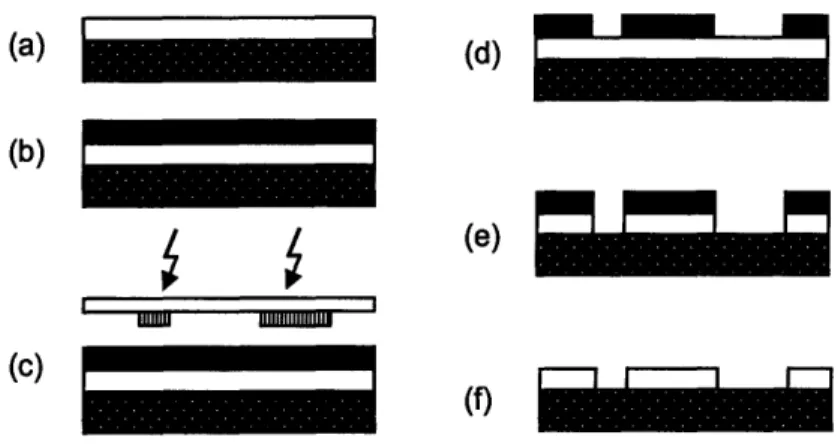
The basic steps of UV photolithography are shown in Figure 3. The process begins with a clean substrate on which a thin oxide film has been deposited. Alternatively, nitride,
polycrystalline silicon, aluminum, or noble metal films may also be used. Deposition techniques include thermal oxidation, evaporation, sputtering, and various chemical vapor deposition methods. A photoresist is then applied using one of a few possible methods: spin coating, electrochemical coating, spray coating, or casting.18 In the most common process, spin coating,
a few milliliters of resist are applied to the wafer which is then spun to spread the resist into a uniform layer. Photoresists can be divided into two types: positive and negative. In positive resists, areas exposed to light become more soluble and are thus removed. In negative resists, polymerization occurs in the regions exposed to light, and the unpolymerized regions are
subsequently removed. After applying the resist, the wafer is exposed to UV light and developed. The film is then etched using wet or dry etching techniques, and the resist is stripped off to leave the patterned film. Additional steps for priming, baking, and rinsing may also be added.17
(a) (d)
(b)
(e)
nlU uIus us11111
Figure 3. Photolithography processing steps: (a) apply film for patterning to clean substrate; (b) spin-coat resist onto film; (c) expose to UV; (d) develop; (e) etch film; (f) strip resist'7
2.2.2 Plasma Etching
Plasma etching is a dry etching technique that involves the excitation and ionization of gases in a vacuum chamber. These gases in turn react chemically and physically with the film surface. This method allows for more accurate anisotropic etching and thus the ability to pack structures more closely.18 In recent years, high aspect ratio micromachining (HARMS)
technologies have been developed that use deep reactive ion etching (DRIE) to etch channels of even greater depths. Channels as deep as 40 and 400 pm have been etched with widths within + 0.1 and 1 pm, respectively.19
2.2.3 Replica Molding
Replica molding, as its name implies, is a method of fabricating stamps and molds by replicating the three-dimensional pattern from a solid substrate onto an elastomer. To produce the replica mold, the prepolymer is cast over the patterned master and cured. The most
commonly used elastomers are siloxane-based polymers such as poly(dimethylsiloxane) (PDMS). After curing, the newly patterned elastomer is peeled off; the use of an elastomer allows for
easier separation from the master, which is typically produced from a rigid material. The polymer mold can then be used to pattern other materials. Features on the order of tens of
nanometers can be successfully reproduced using this technique.'6
2.3 Draper's Technology
At the Charles Stark Draper Laboratory, researchers have applied microfabrication techniques to the problem of engineering microvasculature for the regeneration of tissues and organs. Masks have been designed based on microfluidic models of blood flow. These masks are used to pattern silicon masters using traditional microfabrication techniques. Soft
lithography and polymer processing techniques are then used to make polymeric molds that are subsequently joined to form the final closed microvascular networks. The basic fabrication steps are shown below in Figure 4.
far1w w - m
(d)
(b)
SI(e I
(c)
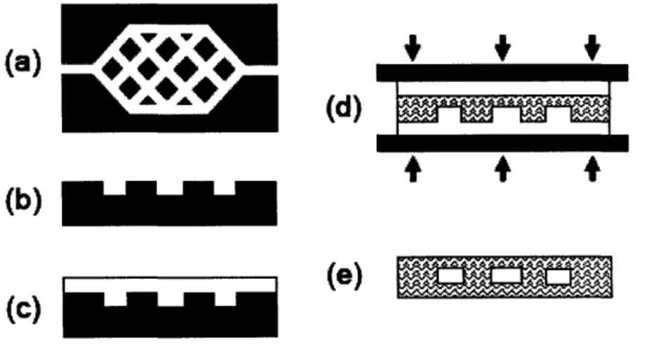
Figure 4. Basic steps in fabricating Draper's microvascular networks: (a) design a mask using microfluidic models; (b) use the mask to pattern a silicon substrate; (c) fabricate a PDMS template via replica molding; (d) fabricate a PLGA template via compression micromolding; (e) thermal fusion bond PLGA templates to form a closed network of channels22
r
2.3.1 Fabrication of Master Molds
The fabrication of Draper's microvascular networks begins with the designing and printing of photolithography masks. Fluid dynamic models (Figure 5) are produced to approximate the behavior of blood flow in target organs. These models account for physiologically important parameters including the non-Newtonian rheology of blood,
hematocrit, flow rate, pressure, and shear stress.20'2 1 These fluid dynamic models are then used
to design masks which are subsequently used to pattern silicon master molds (Figure 6) using photolithography and plasma etching.
Figure 6. Scanning electron micrograph of a vascular network etched on a silicon wafer; reprinted with kind permission from Springer Science and Business Media21
2.3.2 Fabricating Polymer Templates
Once a silicon master has been fabricated, polymer templates can be produced using a variety of techniques. PDMS templates are produced via replica molding; after casting, the prepolymer is cured at 60-650C for three to four hours.21,22 Plasma bonding is then used to bond a patterned PDMS sheet to an unpatterned sheet resulting in a network of closed channels
(Figure 7).21 Alternatively, PDMS templates can be used as molds for the patterning of other polymers. Poly(lactic-co-glycolic acid) (PLGA) templates have been produced via compression
micromolding. PLGA pellets are placed between PDMS molds and heated well above the glass transition temperature. A compressive force is then applied to compression mold the PLGA melt into the pattern on the PDMS templates. Thermal fusion bonding is then used to create closed PLGA networks. This is done by the contacting of two layers and then subsequent heating to just above the glass transition temperature; the interdiffusion of polymer chains bonds the two
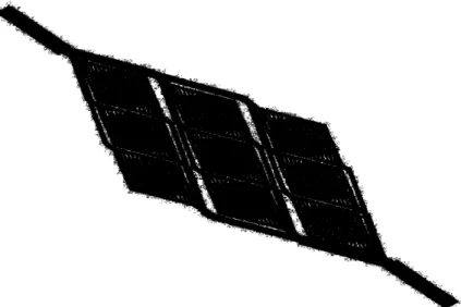
Figure 7. Microfabricated PDMS network with a surface area of 13.69 cm2, internal volume of 21.3 mm3
, channel depth of 35 pm, and channel widths ranging from 35 pm to 5 mm; reprinted with kind permission from Springer Science and Business Media 21
2.3.3 Cell Seeding of Networks
Short term cell viability and proliferation have been demonstrated in fabricated PDMS networks. The networks were sterilized, coated with purified collagen, and then seeded with immortalized human microvascular endothelial cells (HMEC-1). Seeded networks were
incubated at 37°C with 5% CO2, and media was perfused through the network during incubation using a system similar to the one shown in Figure 8. Cells reached confluence after
Figure 8. Schematic of culture system used with PDMS networks21
(a [b
Figure 9. (a) Confluent layer of HMEC-1 cells in a PDMS network after approximately 1 week of culturing
(b) HMEC-1 cells after 2 weeks of culturing; reprinted with kind permission from Springer Science and
3 Technical Challenges & Considerations
Development of Draper's microvascular networks is still in the initial stages. Several technical challenges remain to be overcome before the technology can be commercialized including the selection of a final material, refinement of the network geometry, incorporation of porosity, selection of surface coatings or modifications, optimization of cell culture on scaffolds, and formulating the perfusion media. While some potential solutions for these challenges already exist, research in these areas continues to produce new ideas for materials and methods. Further experimentation will be necessary to determine whether current options are sufficient or if further development of complementary technologies will be required to address Draper's needs.
3.1 Material Selection
When developing scaffolds for incorporation into the body, the efficacy is greatly affected by the choice of material. Polymers are widely used in biomedical applications due to the similarity of their physical properties to those of soft tissue. Additionally, polymers allow for a large degree of tailoring of properties. While biodegradable polymers are typically preferred due to their ability to degrade as new tissue is formed, non-biodegradable polymers are still used for many applications which require mechanical properties not offered by biodegradable
polymers.23
For Draper's networks, a biodegradable polymer is desired for the final material selection. While the biocompatibility of the material is important, some properties such
anti-thrombogenicity and cell adherence can be addressed with surface modifications if necessary. Degradation properties will also play a key role in the decision making process. Considering the fact that the proliferation phase of wound healing lasts for three weeks, the microvascular
network's degradation rate should be close to this length of time to provide support without inhibiting the wound healing process. The method of degradation and degradation products will also be important due to channel patency concerns and the perfusion of biomolecules through the networks. The necessary mechanical properties for the networks still remain to be determined. Research on engineering microvascular grafts typically stresses the importance of burst strength, with values greater than that of saphenous veins (1700 mmHg) being desirable.24 The
compliance of blood vessels is also important as mismatch can induce intimal hyperplasia, thus reducing patency. Compliance values for host artery and saphenous vein grafts are
approximately 5.9 and 4.4% per mmHg x 10-2, respectively.25 While this may provide general guidelines for appropriate mechanical properties, values are likely to differ for microvasculature. Additionally, Draper's networks are not designed to be permanent; they must provide
mechanical support only until host cells have incorporated the networks into existing vasculature. PDMS, a non-biodegradable polymer, was used for early phases of research at Draper Labs because of its ease of use in fabricating microfluidic devices.19 Work has since been adapted to biodegradable polymers such as PLGA and poly(glycerol sebacate) (PGS).22,26 While the ability to apply Draper's technology to biodegradable polymers has been demonstrated, the selection of a final material with appropriate properties remains to be made. A wide range of biodegradable polymers have shown potential for use in tissue engineering applications, and researchers continue to develop polymers and copolymers with new combinations of properties. A few of the main synthetic biodegradable polymers have been listed in Table 1 and will be
discussed along with natural polymers in considering the desired material properties for Draper's vascularized networks.
Melting Point Glass Degradation
Polymer Transition (C) Strength Time Degradation Products
("C)
Transition ("C)
Time
7.0 GPa
Poly(glycolic acid) 225 -230 35 -40 (modulus) 6-12 months Glycolic acid
(modulus)
2.7 GPa
Poly(1-lactic acid) 173 -178 60 -65 (modulus) >2 years L-lactic acid
(modulus)
0.4 GPa
Poly(caprolactone) 58- 63 -65 - -60 (modulus) >2 years Caproic acid
(modulus)
8-40 MPa Lysine, glycolic acid,
Polyurethane* -- - 1-2 months
(tensile strength) caproic acid
Sufficient for load Diols, acids, &
Poly(ortho ester)** - -10 -10 Days to months
bearing perntaerythritol 1.3 MPa 12 months(in
Poly(anhydride)*** -- -MPa 12 months(in Dicarboxylic acids
(modulus) vitro)
Poly(glycerol 0.3 MPa
sebacate) 5; 38 -- (modulus) 2 months Sebacic acid, glycerol
sebacate)
(modulus)
* Based on lysine diisocyanate & poly(glycolide-co-y-caprolactone)
** POE IV prepared with 5 mole % lactide, 3,9-diethylidene-2,4,8,10-tetraoxaspiro[5,5]undecane, and n-octanediol, n-decanediol, or n-dodecanediol
** Poly[1,6-bis(carboxyphenoxy)hexane]
3.1.1 Aliphatic Polyesters
Poly(glycolic acid) (PGA), poly(lactic acid) (PLA), and poly(caprolactone) (PCL) are three of the most commonly used biodegradable polymers in biomedical research and
applications. PGA is a highly crystalline, and thus inelastic, polymer with a high tensile strength and modulus. Because of its rapid degradation rate and low solubility in common organic
solvents, it is often combined with PLA to form PLGA.14 PLA, a semi-crystalline polyester, also possesses a high tensile strength and modulus but degrades at a slower rate and is easier to process.30 Lactide has three isomers - L-lactide (LLA), D-lactide (DLA), and meso-lactide
-with LLA being preferentially metabolized by the body.27,30
By varying the ratios of PGA and PLA isomers used, PLGA with varying mechanical and chemical properties can be obtained. PLGA is currently used in many formulations of
degradable sutures.30 A 90% glycolide and 10% L-lactide formulation sold by Ethicon as
Degradation of PLGA by hydrolysis leads to the homogeneous erosion of amorphous and then crystalline regions of the copolymer.27
While certain properties of PLGA can be tailored simply based on the composition, factors related to the interaction of PLGA with biomolecules and cells require more complicated approaches for tailoring. Porous PLGA structures result in very rapid leaching of angiogenic factors after incorporation. To enable slower and more controlled release of factors, PLGA microspheres, beads, and bioactive foams have been fabricated.14 Another concern related to biomolecules is their potential inactivation by the local accumulation of acidic degradation products.27 These acidic degradation products have also been associated with adverse tissue reactions in bony sites of the body.32
Finally, PLGA exhibits a limited affinity for cells. Thus, additional coatings or surface modifications may be required to ensure proper cell attachment
and growth in certain tissue engineering applications.26 While these concerns may necessitate further processing techniques, they can still be addressed.
Other in vivo degradation properties of PLGA have proven to be more unavoidable, which have caused it to be viewed as less than ideal for many tissue engineering applications
such as drug delivery. For instance, after implantation, PLGA undergoes a lag phase with minimal mass change. This is followed by a rapid loss of mass via bulk degradation.
Mechanical strength, on the other hand, undergoes a sharp drop immediately after implantation. Furthermore, as PLGA degrades, it swells and deforms significantly.33 These degradation properties could potentially compromise flow through fabricated channels, thus suggesting that another synthetic polymer may be more appropriate for use in Draper's networks.
Polycaprolactone (PCL) is another aliphatic polyester that has been widely studied in addition to PLA and PGA. It is semicrystalline and has a much slower degradation rate, on the
order of two to three years.27 Therefore, it lends itself to more long term drug delivery
applications such as the contraceptive device, Capronor, which delivers zero-order release of levonorgestrel for over a year.3 0 PCL also differs from other aliphatic polyesters in having
extremely low glass transition and melting temperatures but a high decomposition temperature of 3500C; this could allow for easier processing of the polymer. While PCL homopolymers
degrade too slowly to be practical in this application, PCL can be used to form a wide range of compatible polymer blends.34 Many of these are now being studied for use in applications including bone and vascular tissue engineering.35,36' 37'38
3.1.2 Polyurethanes
Polyurethanes (PUs) are a class of elastomers which have been used in larger diameter vascular grafts and are now being developed for use in smaller diameter grafts. These polymers are highly attractive for vascular tissue engineering applications due to their elastic and
compliant mechanical properties and biocompatibility. Many difficulties were encountered in the initial development of PUs including hydrolytic instability, susceptibility to oxidative degradation, and the toxicity of degradation products derived from some diisocyanate
components. Researchers have since developed newer generations of PUs that eliminate these issues.27,39
Several PUs and PU composites are now in various stages of development. Thoratec Corporation has commercialized a polyetherurethaneurea vascular access graft and is now conducting clinical trials on the use of the material for small-diameter coronary bypass grafts. Cardiotech International has also developed a family of poly(carbonate-urea)urethanes under the trademarked name Chronoflex®.39
Saad et al. have developed a group of block copolyesters termed DegraPol". These degradable polyesterurethanes are based on diisocyanates of PHB/HV-diol and PCL-diol or Diorez®. While J774 macrophages and 3T3 fibroblasts cultured on Degrapol® exhibited lower levels of cell adhesion and growth than cells cultured on tissue culture polystyrene (TCPS), fibroblasts on Degrapol" produced more ECM. When implanted subcutaneously in rats,
moderately thick fibrous capsules (60-250 jm) formed. Additionally, implants were reduced to approximately 50% of their molecular weight after one year.36 While these results suggest moderate biocompatibility, the degradation rate may be slower than desired.
Another group of poly(ester-urethane)ureas (PEUUs) have been developed by Guan et al. These PEUUs are synthesized from PCL and 1,4-diisocyanotobutane with lysine ethyl ester or putrescine chain extenders. Excellent mechanical properties and human umbilical vein
endothelial cell (HUVEC) viability and proliferation have been demonstrated on unmodified and RGDS-modified PEUUs.
Williamson et al. have developed a porous scaffold for small diameter grafts using gravity spun PCL fibers wrapped around a stainless steel mandrel and coated with electrospun PU. The PCL fibers are spun with a fiber-to-fiber gap of 1-5 jim, and the PU anti-luminal surface is characterized by 10-30 pm pores. HUVECs and smooth muscle cells (SMCs) both exhibited good attachment to the scaffolds.38
PU composites are also being developed for use in skin substitutes. For example, Bruin et al. have constructed lysine diisocyanate (LDI)-based poly(glycolide-co-&-caprolactone) urethane networks which degrade within four to eight weeks. Ninety to 250 jIm pores were
formed using salt leaching techniques, and a non-porous polyetherurethane layer was glued on top to form a bilayered skin substitute approximately 2 mm thick.40
3.1.3 Poly(ortho esters)
Poly(ortho esters) (POEs), developed by Alza Corporation, are a group of hydrophobic polymers with hydrolytically sensitive backbones. The high degree of hydrophobicity prevents water penetration and results in slow degradation by surface erosion. Consisting of four different families, these polymers have primarily been developed for use in drug delivery. POE I is synthesized from diols and diethoxytetrahydrofuran, POE II from diols and a diketene acetal, and POE III from a triol and ortho ester. POE IV is a modification of POE II that includes segments of lactic or glycolic acid in the backbone to aid in degradation of the polymer.3 0
Of the four families, POE IV is the only polymer which appears to have the
characteristics necessary for commercialization. Synthesis of POE IV is simple and reproducible, the polymer possesses appropriate thermal and mechanical properties, and degradation rates can be varied from a few days to months. Degradation occurs through surface erosion, resulting in the linear weight loss and release of lactic and propionic acid. The mechanism for degradation also prevents the interior of the polymer matrix from becoming highly acidic as is common with PLGA.29 While surface erosion would allow networks to maintain their structure and strength as they degraded, the high degree of hydrophobicity in POE IV could potentially inhibit the proper delivery of nutrient medium to cells within the scaffold.
3.1.4 Polyanhydrides
Polyanhydrides are another group of surface-eroding polymers. Because of their poor mechanical properties and rapid degradation rate, they have been studied primarily for use in drug delivery applications. Their degradation, which is dependent on both polymer structure and the monomers used, can be slowed by the incorporation of aromatic carboxylic acids in place of
aliphatic ones.30 Mechanical properties can also be significantly enhanced by copolymerization
with polyimides. In these poly(anhydride-co-imides), hydrolysis of the anhydride bonds occurs first, followed by the imide bonds.27
3.1.5 Poly(glycerol sebacate)
As previously mentioned, Draper has also fabricated vascularized networks using PGS. PGS, a biodegradable and biocompatible elastomer developed by Wang et al., possesses
mechanical properties similar to those of veins.26 It has been suggested that it degrades primarily through surface erosion since it exhibits a gradual loss of mass and mechanical strength.28'33 Endothelial cells adhere to PGS, but surface modification with GRGDS peptides increases uniformity of distribution and adhesion.26 The in vivo biocompatibility of PGS has been tested, and it exhibits responses comparable to, if not better than, PLGA for inflammation and fibrous capsule formation.28
While the mechanical properties of PGS would be more relevant to blood vessel grafts immediately connected to a host's vasculature, a stronger material may allow earlier inosculation
of microvascular networks by mechanically supporting the blood vessels before they fully mature. Another potential advantage of PGS is that it exhibits surface erosion but also has a hydrophilic surface.28 Fabrication of PGS microvascular networks has already been
accomplished, and although the size and uniformity of the pores is unknown, porous scaffolds have also been fabricated separately through salt leaching.26' 28
3.1.6 Natural Polymers
The use of natural polymers in tissue engineering applications has obvious appeal due to their excellent biocompatibility. For example, collagen, the most abundant protein in the human body, has been incorporated into various skin substitutes including Integra, Apligraf, and Orcel.30
Benefits of using collagen in matrices include its natural abundance in skin, promotion of cellular adhesion and proliferation, and its ability to be cross-linked to increase mechanical strength. Because collagen is typically present in later stages of wound repair, some have suggested that it may not be the optimal material for cell migration.12 However, in addition to
cell surface receptors, type I collagen is highly porous (50-150 pm pores); this suggests that the late appearance of collagen in wound repair may not be a substantial issue.14 An additional concern with regards to the use of collagen may be its high thrombogenicity.30
Another natural polymer highly researched for wound healing applications is hyaluronic acid. While it is a known mitogen and motogen of mesenchymal and epithelial cells, cell adherence to the material is poor.14'30 Additionally, its degradation products are angiogenic, but HA itself inhibits endothelial cell proliferation. Modifications to address these issues include adding RGD sequences and cross-linking with chitosan, another GAG. Chitosan attracts
neutrophils and activates macrophages. The HA-chitosan combination has been used in research to make human skin equivalents.14 Although natural polymers are typically more biocompatible
than synthetic ones, they also raise additional concerns such as antigenicity and batch-to-batch variation.30
3.2 Network Geometry
While microfabrication techniques have been successfully used to create three
dimensional polymer networks, a significant amount of process refinement still remains to be carried out. One example is the further enhancement of channel geometry. Currently, channels with varying widths but uniform depths have been fabricated, resulting in rectangular cross-sections (Figure 10). While the effect of geometry on flow, pressure changes, and turbulence is currently being assessed, it can be assumed that, considering the actual geometry of blood vessels, cylindrical channels would be preferred. The processing changes required to accomplish this are fairly straightforward; cylindrical channels can be formed by bonding two patterned polymer templates, each with a semicircular trench.21 Though straightforward, the
micromachining of semi-circular channels may be a lengthy process as it requires the deposition and etching of many layers.
Figure 10. Cross-section of a bonded PGS capillary; reprinted with permission from Mary Ann Liebert, Inc.26
Enhancement of the three dimensional architecture of networks may also be worth consideration. Currently, two dimensional networks are stacked to form the three dimensional networks; the inlets and outlets connect the ends of all of the two dimensional networks.
Therefore, the branching of the vasculature still primarily occurs in two dimensions. Additional studies will need to be conducted to assess the effect of this on the efficacy of the networks.
3.3 Porosity
Another related technical challenge that must be addressed is the incorporation of porosity into the matrix surrounding the networks in order to facilitate the migration of cells and
diffusion of perfusion media throughout the matrix. Techniques for the fabrication of porous scaffolds via solvent casting are currently being investigated at Draper for this purpose.
Considering the processes involved in fabricating the vascularized networks, such as replica and compression molding, steps for introducing porosity will likely be carried out after channels are formed. Additionally, pores of differing sizes may be desired for different areas of the construct. Macropores (-500 pm) regulate cell type and seeding density, mesopores (-20-30 ipm) allow for
the infiltration of microvessels, and micropores (< 10 pin) regulate molecular transport.14 Studies by Matsuda and Nakayama have found that for a range of micropored segmented polyurethane films (SPU) with constant pore area per unit area, pore diameters between 18 and
50 rpm were optimal for endothelialization with bovine thoracic aorta ECs. Complete
endothelialization was achieved after two days for all pore sizes tested between 18 and 100 pm while migration through 9 pm pores was significantly retarded. Other studies have shown that SPU prostheses with pores sizes less than 15 pm, between 15-45 lm, and greater than 50 ipm result in minimal, fibrohistiocytic, and organized fibrous tissue growth, respectively.41
In fabricating complete devices for tissue engineering applications, pores of at least 100 pm would be desired in the polymer scaffold surrounding the channel networks. Here the porosity would serve mainly to facilitate the migration of cells and ingrowth of tissue. In the
case of materials such as PU, porosity could also increase the flexibility of the material and thus allow for better compliance matching with blood vessels.41 Depending on how well media is able to diffuse through the polymer used, micropores may also be formed in the channel walls to provide initial vascular permeability. Blood vessels must exhibit some degree of permeability to allow the transport of macromolecules. This is especially important for Draper's channel
networks as the perfusion medium will not only deliver nutrients and oxygen, but also growth factors to enhance angiogenesis and wound healing in the area. Permeability, however, must be carefully controlled to prevent imbalances in oncotic pressure, osmotic pressure due to
macromolecules, which can lead to edema.3
3.4 Coatings and Surface Modifications
Coatings and surface modifications of tissue-engineered devices typically serve two main purposes: providing an anti-thrombogenic surface and/or promoting specific cellular functions.
The issue of thrombogenicity arises with all tissue engineering devices that are in direct contact with blood. While a variety of anti-thrombogenic coatings are available and used in vascular grafts, using ECs as a coating may be preferred for Draper's technology. While this does present added challenges and complications, cells will eventually be necessary components in the networks if any significant amount of tissue synthesis is to be carried out in vitro. In addition to issues regarding maintaining cultures, additional coatings or surface modifications may be required to promote cell attachment and growth on the channels and in porous regions.
Materials which promote cell attachment typically have receptors for many different cell types. Additionally, simple methods for coating various materials with preclot, fibronectin,
improve cell selectivity on various parts of the networks, surface modification with cell adhesion ligands may be preferred over coatings of actual cell adhesive materials. In considering surface modifications, the specific ligand type, spacer length, and peptide surface concentration are all
important factors.
Hubbell et al. touch upon each of these issues in their study of the attachment of human foreskin fibroblasts (HFFs), human vascular smooth muscle cells (HVSMCs), HUVECs, and
quiescent and activated human blood platelets.43 Glycophase glass and PEG-modified PET surfaces were functionalized at concentrations of 10 pmol/cm2 and 20 finol/cm2, respectively, with four different peptide sequences: Arg-Gly-Asp (RGD), Tyr-Ile-Gly-Ser-Arg (YIGSR), Pro-Asp-Ser-Gly-Arg (PDSGR), and Arg-Glu-Asp-Val (REDV). RGD is a cell-binding ligand found on many cell adhesion proteins while YIGSR and PDSGR are found on laminin and REDV is found on fibronectin. Results suggested that HVSMCs require higher concentrations of ligand (>20 fmol/cm2) than HFFs, and all cell types require higher concentrations of PDSGR than RGD and YISGR to adhere to surfaces. Additionally, REDV selectively promotes the adhesion of HUVECs. These results are consistent with other studies showing that at least 10 fmol/cm2 GRGDY is required for human fibroblast spreading. While platelets have also been shown to adhere to RGD-functionalized surfaces, it was suggested that this is only the case with longer spacer lengths.43 The results from this study suggest that REDV may be an appropriate ligand for the surface modification of channels while RGD or YIGSR could be used to modify the porous regions surrounding the channels.
3.5 Cell Seeding
When pre-seeding vascular networks in vitro, endothelial cells will likely be the primary cells of concern as they line all blood vessels and are the only cells found in small capillaries.44 However, a combination of cells may be needed to optimize the subsequent growth of cells and tissue on the networks. For example, much research indicates that while blood vessel and tissue growth can be induced with ECs alone, the vessels produced are typically unstable and do not last very long.3 Additionally, human ECs require the addition of angiogenic factors such as phorbol 12-myristate 12-acetate (PMA), basic fibroblast growth factor (bFGF), VEGF, and fetal calf serum (FCS) to form blood vessels. Some factors, including PMA, are known to promote tumors and thus not likely to be used in clinical applications. Better results, however, have been achieved through co-culturing with fibroblasts. Fibroblasts are able to synthesize greater
quantities of ECM and enable the formation of more stable, functional blood vessels.45 The synergistic effects of co-culturing cells are likely due to cell-cell and cell-matrix interactions, both of which help to regulate angiogenesis. HUVECs produce a very limited amount of ECM when cultured alone while fibroblasts are capable of producing substantial ECM when cultured alone.4 5 Thus, in addition to providing cell-cell interactions, co-culturing of cell
types may also allow for improved EC cell-matrix interactions on synthetic polymer networks. While the majority of research on the development of vasculature in vitro focuses on various types of differentiated ECs, some researchers are now suggesting the use of endothelial progenitor cells (EPCs) instead. EPCs, which differentiate into ECs, can be isolated from adult peripheral blood.46 While they are found in extremely low concentrations, cultured populations can be doubled more than 1,000 times. Mature ECs, on the other hand, senesce after
differentiation, migration, and/or proliferation without additional angiogenic factors.46 The
seeding of EPC-derived ECs and SMCs onto PGA-PLLA scaffolds in studies by Wu et al. have resulted in the formation of layered tissue containing microvessels. Other studies have also been conducted to demonstrate the endothelialization of gelatin-coated PU grafts by EPCs.4 7
In considering the application of vascular networks to cutaneous wound healing applications, seeding with keratinocytes could also be used to enhance reepithelialization. If used, these cells would be seeded on the top surface of the microvascular networks after seeding with ECs and fibroblasts. The effects of keratinocyte seeding density have been previously studied by Butler et al. using a collagen-GAG matrix. In order to achieve confluence of
uncultured autologous keratinocytes in two weeks, a seeding density of at least 100,000 cells/cm2 was required. This seeding density allowed for a 30-fold expansion over the donor site surface area.48 A subsequent study revealed that proliferation increases when autologous keratinocytes are cultured prior to seeding.13 Thus confluence can be achieved with lower seeding densities if cultured keratinocytes are used instead. After the determination of cell sources and cell types to be cultured on Draper's microvascular networks, additional experiments will be necessary to optimize seeding densities for each cell type.
3.6 Perfusion Media
The optimization of medium formulations for perfusion also presents a significant challenge. Appropriate combinations and concentrations of biomolecules must be determined in order to sustain cell viability, regulate specific cell functions, and promote angiogenesis.
Furthermore, the incorporation of antimicrobial components such as silver nanoparticles may be desirable as this could eliminate the need for additional antibiotics. In order to promote
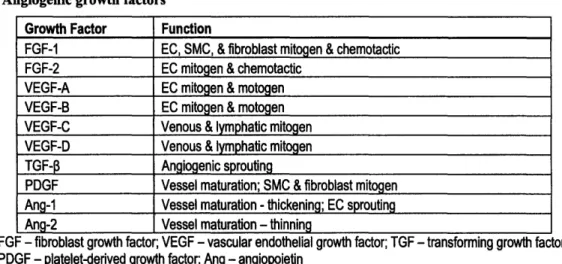
angiogenesis, a variety of growth factors should be considered for addition to the perfusion media. Table 2 lists the major growth factors known to be involved in angiogenesis. Because angiogenesis is an essential part of wound repair, many of the same growth factors are found in healing wounds (Table 3).
Table 2. Angiogenic growth factors14'49 Growth Factor Function
FGF-1 EC, SMC, & fibroblast mitogen & chemotactic
FGF-2 EC mitogen & chemotactic VEGF-A EC mitogen & motogen
VEGF-B EC mitogen & motogen VEGF-C Venous & lymphatic mitogen
VEGF-D Venous & lymphatic mitogen
TGF-P3 Angiogenic sprouting
PDGF Vessel maturation; SMC & fibroblast mitogen
Ang-1 Vessel maturation - thickening; EC sprouting Ang-2 Vessel maturation - thinning
FGF - fibroblast growth factor; VEGF - vascular endothelial growth factor; TGF - transforming growth factor;
Table 3. Wound healing growth factors9'10, 50
Family Growth Factor Primary Sources Primary Target Cells & Effect
EGF Platelets Keratinocyte motogen & mitogen
Epidermal growthEpidermal growth TGF-a Macrophages; keratinocytes Keratinocyte motogen & mitogen HB-EGF Macrophages
Keratinocyte & fibroblast mitogen FGF-1 (aFGF)
Fibroblast growth FGF- (FGF) Macrophages; endothelial
FGF-2 (bFGF) cells Angiogenesis; fibroblast mitogen
factor (FGF) FGF-4
FGF-7 (KGF) Fibroblasts Keratinocyte motogen & mitogen
TGF-31 Keratinocyte motogen; chemotactic for Transforming growth Platelets; macrophages macrophages & fibroblasts; ECM synthesis &
factor P (TGF-P) TGF-12 remodeling
TGF-P3 Macrophages Antiscarring
Platelet-derived Chemotactic for macrophages & fibroblasts;
growth factor (PDGF) / PDGF keratinocytes macrophage activation; fibroblast mitogen; matrix
vascular endothelial production
growth factor (VEGF) VEGF Macrophages; keratinocytes Angiogenesis
Insulin-like growth IGF-1 Plasma; platelets Endothelial cell & fibroblast mitogen
factor (IGF)
Interleukins (ILs) IL-la & [ Neutrophils Early activator of growth factor expression in
macrophages, keratinocytes, & fibroblasts
Colony stimulating CSF-1 Multiple cells Macrophage activation and granulation tissue
factor (CSF) ellsproduction
Other TNF-a Neutrophils Similar to IL-ls
Other Activin Fibroblasts; keratinocytes Unknown
Other CTGF (CCN2) Fibroblasts; endothelial cells Fibroblasts; downstream of TGF-31
HB-EGF - heparin-binding epidermal growth factor; aFGF - acidic FGF; bFGF - basic FGF; KGF - keratinocyte growth factor;
TNF - tumor necrosis factor; CTGF - connective tissue growth factor
Fibroblast growth factor (FGF), the first angiogenic growth factor to be discovered, is part of a family of growth factors consisting of at least 20 molecules. FGFs are produced by vascular ECs and SMCs and have been found to stimulate both EC and fibroblast growth.
FGF-1, also known as acidic FGF (aFGF), is involved in capillary progression, wound healing, and tumor progression.49 FGF-2, also referred to as bFGF, is essential in early wound repair
processes occurring in the first three days.l0 Together with vascular endothelial growth factor
(VEGF), these two growth factors bind to fibrin to retain bioactivity and synergistically promote angiogenesis.12,49
Included in the platelet-derived growth factor (PDGF) / VEGF family are the growth factors PDGF, VEGF, and PlGF.,949 PDGF is both the most potent mitogen in serum for mesenchymally-derived cells, such as fibroblasts and SMCs, and a strong chemotactic for inflammatory cells. It consists of two polypeptide chains, A or B chains, which give rise to its three isoforms: PDGF-AA, PDGF-BB, and PDGF-AB.51 Its concentration in human serum is estimated at 15-50 ng/mL with maximum chemotaxis or activation of various cell types at varying concentrations. Lower concentrations of 1-5 ng/mL and 10-20 ng/mL are optimal for neutrophil chemotaxis and SMC and fibroblast attraction, respectively. A concentration of 20 ng/mL allows for maximum monocyte chemotaxis, and even higher concentrations of 20-40 ng/mL activate neutrophils.52 Johnson & Johnson Wound Management Worldwide produces the only FDA-approved recombinant human growth factor product currently on the market in its REGRANEX® (becaplermin) gel. With 0.1% recombinant human PDGF-BB (rhPDGF-BB), REGRANEX® promotes the healing of diabetic foot ulcers.9'53 VEGF is another growth factor
essential to proper wound healing with VEGF-A, or VEGF165, being the most common human isoform.49 It induces the local formation of blood vessels and thus becomes important once granulation tissue begins to form around days four to seven.10'32 The regulation of VEGF is affected by conditions such as hypoxia and a wide range of other growth factors including PDGF, tumor necrosis factor a (TNF-a), FGF-2, FGF-4, FGF-7, transforming growth factor P (TGF-p), angiotensin-2, insulin-like growth factor-i (IGF-1), interleukin-1 (IL-1), IL-6, IL- 10, IL- 13, Ang-1, and Ang-2.49
The TGF-3 family is another group of growth factors involved in angiogenesis and wound healing. TGF-01, TGF-p2, and TGF-P3 are all present in low, if not undetectable, levels
elevates, with 31 having the highest concentration of about 30 ng/mL. P2 and TGF-P3 are found in much lower concentrations of 1-1.5 and 1-2 ng/mL, respectively.54 It has been suggested that TGF-31 and TGF-02 are related to scarring and inflammation in adults.9'49 This is due to observations that the factor is present in high concentrations in adult wounds in which these two phenomena occur but in relatively low concentrations in embryonic wounds, which heal without these phenomena. TGF-P31 and TGF-P2 concentrations can both be down-regulated in wounds through the application of TGF-P3.50 In addition to this function, studies by
Bandyopadhyay et al. have also suggested that TGF-P3 may be involved in the regulation of epidermal and dermal cell migration during wound healing. They have observed that plasma (from unwounded skin) promotes the migration of dermal fibroblasts and human dermal microvascular ECs (HDMECs) while serum (from wounded skin) promotes the migration of keratinocytes. This effect has been attributed to the increased levels of TGF-13 in serum.54
The therapeutic use of growth factors must be carefully controlled to prevent pathological angiogenesis. For example, excessive amounts can lead to fibrovascular growth.14 However, the external regulation of growth factors may prove to be a difficult task due to interactions with different receptors and other growth factors. As previously mentioned, VEGF is affected by many growth factors, and TGF-P3 can regulate TGF-131 and TGF-P2. Other examples of these interactions include the ability of TGF-1 to stimulate or inhibit angiogenesis depending on whether its receptor is activin-like kinase type-1 or type 5 (ALK-1 or -5) and the ability of Ang-2 to antagonize Ang-l but also promote neovascularization when combined with VEGF. 14,49
In developing a formulation for perfusion media, a balance will need to be found between using more growth factors to promote a faster response and using fewer factors to limit costs and unknown effects from growth factor interactions. PDGF, VEGF, and TGF-P3 will likely be the
primary components used in promoting angiogenesis. The addition of FGF-2 may not be essential as it is more important in the very early stages of wound healing. TGF-l1 and TGF-32 may also be excluded to limit scarring and inflammation as the wound heals. For chronic wound healing applications, the addition of EGF, TGF-a, and FGF-7 may be desired as these factors have been shown to promote reepithelialization when applied exogenously.50
Another issue of concern is the concentration of growth factors required to achieve a positive response. In vitro, human endothelial cells require a greater amount of growth factors
than animal equivalents such as bovine or rat cell cultures.45 If large concentrations of factors are required over extended periods of time to promote wound healing, this method of treatment may prove to be economically infeasible. On the other hand, one benefit of the incorporation of growth factors into the medium instead of the scaffold itself is that concentrations can be varied as the wound enters different phases of tissue growth and remodeling. Ideally, the
concentrations of the various factors in the perfusion media will match those found naturally in acute wounds. While studies to determine these values have yielded widely varying results, they
provide a starting point for estimating appropriate concentrations.52,54,55,56'57 Based on these studies, a suggested preliminary formulation to be tested is as follows: 50 ng/mL PDGF, 3 ng/mL VEGF, 2 ng/mL TGF-p3, 2 ng/mL TGF-a, and 50 pg/mL EGF.
Finally, flow characteristics of the media as it is perfused throughout the networks must also be considered. One characteristic which has already been addressed to some extent by the microfluidic models is shear stress on vessel walls. Areas of lower shear are associated with the development of atherosclerosis. Thus, Draper has designed its networks to have constant shear rates throughout the networks.20 Shear stress is also an important factor due to its effects on EC
Additionally, higher levels of shear stress result in decreased cell proliferation, the up-regulation of the expression of some genes, and the down-regulation of others. Cyclic stretch resulting from pressure pulses is another important factor. While cyclic stretch can also contribute to EC elongation and orientation like shear stress, other effects contrast with those produced by shear stress.5 s While all cell studies of Draper's networks to date have used constant flow for
perfusing media, further experiments should be conducted to determine whether a pulsatile flow yields better results.