Article
ARP3 Controls the Podocyte Architecture at the
Kidney Filtration Barrier
Graphical Abstract
Highlights
d
ARP3-dependent actin assembly is required for podocyte
process formation
d
Arp2/3 thereby links process formation, podocyte adhesion
and mechano-adaptation
d
Arp2/3 function is regulated by a reciprocal interplay with
actomyosin
Authors
Christoph Schell, Benedikt Sabass,
Martin Helmstaedter, ..., Jo¨rn Dengjel,
Manuel Rogg, Tobias B. Huber
Correspondence
[email protected]
In Brief
Glomerular podocytes maintain the
kidney filtration barrier. Schell et al. show
that the Arp2/3 machinery is required for
podocyte process formation and
adaptation to filtration barrier mechanical
requirements via reciprocal interplay with
the actomyosin network. Dendritic actin
network loss results in actomyosin
cytoskeleton activation and decreased
adhesion, leading to podocyte
detachment.
Schell et al., 2018, Developmental Cell47, 741–757
December 17, 2018ª 2018 The Authors. Published by Elsevier Inc. https://doi.org/10.1016/j.devcel.2018.11.011
Developmental Cell
Article
ARP3 Controls the Podocyte Architecture
at the Kidney Filtration Barrier
Christoph Schell,1,2,3Benedikt Sabass,4Martin Helmstaedter,2Felix Geist,2Ahmed Abed,2Mako Yasuda-Yamahara,2,5
August Sigle,2Jasmin I. Maier,1,2Florian Grahammer,2,6Florian Siegerist,7Nadine Artelt,7Nicole Endlich,7
Dontscho Kerjaschki,8Hans-Henning Arnold,9Jo¨rn Dengjel,10,11,12Manuel Rogg,2,13and Tobias B. Huber2,6,10,13,14,*
1Institute of Surgical Pathology, Medical Center – University of Freiburg, Faculty of Medicine, University of Freiburg, Freiburg 79106, Germany 2Department of Medicine IV, Medical Center – University of Freiburg, Faculty of Medicine, University of Freiburg, Freiburg 79106, Germany 3Berta-Ottenstein Programme, Faculty of Medicine, University of Freiburg, Freiburg 79106, Germany
4Institute of Complex Systems-2, Forschungszentrum J€ulich, J€ulich 52428, Germany 5Department of Medicine, Shiga University of Medical Science, Otsu, Shiga 520-2192, Japan
6III. Department of Medicine, University Medical Center Hamburg-Eppendorf, Hamburg 20246, Germany 7Department of Anatomy and Cell Biology, University Medicine Greifswald, Greifswald 17487, Germany 8Department of Pathology, Medical University of Vienna, Vienna 1090, Austria
9Cell and Molecular Biology, Technical University of Braunschweig, Braunschweig 38106, Germany
10BIOSS Center for Biological Signaling Studies, Albert-Ludwigs-University Freiburg, Freiburg 79106, Germany 11Department of Biology, University of Fribourg, Fribourg 1700, Switzerland
12Department of Dermatology, Medical Center – University of Freiburg, Faculty of Medicine, University of Freiburg, Freiburg 79104, Germany 13These authors contributed equally
14Lead Contact
*Correspondence:[email protected]
https://doi.org/10.1016/j.devcel.2018.11.011
SUMMARY
Podocytes, highly specialized epithelial cells, build
the outer part of the kidney filtration barrier and
withstand high mechanical forces through a complex
network of cellular protrusions. Here, we show that
Arp2/3-dependent actin polymerization controls
actomyosin contractility and focal adhesion
matura-tion of podocyte protrusions and thereby regulates
formation, maintenance, and capacity to adapt to
mechanical requirements of the filtration barrier.
We find that N-WASP-Arp2/3 define the development
of complex arborized podocyte protrusions
in vitro
and
in vivo. Loss of dendritic actin networks results
in a pronounced activation of the actomyosin
cyto-skeleton and the generation of over-maturated but
less efficient adhesion, leading to detachment of
po-docytes. Our data provide a model to explain
podo-cyte protrusion morphology and their mechanical
stability based on a tripartite relationship between
actin polymerization, contractility, and adhesion.
INTRODUCTION
Cellular protrusion formation is involved in many important bio-logical functions ranging from tumor metastasis to embryogen-esis (Ladoux et al., 2016, Bravo-Cordero et al., 2012; Hunter and Fernandez-Gonzalez, 2017; Krause and Gautreau, 2014). Among the best-studied cellular protrusions in 2D is the ‘‘clas-sical’’ lamellipodium, which is generated in a well-coordinated interplay of polymerization, crosslinking and severing of actin
fil-aments (Krause and Gautreau, 2014). Various protrusion types have been identified and differ not only in their molecular compo-sition, but also in their context-dependent properties (Inagaki and Katsuno, 2017; Di Martino et al., 2016; Leijnse et al., 2015). However, not much is known about how extremely fine protrusions can produce outstanding mechanical durability while maintaining adaptive capacity in integrated tissues and organs.
Here, we focused on the podocyte, which is essentially involved in the formation of the kidney filtration barrier by enwrap-ping glomerular capillaries with its specialized cellular protru-sions (namely, foot processes [FPs]; Pavenst€adt et al., 2003). As podocyte FPs are required to maintain morphological stability and cell-matrix adherence against physical filtration forces, they serve as an ideal model to study the contribution of protrusion phenotypes and reciprocal cell-matrix interactions. Retraction and simplification of the complex podocyte FP network is often linked to detachment of podocytes from the glomerular base-ment membrane and presents a key feature of glomerular dis-ease (Grahammer et al., 2013; Kriz et al., 2013). During glomerular development, podocytes undergo dramatic morphogenetic al-terations, transforming from cuboidal epithelial cells into arbor-ized podocytes with a network of highly branched FPs (Schell et al., 2014; Welsh and Saleem, 2011). Based on the identification of disease-causing mutations affecting core components of the actin cytoskeleton (e.g., ACTN4 or INF2), the concept of a tightly controlled cellular morphology for podocyte function has evolved (Brown et al., 2010; Kaplan et al., 2000). However, it is still incom-pletely understood how the podocyte process networks form, which molecular machineries are involved, and if different cyto-skeletal pools exert specific functions.
Here, we show that the Arp2/3 complex controls the integrity of the kidney filtration barrier by influencing two distinct, yet interlinked processes. First, Arp2/3 complex-dependent actin
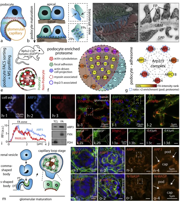
Figure 1. The Arp2/3 Complex Presents a Central Node in the Network of Cytoskeletal Proteins in Podocytes (A) Schematic depicting how podocytes reside on the outer surface of glomerular capillaries.
(B) During glomerular maturation podocytes undergo profound cell shape changes (GBM, glomerular basement membrane; FP, podocyte foot process). (C) Colorized scanning electron microscopy (SEM) image of podocyte protrusions illustrating the complex interdigitating network of podocyte FPs covering glomerular capillaries.
(D) Transmission electron microscopy (TEM) demonstrates FPs attaching to the glomerular basement membrane (GBM; cap - glomerular capillary). Slits between individual FPs are bridged by specialized cell-cell contacts known as slit diaphragms.
(E) Schematic describing the strategy for isolation of genetically labeled primary podocytes and subsequent mass spectrometry (MS) analysis. Following single-cell isolation and further FACS-based purification, respective single-cell populations are analyzed.
(legend continued on next page)
polymerization critically determines the arborization and complexity level of podocyte FPs. Second, Arp2/3-controlled focal adhesion (FA) and traction forces influence podocyte adhe-sion. The Arp2/3 complex provides actin nucleation and fila-ment-branching properties to generate highly branched actin networks, which are essential for the formation and propulsion of lamellipodia (Rotty et al., 2013). It was only recently demon-strated that Arp2/3 also influences the alignment of cell-matrix adhesions and thereby affects cell-matrix-dependent haptotaxis (Wu et al., 2012). In the classical model, Arp2/3-complex-medi-ated filament assembly is activArp2/3-complex-medi-ated by nucleation-promoting factors (NPFs such as SCAR/WAVE and N-WASP) downstream of CDC42- and RAC1-dependent signaling axes (Campellone and Welch, 2010). Advanced models challenged this view, as they identified competition between the Arp2/3 complex and other nucleation machineries (such as the formin protein family) defining critical cellular responses (Lomakin et al., 2015; Wu et al., 2017). Despite many years of research on Arp2/3, little is known about its role in vivo beyond the importance for classical lamellipodia and adhesion structures.
RESULTS
The Arp2/3 Complex Presents a Central Node in the Network of Cytoskeletal Proteins in Podocytes
Given the extensive characterization of the Arp2/3 complex in vitro, we reasoned that Arp2/3-dependent branched actin networks might be involved in process formation as well as FA initiation in glomerular epithelial cells. Podocytes derive from the metanephric mesenchyme (Schell et al., 2014) and exhibit a rather cuboidal morphology in early glomerular development. With ongoing matu-ration, cells shift their polarization pattern and extend numerous cellular protrusions (podocyte processes) toward the glomerular basement membrane (Figures 1A and 1B). These thin processes are interconnected with extensions from adjacent podocytes and provide anchorage to the glomerular basement membrane (protrusions directly evolving out of the cell body are termed pri-mary processes, whereas further arborized and terminally branched protrusions are termed secondary, or FPs;Figures 1C
and 1D). Podocytes reside on the outer surface of glomerular cap-illaries and are exposed to high physical filtration forces. Special-ized adhesion machineries are required to prevent detachment of those cells into the urinary space. We hypothesized that the structure and composition of the actin cytoskeleton network might interconnect FP development with efficient adhesion regulation. To describe the composition of the podocyte cytoskeleton, we made use of our recently developed genetic-reporter-based pro-tocol for highly efficient single-cell isolation and downstream mass spectrometry (MS) applications (Figure 1E). Based on our initial hypothesis, we aimed to identify proteins or complexes that are known to act at the interface of cellular protrusion forma-tion and FA regulaforma-tion. Here, GFP-labeled podocytes were iso-lated from genetic reporter strains and after fluorescence-acti-vated cell sorting (FACS)-based enrichment, MS analysis was performed. Filtering this dataset for podocyte-enriched cytoskel-etal components involved in actin binding, protrusion formation, and FAs resulted in a list of 114 proteins, where the N-WASP/ Arp2/3 complex axis appeared as a central node (Figures 1F and S1;Table S1; comparing podocytes versus non-podocytes, i.e., mesangial and endothelial cells). Employing proteomics of enriched adhesion complexes (focal adhesome), cell fractionation protocols, as well as immunofluorescence, we could furthermore demonstrate that ARP3 is enriched in the adhesion complex of podocytes ( Fig-ures 1G–1J and S2;Table S2). These observations once more corroborated our initial hypothesis that the Arp2/3 complex might function at the interface of propulsive actin network generation and the FA complex regulation in podocytes. Using confocal micro-scopy on human kidney tissue, N-WASP and ARP3 were detected in the podocyte compartment (enriched in podocytes as indicated by co-labeling with the podocyte specific protein SYNPO;Figures 1K, 1L, andS2). Formation and generation of podocyte FPs occurs during glomerular maturation at the so-called capillary loop stage (Figure 1M). Immunofluorescence studies on different maturational stages of glomeruli revealed that ARP3 and N-WASP accumulated at the basal compartment of podocytes where FP specification and arborization occurs (Figures 1N and 1O). This localization pattern was also detected in completely maturated glomeruli of adult mice, where N-WASP except for glomeruli also showed minor
(F) Proteome network analysis of podocyte-enriched proteins revealed high abundance of proteins contributing to distinct functional subsets such as actin binding or focal adhesion-associated proteins. Filtering of the enriched proteins for the indicated functional subsets confirmed components of the Arp2/3 complex as a central node (color coded: enrichment scores and functional groups are indicated inTable S1; proteins were defined as podocyte enriched by a detection ratio >2 for podocytes to non-podocyte glomerular cells).
(G) Focal adhesome proteomics generated out of immortalized human podocytes detected multiple members of the Arp2/3 complex as enriched components of the focal adhesome (FA) (enrichment scores are indicated inTable S2; proteins were hierarchically ranked according to their detection scores).
(H and I) Immunofluorescence of spreading primary wild-type podocytes revealed co-localization of ARP3 and the focal adhesion protein PAXILLIN during early spreading phases (white line indicates region for intensity line scan analysis; RFI, relative fluorescence intensity; FA, focal adhesion).
(J) Western blot analysis of focal adhesion (FA)-enriched cell fractions or total cell lysate (TCL) confirmed ARP3 as a FA-enriched protein in podocytes. PXN (Paxillin) and PDH (Pyruvate dehydrogenase E1) were used to demonstrate successful separation of the focal adhesion fraction from the whole cell compartment (see alsoFigure S2).
(K) Confocal laser scanning microscopy of human glomeruli confirmed distinct localization of the nucleation-promoting factor N-WASP in the podocyte compartment (dashed line boxes indicate areas of higher magnification inserts).
(L) A similar enrichment was also observed for ARP3 employing confocal laser scanning microscopy (inserts in (L) show different levels of z stack series 0.43 micrometers apart; dashed outlines indicate the podocyte compartment; synpatopodin (SYNPO) was used as a marker for the podocyte compartment). (M) Schematic depicting stages of glomerular development (podocyte progenitors and podocytes are shown in green; endothelial cells and capillary loops are depicted in orange).
(N and O) At late capillary loop stage, ARP3 (N) and N-WASP (O) accumulated at the basal compartment of maturating podocytes, where podocyte FP formation takes place (in earlier developmental stages, this prominent accumulation was not apparent; dashed line boxes indicate inserts; arrows indicating the basal podocyte compartment; pod, podocyte; cap, capillary; NIDOGEN was employed as a marker of the glomerular basement membrane; mice glomeruli were analyzed at p0).
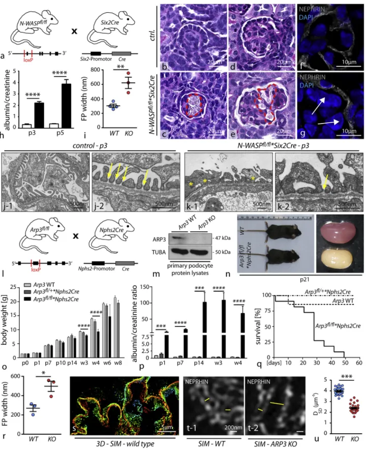
Figure 2. N-WASP and ARP3 Are a Prerequisite for Ordered Podocyte DevelopmentIn Vivo (A) Schematic illustrating the crossing strategy for the generation of N-WASPfl/fl
*Six2Cre knockout mice: Cre recombination targets all tissues deriving from the metanephric mesenchyme, i.e., the whole nephron including podocytes.
(legend continued on next page)
accumulations in distal nephron parts (Figure S2). Together, these findings indicate a substantial requirement of N-WASP and ARP3 in the podocyte FP compartment not only during development but also in completely maturated glomeruli.
N-WASP and ARP3 Are a Prerequisite for Ordered Podocyte Development In Vivo
To test the relevance of N-WASP/Arp2/3-generated actin net-works for the formation of podocyte FPs, we employed a set of conditional knockout mouse models. We previously observed that deletion of N-WASP using the podocyte-specific Nphs2Cre line resulted in a delayed onset of proteinuria, starting at 3 weeks after birth (Schell et al., 2013). It is known from earlier studies that the hNPHS2 promotor exerts activity starting at embryonic day E14.5 onward and specifically targets maturating podocytes at the late capillary loop stage (Moeller et al., 2002). Hence, com-plete and efficient deletion in early podocyte progenitors cannot be predicted. To circumvent potential compensatory actions of other actin NPFs, we employed the Six2Cre deleter strain (E11.5;Figure S3), which targets the whole nephron including podocyte progenitors from early nephron and glomerular matu-ration onward (Kobayashi et al., 2008). Here, we observed that loss of N-WASP resulted in conspicuous glomerular capillary aneurysms (Figures 2B–2G), a phenotype associated with disturbed podocyte process formation (Hartleben et al., 2013). The impact on the integrity of the kidney filtration barrier was marked as respective knockout animals exhibited proteinuria early after birth (Figure 2H). To assess the morphology of podo-cyte FPs, we employed electron microscopy and detected marked simplification of FP morphology in N-WASP*Six2Cre knockout animals (Figures 2I–2K), indicating the prerequisite role for N-WASP in this morphogenetic process. Of note, primary processes appeared not to be affected. Aside from the impact of N-WASP deletion on the glomerular compartment, we observed significant reduction in kidney and body weight of respective knockout mice (Figure S3). This effect might be attributed to the deletion of N-WASP throughout the whole nephron ( Fig-ure S3, as previously shown [Reginensi et al., 2013]). To abolish
Arp2/3 complex-mediated actin nucleation, the nucleation core component Arp3 was deleted by the use of the well-established Nphs2Cre line, which initiates recombination at the late capillary loop stage during glomerular development (Figures 2L and 2M). Loss of ARP3 in podocytes resulted in high levels of proteinuria already at birth, accompanied by decreased birth weight gain (Figures 2N–2P). This phenotype drastically progressed to chronic kidney disease characterized by glomerular sclerosis as well as overall reduced survival (Figures 2Q andS4). Remark-ably, loss of ARP3 resulted in global simplification of podocyte FPs in a similar manner as loss of N-WASP, which we demon-strated by transmission electron microscopy (TEM) (Figures 2R andS4). Of note, primary processes were not obviously affected in terms of morphology and size (in line with our observations in the NWASP*SixCre model). In addition, we also employed a recently established super resolution microscopy technique (Figures 2S–2U andS4) to visualize and quantitate FPs of wild-type and respective knockout animals (Siegerist et al., 2017). These studies corroborated our initial observation by TEM and overall support our initial hypothesis that propulsive actin net-works, as provided by the N-WASP/Arp2/3 complex axis, are involved in the complex generation of podocyte FPs and accu-rate formation of the kidney filtration barrier. Of note, Arp3 knockout podocytes did not exhibit major differences in the expression of podocyte-specific proteins (Figure S4).
ARP3-Mediated Actin Polymerization Is Required for Efficient Protrusion Formation
Podocyte FPs are specialized cellular protrusions, which inter-connect adjacent podocytes to each other and enclose glomer-ular capillaries (Figures 1A–1D). We devised a simplified in vitro model to assess protrusion formation of primary podocytes ( Fig-ures 3A and 3B) and observed that finely arborized protrusions are mainly F-actin based as their in vivo counterparts (Suleiman et al., 2017). Blockage of actin polymerization using latrunculin resulted in a marked reduction of generated protrusions (Figures 3C and 3D). In order to establish a primary cell culture system of high purity, we made use of a genetically encoded reporter
(B–G) Histological evaluation revealed dilated and aneurysmal transformed glomerular capillaries indicating defective enclosing of podocyte foot processes ([B and D] glomeruli from control animals; [C and E] aneurysmatic capillaries in N-WASP*Six2Cre knockout animals; red dotted lines highlight areas of dilated glomerular capillaries). Immunofluorescence for the podocyte marker NEPHRIN also demonstrated the defective invagination of podocytes toward the capillary compartment ([F] shows an example of a respective control animal, while in G impaired invagination in N-WASP*SixCre knockout animals is shown; indicated by white arrows).
(H) Evaluation of albumin to creatinine ratio (mg/mg) detected proteinuria in respective knockout animals at p3 and p5 (at least 3 animals at each time point were analyzed; ****p < 0.0001).
(I) Quantification of foot process (FP) width by TEM showed pronounced effacement and simplification in respective N-WASP KO animals (n = 3–4 animals were analyzed; **p < 0.01).
(J and K) Transmission electron microscopy of wild type (J) and of N-WASP KO (K) mice detected misconfigured podocyte FPs, without proper slit diaphragm junctions (yellow asterisks indicate misconfigured FPs; yellow arrows highlight SD junctions).
(L) Schematic depicting the crossing scheme for the generation of podocyte-specific Arp3 deletion using the Nphs2Cre line.
(M) Western blot of FACS-sorted primary podocyte confirmed loss of ARP3 in respective knockout cells (TUBA: alpha-tubulin was used as a loading control). (N) Arp3 knockout animals showed shrunk and pale kidneys at p21.
(O) Body weight gain was decreased in Arp3 KO animals beginning with 3 weeks of age; respective heterozygous animals were unaffected (****p < 0.0001). (P) Measurement of albumin to creatinine ratio (mg/mg) detected increased levels of proteinuria in Arp3 knockout animals already at p1. Proteinuria persistently showed high levels for the whole observational period of 4 weeks (at least 6 animals per time point and genotype were analyzed; ***p < 0.001, ****p < 0.0001). (Q) Kaplan-Meier analysis revealed that 50% of respective Arp3 knockout animals died within 30 days after birth.
(R) Quantification of FP width in Arp3 knockout animals revealed marked simplification and effacement (n = 3 animals per genotype were analyzed; *p < 0.05). (S–U) Overview of 3D SIM microscopy in respective wild-type animals (S); single plane zoom-ins (T-1 for wild type animals, and T-2 for respective Arp3 KO animals) illustrate misconfigured filtration barrier morphology indicative of aberrant FP architecture. (U) Quantification of SIM data (measurements were accu-mulated over 2 individual animals per genotype; ***p < 0.001). All data are represented as mean ± SEM.
(legend on next page)
system (Figure 3E). Loss of N-WASP or ARP3 resulted in a marked decrease of protrusion formation under 3D-culture con-ditions (Figures 3F–3H and S4). When we classified the morphology of respective protrusions (Figure 3I), we found that wild-type podocytes produced a higher level of complexity in terms of terminally branched protrusions compared to either N-WASP or Arp3 primary knockout podocytes (Figure 3J). This inability for efficient and complex protrusion formation was also underlined by an overall decrease in length of those cellular protrusions (Figure 3K). Altogether, loss of ARP3 and N-WASP in primary podocytes resulted in overall shorter and less arborized protrusions. These findings were coupled by alteration in the structure of the F-actin cytoskeleton characterized by decreased levels of cortical actin and diffuse accumulation of coarse cyto-plasmic actin speckles in Arp3 knockout podocytes (Figures 3L– 3O). Similar findings of defective lamellipodia protrusion forma-tion were also observed in 2D-culture environments in respective Arp3 knockout podocytes (Figure S4). Morphological assess-ment of FP structure and complex arborization levels in Arp3 knockout animals additionally revealed an overall simplification of FPs in vivo, paralleling our in vitro observations using respec-tive primary podocytes in 3D-culture conditions (Figures 3P, 3Q, and2R–2U). Together, the N-WASP/Arp2/3 axis appears to act as a prerequisite for efficient and complex arborized cellular pro-trusion formation of podocytes in vitro and FP formation in vivo (in the following the term protrusion will be used for 3D cellular extensions in vitro, and the term FPs for the in vivo situation). Loss of ARP3 Results in Altered Focal Adhesion Morphology and Increased Actomyosin Activity
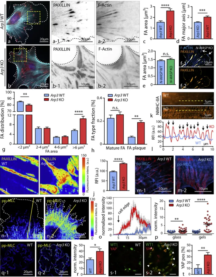
As efficient protrusion formation also involves interaction of cells with their surrounding extracellular matrix (ECM), we aimed to analyze how the N-WASP/Arp2/3 axis might affect FA function and respective morphology. Here, we observed that loss of ARP3 but not of N-WASP in primary podocytes re-sults in the formation of FAs with distorted morphology and a
plaque-like appearance. These FAs were linked to each other via dense F-actin webs (Figures 4A–4F and S4). The overall number of FA sites as well as their composition appeared not to be altered in Arp3 knockout cells, but the morphology was shifted toward plaque-like FA sites at the expense of nascent adhesions (Figures 4G, 4H, S4, and S5). These plaque-like FAs displayed an increased density and altered localization pattern of FA proteins such as PAXILLIN (Figures 4I and 4J). Remarkably, primary N-WASP knockout cells exhibited an un-altered FA morphology and distribution pattern, accompanied by prominent lamellipodial protrusions (Figures 4E, 4F, and S5). This difference between protrusion deficiency in 3D-culture conditions (N-WASP and ARP3) and FA morphology (only ARP3) might be explained by compensatory upstream NPFs in 2D conditions. Indeed, the NPF WAVE2 was detected at the lamellipodial leading edge of WT and N-WASP KO primary podocytes. Inhibition of the RAC1/WAVE2/Arp2/3 signaling axis in N-WASP KO podocytes resulted in a phenocopy of the dis-torted FA morphology of ARP3 knockout podocytes, support-ing a compensatory concept of NPFs in a contextual manner (Figure S5). As FA function is directly linked to tensional control of the actomyosin cytoskeleton, we assessed the distribution of non-muscle myosin II (NMHC-IIA) and actinin-4 (ACTN4) in Arp3 wild-type versus knockout cells (Figures 4K, 4L, and S5). Here, we observed sarcomere-like distribution patterns for both proteins in Arp3 knockout cells, suggestive for increased inherent tension levels. We correlated phosphory-lated levels of myosin-light-chain (MLC) with respective geno-types and observed an increased activity and circular distribu-tion of activated actomyosin cytoskeleton in Arp3 knockout podocytes (Figures 4M–4O), whereas a more longitudinal pattern for pp-MLC was detected in wild-type cells. The indi-vidual levels of pp-MLC also appeared to be modulated by the substratum stiffness, as knockout cells exhibited even higher pp-MLC levels on softer surfaces when compared to control cells (Figure 4P). Similar observations were also made
Figure 3. ARP3-Mediated Actin Polymerization Is Required for Efficient Protrusion Formation
(A and B) Primary podocytes in 3D culture conditions form protrusions with increasing levels of branching; immunofluorescence staining revealed that terminally branched protrusions consisted mainly of filamentous actin, whereas major protrusions also showed positivity for tubulin (B). (C) Acute treatment with the actin polymerization inhibitor Latrunculin A markedly suppressed protrusion formation, as shown by a quantifcation for main protrusions (D) (n = 38–41 analyzed cells; ****p < 0.0001).
(E) Schematic depicting the generation of a fluorescence-based reporter line: after podocyte-specific Cre-recombination mGFP is selectively expressed in only the glomerular epithelial cell compartment. Single-cell isolation and further FACS-based purification results in a primary culture system with proven genetic origin. (F and G) 3D reconstruction of primary podocytes in 3D culture systems (wild typ podocytes [F]) unmasked the deficiency of Arp3 knockout podocytes (G) to form terminally branched protrusions.
(H) Color code describing segmentation of individual protrusion classes into main, primary, and secondary order protrusions.
(I) Loss of ARP3 and N-WASP resulted in significantly less cells with branching protrusion phenotypes (at least 150 cells per condition were analyzed, and data were accumulated over at least 3 experiments; n.s., non significant, ***p < 0.001, ****p < 0.0001).
(J and K) Reconstructed images of individual z stacks were used as a basis for the segmentation analysis (J) and length measurements of protrusions (K): due to ARP3 and N-WASP deficiency, higher order branched protrusions were diminished and apparent protrusions were significantly shorter compared to control conditions (17 wild-type, 32 Arp3 KO, and 20 N-WASP KO primary podocytes were reconstructed and analyzed; n.s., non significant, *p < 0.05, **p < 0.01, ****p < 0.0001).
(L–O) Analysis of the cortical actin cytoskeleton of wild-type (L) and respective KO (M) cells under 3D culture conditions. Representative line scans (N) of fila-mentous actin intensities in single planes demonstrated altered levels of cortical actin due to loss of ARP3 (19 wild-type and 16 Arp3 KO cells were reconstructed, and ratios between cortical and cytoplasmic actin intensities were measured at multiple localizations (O); orange dashed lines indicate areas of line scan analysis, black arrows mark individual representative cell borders; ****p < 0.0001; MFI, mean fluorescence intensity).
(P) Scanning electron microscopy (SEM) detected pronounced simplification, reduced branching and misconfiguration of podocyte processes in Arp3 KO ((P-1) shows a representative image of wild type controls, whereas (P-2) and (P-3) demonstrate representative images from Arp3 KO animals; colorized SEM image (P-3) depicting segmentation of podocyte processes, each colorized dashed line indicates one individual major process; white arrows and red asterisks mark secondary processes). (Q) Podocyte process branching was quantified from semi-planar SEM areas (n = 5 wild-type and 4 Arp3 KO mice were analyzed at p8; ****p < 0.0001). All data are represented as mean ± SEM.
Figure 4. Loss of ARP3 Results in Altered Focal Adhesion Morphology and Increased Actomyosin Activity
(A and B) Staining for the focal adhesion (FA) component PAXILLIN (red) revealed confluent FA clusters in primary Arp3 KO podocytes (B-1), which were connected via filamentous actin webs (wild type (A) and KO (B)). F-actin (blue) was stained by Phalloidin.
(legend continued on next page)
for mechanical tension markers pp-MLC and YAP on glomer-ular sections in vivo (Figures 4Q–4T and S5). These finding together indicate that a loss of ARP3-mediated actin branching results in increased intracellular tension linked to elevated levels of pp-MLC and nuclear translocation of YAP in podo-cytes in vivo. Our in vitro and in vivo results imply an interde-pendent crosstalk between the podocyte actin cytoskeleton composition and inherent intracellular tension levels, as pro-vided by the actomyosin cytoskeleton.
Competition between Different F-Actin Networks Modulates Protrusion Phenotypes and FA Morphology of Podocytes
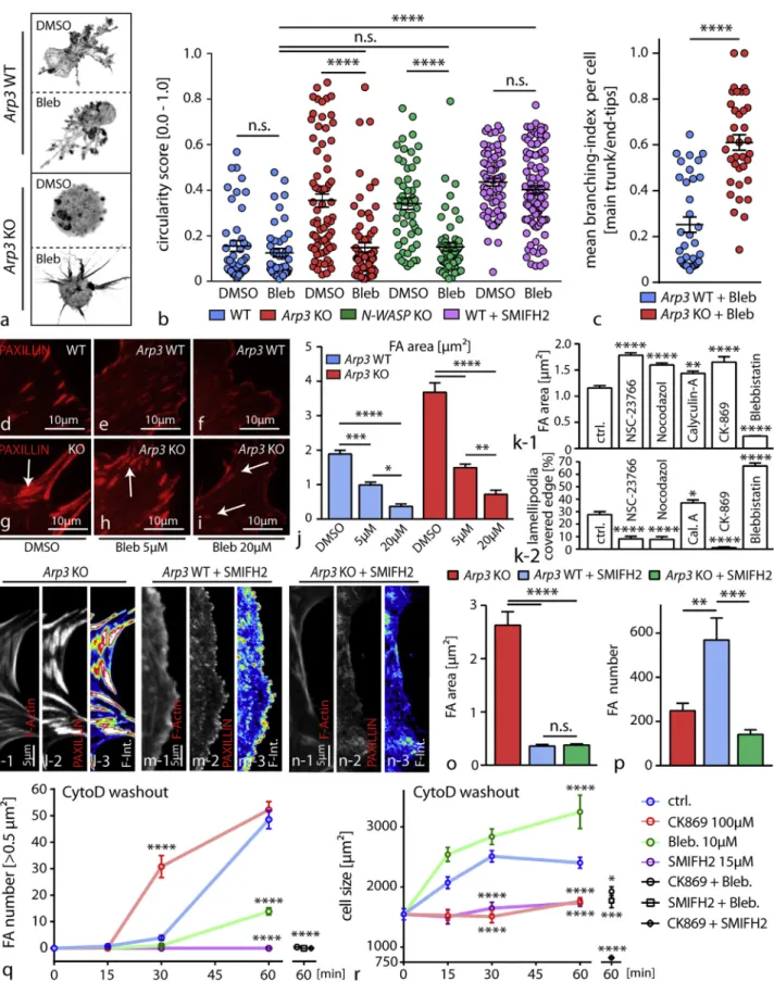
Next, we addressed the question of whether and how Arp2/3-dependent actin networks compete with actomyosin-mediated contractility and how this interplay might translate into modula-tion of essential parameters such as protrusion formamodula-tion and cell-matrix interaction. First, Arp3 as well as N-WASP knockout cells were seeded in 3D matrices and pretreated with blebbistatin to assess protrusion formation and their branching complexity. In both conditions, inhibition of myosin-2 resulted in a mitigation of the protrusion defect (Figures 5A, 5B, and S5). Similar results were also observed upon blockage of the Arp2/3 complex using specific inhibitory compounds in primary cells and to some extent in tissue culture approach (Figure S5). The Arp2/3 complex differs in its mode of actin polymerization from other actin nucleators (such as formins) by providing a branched actin network instead of only linearly polymerized actin filaments (Campellone and Welch, 2010). To elucidate the mini-mal requirement for actin-dependent protrusion formation in our system, we inhibited formin function and observed a phenocopy in terms of defective protrusion formation. In contrast,
co-treat-ment with the myosin-2 inhibitor blebbistatin could not revert or modify this phenotype as observed for Arp3 or N-WASP knockout cells (Figure 5B). Interestingly, the branching level of protrusions (ratio of major to minor protrusions) in terms of arbor-ization correlated to the presence or absence of ARP3, indicating that formin-mediated actin polymerization appears as a prereq-uisite for protrusion formation, whereas Arp2/3 predominantly determines the level of terminally branched protrusions and influences myosin-2 tension (Figures 5B and 5C).
As a loss of ARP3 resulted in the formation of FA plaques ( Fig-ures 4A–4D), we reasoned that increased actomyosin contrac-tility might modulate this phenotype. In fact, inhibition of myosin-2 with blebbistatin significantly reversed the formation of FA plaques in a dose-dependent manner and also mitigated migratory deficits of Arp3 knockout cells (Figures 5D–5J and S6). To exclude at this point that the FA-plaque phenotype is rather a result from generally inhibited actin polymerization, we tested a series of different Arp2/3 inhibitors as well as other chemical compounds affecting different modes of actin poly-merization. Interestingly, we detected predominant formation of FA plaques only in Arp2/3-inhibited cells, underlining the specificity of branched actin networks involved in the generation of the FA-plaque phenotype (Figure S6). These observations were corroborated by complementary experiments where acto-myosin contractility was titrated either by treatment with the phosphatase-inhibitor calyculin-A or inhibited with blebbistatin in wild-type cells (Figures 5K andS6). Moreover, employing in-hibitors for RAC1 (as an established upstream regulator) or the Arp2/3 complex itself revealed that there is an inverse correlation between Arp2/3-dependent lamellipodia formation and FA size (Figure 5K). At this point, we asked whether and how a compet-itive interplay between the Arp2/3 complex and formin type actin
(C and D) Quantification of average focal adhesion area (C) and major axis (D) showed increased values for both parameters in respective KO cells (25 wild-type and 28 Arp3 KO cells were analyzed; ***p < 0.001, ****p < 0.0001).
(E and F) Average focal adhesion area (E) and lamellipodium formation (F) was not obviously impaired in primary N-WASP KO podocytes in 2D culture conditions (n = 36 cells per genotype were analyzed, n.s., non significant; FA were stained by PAXILLIN and F-actin by phalloidin; white arrows indicate lamellipodia). (G) Analysis of focal adhesion clusters due to their individual size indicated a shift toward enlarged adhesion clusters in Arp3 KO podocytes (25 WT and 26 Arp3 KO cells were analyzed; **p < 0.01, ****p < 0.0001).
(H) This shift in FA size was paralleled by a change in the distribution of individual FA classes: loss of Arp3 resulted in the accumulation of FA plaques (n = 18 cells per genotype were analyzed; n.s., non significant, **p < 0.01).
(I and J) Analysis of PAXILLIN fluorescence intensities (FI) in wild type podocytes (I-1) and in respective Arp3 KO cells (I-2) revealed altered intensity patterns and increased mean PAXILLIN intensity (J) per focal adhesion (n = 100 mature FA per genotype were analyzed; ****p < 0.0001).
(K and L) Immunofluorescence for NMHC-IIA (K) demonstrated an increased sarcomere-like pattern of actin fibers (L) in respective Arp3 KO podocytes (yellow lines indicate positions for line scan measurements, and black arrows indicate myosin-2 peaks).
(M) Immunofluorescence staining for pp-MLC and the focal adhesion marker PAXILLIN illustrated prominent, fiber-like accumulations of pp-MLC linked to FAs in Arp3 KO podocytes.
(N) Staining for pp-MLC revealed increased MLC activation levels in Arp3 KO podocytes, condensed in arc regions between pseudopodial protrusions. Also a ring-like distribution was observed in the majority of Arp3 KO podocytes. (Yellow lines indicate representative positions for line scan measurements). (O) Line scan profiles for pp-MLC showed highest activation levels close to the cell edge in Arp3 KO cells (values represent mean intensities of 6 cells per ge-notype; gray error bars represent SEM).
(P) Quantification of fluorescence intensity for pp-MLC on either glass or soft gels revealed higher MLC activation levels for Arp3 KO podocytes (37 wild-type and 41 Arp3 KO cells on glass; 31 wild-type and 32 Arp3 KO cells on gels were analyzed; **p < 0.01, ****p < 0.0001).
(Q and R) Staining for pp-MLC in glomeruli from either wild-type (Q-1) or Arp3 KO (Q-2) animals showed high levels of phosphorylated MLC in the podocyte compartment, as visualized with the podocyte-specific marker NEPHRIN (mean podocyte pp-MLC intensity of n = 4 WT and 4 KO animals were statistically analyzed; *p < 0.05; the mean pp-MLC intensity per animal was calculated from mean pp-MLC intensity of the segmented podocyte compartment of at least 20 glomeruli per animal).
(S and T) Immunofluorescence staining for the mechanical tension marker YAP in glomeruli from either wild-type or Arp3 KO (S) animals showed increased percentages of YAP-positive podocyte nuclei in KO animals (T). Podocyte nuclei were visualized by the podocyte-specific marker WT1 (the mean percentage of YAP-positive podocyte nuclei per glomerulus of n = 5 WT and 5 KO animals were analyzed; white arrow bars indicate podocyte nuclei with co-occurring positivity for YAP, **p < 0.01; the percentage of YAP positive podocyte nuclei per glomerulus per animal was calculated from at least 20 glomeruli per animal). All data are represented as mean ± SEM.
Figure 5. Competition between Different F-Actin Networks Modulates Protrusion Phenotypes and FA Morphology of Podocytes
(A and B) Decrease of intracellular tension in Arp3 KO primary podocytes with the myosin-2 inhibitor blebbistatin (Bleb) resulted in the formation of simplified cellular protrusions compared to blebbistatin-treated WT cells (A). Cell morphology and thereby protrusion generation was quantified by circularity scores (legend continued on next page)
nucleation might influence FA morphology. Inhibition of formins resulted in a significant dissolution of FA plaques in Arp3 knockout cells. Interestingly, a similar response was observed in wild-type cells but was accompanied by a pronounced in-crease in numbers of nascent FAs (Figures 5L–5P). These find-ings indicate that formin type nucleation machinery and the Arp2/3 complex feed into different FA types via generation of distinct actin networks. To further extend these observations, we performed washout and co-treatment experiments after dissolution of the actin cytoskeleton with cytochalasin D treat-ment. Here, control cells showed a time-dependent expansion in cell size accompanied by increasing numbers of FAs (Figures 5Q and 5R). In contrast, blockage of the Arp2/3 complex, myosin-2, or formins revealed again an inverse correlation of cell size expansion and assembly of FAs. Altogether, these ob-servations suggest that an interplay between the formin type nucleation machinery, the Arp2/3 complex, and myosin-2 activ-ity critically defines cellular protrusion formation and at the same time influences the assembly rate as well as maturational level of cell-matrix adhesions.
Loss of ARP3 Leads to Decreased Adhesive Properties of Podocytes under Mechanical Stress Conditions Up to this point, we established a central role for Arp2/3 in podo-cytes linking protrusion formation, actomyosin tension, and FA maturation. However, the in vitro experiments were performed under static conditions. Podocytes reside on the outer surface of glomerular capillaries and are therefore exposed to high levels of physical filtration forces (Endlich and Endlich, 2006). To simu-late this continuous physical stress, we cultured primary knockout podocytes under cyclic-isometric stress conditions (Figure 6A). Interestingly, this intervention led to a rearrangement in the F-actin cytoskeleton of wild-type cells characterized by an increased density of ventral stress fibers, whereas Arp3 knockout cells did not show an adaptive response (Figures 6B–6F andS6). Of note, wild-type cells still showed a lower stress fiber density compared to Arp3 knockout cells. In addi-tion, FA morphology showed an adaptive change in wild-type
cells under stress conditions while in ARP3-deficient cells, FA plaques remained stable and appeared unaffected (Figures 6G–6L). Together with our previous observations of increased actomyosin cytoskeleton activity, we wanted to directly test ex-erted traction forces of Arp3 knockout cells. Here, we observed that primary podocytes deficient in ARP3 exerted significantly higher traction forces on the underlying substratum paralleling the prior results of increased actomyosin activity in vitro and in vivo (Figures 6M–6O and4N–4T). Surprisingly, we detected an increased tendency of detachment of Arp2/3-inhibited podo-cytes under mechanical stress conditions (Figures 6P, 6Q, and S6). Altogether, those observations suggested that a loss of Arp2/3 function results in a hyper-contractile status essentially impairing adaptive capacity in situations of continous me-chano-physical stress conditions.
ARP3 Is Required for Efficient Podocyte Adhesion and Maintenance of Foot Process Morphology In Vivo To transfer these observations to the in vivo setting, we analyzed our in vivo models for signs of podocyte detachment and could detect loss of WT1-positive cells from the glomerulus into the urine of respective Arp3 knockout animals (Figures 7A– 7D). To address the question, whether these mechanisms are not only required during glomerular maturation but might also be relevant to maintenance conditions in fully developed podo-cytes, we generated an inducible knockout model for Arp3 specifically in podocytes (Figure 7E). Here, we observed a pro-nounced reduction of podocytes per glomerulus indicative of podocyte detachment (Figures 7F and 7G), paralleled by increasing levels of proteinuria and glomerular sclerosis ( Fig-ures 7H and 7I). Assessment of podocyte FP morphology employing scanning electron microscopy as well as super-res-olution microscopy revealed marked simplification of podocyte FPs (Figures 7J–7L and S6). Together, these experiments in vitro and in vivo support the concept that ARP3-dependent actin networks not only determine the complexity level of podo-cyte protrusions (in vitro) or FPs (in vivo) but also are required for the establishment of efficient and mechano-adaptive podocyte
(B). Quantification showed that co-treatment with blebbistatin results in a decrease of the circularity score in Arp3 as well as N-WASP KO podocytes, reflecting a more protrusive phenotype. Suppression of protrusion formation by SMIFH2-mediated formin inhibition is resistant to co-treatment with blebbistatin. (44 DMSO and 43 blebbistatin treated wild-type cells; 73 DMSO and 70 blebbistatin treated Arp3 KO cells; 50 DMSO and 53 blebbistatin treated N-WASP KO cells; 70 SMIFH2+DMSO and 100 SMIFH2+blebbistatin treated wild-type cells were analyzed; n.s., non significant, ****p < 0.0001).
(C) Definition of a branching index (major protrusion per end tips (minor protrusions) allowed a more detailed analysis of protrusion complexity and quality. Quantification for this branching index revealed simplified cellular protrusions with reduced branching levels for blebbistatin treated Arp3 KO cell podocytes compared to blebbistatin treated WT cells. (35 WT and 36 Arp3 KO cells were analyzed; ****p < 0.0001).
(D–J) Inhibition of myosin-2 via blebbistatin resulted in a dose-dependent decrease in focal adhesion (FA) size in Arp3 knockout podocytes (WT - (D-F) and Arp3 KO - (G-I)), indicating that FA plaques represent myosin-2-dependent structures in these cells. Quantification of FA area was performed ([J]; white arrows indicating FA sites; 20 cells per condition were analyzed; *p < 0.05, **p < 0.01, ***p < 0.001, ****p < 0.0001).
(K) Correlation of average focal adhesion (FA) size to the level of lamellipodium formation shows an inverse correlation between these cellular processes, indicating also an inverse correlation between Arp2/3 and (acto-) myosin-2 activity as an underlying mechanism. Calyculin-A and blebbistatin were used, respectively, to either activate or inhibit myosin-2 activity. Rac1 inhibition by NSC-23766 or CK-869 was used to inhibit Arp2/3-mediated actin nucleation; Nocodazol was used to stabilize focal adhesions (FA) in a myosin-independent fashion. (24 to 44 immortalized human podocytes were analyzed per condition; *p < 0.05, **p < 0.01; ****p > 0.0001).
(L–P) Treatment with the formin inhibitor SMIFH2 resulted in a dissolution of mature FAs, which was accompanied by an increase in the number of nascent adhesions in wild-type cells (M) and a loss of focal adhesion formation in Arp3 KO podocytes (N) (compared to untreated Arp3 KO cells - (L); n.s., non significant; **p < 0.01, ***p < 0.001, ****p < 0.0001). (O) Focal adhesion area and (P) FA number were quantified.
(Q and R) Washout experiments employing cytochalasin-D and co-treatments with inhibitors for myosin-2 activity (blebbistatin), formin nucleation (SMIFH2), or Arp2/3 nucleation (CK-869) revealed specific actin network activities as a prerequisite for the inverse correlation between dynamic focal adhesion maturation (Q) and cell size spreading (R) of podocytes (31–104 cells per condition were analyzed for FA maturation and 40–215 cells were analyzed for cell size; *p < 0.05; ***p < 0.001; ****p > 0.0001). All data are represented as mean ± SEM.
adhesion properties under conditions of physical stress (Figures 6and7M–7O).
DISCUSSION
The complex cellular architecture of glomerular epithelial cells (namely podocytes), consisting of primary and secondary pro-cesses, is intimately linked with their function in providing the integrity of the kidney filtration barrier. Despite significant
prog-ress in podocyte biology research, it is still incompletely under-stood how those cellular protrusions are developed and main-tained during later life (Schell et al., 2014; Pavenst€adt et al., 2003; Liapis et al., 2013). Here, we employed in vitro and in vivo models to understand the contribution of Arp2/3-generated actin networks to control podocyte process networks. The Arp2/3 complex is an extensively in vitro characterized molecular ma-chinery generating propulsive actin polymerization and branch-ing, which is involved in diverse cellular processes such as
Figure 6. Loss of ARP3 Leads to Decreased Adhesive Properties of Podocytes under Mechanical Stress Conditions (A) Schematic illustrating the application of cyclic stretch on primary podocytes by culture on a flexible membrane.
(B–E) Immunofluorescence staining of the actin cytoskeleton in wild-type (B and C) and knockout podocytes (D and E) analyzed under non-stressed and stressed conditions, respectively.
(F) Quantification of stress fibers under stressed conditions revealed an adaptive deficit of actin filaments in Arp3 knockout podocytes when compared to wild-type controls (at least 50 cells per condition were analyzed; n.s., non significant, **p < 0.01;****p < 0.0001).
(G–L) Analysis of focal adhesion morphology ((G and H) depict wild type podocyte FAs; (I and J) depict Arp3 KO podocytes) under stressed conditions indicated an increase in focal adhesion size in wild-type cells ((K) - FA area quantification and (I) - FA major axis quantification for each condition), whereas no changes were observed in respective knockout podocytes (at least 11 cells per condition and genotype, n.s., non significant, *p < 0.05; **p < 0.01).
(M and N) Quantification of traction force microscopy (M) on collagen-coated soft gel matrices showed increased traction forces in Arp3 KO podocyte and increased strain energy (N) (22 WT and 32 KO cells, ***p < 0.001, ****p < 0.0001).
(O) Pseudo-colored force maps of wild-type and Arp3 KO podocytes on soft matrices (note difference in individual traction scale between wild-type and KO cells). (P) Immunofluorescence staining of Arp2/3 inhibited podocytes under stressed conditions. (Q) Quantification of adherent cells under stressed conditions (n = 3 individual experiments; ***p < 0.001). All data are represented as mean ± SEM.
migration or endocytosis (Swaney and Li, 2016; Soderling, 2009). However, it is still poorly understood which specific role this complex exerts in different biological contexts and cell types in vivo. Remarkably, we observed that the Arp2/3 complex influ-ences not only the complexity level of cellular protrusions of po-docytes (in vitro and in vivo) but also modulates their mechano-physical adaptive capacity under dynamic stress conditions.
Based on the identification of causative mutations for genetic podocyte diseases, previous work showed the relevance of certain actin-cytoskeleton related proteins (Perico et al., 2016). Based on 2D studies with cultured cells, it is well established that lamellipodial protrusions depend on propulsive dendritic actin meshworks generated by the Arp2/3 complex (Bisi et al., 2013; Dang et al., 2013). This mode of actin nucleation also seems to be required for the formation of more complex cellular protrusions in vivo, like it was recently shown for neuronal den-drites and oligodendrocytes (Zuchero et al., 2015; Kim et al., 2015; Kim et al., 2013). Given their highly arborized cellular pro-trusions, podocytes have several morphological features with these cells in common. However, biological function and expo-sure to physical forces differ strongly between these cell types (e.g., exposure to filtration forces in the case of podocytes), sug-gesting cell-specific mechanisms for protrusion formation and maintenance. Employing in vitro as well as in vivo models, we could delineate how N-WASP and the Arp2/3 complex influence the morphology and architecture of podocyte FPs (Figures 1,2, and3). Based on our in vivo observations, the onset and manifes-tation of phenotypes (comparing Arp3 and N-WASP podocyte-specific deletion) might be explained by the upstream-regulatory role of N-WASP and potential compensatory NPFs (e.g., WAVE2; as shown inFigure S5). Remarkably, we observed an indispens-able role of formin-mediated actin polymerization in phases of initial 3D protrusion formation, whereas Arp2/3-dependent processes are mainly driving the complexity level of generated protrusions (Figures 2,3, and5). Besides established models for F-actin network modulation (such as Rho-GTPase signaling; Figures 7andS7), recent reports proposed an additional model where different actin polymerization machineries are in competi-tion for limited G-actin pools (Burke et al., 2014; Suarez et al., 2015; Rotty et al., 2015; Suarez and Kovar, 2016). In line with this, we identified formin-mediated actin polymerization as a competing factor in this network, modulating podocyte protru-sion as well as FA formation (Figure 4). To date, the atypical for-min INF2 is the best characterized forfor-min family member in the context of podocytes based on the identification of INF2 dis-ease-causing mutations (Brown et al., 2010). Previously, INF2 mutations were characterized to affect the interaction with mDIA, subsequently leading to altered RhoA signaling (Sun et al., 2011). Remarkably, knockin mice bearing disease-causing INF2 mutations do not show any obvious phenotype under basal conditions, in particular normal podocyte FP morphology was reported (Subramanian et al., 2016). However, stressing this sys-tem resulted in the collapse of podocyte FP architecture, high-lighting the concept of balanced actin cytoskeleton pools to maintain podocyte FP morphology. Based on our results, it may be hypothesized that deletion of formins in vivo might result in a predominant defect of primary process formation (although, to our knowledge, there are no systematic studies published focusing on formin deletion in vivo). Consistently, the correlation
of our in vitro and in vivo findings indicate that Arp2/3-controlled process formation predominantly affects most terminal pro-cesses instead of primary propro-cesses or protrusions (Figure 2). Together, these findings suggest that Arp2/3-mediated actin polymerization is essentially involved in critical morphogenetic programs as observed during the complex process of glomerular maturation, leading from only cuboidal epithelial cells to highly arborized podocytes (Figures 1and7).
Aside from the formation of complex, arborized networks of interdigitating FPs, podocytes are required to withstand high levels of physical forces given their extreme anatomical position on the surface of glomerular capillaries (Endlich and Endlich, 2006). The relevance of podocyte adhesion is exemplified by the identification of disease-causing mutations affecting certain integrin subunits as well as a series of genetic models targeting different components of the cell-matrix adhesion machinery (Has et al., 2012; Sachs et al., 2006; Pozzi et al., 2008). However, the complex and reciprocal interplay between cell-matrix adhe-sion and inherent contractility control is still unclear. Here, we demonstrated a high abundance of Arp2/3 complex components in the focal adhesome of podocytes in vivo and in vitro (Figure 1), highlighting a close link between this cytoskeletal machinery and the cell-adhesion nexus. Only very recently, it was shown that the Arp2/3 complex is influencing the alignment of FA complexes in a migration mode termed as haptotaxis (Wu et al., 2012), and it was demonstrated that ARP3 is interacting with FA components such as VINCULIN and FAK (Chorev et al., 2014; Serrels et al., 2007). Furthermore, we observed the formation of large FA plaques at the expense of smaller nascent FAs in Arp3 knockout cells ( Fig-ure 4). Based on biophysical models, retrograde actin flux due to Arp2/3-generated propulsive actin polymerization presents a critical factor in the maturation cycle of FAs by initiating nascent adhesions (Choi et al., 2008; Wu et al., 2017; Wu et al., 2012). In this context, it is of interest that Arp3 knockout cells were able to initialize nascent FAs but presented with hyper-maturated, plaque-like FAs (Figure 4). These observations indicate that the Arp2/3 complex might not be solely required for the initialization of nascent FAs but also for later maturational processes and, thereby, might influence the turnover of FAs (Wu et al., 2012). Remarkably, ARP3-deficient cells exhibited concomitantly an activated actomyosin cytoskeleton and exerted high traction forces (Figures 3,4, and6). We reasoned that loss of branched actin networks provokes increased inherent tension levels, and indeed lowering the activity of myosin-2 resulted in a reversal or at least mitigation of observed phenotypes such as FA plaques or impaired 3D protrusion formation (Figure 4). Besides established models for F-actin network modulation (such as Rho-GTPase signaling), recent reports proposed an additional model where different actin polymerization machineries are in competition for limited G-actin pools (Figures 7andS7) (Leijnse et al., 2015; Liapis et al., 2013; Lomakin et al., 2015). In line with this, we identified formin-mediated actin polymerization as a competing factor in this network, modulating podocyte protru-sion as well as FA formation (Figure 4).
By combining our in vitro and in vivo observations we demonstrated that ARP3-deficient cells show only impaired adhesive properties under dynamic stress conditions, despite large FA plaques and increased actomyosin activity (Figures 4and6). These findings indicate that a loss of branched actin
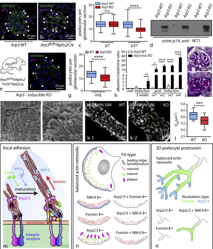
Figure 7. ARP3 Is Required for Efficient Podocyte Adhesion and Maintenance of Foot Process Morphology (A and B) Immunfluorescence for WT1-positive podocytes in wild type and Arp3 knockout animals.
(C) Quantification of podocyte number per glomerulus a pronounced decrease in conditional Arp3 knockout animals, indicating significant detachment of po-docytes (at least 106 glomeruli of 3 mice per genotype and age; n.s., non significant, ****p < 0.0001).
(D) Western blot for WT-1 positive cells in urine detected only detached podocytes in respective knockout animals. (E) Schematic illustrating the crossing strategy for the generation of tetracycline inducible Arp3fl/fl
*hNphs2*rtTA*TetOCre knockout animals. (F) Immunofluorescence for WT1-positive podocytes in Arp3 inducible knockout animals.
(legend continued on next page)
networks not only results in a compensatory prevalence of formin-mediated actin polymerization but also might impede the adaptive capacity of podocytes in situations of continuous mechano-physical stress. A similar model was recently pro-posed for disease-causing mutations in the ACTN4 gene, and we could also establish a reciprocal interplay between podo-cyte FA proteins and actomyosin activity (Schell et al., 2017; Feng et al., 2018).
The concept of a tripartite relationship between FA maturation status, traction force, and ECM linkage/sensing suggests a model for FP effacement and consecutive podocyte detach-ment. Altering one component (e.g., impairing branched actin networks) will result in a compensatory response (increased ten-sion, altered FA morphology), which might transiently tighten adherence toward the glomerular basement membrane, but ultimately impedes required mechanical adaptive plasticity in conditions of physical stress.
STAR+METHODS
Detailed methods are provided in the online version of this paper and include the following:
d KEY RESOURCES TABLE
d CONTACT FOR REAGENT AND RESOURCE SHARING
d EXPERIMENTAL MODEL AND SUBJECT DETAILS
B Animals
B Human Kidney Samples
d METHOD DETAILS
B Genotyping
B Histology and Immunofluorescence Staining on Cryo-sections
B Electronmicroscopy & Scanning Electron Micro-scopy (SEM)
B Super Resolution Microscopy (SIM)
B Measurement of Urinary Albumin and Creatinine
B Glomerular and Podocyte Preparation for Western Blot and MS Applications
B Mass Spectrometry and MaxQuant-Based Identifi-cation
B Focal Adhesion (Adhesome) Complex Isolation
B Isolation of Primary Podocytes and Culture Conditions
B 3D Protrusion Formation of Primary Podocytes and Cortical Actin Fluorescence Measurement
B Immunofluorescence on Primary Podocytes
B Isometric Cycling Stress (Flexcell)
B Traction Force Microscopy
B Random Single Cell Migration Assays
d QUANTIFICATION AND STATISTICAL ANALYSIS
B Statistical Analysis
B Glomerluar Sclerosis Index
B Immunofluorescence Quantification
B Quantification of Foot Process Morphology and Branching Index
B Quantification of Focal Adhesion Morphology and Analysis of IF Intensities
B Cell Migration
B Protein Enrichment and Functional Annotation Analysis
d DATA AND SOFTWARE AVAILABILITY
SUPPLEMENTAL INFORMATION
Supplemental Information includes seven figures and two tables and can be found with this article online athttps://doi.org/10.1016/j.devcel.2018.11.011.
ACKNOWLEDGMENTS
We would like to thank Charlotte Meyer and Betina Kiefer for their expert tech-nical assistance. In addition, we would like to express our gratitude to all mem-bers of our laboratories, to Sebastian Arnold, and to the Life Imaging Center (LIC) of the University of Freiburg for helpful discussions and support. This study was supported by the German Research Foundation (DFG): CRC 1140 (to T.B.H.), CRC 1192 (to T.B.H.), and CRC 992 (to T.B.H.), Heisenberg program (to T.B.H.), HU 1016/5-1 and HU 1016/8-1 (to T.B.H.); by the Euro-pean Research Council (ERC grant 616891 to T.B.H.) and by the H2020-IMI2 consortium BEAt-DKD (115974; to T.B.H.); by the BMBF (Bundesminis-terium f€ur Bildung und Forschung) STOP-FSGS 01GM1518B (to N.E.) and BMBF STOP-FSGS 01GM1518C (to T.B.H.); by the Excellence Initiative of the German Federal and State Governments (BIOSS to T.B.H. and the Frei-burg Institute for Advanced Studies [FRIAS] to T.B.H.); by the Alexander von Humboldt Foundation (to A.A.); by the German Society of Nephrology (DGFN to C.S.); by the Else Kro¨ner Fresenius Stiftung, NAKSYS (to C.S., M.R., and T.B.H.) and Matriglom A_09 (to C.S.); and by the Berta-Ottenstein Programme, Faculty of Medicine, University of Freiburg (to C.S.).
AUTHOR CONTRIBUTIONS
C.S., M.R., F. Geist, J.D., B.S., and T.B.H. conceived and analyzed experi-ments. C.S., M.R., and T.B.H. supervised the study. C.S., M.R., F. Geist, M.H., A.A., M.Y.-Y., J.I.M., A.S., F.S., N.A., N.E., and D.K. performed experi-ments. H.H.A., F. Grahammer. and J.D. provided critical reagents. C.S. and M.R. prepared figures and tables. C.S., M.R., and T.B.H. wrote the manuscript with input and discussion from all authors.
DECLARATION OF INTERESTS
T.B.H. declares to be a member of the scientific advisory board of Goldfinch Bio and to have a research grant from Fresenius Medical Care/ Unicyte. N.E. declares that PEMP (podocyte exact morphology measurement procedure) that has been used for this manuscript is registered for a patent. N.E. and F.S. are among the founders of the Start-UP NIKOKA, which will commer-cialize PEMP. All other authors declare no competing interests.
(G) Quantification of podocyte numbers in inducible Arp3 knockout animals.
(H) Albumin to creatinine ratio (mg/mg) measurements detected increased levels of proteinuria already in the second week of doxycycline induction: levels increased furthermore over a period of 4 weeks (3–11 animals per time point and genotype were analyzed; *p < 0.05, **p < 0.01, ****p < 0.0001).
(I) PAS staining demonstrated focal segmental glomerulosclerosis in inducible Arp3 KO animals.
(J) Scanning electron microscopy demonstrates simplified FP morphology in Arp3 inducible knockout animals.
(K and L) SIM revealed aberrant kidney filtration morphology correlating to simplified FPs in Arp3 inducible knockout animals (measurements were accumulated over 2 individual animals per genotype; ***p < 0.001).
(M–O) Graphical summary of proposed Arp2/3-mediated functions in podocytes (M): nucleation of dendritic actin networks by the Arp2/3 complex promotes FA assembly at cell protrusion sites and controls FA maturation by limiting non-muscle myosin II (NM-II) complex activity (N). (O) Arp2/3 allows the development of podocyte protrusions by modulating NM-II activity and directly promotes complex arborization of podocyte protrusions in vitro and in vivo. All data are represented as mean ± SEM.
Received: November 29, 2017 Revised: September 3, 2018 Accepted: November 1, 2018 Published: November 29, 2018
REFERENCES
Bechtel, W., helmst€adter, M., Balica, J., Hartleben, B., Kiefer, B., Hrnjic, F., Schell, C., Kretz, O., Liu, S., Geist, F., et al. (2013). Vps34 deficiency reveals the importance of endocytosis for podocyte homeostasis. J. Am. Soc. Nephrol. 24, 727–743.
Berginski, M.E., Vitriol, E.A., Hahn, K.M., and Gomez, S.M. (2011). High-reso-lution quantification of focal adhesion spatiotemporal dynamics in living cells. PLoS One 6, e22025.
Bisi, S., Disanza, A., Malinverno, C., Frittoli, E., Palamidessi, A., and Scita, G. (2013). Membrane and actin dynamics interplay at lamellipodia leading edge. Curr. Opin. Cell Biol. 25, 565–573.
Boerries, M., Grahammer, F., Eiselein, S., Buck, M., Meyer, C., Goedel, M., Bechtel, W., Zschiedrich, S., Pfeifer, D., Lalo€e, D., et al. (2013). Molecular fingerprinting of the podocyte reveals novel gene and protein regulatory net-works. Kidney Int. 83, 1052–1064.
Bravo-Cordero, J.J., Hodgson, L., and Condeelis, J. (2012). Directed cell inva-sion and migration during metastasis. Curr. Opin. Cell Biol. 24, 277–283.
Brown, E.J., Schlo¨ndorff, J.S., Becker, D.J., Tsukaguchi, H., Tonna, S.J., Uscinski, A.L., Higgs, H.N., Henderson, J.M., and Pollak, M.R. (2010). Mutations in the formin gene INF2 cause focal segmental glomerulosclerosis. Nat. Genet. 42, 72–76.
Burke, T.A., Christensen, J.R., Barone, E., Suarez, C., Sirotkin, V., and Kovar, D.R. (2014). Homeostatic actin cytoskeleton networks are regulated by as-sembly factor competition for monomers. Curr. Biol. 24, 579–585.
Campellone, K.G., and Welch, M.D. (2010). A nucleator arms race: cellular control of actin assembly. Nat. Rev. Mol. Cell Biol. 11, 237–251.
Chatr-Aryamontri, A., Oughtred, R., Boucher, L., Rust, J., Chang, C., Kolas, N.K., O’donnell, L., Oster, S., Theesfeld, C., Sellam, A., et al. (2017). The BioGRID interaction database: 2017 update. Nucleic Acids Res. 45, D369–D379.
Choi, C.K., Vicente-Manzanares, M., Zareno, J., Whitmore, L.A., Mogilner, A., and Horwitz, A.R. (2008). Actin and alpha-actinin orchestrate the assembly and maturation of nascent adhesions in a myosin II motor-independent manner. Nat. Cell Biol. 10, 1039–1050.
Chorev, D.S., Moscovitz, O., Geiger, B., and Sharon, M. (2014). Regulation of focal adhesion formation by a vinculin-Arp2/3 hybrid complex. Nat. Commun.
5, 3758.
Cotta-de-Almeida, V., Westerberg, L., Maillard, M.H., Onaldi, D., Wachtel, H., Meelu, P., Chung, U.I., Xavier, R., Alt, F.W., and Snapper, S.B. (2007). Wiskott Aldrich syndrome protein (WASP) and N-WASP are critical for T cell develop-ment. Proc. Natl. Acad. Sci. USA 104, 15424–15429.
Cox, J., and Mann, M. (2008). MaxQuant enables high peptide identification rates, individualized p.p.b.-range mass accuracies and proteome-wide pro-tein quantification. Nat. Biotechnol. 26, 1367–1372.
Dang, I., Gorelik, R., Sousa-Blin, C., Derivery, E., Gue´rin, C., Linkner, J., Nemethova, M., Dumortier, J.G., Giger, F.A., Chipysheva, T.A., et al. (2013). Inhibitory signalling to the Arp2/3 complex steers cell migration. Nature 503, 281–284.
Di Martino, J., Henriet, E., Ezzoukhry, Z., Goetz, J.G., Moreau, V., and Saltel, F. (2016). The microenvironment controls invadosome plasticity. J. Cell Sci. 129, 1759–1768.
el Nahas, A.M., Bassett, A.H., Cope, G.H., and Le Carpentier, J.E. (1991). Role of growth hormone in the development of experimental renal scarring. Kidney Int. 40, 29–34.
Endlich, N., and Endlich, K. (2006). Stretch, tension and adhesion - adaptive mechanisms of the actin cytoskeleton in podocytes. Eur. J. Cell Biol. 85, 229–234.
Eppig, J.T. (2017). Mouse genome informatics (MGI) resource: genetic, genomic, and biological KnowledgeBase for the Laboratory Mouse. ILAR J.
58, 17–41.
Feng, D., Notbohm, J., Benjamin, A., He, S., Wang, M., Ang, L.H., Bantawa, M., Bouzid, M., Del Gado, E., Krishnan, R., et al. (2018). Disease-causing mutation in alpha-actinin-4 promotes podocyte detachment through maladaptation to periodic stretch. Proc. Natl. Acad. Sci. USA 115, 1517–1522.
Fraley, S.I., Feng, Y., Krishnamurthy, R., Kim, D.H., Celedon, A., Longmore, G.D., and Wirtz, D. (2010). A distinctive role for focal adhesion proteins in three-dimensional cell motility. Nat. Cell Biol. 12, 598–604.
Grahammer, F., Schell, C., and Huber, T.B. (2013). The podocyte slit diaphragm–from a thin grey line to a complex signalling hub. Nat. Rev. Nephrol. 9, 587–598.
Hartleben, B., Widmeier, E., Suhm, M., Worthmann, K., Schell, C., Helmst€adter, M., Wiech, T., Walz, G., Leitges, M., Schiffer, M., et al. (2013). aPKCl/i and aPKCz contribute to podocyte differentiation and glomerular maturation. J. Am. Soc. Nephrol. 24, 253–267.
Has, C., Sparta`, G., Kiritsi, D., Weibel, L., Moeller, A., Vega-Warner, V., Waters, A., He, Y., Anikster, Y., Esser, P., et al. (2012). Integrina 3 mutations with kid-ney, lung, and skin disease. N. Engl. J. Med. 366, 1508–1514.
Horton, E.R., Byron, A., Askari, J.A., Ng, D.H.J., Millon-Fre´millon, A., Robertson, J., Koper, E.J., Paul, N.R., Warwood, S., Knight, D., et al. (2015). Definition of a consensus integrin adhesome and its dynamics during adhesion complex assembly and disassembly. Nat. Cell Biol. 17, 1577–1587.
Hunter, M.V., and Fernandez-Gonzalez, R. (2017). Coordinating cell move-ments in vivo: junctional and cytoskeletal dynamics lead the way. Curr. Opin. Cell Biol. 48, 54–62.
Inagaki, N., and Katsuno, H. (2017). Actin waves: origin of cell polarization and migration? Trends Cell Biol. 27, 515–526.
Kaplan, J.M., Kim, S.H., North, K.N., Rennke, H., Correia, L.A., Tong, H.Q., Mathis, B.J., Rodrı´guez-Pe´rez, J.C., Allen, P.G., Beggs, A.H., et al. (2000). Mutations in ACTN4, encoding alpha-actinin-4, cause familial focal segmental glomerulosclerosis. Nat. Genet. 24, 251–256.
Kim, I.H., Racz, B., Wang, H., Burianek, L., Weinberg, R., Yasuda, R., Wetsel, W.C., and Soderling, S.H. (2013). Disruption of Arp2/3 results in asymmetric structural plasticity of dendritic spines and progressive synaptic and behav-ioral abnormalities. J. Neurosci. 33, 6081–6092.
Kim, I.H., Rossi, M.A., Aryal, D.K., Racz, B., Kim, N., Uezu, A., Wang, F., Wetsel, W.C., Weinberg, R.J., Yin, H., et al. (2015). Spine pruning drives anti-psychotic-sensitive locomotion via circuit control of striatal dopamine. Nat. Neurosci. 18, 883–891.
Kobayashi, A., Valerius, M.T., Mugford, J.W., Carroll, T.J., Self, M., Oliver, G., and Mcmahon, A.P. (2008). Six2 defines and regulates a multipotent self-re-newing nephron progenitor population throughout mammalian kidney devel-opment. Cell Stem Cell 3, 169–181.
Krause, M., and Gautreau, A. (2014). Steering cell migration: lamellipodium dynamics and the regulation of directional persistence. Nat. Rev. Mol. Cell Biol. 15, 577–590.
Kriz, W., Shirato, I., Nagata, M., Lehir, M., and Lemley, K.V. (2013). The podo-cyte’s response to stress: the enigma of foot process effacement. Am. J. Physiol. Renal Physiol. 304, F333–F347.
Kuo, J.C., Han, X., Hsiao, C.T., Yates, J.R., 3rd, and Waterman, C.M. (2011). Analysis of the myosin-II-responsive focal adhesion proteome reveals a role for beta-Pix in negative regulation of focal adhesion maturation. Nat. Cell Biol. 13, 383–393.
Ladoux, B., Me`ge, R.M., and Trepat, X. (2016). Front-rear polarization by me-chanical cues: from single cells to tissues. Trends Cell Biol. 26, 420–433.
Leijnse, N., Oddershede, L.B., and Bendix, P.M. (2015). An updated look at actin dynamics in filopodia. Cytoskeleton 72, 71–79.
Liapis, H., Romagnani, P., and Anders, H.J. (2013). New insights into the pa-thology of podocyte loss: mitotic catastrophe. Am. J. Pathol. 183, 1364–1374.
Lomakin, A.J., Lee, K.C., Han, S.J., Bui, D.A., Davidson, M., Mogilner, A., and Danuser, G. (2015). Competition for actin between two distinct F-actin