HAL Id: hal-03231276
https://hal.univ-lorraine.fr/hal-03231276
Submitted on 20 May 2021
HAL is a multi-disciplinary open access
archive for the deposit and dissemination of
sci-entific research documents, whether they are
pub-lished or not. The documents may come from
teaching and research institutions in France or
abroad, or from public or private research centers.
L’archive ouverte pluridisciplinaire HAL, est
destinée au dépôt et à la diffusion de documents
scientifiques de niveau recherche, publiés ou non,
émanant des établissements d’enseignement et de
recherche français ou étrangers, des laboratoires
publics ou privés.
Artificial intelligence assistance for fetal head biometry:
Assessment of automated measurement software
G. Ambroise Grandjean, G. Hossu, C. Bertholdt, P. Noble, O. Morel, G.
Grangé
To cite this version:
G. Ambroise Grandjean, G. Hossu, C. Bertholdt, P. Noble, O. Morel, et al.. Artificial intelligence
as-sistance for fetal head biometry: Assessment of automated measurement software. Diagnostic and
In-terventional Imaging, Elsevier, 2018, 99 (11), pp.709-716. �10.1016/j.diii.2018.08.001�. �hal-03231276�
ORIGINAL
ARTICLE
/Obstetric
imaging
Artificial
intelligence
assistance
for
fetal
head
biometry:
Assessment
of
automated
measurement
software
G.
Ambroise
Grandjean
a,∗,
G.
Hossu
b,
C.
Bertholdt
c,
P.
Noble
c,
O.
Morel
b,
G.
Grangé
caInserm,IADI,universitédeLorraine,54500Vandœuvre-lès-Nancy,France bInserm,CIC,universitédeLorraine,CHRUdeNancy,54000Nancy,France
cDepartmentofobstetrics,Port-Royal,hôpitalCochin,AssistancePublique—Hôpitauxde
Paris,75014Paris,France
KEYWORDS Fetalbiometry; Artificialintelligence (AI); Automatic measurement software; Agreementstudy; Reproducibility Abstract
Purpose:To evaluate the feasibility and reproducibility of artificial intelligence software (Smartplanes®)toautomaticallyidentifythetransthalamicplanefrom3Dultrasoundvolumes andtomeasurethebiparietaldiameter(BPD)andheadcircumference(HC)infetus.
Material andmethods:Thirty fetuses wereevaluated at 17—30weeks’gestation. For each fetus two three-dimensional (3D) volumes of the fetal head along with one conventional two-dimensional (2D) image of the transthalamic plane were prospectively acquired. The Smartplanes® softwareidentifiedthetransthalamicplanefromthe3Dvolumesandperformed BPDandHCmeasurementsautomatically(3Dauto).Twoexperiencedsonographersalso mea-suredBPDandHCfrom2Dimagesandfromthe3Dvolumes.Measurementswere compared usingBland-Altmanplots.Interclasscorrelationcoefficient(ICC)was usedtoevaluate intra-andinterobserverreproducibility.
Results:Foreachseriesofmeasurements,intra-andinterobserverreproducibilityrateswere highwithICCvalues>0.98.The95%confidenceintervalsbetweentheBPDmeasurementswere 2mm(3Dversus2D)and4mm(3Dautoversus2D)andtheHCmeasurementswere7.5mm(3D versus2D)and11mm(3Dautoversus2D).
Conclusion:FetalheadmeasurementsobtainedautomaticallybySmartplanes® softwarefrom 3Dvolumesshowgoodagreementwiththoseobtainedbytwoexperiencedsonographersfrom conventional2Dimagesand3Dvolumes.Thereproducibilityofthesemeasurementsissimilar tothatobservedbyexperiencedsonographers.
©2018Soci´et´efranc¸aisederadiologie.PublishedbyElsevierMassonSAS.Allrightsreserved.
∗Correspondingauthor.
E-mailaddress:g.ambroise@chru-nancy.fr(G.AmbroiseGrandjean).
https://doi.org/10.1016/j.diii.2018.08.001
710 G.AmbroiseGrandjeanetal.
Ultrasound machines have nowintegrated software to
facilitateandoptimizefetalmeasurements. However,this
softwareis stillsemi-automaticandtheacquisition ofthe
correctultrasoundplanestillreliesontheoperator.Image
patternrecognitionandmachinelearningalgorithmsenable
this software to identify the optimal placement of the
calipers[1,2].
Three-dimensional (3D) ultrasound also has a role in obtainingplanesforfetalbiometricmeasurements.The tri-planefunction,usingasweepoftheultrasoundbeam,allows avolumetobeobtainedfromwhichthecorrect measure-mentplanescanbeextractedandcalipersplacedatalater moment[3].
The developmentof artificialintelligencesoftwarehas allowedthecombination of3Dvolumeanalysisandimage recognitiontoextractthecorrectmeasurementplanesfrom a 3D volume for further optimal caliper placement. This technique of measuring is still at an experimental stage andhascurrentlyonlybeenshowntoperformoptimallyin thesecondtrimester.Ahighdegreeofagreementbetween the measurements obtained via the software and those obtainedconventionallyfromtwo-dimensional(2D)images was recently demonstrated [4]. This study also demon-stratedthatsubsequentmeasurementswereidenticalwhen the software was applied repeatedly to the same fetal volume[4]. However,a certain amount of variability was observedwhenthesoftwarewasappliedtotwodistinct3D volumesthatwereacquiredatthesameultrasound exam-ination[4].Thisvariabilityisinlinewithinherentchanges thatoccur duringthe acquisition ofa volumesuchasthe angleofthetransducerduringasweeporduetomaternal orfetalmovements.Theanalysisofthisvariabilityshowsa reproducibilitywhichisslightlysuperior[4]tothatseenin comparing2Dreproducibility.
The purposeofthisstudy wastoevaluatethe feasibil-ity and reproducibility of using the artificial intelligence software, Smartplanes® to automatically identify the transthalamicplanefrom3Dultrasoundvolumesandto mea-surethebiparietaldiameter(BPD)andheadcircumference (HC)infetus.
Materials
and
Methods
Inclusion
criteria
Thiswasaprospectivecross-sectionalpilotstudycarriedout
atamultidisciplinaryprenataldiagnosticcenter.Allwomen
undergoingultrasoundexaminationwithsonographerA,an
experiencedsonographer,foraperiodofonemonth,were
included. The inclusion criteria were: maternal age≥18
yearsold, gestational age between 16 and 30 weeks and
asingletonpregnancy.Exclusioncriteriawere:suspectedor
knownmalformationofthehead,thefetalhead
inaccessi-bleonultrasound,refusaltoparticipateinthestudy.High
BMIhamperingtheperformanceofultrasoundexamination,
onlywomenwithabodymassindex(BMI)<25kg/m2were
included.
The biometric images and the 3D ultrasound volumes
wererecordedanonymously.Theobservationalstudyusing
anonymizeddatadidnotrequireInstitutionalReviewBoard
approval.
Imaging
protocol
All examinationswere performed usinga Resona7®
(Min-dray) ultrasound unit equipped with a D8-4U probe.
This ultrasound machine includes a software program,
Smartplanes®,which enablestheautomatedidentification
ofthecorrectscanningplaneswithintheheadvolumeand
the automatic positioning of the calipers and ellipse for
measuringthebiparietaldiameter(BPD)andhead
circum-ference (HC). The software hasa large stored dataset of
5000imagescorrespondingtothedesiredultrasoundplanes
forbiometricmeasurements.Ithassampleimages
demon-strating correct caliper placement and others where the
caliperplacement is erroneous.Using this data-bank, the
artificialintelligenceprogramisabletoselectthe
transtha-lamic plane andmake the biometric measurementswhile
takingintoaccountthevariabilityoftheanatomical
struc-turesandthefluctuationsinthecharacteristicsoftheimage
(Fig.1).
Foreachfetus,operatorA(G.G.),whowasa gynecolo-gistwithstrongexperienceinobstetricultrasound,recorded a 2D image of the transthalamic plane and alsoacquired two3D volumes of thehead using theSmartplanes® soft-ware.
The3Dvolumewasacquiredfromatransverseplan con-taining the septum cavum pellucidum anteriorly and the cerebellumposteriorlyusinga5-MHzprobewithharmonics. Aftereach3Dvolumewasobtained,theSmartplanes® soft-wareidentifiedthetransthalamicplaneandpositionedthe calipersbetweentheouterborderoftheproximalparietal boneandtheinnerborderofthedistalparietalbone(outer toinner)inordertomeasuretheBPD.Anellipsewas posi-tionedaroundtheouterborderoftheskulltomeasureHC. Byobtainingthesemeasurements,theprogramwasableto registerthattheautomaticanalysiswassuccessfulandthis allowedthevolumetobestored.Whenmeasurementswere notobtainedautomatically,thevolumewasnotstored.The failurewasrecorded,andafurthervolumeacquisitionwas performed inordertoobtain measurementsandstorethe volume.
Afterallimageshadbeencollected,measurementswere performedmanuallybyoperatorAandbyoperatorB(G.A.) whowasamidwifespecialistin 2Dand3D ultrasound.2D imagesand3Dvolumeswererandomlypresentedtothetwo operatorstominimizerecallbias.Eachoperatorwasblinded tothemeasurementsmadebytheotheroperator.BPDand HCmeasurementswereperformedtwicebyeachoperator oneach2Dtransthalamicimage(2Dmeasurements). They alsoexaminedthe3Dvolumestoobtainthetransthalamic plane andthen measured BPDand HC(3Dmeasurements) (Supplementarydata).Automatedmeasurementsobtained bytheSmartplanes® softwarewerealsorecorded(3Dauto measurements).InordertomeasuretheBPD,theoperators appliedtheir usual practiceof positioningthe caliperson theouterbordersoftheparietalbones(outertoouter).This wasdifferentfromSmartplanes® measurementof‘‘outerto inner’’andthereforeasystematicdifferencebetweenthe measurementsofslightlylessthan2mmwasexpected.For HCmeasurementthemethodusedbytheoperatorsandthe software wasidentical. The study flow chart is shown on Fig.2.
Figure1. ResultsobtainedafteranalysisofaheadvolumeusingSmartPlanes® software.Transcerebellarplane,transthalamicplane, mediansagittalplane,andtransventricularplane.
Figure2. Studyflowchartdiagram.
OperatorAperformedimageacquisitionsinordertomeet
thequalitycriteriaof theIntergrowth21ststudy [5].The
twoexperiencedoperatorsthen independentlydidquality controlwhenmeasurementswerecarriedout.Onlyimages thatmettheintergrowthqualitycriteriawereusedforthis study.
Statistical
analysis
Theconcordancebetweenthedifferentmeasurements(2D,
3Dand3Dauto)wasevaluatedbycomparingthedifferent
seriesofmeasurementstotheaverageofthemeasurements
712 G.AmbroiseGrandjeanetal.
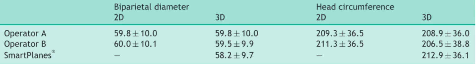
Table1 Biparietal diameter and head circumference values obtained by two independent operators and using
SmartPlanes® technology.
Biparietaldiameter Headcircumference
2D 3D 2D 3D
OperatorA 59.8±10.0 59.8±10.0 209.3±36.5 208.9±36.0
OperatorB 60.0±10.1 59.5±9.9 211.3±36.5 206.5±38.8
SmartPlanes® — 58.2±9.7 — 212.9±36.1
2Dindicatestwo-dimensional.3Dindicatesthree-dimensional.Resultsareexpressedinmmasmean±standarddeviation.
image.Intraoperatorreproducibilitywasevaluatedby
com-paringvaluesobtainedwithrepeatedmeasurementsofthe
same parametersby each operator andthe Smartplanes®
software.Theinteroperatorreproducibilitywasassessedby
comparingthe average of the two3D measures obtained
by operator B and the average of the 3D auto measures
obtained by the software against the average of the 3D
measurementsobtainedby operator A. The minimal
sam-plesize to assess reproducibility for these measurements
wasestimatedat30fetuses.Variabilitybetweentheseries
ofmeasurementswasassessedusinginterclasscorrelation
coefficient (ICC)withits 95% confidence interval(CI)and
Bland-Altmanplots.SignificancewasestimatedusingFtest.
Athreshold<5%wasconsidered significant.The statistical
analysiswasperformedwithRsoftwareversion3.3.1.
Results
A total of 30 patients were included in the study. The
meangestationalageofthefetuseswas23weeks±3.2(SD)
(range:17—29weeksgestation).
In2/60acquisitions(3.3%),theinitiallyobtained3D
vol-ume did not permit the Smartplanes® software to obtain
theBPDandHCmeasurements.However,asecond
acquisi-tioncarriedoutimmediatelyafterwardsenabledallofthe
plannedmeasurementstobemade.The totaldurationfor
bothvolumeacquisitionandautomatedanalysiswasalways
lessthan10s.Table1presentsthemeansandstandard devi-ationsofthemeasurementsmadethetwooperatorsandthe software.
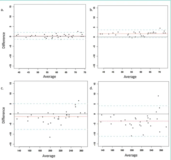
The95% confidenceintervals(95%CI)ofthedifference observedbetween2Dmeasurementsand3Dmeasurements ofBPDandHCwere2and7.5mm,respectively.These inter-vals were 4 and 11mm, respectively for the differences betweenmeasurementsobtainedbythesoftware(3Dauto) andtheconventional2Dmeasurements(Fig.3).
Regarding the intraobserver reproducibility, the ICCs weregreaterthan0.99forcomparisonofthemeasurements 3D and 3D auto obtained fromthe two separate volumes acquiredduringthesameultrasoundexaminationbythetwo operatorsandthesoftware(Table2).RegardingBPD,the95% CIforthedifferenceobservedbetweentherepeated mea-surementswasof1mmforthetwooperators(3Dmeasures) and2.5mmforthesoftware(3Dautomeasures).Regarding HC,the95%CI forthe differenceobserved was5and7.5 mm,respectivelyfor the3Dmeasurementsof operatorsA andBand10mmforthe3Dautosoftware(Fig.4).
Regardinginterobserverreproducibility,ICCswere>0.90 for comparisons of 3D measurements by operator A with 3DmeasurementsbyoperatorBand3Dautomeasurements (Table3).The95%CIforBPDmeasurementsbetween oper-atorAcomparedwithoperatorBandthe3Dautosoftware were1.5and3mm,respectively.ForHCmeasurementsthe intervalswere6and9mmrespectively(Fig.5).
Discussion
Thereproducibilityofasetofmeasurementshasanimpact
onitsaccuracy and therefore itspredictive value. This is
particularly important in the second and third trimesters
whenfetalmeasurementsarebeingusedtopredictgrowth
and mayresultin importantdecisionsregarding timing of
delivery.The accuracy of thesemeasurementsis affected
byintraoperatorandinteroperatorvariability,especiallyin
a busy hospital setting where measurements to monitor
growthmaybeperformedbydifferentoperators[6].
Smartplanes® softwaremakesitpossible anautomated search oftransthalamicplane froma 3Dvolumeandthen automatic measurement of BDP and HC values. This has thepotentialadvantageofstandardizingthemeasurement techniqueandthereforeminimizinginteroperator variabil-ity.
This study shows that measurements obtained using Smartplanes® from a 3D volumeagree closely with those obtainedbytwoexperiencedsonographersfrom2Dimages and3Dvolumes.Theobservedvariabilityislowerthan pre-viouslyreportedforcomparisonsbetween2Dmeasurement andthosemeasuredmanuallyfrom3Dvolumes[3]. Regard-ingall comparisonsmade inour study,thereproducibility wasgreateror equaltothatreportedinalargerscale 2D study[7].Theintraoperatorreproducibilityofthesoftware isveryslightlylowerthanthatobservedbythetwoexpert sonographers,however,itisstillveryhigh.
MeasurementsoftheBPDyieldedasystematicdifference ofabout1.5mmbetweenthosetakenbythesonographers andthesoftware.Thiscanbeexplainedbythediscordance in caliper placementbetween the sonographers (outer to outer) and the software (outer to inner). Any changes in the usual measurement techniques of a sonographer could theoretically have an impact onthe reproducibility ofthosemeasurements.Itisbecauseofthisthatthe oper-ators applied their usual technique of BDP measurement. Whilethereisaninternationalagreementregarding place-mentoftheellipseforHCmeasurement,recommendations
Figure3. Graphsshowagreementbetween3Dmeasurements(meanoperatorB)and2DconventionalmeasurementsforBPD(a)andHC (c)andbetweenautomatedmeasurements(meanSmartplanes®)and2DconventionalmeasurementsforBDP(b)andHC(d).Differenceand averageexpressedinmillimeters.
Table2 Intraobserver variability for fetal head measurements obtained by two independent operators and using
SmartPlanes® technology.
OperatorA OperatorB SmartPlanes®
BPD 0.998<ICC<0.999 0.997<ICC<0.998 0.994<ICC<0.997
HC 0.997<ICC<0.998 0.996<ICC<0.998 0.990<ICC<0.995
BPDindicatesparietaldiameter.HCindicatesheadcircumference.ICCindicatesintraclasscorrelationcoefficient.
concerningtheBPDmeasurementsvary.Thisexplains why
thesoftwarewasprogramedtomeasuretheBPDdifferently.
Thefetalpositionmadeitpossibletofindanacquisition
planeclosetotheexpectedscanningplaneforall60head
volumesstudied.Thesoftwareprovidedcorrectcaliper
posi-tionsafterone,oroccasionallytwo, acquisitionattempts.
Thus,theuseofSmartplane® softwaresignificantlylimited
constraintsassociatedwithprobemanipulationand
orien-tationoftheultrasoundbeam.
Acquisitionof an ultrasoundvolumeisusually asimple
procedure,evenfor lessexperienced operators.However,
714 G.AmbroiseGrandjeanetal.
Figure4. GraphsshowintraobservervariabilityfortheBPD(a)andHC(d)measurementsofoperatorA,theBPD(b)andHC(e) measure-mentsofoperatorBandtheBPD(c)andHC(f)measurementsofSmartplanes®.Differencesandaveragesareexpressedinmillimeters.
Table3 Interobservervariabilityforfetalheadmeasurements.
OperatorAvs.OperatorB OperatorAvs.SmartPlanes®
BPD 0.998<ICC<0.999 0.807<ICC<0.997
HC 0.996<ICC<0.998 0.857<ICC<0.996
BPDindicatesparietaldiameter.HCindicatesheadcircumference.ICCindicatesintraclasscorrelationcoefficient.Thecomparisonwas madeonthemeanoftwomeasurementsobtainedfromrepeatedvolumeacquisitionbyoperatorAorBorusingSmartplanes® software.
themeasurements requireskill, time, knowledge of fetal
anatomyand mastery of the volume-processing software.
TheseresultsconfirmthattheSmartplane® automatic
mea-surementsoftwareallowsaccurate,reproducibleandrapid
biometricmeasurementstobetakenfrom3Dvolumes.
Fur-thermore,unlikethe5D technology[8],Smartplane® does
notrequirethatanatomicallandmarksbeplacedinthe vol-ume to obtain head measurements therefore potentially overcoming the inaccuracies or the failures of measure-ments associated with the inexperienced operator. This, however, must be confirmed by further studies using the Smartplane® technologytocompareaccuracy of measure-mentsusing3Dvolumeacquisitionsobtainedbyexperienced andinexperiencedoperators.
Ourresults areconsistentwiththoseobtained with5D technology[4]. Thatstudy, using 5D softwareon a series of 120 fetal head measurements, demonstrated that the software was successful in obtaining standard fetal head
measurements98.3%ofthetimewithsignificanttimesaving comparedtostandard2Dbiometry.Theirintra-and interob-serverreproducibilitydatawasalsosimilartoours,showing results that were slightly higher than those observed in 2D.
However, these studies have both been performed on 3D volumes where the acquisition plane was optimal. It has been demonstrated, in a study comparing a series of non-automated measurements taken from 2D images and 3D volumes, that past a certain angle between the acquisition plane and the measurement plane, it is not always possible toreconstruct a 2D image toachieve the same quality as obtained by the real-time 2D image and therefore the concordance of biometric measurements is not guaranteed. Fetal position, especially in the third trimester, does not always allow for optimal acquisition of3Dvolumes,thereforetheSmartplane® softwareshould also be evaluatedcomparing different acquisition angles.
Figure5. GraphsshowinterobservervariabilitybetweenoperatorAandoperatorBofBPD(a)andHC(c)3Dmeasurements,andbetween operatorAandSmartplanes® ofBPD(b)andHC(e)3Dmeasurements.Differenceandaverageareexpressedinmillimeters.
In3D,theoperatoronlyhastopositiontheprobeopposite
the cephalic pole. It was previously demonstrated that
thetimerequiredtoacquirethevolumeswassignificantly
shorter than that required for the acquisition of a 2D
measurement set including femoral length, cephalic and
abdominal perimeters: 45s versus 117s, respectively [3].
The difference between 3D and 2D might be even more importantforaninexperiencedsonographer.
In conclusion, advancesin image recognitionand algo-rithmshaveallowedthedevelopmentofsoftwarecapable of automaticallyselecting thecorrectplane and perform-ing measurements of the fetal head which aresimilar to thoseobtainedbyexperiencedsonographers.Morestudies needtobedonetoconfirmuseofthistechnologyina vari-etyofclinicalsettings.However,theseresultshaveopened
thedoortothepracticeofultrasoundassistedbyartificial intelligence.
Appendix
A.
Appendix
A
Supplementary
data
Supplementary data associated with this
arti-cle can be found, in the online version, at
https://doi.org/10.1016/j.diii.2018.08.001.
Disclosure
of
interest
716 G.AmbroiseGrandjeanetal.
References
[1]YazdiB,ZankerP,WangerP,SonekJ,PintofflK,HoopmannM,
etal.Optimalcaliperplacement:manualvsautomated
meth-ods.UltrasoundObstetGynecol2014;43:170—5.
[2]ZaludI,GoodS,CarneiroG,GeorgescuB,AokiK,GreenL,etal.
Fetal biometry:a comparison between experienced
sonogra-phersandautomatedmeasurements.JMaternFetalNeonatal
Med2009;22:43—50.
[3]Sarris I, Ohuma E, Ioannou C, Sande J, Altman DG,
Papa-georghiou AT, et al. Fetal biometry: how well can offline
measurements from three-dimensional volumes substitute
real-time two-dimensional measurements? Ultrasound Obstet
Gynecol2013;42:560—70.
[4]RizzoG,AielloE,MariaElenaPietrolucci,ArduiniD.The
feasi-bilityofusing5DCNSsoftwareinobtainingstandardfetalhead
measurements from volumes acquired by three-dimensional
ultrasonography:comparisonwithtwo-dimensionalultrasound.
JMaternFetalNeonatalMed2016;29:2217—22.
[5]Papageorghiou AT, Ohuma EO, Altman DG, Todros T, Ismail
LC,LambertA,etal.Internationalstandardsforfetalgrowth
based on serial ultrasound measurements: the Fetal Growth
LongitudinalStudyoftheINTERGROWTH-21stProject.Lancet
2014;384:869—79.
[6]HamdaouiN, LesieurE,ManciniJ,Dabadie A,Bourderionnet
V,PicoH,etal.HermanscoreinprenatalscreeningforDown
syndrome:cana juniorassess asenior?DiagnIntervImaging
2017;98:155—60.
[7]SarrisI,IoannouC,ChamberlainP,OhumaE,RosemanF,Hoch
L,etal.Intra-andinterobservervariabilityinfetalultrasound
measurements.UltrasoundObstetGynecol2012;39:266—73.
[8]Rizzo G, Pietrolucci ME, Capece G, Cimmino E, Colosi E,
Ferrentino S, et al. Satisfactory rate of post-processing
visualization of fetal cerebral axial, sagittal, and coronal
planes from three-dimensional volumes acquired in routine
second trimester ultrasound practice by sonographers of
peripheral centers. J Matern Fetal Neonatal Med 2011;24: