Publisher’s version / Version de l'éditeur:
Journal of Leukocyte Biology, 79, November 2, pp. 339-350, 2006
READ THESE TERMS AND CONDITIONS CAREFULLY BEFORE USING THIS WEBSITE.
https://nrc-publications.canada.ca/eng/copyright
Vous avez des questions? Nous pouvons vous aider. Pour communiquer directement avec un auteur, consultez la première page de la revue dans laquelle son article a été publié afin de trouver ses coordonnées. Si vous n’arrivez pas à les repérer, communiquez avec nous à PublicationsArchive-ArchivesPublications@nrc-cnrc.gc.ca.
Questions? Contact the NRC Publications Archive team at
PublicationsArchive-ArchivesPublications@nrc-cnrc.gc.ca. If you wish to email the authors directly, please see the first page of the publication for their contact information.
NRC Publications Archive
Archives des publications du CNRC
This publication could be one of several versions: author’s original, accepted manuscript or the publisher’s version. / La version de cette publication peut être l’une des suivantes : la version prépublication de l’auteur, la version acceptée du manuscrit ou la version de l’éditeur.
For the publisher’s version, please access the DOI link below./ Pour consulter la version de l’éditeur, utilisez le lien DOI ci-dessous.
https://doi.org/10.1189/jlb.1004600
Access and use of this website and the material on it are subject to the Terms and Conditions set forth at
Mast cells, which interact with Escherichia coli, up-regulate genes
associated with innate immunity and become less responsive to
Fc(epsilon)RI-mediated activation
Kulka, Marianna; Fukuishi, Nobuyuki; Rottem, Menachem; Mekori, Yoseph
A.; Metcalfe, Dean D.
https://publications-cnrc.canada.ca/fra/droits
L’accès à ce site Web et l’utilisation de son contenu sont assujettis aux conditions présentées dans le site
LISEZ CES CONDITIONS ATTENTIVEMENT AVANT D’UTILISER CE SITE WEB.
NRC Publications Record / Notice d'Archives des publications de CNRC:
https://nrc-publications.canada.ca/eng/view/object/?id=029ad9ad-cc50-4bad-815a-dda28a7ea811
https://publications-cnrc.canada.ca/fra/voir/objet/?id=029ad9ad-cc50-4bad-815a-dda28a7ea811
Mast cells, which interact with Escherichia coli, up-regulate
genes associated with innate immunity and become less
responsive to FcRI-mediated activation
Marianna Kulka,* Nobuyuki Fukuishi,
†Menachem Rottem,
†Yoseph A. Mekori,
‡and Dean D. Metcalfe
†,1*Allergy-Immunology Division, Northwestern University Feinberg School of Medicine, Chicago, Illinois;
†Laboratory
of Allergic Diseases, National Institute of Allergy and Infectious Diseases, National Institutes of Health, Bethesda,
Maryland; and
‡Department of Medicine, Meir General Hospital, Kfar-Saba, and the Sackler School of Medicine,
Tel-Aviv University, Israel
Abstract:
Mast cells, which are associated with T
helper cell type 2-dependent inflammation, have
now been implicated in the innate immune
re-sponse. To further characterize how mast cells are
programmed to respond to infectious organisms,
we used expression profiling using DNA
microar-ray analysis of gene expression by human mast cells
(huMC) during ingestion of Escherichia coli and
examined immunoglobulin E (IgE)-mediated
de-granulation. Analysis of data revealed that specific
groups of genes were modulated, including genes
encoding transcription factors, cell signaling
mol-ecules, cell cycle regulators, enzymes, cytokines,
novel chemokines of the CC family, adhesion
mol-ecules, and costimulatory molecules.
Enzyme-linked immunosorbent assay analysis confirmed the
production of tumor necrosis factor and the
che-mokines CC chemokine ligand (CCL)-1/I-309,
CCL-19/macrophage-inflammatory protein-3
(MIP-3), and CCL-18/MIP-4; flow cytometry
confirmed the up-regulation of carcinoembryonic
antigen-related cell adhesion molecule 1, the
inte-grin CD49d, and CD80. Coincubation with E. coli
down-regulated Fc receptor for IgE I (FcRI)
ex-pression and FcRI-mediated huMC
degranula-tion. These data are consistent with the concept
that bacterial exposure directs mast cell responses
toward innate immunity and away from
IgE-mediated effects. J. Leukoc. Biol. 79: 339 –350;
2006.
Key Words:
IgE
䡠MIP-3
䡠CCL-19
䡠CCL-18
INTRODUCTION
In the effort to understand innate immunity and the means by
which protection against infectious organisms is accomplished,
recent attention has been directed to mast cells, which occupy
tissues that interface the external environment, including the
respiratory tract, skin, and gastrointestinal tract, placing them
in a unique position to encounter invading organisms,
includ-ing bacteria, and orchestrate a response.
To date, mast cells have been reported to respond to bacteria
through pattern recognition receptors [1] and through
phago-cytosis, the latter, first reported to occur in mast cells as early
as 1923 [2]. The recognition and ingestion of bacteria by mast
cells have now been well-documented and occur through
com-plement receptors [3] and immunoglobulin G (IgG) [4] and via
direct interaction with type 1 fimbriated bacteria [5], which
when ingested by human mast cells (huMC), undergo a
signif-icant loss of viability, and mast cells respond to this interaction
by secreting tumor necrosis factor (TNF) and interleukin (IL)-6
[6 – 8]
To further explore how huMC focus their response to
bac-teria and whether this process directs mast cells away from
IgE-mediated events, we examined the expression of 13,971
genes in mast cells internalizing Escherichia coli, comparing
the pattern of gene expression with that of resting mast cells.
As will be shown, the response of mast cells to this bacteria is
global and includes up-regulation of genes involved in
tran-scription, cell signaling, the cell cycle, enzyme pathways,
adhesion, and surface molecule expression, and huMC
simul-taneously become less responsive to IgE-mediated activation.
These biologic changes in mast cells are an interesting adjunct
to the hygiene hypothesis, which says that bacterial exposure
directs the immune system toward T helper cell type 1
(Th1)-and away from Th2-mediated inflammation.
MATERIALS AND METHODS
E. coli
The E. coli K12 strain, purchased from American Type Culture Collection (Manassas, VA), was cultured in 8 g/l trypton (Fisher Scientific, Fair Lawn, NJ) and 0.5 g/l NaCl (Sigma-Aldrich, St. Louis, MO). This media (20 L), in a sterilized, 50 ml tube, was inoculated with E. coli, incubated at 37°C for 16 h with constant shaking at 250 rotations per minute (rpm), and spread on a 1.5%
1Correspondence: NIH/NIAID/LAD, Bldg. 10, Rm. 11C205, 10 Center Drive, MSC 1881, Bethesda, MD 20892-1881. E-mail: dmetcalfe@niaid.nih.gov
Received October 20, 2004; revised September 12, 2005; accepted Sep-tember 14, 2005; doi: 10.1189/jlb.1004600
agar plate containing Luria-Bertani (LB) broth (Molecular Biologicals, Inc., Columbia, MD) using a platinum loop. Single colonies of bacteria were picked and placed in 50 ml sterilized tubes containing 20 ml bacteria culture media. These cultures were incubated at 37°C for 16 h and then centrifuged at 3000 rpm for 15 min. The supernatant was discarded, and the E. coli pellet was resuspended in 20 ml phosphate-buffered saline (PBS) and centrifuged at 3000 rpm for 15 min. The supernatant was discarded, and the concentration of E.
coli was adjusted to 5 ⫻ 109bacteria/ml with PBS. Additionally, Alexa Fluor 488-labeled E. coli (K12 strain, Molecular Probes, Inc., Eugene, OR) was used in some confocal experiments.
Cell cultures
Laboratory of Allergic Diseases Cell 3 (LAD3) huMC [9], which have similar characteristics to its sister cell lines (LAD1 and LAD2 cells) and primary cultured mast cells, were maintained in StemPro-34 serum-free medium (SFM; Invitrogen Corp., Carlsbad, CA), supplemented with 2 mM L-glutamine, 50 g/ml streptomycin, 100 IU/ml penicillin (Biosource International, Camarillo, CA), and 100 ng/ml recombinant human stem cell factor (rhSCF; PeproTech, Inc., Rocky Hill, NJ).
Human peripheral blood-derived CD34⫹cells were cultured in StemPro-34 SFM supplemented with 2 mM L-glutamine, 50 g/ml streptomycin, 100 IU/ml penicillin, 100 ng/ml SCF, and 100 ng/ml rhIL-6 (PeproTech, Inc.). rhIL-3 (30 ng/ml) was added for the first week. Half of the culture medium was replaced every 7 days. Cultures at 8 –10 weeks consisted of greater than 99% huMC.
E. coli
internalization by mast cells
huMC were centrifuged at 1000 rpm for 5 min, the supernatant discarded, and the cells resuspended in cell culture media adjusted to 5 ⫻ 105cells/ml. The cell suspension (5 ml) was then added to a 25-cm2culture flask and incubated for 30 min at 37°C in a 5% CO2incubator, and 5 l 5 ⫻ 109bacteria/ml was added (bacteria-to-cell ratio of 10:1). The cell-bacteria suspension was then maintained at 37°C in a 5% CO2incubator for times specified. After incuba-tion, the cell-bacteria suspension was centrifuged at 1000 rpm for 5 min, and supernatant was discarded. The pellet was resuspended in 20 ml cell culture media containing antibiotics and centrifuged at 1000 rpm for 5 min. The procedure was repeated, and the pellet was resuspended in 2.5 ml culture media containing antibiotics and incubated for times indicated.
Confocal scanning microscopy
After culture of huMC with E. coli for 5, 15, and 30 min, the cells were fixed and permeabilized using IntraPrep permeabilization reagent (Immunotech, Marseille, France) as per the manufacturer’s instructions. Permeabilized cells were then incubated with rabbit anti-E. coli K12 strain antiserum (a kind gift of Denka Seiken, Tokyo, Japan) for 15 min, the cell suspension was centrifuged at 1200 rpm for 5 min, and the supernatant was discarded. Cells were next resuspended with 1 ml PBS and centrifuged at 1200 rpm for 5 min, and the supernatant was discarded. This procedure was repeated, the pellet suspended in 1 ml PBS, incubated with Alexa Fluor 488-conjugated anti-rabbit IgG (Molecular Probes Inc.) for 15 min, suspended in 0.5% paraformaldehyde in PBS, and placed on poly-L-lysine-coated chambered coverglass slips (Nalgene Nunc International, Naperville, IL), which were examined by a confocal-scanning microscopy Leica TCS-NT/SP laser-confocal-scanning confocal microscope. For characterization of live cells, cells were incubated with anti-Fc receptor for IgE I (FcεRI)-fluorescein isothiocyanate (FITC) or anti-Kit-phycoerythrin (PE) monoclonal antibody (mAb; from BD Biosciences, San Jose, CA) in PBS/0.1% bovine serum albumin (BSA) for 1 h, washed twice with PBS/0.1% BSA, and then analyzed by confocal microscopy.
Microarray analysis
The array chips are custom-made by National Institute of Allergy and Infec-tious Diseases [Bethesda, MD; human sequence chip series “sa” (Hssa)] and consist of 13,971 oligonucleotides, each of which represents a unit gene cluster. All of these elements are 70 mer oligonucleotides synthesized by Qiagen Operon Inc. (Valencia, CA), which hybridize human cDNA synthesized from a human mRNA library.
Total RNA was extracted from three separate phagocytosis experiments for each time-point. RNA was then purified using an RNeasy mini-kit (Qiagen Inc., Valencia, CA). For probe generation, RNA was converted to
double-stranded cDNA by reverse transcription (RT) and Cy3- or Cy5-labeled. Oligo dT 20-mer was first annealed to the RNA, and then RT was performed using Superscript II RT (Invitrogen Corp.). Cy3-labeled deoxyuridine triphosphate (dUTP; Amersham Biosciences AB, Uppsala, Sweden) was added along with unlabeled deoxy-unspecified nucleoside 5⬘-triphosphates (dNTPs) to make Cy3-labeled probe for E. coli-nonexposed samples (control), and Cy5-labeled dUTP (Amersham Biosciences AB) was added along with unlabeled dNTPs to make Cy5-labeled probe for E. coli-exposed samples. The probes were then purified using a Vivaspin 30K centrifugal filter device (Vivascience AG, Hannover, Germany) in Tris-EDTA buffer and quantitated at 550 nm for Cy3 or at 650 nm for Cy5. The microarray chip was incubated with blocking mixture containing 5⫻ saline sodium citrate (SSC), 1% BSA, and 0.1% sodium dodecyl sulfate (SDS) at 42°C for 1 h, and the hybridization was performed by adding 50 pmol-labeled probes to a reaction mix, which included 10 g human Cot-1 DNA, 1 g Poly dA40 – 60, 4 g yeast t-RNA (Invitrogen Corp.), 5⫻ SSC, and 0.1% SDS in 25% formamide solution. The mixture was heated at 98°C for 2 min, applied to a microarray slip after cooling, and incubated overnight at 42°C. The arrays were then sequentially washed in 1⫻ SSC and 0.05% SDS and 0.1⫻ SSC. Next, the arrays were centrifuged for 5 min at 500 rpm for drying and scanned with a GenePix 4000A microarray scanner (Axon Instruments, Inc., Union City, CA). Data were analyzed using a mAdb program (located at http://nciarray.nci.nih.gov/), and the data, with P ⱕ 0.02 compared with control and a signal-to-noise ratio 2.0 or greater, were selected for further analysis.
For determination of gene induction following FcεRI activation, huMC were sensitized overnight with 100 ng/mL biotin-IgE and then stimulated with 100 ng/mL streptavidin for 3 h. Total RNA was isolated from each preparation by the RNeasy mini-kit (Qiagen Inc.) The cDNA probes were generated and hybridized to the Affymetrix gene chip human genome U133 (HG-U133) according to the manufacturer’s protocol (Affymetrix, Santa Clara, CA; http:// www.affymetrix.com/support/technical/datasheets/human_datasheet.pdf). Global scaling was done by all probes and analyzed using Affymetrix GCOS 1.2. Genes with P ⱕ 0.02, compared with control and a signal-to-noise ratio 2.0 or greater, were selected for further analysis.
Real-time polymerase chain reaction (PCR)
The microarray results were validated using real-time PCR on an ABI7500 SDS system. The cDNA templates were prepared independently from cDNA templates, which were used for the microarray hybridizations. At 10 ng, an equivalent of cDNA was used in each quantitative PCR assay. Primer sets for PCR amplifications were designed using the Primer Express software (Perkin-Elmer Applied Biosystems, Boston, MA). All reactions were performed in triplicate for 40 cycles as per the manufacturer’s recommendation. Relative quantitation was performed using standard curves for each primer set. Samples are normalized using the geometric mean of the glyceraldehyde 3-phosphate dehydrogenase (GAPDH) housekeeping gene, and all data are reported as the fold induction over untreated control cells using the comparative threshold cycle method.Cytokine, chemokine, and -hexosaminidase
assays
Human I-309 [CC chemokine ligand 1 (CCL1)], macrophage-inflammatory protein-4 (MIP-4)/pulmonary and activation-regulated chemokine (PARC; CCL18), MIP-3 (CCL19), and TNF were measured using commercial enzyme-linked immunosorbent assay (ELISA) kits or commercial ELISA development sets (R&D Systems, Minneapolis, MN). Lowest limits of detection for the ELISA kits were 0.71 pg/mL (I-309), 10 pg/mL (MIP-4), 10 pg/mL (MIP-3), and 0.12 pg/mL (TNF). MIP-1␣, MIP-1, monocyte chemoattractant protein-1 (MCP-1), and regulated on activation, normal T expressed and secreted (RANTES) were measured using the BD™ cytometric bead array (CBA) human chemokine kit II and analyzed on a FACSarray (BD Biosciences). Lowest limits of detection for the CBA assay are 0.05 pg/mL (RANTES), 4.08 pg/mL (MIP-1␣), 3.27 pg/mL (MIP-1), and 0.79 pg/mL (MCP-1). For determination of degranulation, 1 ⫻ 106cells were incubated with 10 ⫻ 106bacteria (2 l 5⫻109bacteria/mL) or 2 l LB broth (as control) for 8 h at 37°C in antibiotic-free media, washed with media containing antibiotics, and then sensitized overnight with 1 g/mL IgE-biotin. Cells were washed and resuspended in Hepes buffer (10 mM HEPES, 137 mM NaCl, 2.7 mM KCl, 0.38 mM Na2HPO4.7H2O, 5.6 mM glucose, 1.8 mM CaCl2.2H2O, 1.3 mM MgSO4.7H2O,
0.04% BSA, pH 7.4) and then stimulated with streptavidin and incubated at 37°C for 30 min. The -hexosaminidase in the supernatants and cell lysates was quantified by hydrolysis of p-nitrophenyl N-acetyl--D-glucosamide (Sigma-Aldrich) in 0.1 M sodium citrate buffer (pH 4.5) for 90 min at 37°C. The percentage of -hexosaminidase release was calculated as percent of total content. Chemokine content was measured using the CBA assay as above.
Flow cytometry
For analysis of expression of surface molecules, cells were incubated with anti-human leukocyte antigen (HLA)-DR, -DP, and -DQ FITC-conjugated and anti-CD49d PE-conjugated antibody (BD Biosciences) for 30 min on ice and analyzed by fluorescein-activated cell sorter. In the case of CD80 and carci-noembryonic antigen-related cell adhesion molecule 1 (CEACAM1), cells were incubated with anti-CD80 (BD Biosciences) or anti-CEACAM1 (CD66a; Axxora LLC, San Diego, CA) and then anti-mouse IgG PE-conjugated antibody (Southern Biotech, Birmingham, AL). For intracellular analysis of tryptase and chymase content, huMC were fixed with 4% paraformaldehyde for 5 min, permeabilized with PBS/0.1% saponin, blocked with PBS/5% milk/0.1% sa-ponin, and incubated with rabbit antitryptase, mouse antichymase-biotin, or appropriate isotype antibody (all from Chemicon International, Temecula, CA) for 1 h at 4°C. Cells were washed twice with PBS/0.1% saponin and incubated with anti-rabbit-allophycocyanin or streptavidin-PE (both from BD Bio-sciences) before analysis by flow cytometry.
Mast cell bactericidal assay
Viability of internalized bacteria was determined by measuring colony-forming units (CFU) as described [10]. huMC (0.3⫻106/ml) were grown overnight in StemPro medium (antibiotic-free) at 37°C with 5% CO, then exposed to 3.0 ⫻ 107bacteria for 30 min at 37°C. huMC were washed twice with StemPro (antibiotic-free) to remove adherent or noninternalized bacteria. Cells were rapidly frozen and thawed to release internalized bacteria, and viable bacteria were quantitated by determining the number of CFU from serial dilutions of the cell lysates plated on MacConkey agar. The viability of the huMC during the incubation period was monitored periodically by Trypan blue dye exclusion assay and determined to be consistently ⬎90%.
Statistical analysis
Data in the text and figures are expressed as the mean ⫾SEM. Statistical comparisons were carried out using ANOVA and the Student’s t-test. P values less than 0.05 were considered significant.
RESULTS
Binding and internalization of E. coli by huMC
LAD huMC are sister cell cultures (1–3) obtained from a
patient with severe systemic mast cell disease [9]. They have
many characteristics in common with primary CD34
⫹progen-itor-derived, cultured huMC. Specifically, these cells stain
metachromatically with toluidine blue (Fig. 1A); contain
his-tamine (5.3⫾1.2 pg/cell), tryptase (Fig. 1B), and chymase (Fig.
1C); express surface Kit and Fc
ε
RI (Fig. 1, D–F); require SCF
for growth; and degranulate in response to Fc
ε
RI cross-linking.
LAD3 mast cells were used in the studies below.
Murine and cord blood-derived huMC are reported to attach
to and internalize bacteria [5–7]. Therefore, we first verified
that LAD huMC were capable of internalizing bacteria and
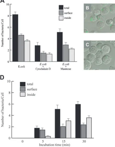
then characterized the process. As can be seen in Figure 2,
confocal scanning microscopy shows that E. coli (green)
ad-hered to the surface of and were internalized into mast cells
(Fig. 2, A and B). Adherence and internalization could be
inhibited by pretreatment with cytochalasin D or mannose (Fig.
2, A and C). A time course of the interaction between mast
cells and live E. coli, which starts with adhesion of the bacteria
to the cell surface, followed by internalization, is shown in
Figure 2D. Taken together, these results indicate that huMC
recognize and attach to E. coli. On average, and by 30 min, four
bacteria were internalized by each huMC.
The viability of the internalized bacteria was measured to
determine if adhesion and internalization were microbicidal.
After allowing bacteria to be internalized for 30 min in the
presence of mannose or cytochalasin D (see Materials and
Methods), mast cells were lysed, and bacteria were plated onto
agar plates. Although each huMC internalized 3.8 ⫹ 0.2
bacteria/huMC (Fig. 2D), only 1.9 ⫹ 0.4 (34%) of those
bac-teria were able to grow and form colonies. If huMC had been
Fig. 1.Characterization of LAD huMC. LAD cells were fixed and stained with toluidine blue (A) or were fixed, permeabilized, and stained with antitryptase or anti-chymase mAb and examined by flow cytometry (B–C). Live LAD cells were incubated with anti-FcεRI-FITC or anti-Kit-PE mAb and analyzed by confocal micros-copy (D–F). DIC, Differential in-terference contrast.
preincubated with cytochalasin D, only 0.3 ⫹ 0.1 (8%)
bac-teria/huMC formed colonies. Mannose did not have a
signifi-cant effect, as 1.0 ⫹ 0.5 (26%) bacteria/huMC formed
colo-nies. These data suggest that once internalized, huMC are able
to destroy the E. coli.
Gene expression by huMC following exposure to
E. coli
A critically important outcome of the interaction of opsonized
bacteria with mast cells would be the up-regulation of families
Fig. 2. Confocal scanning microscopy analysis of the effects of cytochalasin D and mannose on huMC internalization of bacteria. (A) The effects of 4 M cytochalasin D and 10 mM mannose pretreatment on adhesion and internalization of fluoro-labeled E.
coli by mast cells. (B) Mast cells incubated with fluoro-labeled E. coli. (C) Cytochalasin D-treated (4 M) mast cells incubated with
fluoro-labeled E. coli. In all the above, mast cells were incubated with tenfold bacterial particles for 30 min. (D) Time course of internalization of live E. coli by huMC, as analyzed by confocal scanning microscopy. Mast cells were exposed to live E. coli, and cells and bacteria were fixed and stained with FITC-labeled anti-E. coli antibody.
Fig. 3. Annotation distribution of activated genes at 4, 8, and 24 h of incubation of live E. coli with huMC, which were elevated at least twofold at P ⬍ 0.02. Definitions of the gene families are based on the Gene Bank annotation for each gene, which at each time-point, was averaged (n⫽3 separate phagocytosis experiments) and selected for display if greater than twofold up-regulated at P ⬍ 0.02.
TABLE 1. Genes Up-Regulated following E. coli Exposure
Gene Protein description
Fold induction 4 h 8 h 24 h Antigen presentation CD80 CD80 antigen (B7-1) 2.36 1.81 2.74 CD86 CD86 antigen (B7-2) 6.32 1.05 1.30 HLA-DMB MHC-II 3.01 1.58 2.65
CRTAM MHC-I-restricted molecule 2.30 1.75 2.59
HKE4 HLA class II region-expressed gene KE4 2.79 1.66 1.24
Adhesion
CDH1 cadherin, type 1, E-cadherin 1.49 1.28 4.52
ITGA4 CD49d integrin, ␣ 4 2.20 0.99 1.42
ITGA3 CD49c integrin, ␣ 3 ⫺1.27 ⫺1.35 ⫺2.34
ITGAV CD51 integrin, ␣ V (vitronectin receptor) ⫺2.13 ⫺1.32 0.99
ITGB1 CD29 integrin,  1 ⫺1.30 ⫺1.95 ⫺2.59
ITGB4 integrin,  4 2.01 1.49 2.81
ITGB5 integrin,  5 1.23 1.19 3.04
ICAM1 intercellular adhesion molecule (CD54) 1.08 0.88 2.49
ICAM5 intercellular adhesion molecule 5 2.38 2.11 1.28
FBLN2 fibulin 2 2.09 2.12 3.07
CEACAM1 cell adhesion molecule 1 1.15 1.04 3.62
CEACAM3 cell adhesion molecule 3 2.05 1.25 1.67
CEACAM4 cell adhesion molecule 4 1.73 1.41 2.18
CEACAM6 cell adhesion molecule 6 2.11 1.52 2.25
CEACAM8 cell adhesion molecule 8 2.44 1.97 2.05
ARHGDIA GDP dissociation inhibitor (GDI) ␣ 2.43 1.50 1.24
Transcription and signaling
PDE4A phosphodiesterase 4A, cAMP-specific 5.18 4.85 1.23
SH2D2A SH2 domain protein 2A 6.39 4.62 1.41
MT1X metallothionein 1X 3.56 3.48 1.95
MT2A metallothionein 2A 3.67 4.93 2.18
MT1G metallothionein 1G 3.45 3.81 1.76
MT1A metallothionein 1A 3.03 3.20 1.77
MT1F metallothionein 1F 3.33 3.38 1.74
HEY1 hairy/enhancer-of-split related with YRPW 7.40 3.77 1.10
CREM cAMP-responsive element modulator 4.23 6.64 1.46
IRF1 interferon regulatory factor 1 1.00 1.00 2.82
IRF7 interferon regulatory factor 3.78 1.70 10.68
Cytokines, chemokines, and growth factors
SCYA1 chemokine ligand 1 (I-309) 2.52 1.61 1.27
SCYA2 chemokine ligand 2 (MCP-1) 2.69 1.64 1.06
SCYA15 chemokine ligand 15 (MIP-1␦) 0.85 1.05 4.30
SCYA18 chemokine ligand 18 (PARC or MIP-4) 1.38 2.20 3.87
SCYA19 chemokine ligand 19 (MIP-3) 8.27 1.58 3.92
SCYA23 chemokine ligand 23 (CK8) 2.46 2.31 2.05
SCYA24 chemokine ligand 24 (Eotaxin-2) 2.56 2.73 2.73
ISG15 interferon-stimulated protein 2.27 1.95 1.42
MIF macrophage migration inhibitory factor 2.78 1.92 1.27
TNF tumor necrosis factor 2.73 2.20 3.19
Surface receptors
IFNAR1 interferon (␣, , and ) receptor 1 2.38 1.65 2.52
IL4R IL-4 receptor 3.22 2.77 1.18
IL8RA IL-8 receptor, ␣ 3.44 1.45 2.20
IL9R IL-9 receptor 2.53 2.26 1.16
IL10RB IL-10 receptor,  2.25 1.18 1.07
CD22 CD22 antigen, member of siglec family 0.86 1.05 2.15
CD37 CD37 antigen, tetraspanin 1.05 1.23 2.10
CD48 CD48 antigen, bacterial recognition 0.77 0.93 2.44
CD68 CD68 antigen, gram-neg bacterial recognition 1.05 1.18 2.45
C5R1 complement component 5 receptor 1 1.91 2.36 1.16
FCER1A FcεRI, IgE high-affinity receptor, ␣ chain ⫺1.47 ⫺1.48 ⫺1.13
of genes whose products facilitate the ability of mast cells to
respond to the presence of microorganisms and in turn, would
contribute to the initiation of an innate immune response. We
thus examined the global gene expression program engaged by
the interaction of huMC with live E. coli. Using microarray
technology, we selected genes for further study, which were
up-regulated at least twofold in mast cells exposed to E. coli
versus resting cells at the P ⬍ 0.02 level and at 4, 8, and 24 h
(n⫽3– 6). By this process, we identified 251 genes at 4 h, 88
genes at 8 h, and 158 genes at 24 h for further analysis by
grouping as to function.
Significantly up-regulated genes were categorized into 11
groups according to the known functions of their corresponding
proteins: cytokines/chemokines, signaling, cell cycle, cell
sur-face, transcription, enzyme, cytoskeleton, adhesion, protein
transport, DNA/RNA/protein-binding, and unknown (Fig. 3).
The overall pattern of gene up-regulation appeared rather
consistent through 24 h. The largest group of transcripts
up-regulated (16 –20%) coded for enzymes. These genes included
the respiratory chain-related enzymes such as reduced
nico-tinamide adenine dinucleotide dehydrogenase ␣ and 
(five-fold induction), three isoforms of 2⬘-5⬘-oligoandenylate
syn-thetase (two- to 12-fold induction), cytochrome C oxidase
sub-units (threefold induction), and RNase 3 (eosinophil cationic
protein; Table 1). These genes represent an increase in cell
metabolism.
The next largest group of genes up-regulated following mast
cell exposure to E. coli included signaling molecules,
account-ing for 14 –16% of the total number of induced genes. These
included the cAMP responsive element modulator (eightfold
induction), SH2 domain protein 2A (fivefold induction),
phos-phodiesterase 4A (3.5-fold induction), the TNF
receptor-asso-ciated factor 1 (3.4-fold induction), and the interferon (IFN)
regulatory factors 1 and 7 (IRF1 and IRF7; three- to 10-fold
induction; Table 1).
The cytokine/chemokine group accounted for 2– 6% of all
selected genes. In this group, the most strongly induced genes
were TNF (threefold induction), CCL1 (or I-309; 2.5-fold
in-duction), CCL19 (MIP-3; eightfold inin-duction), CCL18
(MIP-4; fourfold induction), CCL24 (eotax2; threefold
in-duction), CCL15 (MIP-1␦; fourfold inin-duction), and MIF
(three-fold induction; Table 1). IL-5, IL-7, and IL-16 were
up-regu-lated two- to threefold, but this induction was not significant
when compared with control (Table 1). These cytokines and
chemokines are associated with innate responses involving
dendritic cells (DC) and lymphocytes [11].
Genes encoding cell-surface receptors accounted for 8 –12%
of up-regulated genes. These receptors included IFNAR1
(2.5-fold induction), IL-4R (three(2.5-fold induction), IL-8RA (three(2.5-fold
induction), IL-9R (2.5-fold induction), IL-10RB (twofold
in-duction), CD22, CD37, CD48, CD68, and complement
compo-nent 5 receptor 1 (Table 1). Expression of the Fc
ε
RI (FCER1A)
and Fc␥RI (FCGR1A) receptors was down-regulated slightly
(onefold reduction).
The gene profile induced in response to bacteria differs from
that generated by IgE-dependent activation of mast cells.
Fc
ε
RI-mediated activation resulted in the expression of
proin-flammatory cytokines and chemokines such as IL-1, IL-3,
granulocyte macrophage-colony stimulating factor (GM-CSF),
CCL5, RANTES, CCL2, MCP-1, CCL3, or MIP-1␣ and CCL4
or MIP-1 (Table 2). MCP-1, MIP-1, MIP-1␣, and RANTES
protein production was confirmed in supernatants from huMC
stimulated with IgE/anti-IgE for 24 h (Fig. 4). Although Fc
ε
RI
activation also induced expression of I-309 and TNF, it did not
induce expression of MIP-3 and MIP-4.
TABLE 1. (Continued)
Gene Protein description
Fold induction 4 h 8 h 24 h Enzymes TPSG1 tryptase ␥ 1 2.33 1.31 2.30 CTSL cathepsin L 1.28 2.20 2.71 CTSD cathepsin D 1.17 0.90 2.31
RNASE3 ribonuclease, RNase A family, 3 (ECP) 0.74 2.18 1.49
OAS3 2⬘-5⬘-oligoadenylate synthetase 3, 100 kDa 1.84 2.98 12.32
PLAA phospholipase A2-activating protein 0.98 ⫺2.13 ⫺2.38
HEXB hexosaminidase B ( polypeptide) ⫺1.59 ⫺2.04 ⫺1.35
Red ⫽ Over two-fold induction and significant when compared with control (Pⱕ0.02); blue ⫽ over two-fold reduction and significant when compared with control (Pⱕ0.02); green ⫽ over two-fold induction or reduction but not significant. MHC, Major histocompatibility complex; GDP, guanosine 5⬘-diphosphate; cAMP, cyclic adenosine monophosphate; SH2, src homology 2; Ck8, chemokine  8; ECP, eosinophil cationic protein.
TABLE 2. Genes Up-Regulated following FcεRI Activation
Gene Protein description Fold induction
SCYA5 CCL5, RANTES 68.59
MCP-1 monocyte chemotactic protein 45.25
SCYA4 CCL4, MIP-1 25.99 SCYA1 CCL1, I-309 22.63 TNFRSF1B TNF receptor 1 13.93 GM-CSF colony-stimulating factor 2 (granulocyte-macrophage) 10.56 SCYA3 CCL3, MIP-1␣ 6.96
NFATC1 NF-ATc transcription factor 6.96
TNF tumor necrosis factor 6.06
IL3 interleukin-3 5.66
FYN FYN oncogene related to SRC 4.59
IL1A interleukin-1␣ 2.00
huMC secrete TNF and chemokines after contact
with E. coli
Microarray results showing up-regulation of I-309, MIP-4, and
MIP-3 by primary huMC derived from human peripheral blood
CD34
⫹cells in response to E. coli were first validated by
real-time PCR (Fig. 5). There was at least a fivefold induction
of mRNA expression of TNF, I-309, MIP-3, and MIP-4 at
8 –24 h after the mast cells had been exposed to E. coli (Fig. 5).
The protein products of the chemokine/TNF genes were then
measured in the supernatants of LAD huMC, which had been
exposed to E. coli for 30 min. Although each mediator was
found to be released in a different time course, all were
significantly elevated 24 h after encountering E. coli (Fig. 6A).
For comparison, huMC derived from human peripheral blood
CD34
⫹cells were similarly incubated with E. coli, and TNF,
I-309, MIP-3, and MIP-4 were measured in the supernatants.
Similar to the LAD huMC, peripheral blood CD34
⫹-derived
mast cells also released these mediators in a time-dependent
manner (Fig. 6B). Thus, these observations indicate that huMC
respond to contact with E. coli by releasing certain
chemo-kines, as well as TNF, validating array data and consistent with
a response that would further innate immunity.
Coincubation with E. coli up-regulates
expression of adhesion and antigen-presenting
molecules
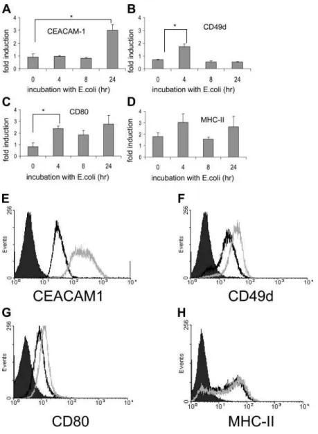
A novel finding of the microarray analysis was that several
adhesion molecules were up-regulated or down-regulated
fol-lowing E. coli coincubation (Table 1). These molecules
in-cluded the 1 integrins and the newly characterized family of
CEACAM molecules. CEACAM1, a receptor known to bind
bacteria in other cell types [12], was up-regulated threefold at
Fig. 4. MCP-1, MIP-1␣, MIP-1, and RANTES re-lease following FcεRI activation of huMC. Mast cells were sensitized with 1 g/mL IgE and then stimulated with 100 ng/mL streptavidin for 24 h, and chemokine levels were measured by the CBA assay. SA, .
Fig. 5. TNF and chemokine mRNA expression induced by mast cell phagocytosis of E. coli particles. Total RNA isolated from peripheral blood CD34⫹ progenitor-derived mast cells after phagocytosis for analysis of TNF and chemokine expres-sion by real-time PCR. GAPDH served as a control for consti-tutive gene expression.
24 h, and CD49d was up-regulated twofold at 4 h. These data
are diagramed in Figure 7. Flow cytometric analysis
con-firmed that CEACAM1 (Fig. 7E) and CD49d (Fig. 7F)
expres-sion is up-regulated after 24 h.
Rodent and huMC are known to express antigen-presenting
molecules such as MHC-II [13–15] and CD80 (B7-1) [16].
Moreover, mouse mast cells are known to present intracellular
antigens to T cells [17, 18]. Our microarray analysis shows that
several of these antigen-presenting molecules are up-regulated
following E. coli coincubation, including CD80 and less so for
MHC-II (Table 1 and Fig. 7), which would enhance a
subse-quent response leading to augmented immunity. Flow
cytomet-ric analysis of CD80 (Fig. 7G) and MHC-II (Fig. 7H) confirms
that surface expression of CD80 is up-regulated at 24 h, and
MHC-II expression is relatively unchanged.
Coincubation with E. coli down-regulates
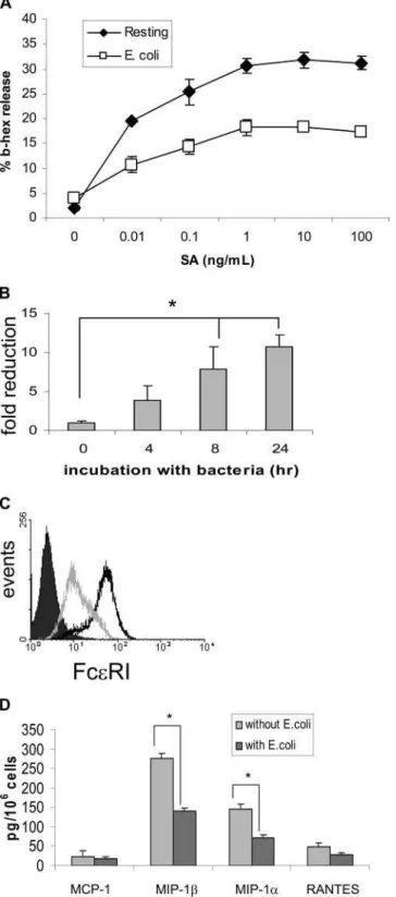
expression of FcRI and degranulation
To examine whether huMC alter their ability to respond to
Fc
ε
RI-mediated signals after coincubation with E. coli, we
exposed huMC to E. coli, activated these mast cells via Fc
ε
RI
with IgE-biotin/streptavidin, and measured degranulation.
Mast cells exposed to E. coli, then activated via Fc
ε
RI,
re-leased significantly less -hexosaminidase (Fig. 8A). Using
the initial analysis criteria of greater-than-twofold induction/
reduction, the microarray did not show any change in Fc
ε
RI
Fig. 6 .TNF and chemokine release from huMC after phagocytosis of E. coli. LAD mast cells (A) and peripheral blood CD34⫹ progenitor-derived mast cells (B) were incubated with tenfold live E.
coli particles for 30 min. The cells were then
incu-bated for several time periods following the removal of E. coli by washing with PBS. Figures represent protein concentrations as measured by ELISA. Data are shown as mean ⫾SEM, n ⫽ 3 separate experi-ments. *, P ⬍ 0.05; **, P ⬍ 0.01.
gene expression. However, when the microarray data were
reanalyzed using a greater-than-onefold induction/reduction
criteria, Fc
ε
RI mRNA expression was down-regulated at 4 h
and 8 h of coincubation with E. coli (Table 1 and Fig. 7B). Flow
cytometry analysis confirmed that huMC expressed less surface
Fc
ε
RI following coincubation with E. coli (Fig. 8C).
Further-more, when huMC were preincubated with E. coli for 24 and
then stimulated via Fc
ε
RI, they released less MIP-1␣ and
MIP-1 compared with untreated cells (Figs. 4 and 8D).
DISCUSSION
Coincubation of huMC with E. coli resulted in bacterial
inter-nalization, as assessed by confocal microscopy (Fig. 2), in
agreement with previous findings [5, 6, 19]. Internalized
bac-teria could not form CFU and were presumably destroyed by
the huMC phagolysosomal system. Furthermore, pretreatment
of huMC with mannose and cytochalasin D inhibited E. coli
internalization. FimH, a mannose-binding subunit, located
preferentially at the type 1 fimbrial tips, is the specific
deter-minant recognized by CD48 on mast cells, and FimH-negative
E. coli mutants exhibit limited mast cell binding [7, 8].
There-fore, pretreatment of huMC with mannose inhibited the
FimH-CD48 interaction and subsequently prevented bacterial
adhe-sion to huMC. Cytochalasin D is a cell-permeable fungal toxin,
which inhibits filamentous actin assembly and mast cell
mem-brane ruffling [20] and has been used to inhibit macrophage
phagocytosis [21].
Microarray analysis showed that the pattern of gene
induc-tion in response to E. coli is prolonged, continuing through at
least 24 h of coincubation with bacteria (Table 1). Transcripts
up-regulated include those coding for enzymes, surface
mole-cules, components of the cytoskeleton, and molecules
associ-ated with signaling, the cell cycle, adhesion, and protein
transport (Fig. 3).
One group of important up-regulated genes included
che-mokines such as CCL1/I-309, CCL18/MIP-4, and CCL19/
MIP-3 (Table 1, Figs. 4 and 5). Mast cells have heretofore not
been known to produce CCL18 and CCL19. CCL1/I-309 binds
CC chemokine receptor 8 on activated Th2 cells, monocytes,
and immature DC and recruits them to target tissues during
inflammation [22]. CCL1/I-309 also binds to human umbilical
vein endothelial cells and induces their chemotaxis, invasion,
Fig. 7. Incubation with E. coli modifies huMC surface mole-cule expression. CEACAM1 (A), CD49d (B), CD80 (C), and MHC-II (D) mRNA expression was assessed by microarray analysis. Surface protein expression of CEACAM1 (E), CD49d (F), CD80 (G), and MHC-II (H) by untreated huMC (black line) or mast cells coincubated (24 h) with E. coli (shaded line) as compared with isotype control (solid curve; n⫽3; *, P⬍0.01).
and differentiation [23]. Therefore, production of CCL1/I-309
by huMC during bacterial infection in the lung and other
tissues would recruit DC and further the immune response.
CCL18/MIP-4, also called PARC, is a new, recently
de-scribed CC chemokine, originally identified as a T cell
che-moattractant produced by DC [24]. Unlike other chemokines,
CCL18/MIP-4 is thought to be produced mainly by monocytes
and DC [25] and is involved in innate immune responses to
staphylococcal enterotoxins [26], pulmonary fibrosis in
sclero-derma [27], and hypersensitivity pneumonitis [28], perhaps by
inducing collagen production by lung fibroblasts [29].
CCL19/MIP-3, also called Epstein-Barr virus-induced
gene 1-ligand chemokine or CK11, is a member of the CC
chemokine family and is constitutively expressed in thymus,
lymph nodes, spleen, and small intestines, and expression in
the bone marrow is inducible by IFN-␥, TNF, and
lippolysac-charide (LPS) [30]. In addition to recruiting macrophage
pro-genitors, MIP-3 is essential in recruiting immature DC and
memory T cells to secondary lympoid organs [31]. Therefore,
during E. coli infection, huMC-derived CCL19/MIP-3 would
recruit DC and memory T cells and initiate an adaptive
im-mune response. Similarly, our study shows that huMC
coincu-bated with E. coli up-regulated expression of TNF [32], which
promotes local inflammation, activates endothelial cells, and is
involved in T cell recruitment to draining lymph nodes during
bacterial infection [1, 33].
The microarray analysis also indicated a profile of cytokine
production in response to bacteria, which noticeably lacked
evidence of up-regulation of cytokines/chemokines including
IL-1, IL-2, IL-3, IL-4, IL-6, IL-8, IL-10, IFN-␥, CCL3, and
CCL4. This pattern differs from Fc
ε
RI-mediated mast cell
activation, where among other cytokines and chemokines,
mes-sage for IL-1, IL-3, GM-CSF, CCL3 (MIP-1␣), CCL4 (MIP-1),
and CCL5 (RANTES) is up-regulated (Table 2). The gene
profile induced by E. coli also differs from the gene profile
induced by selective pathogen ligands such as LPS. In a study
of cord blood-derived mast cells, Okumura et al. [34] found
that LPS up-regulated mRNA for cytokines IL-1, IL-10, IL-15,
CCL5, and CCL8. The majority of genes induced by LPS was
members of the NF-B pathway and did not include I-309,
MIP-3, or MIP-4, suggesting that whole, live E. coli activates
different pathways from LPS alone.
Microarray analysis also showed a previously unknown
pro-file of adhesion molecule up-regulation. The family of novel
adhesion molecules, CEACAMs, in particular, was
up-regu-lated following E. coli exposure (Fig. 6). CEACAMs mediate
binding to and engulfment of Neisseria gonorrhoeae [12] and E.
coli [35] by epithelial cells. We show that huMC express
CEACAM1 and may be involved in internalization and
engulf-ment of E. coli. Similarly, CD49d, a 1 integrin, is also
up-regulated following E. coli exposure (Fig. 6). In the mouse,
CD40d mediates mast cell adhesion to endothelium and is
important for mast cell immigration into the small intestine
during nematode infection [36]. huMC up-regulated genes
in-volved in antigen presentation such as CD80 (Fig. 6). Although
huMC express CD86 and MHC-II, surface protein expression
of these molecules is not up-regulated following E. coli
expo-sure (Fig. 6 and data not shown). However, these data suggest
in total that huMC direct their activation toward responses that
Fig. 8. Coincubation down-regulates huMC degranulation via FcεRI. (A) Untreated huMC and huMC coincubated with E. coli (24 h) were stimulated via FcεRI (see Materials and Methods), and -hexosaminidase (b-hex) was mea-sured to determine degranulation. (B) FcεRI mRNA expression following exposure to E. coli was validated by real-time PCR (*, P⬍0.01). (C) Untreated huMC (solid line) or mast cells coincubated (24 h) with E. coli (shaded line) were assessed for expression of surface FcεRI protein by flow cytometry (solid curve, isotype control; representative of two experiments). (D) Mast cells (1.0⫻106) were incubated with 1.0 ⫻ 108E. coli or LB broth (as control) for 8 h at 37°C in antibiotic-free media, washed with media containing antibiotics, and then sensitized overnight with 1 g/mL IgE-biotin. Cells were washed and stimulated with 10 ng/mL streptavidin and incubated at 37°C for 24 h, and chemokine production was measured by the CBA assay (*, P⬍0.01).
favor those beneficial to the host in addressing an infectious
insult.
Consistent with the hypothesis that huMC reprogram
them-selves following exposure to bacteria, receptor expression was
modified significantly following E. coli exposure. For example,
mast cells up-regulated receptors IL-4, IL-9, and IL-10, which
are known to modulate mast cell differentiation, degranulation,
and cytokine release (Table 1). IL-4 and IL-9 activate mast
cells to proliferate and potentiate their release of
proinflam-matory cytokines [37–39], and IL-10 inhibits Fc
ε
RI expression
and mediator release [40 – 42]. As expected, huMC up-regulate
CD48, which binds E. coli and aids in internalization of
bac-teria [7]. Mast cells retained their ability to degranulate after
exposure to bacteria but less efficiently, possibly as a result of
a decrease in surface expression of Fc
ε
RI (Fig. 7, A–C).
In this report, we thus examine the mRNA profile of gene
expression in huMC, associated with exposure to and
internal-ization of intact bacteria, in this case, E. coli. The
up-regula-tion of genes was global and included genes involved in a
diversity of mast cell functions. Among the genes up-regulated
were chemokines, adhesion molecules, costimulatory
mole-cules, and surface receptors, suggesting a concerted
repro-gramming of the ability of mast cells to respond to a bacterial
insult. Taken together, these data support the concept of a
specific and vigorous mast cell-dependent response to bacteria
to further host defense. IgE-dependent events, which are
crit-ical to the induction of any allergic responses, appear
down-regulated. This direction of mast cell responses to a more
Th1-type inflammation and away from a Th2 allergic response
is an interesting support of the hygiene hypothesis, which
states that exposure to infectious organisms and their products
skews away from atopy.
ACKNOWLEDGMENTS
This work was supported by the intramural program at the
National Institutes of Health Research. The authors thank
Brian Peter Tancowny, Cecilia Sheen, Pazit Salamon, and
Nitza Shoham for their technical assistance. M. K. and N. F.
contributed equally.
REFERENCES
1. Kulka, M., Alexopoulou, L., Flavell, R. A., Metcalfe, D. D. (2004) Acti-vation of mast cells by double-stranded RNA: evidence for actiActi-vation through Toll-like receptor 3. J. Allergy Clin. Immunol. 114, 174 –182. 2. Tsuda, S. (1923) Experimentelle Untersuchungen uber die entzundliche
reaktion der subcutis in beziehung zum individuellen immunitatszustand.
Virchows Arch. Pathol. 247, 123.
3. Sher, A., Hein, A., Moser, G., Caulfield, J. P. (1979) Complement recep-tors promote the phagocytosis of bacteria by rat peritoneal mast cells. Lab.
Invest. 41, 490 – 499.
4. Talkington, J., Nickell, S. P. (2001) Role of Fc ␥ receptors in triggering host cell activation and cytokine release by Borrelia burgdorferi. Infect.
Immun. 69, 413– 419.
5. Malaviya, R., Ross, E. A., MacGregor, J. I., Ikeda, T., Little, J. R., Jakschik, B. A., Abraham, S. N. (1994) Mast cell phagocytosis of FimH-expressing enterobacteria. J. Immunol. 152, 1907–1914.
6. Arock, M., Ross, E., Lai-Kuen, R., Averlant, G., Gao, Z., Abraham, S. N. (1998) Phagocytic and tumor necrosis factor ␣ response of human mast
cells following exposure to gram-negative and gram-positive bacteria.
Infect. Immun. 66, 6030 – 6034.
7. Malaviya, R., Gao, Z., Thankavel, K., van der Merwe, P. A., Abraham, S. N. (1999) The mast cell tumor necrosis factor ␣ response to FimH-expressing Escherichia coli is mediated by the glycosylphosphatidylinosi-tol-anchored molecule CD48. Proc. Natl. Acad. Sci. USA 96, 8110 – 8115. 8. Malaviya, R., Ikeda, T., Ross, E., Abraham, S. N. (1996) Mast cell modulation of neutrophil influx and bacterial clearance at sites of infection through TNF-␣. Nature 381, 77– 80.
9. Kirshenbaum, A. S., Akin, C., Wu, Y., Rottem, M., Goff, J. P., Beaven, M. A., Rao, V. K., Metcalfe, D. D. (2003) Characterization of novel stem cell factor responsive human mast cell lines LAD 1 and 2 established from a patient with mast cell sarcoma/leukemia; activation following aggrega-tion of FcεRI or Fc␥RI. Leuk. Res. 27, 677– 682.
10. Malaviya, R., Ross, E. A., MacGregor, J. I., Ikeda, T., Little, J. R., Jakschik, B. A., Abraham, S. N. (1994) Mast cell phagocytosis of FimH-expressing enterobacteria. J. Immunol. 152, 1907–1914.
11. Rossi, D., Zlotnik, A. (2000) The biology of chemokines and their recep-tors. Annu. Rev. Immunol. 18, 217–242.
12. McCaw, S. E., Liao, E. H., Gray-Owen, S. D. (2004) Engulfment of
Neisseria gonorrhoeae: revealing distinct processes of bacterial entry by
individual carcinoembryonic antigen-related cellular adhesion molecule family receptors. Infect. Immun. 72, 2742–2752.
13. Dimitriadou, V., Mecheri, S., Koutsilieris, M., Fraser, W., Al Daccak, R., Mourad, W. (1998) Expression of functional major histocompatibility complex class II molecules on HMC-1 human mast cells. J. Leukoc. Biol. 64,791–799.
14. Warbrick, E. V., Taylor, A. M., Botchkarev, V. A., Coleman, J. W. (1995) Rat connective tissue-type mast cells express MHC class II: up-regulation by IFN-␥. Cell. Immunol. 163, 222–228.
15. Raposo, G., Tenza, D., Mecheri, S., Peronet, R., Bonnerot, C., Desaymard, C. (1997) Accumulation of major histocompatibility complex class II molecules in mast cell secretory granules and their release upon degran-ulation. Mol. Biol. Cell 8, 2631–2645.
16. Yu, C. K., Chen, C. L. (2003) Activation of mast cells is essential for development of house dust mite Dermatophagoides farinae-induced aller-gic airway inflammation in mice. J. Immunol. 171, 3808 –3815. 17. Tkaczyk, C., Villa, I., Peronet, R., David, B., Mecheri, S. (1999) Fcε
RI-mediated antigen endocytosis turns interferon-␥-treated mouse mast cells from inefficient into potent antigen-presenting cells. Immunology 97, 333–340.
18. Frandji, P., Oskeritzian, C., Cacaraci, F., Lapeyre, J., Peronet, R., David, B., Guillet, J. G., Mecheri, S. (1993) Antigen-dependent stimulation by bone marrow-derived mast cells of MHC class II-restricted T cell hybrid-oma. J. Immunol. 151, 6318 – 6328.
19. Malaviya, R., Navara, C., Uckun, F. M. (2002) Augmentation of mast cell bactericidal activity by the anti-leukemic drug, 4-(3⬘bromo-4'-hydroxyl-phenyl)-amino-6,7-dimethoxyquinazoline. Leuk. Lymphoma 43, 1329 – 1332.
20. Vosseller, K., Stella, G., Yee, N. S., Besmer, P. (1997) c-kit receptor signaling through its phosphatidylinositide-3⬘-kinase-binding site and protein kinase C: role in mast cell enhancement of degranulation, adhe-sion, and membrane ruffling. Mol. Biol. Cell 8, 909 –922.
21. Miller, B. H., Fratti, R. A., Poschet, J. F., Timmins, G. S., Master, S. S., Burgos, M., Marletta, M. A., Deretic, V. (2004) Mycobacteria inhibit nitric oxide synthase recruitment to phagosomes during macrophage infection.
Infect. Immun. 72, 2872–2878.
22. Miller, M. D., Krangel, M. S. (1992) The human cytokine I-309 is a monocyte chemoattractant. Proc. Natl. Acad. Sci. USA 89, 2950 –2954. 23. Bernardini, G., Spinetti, G., Ribatti, D., Camarda, G., Morbidelli, L.,
Ziche, M., Santoni, A., Capogrossi, M. C., Napolitano, M. (2000) I-309 binds to and activates endothelial cell functions and acts as an angiogenic molecule in vivo. Blood 96, 4039 – 4045.
24. Adema, G. J., Hartgers, F., Verstraten, R., de Vries, E., Marland, G., Menon, S., Foster, J., Xu, Y., Nooyen, P., McClanahan, T., Bacon, K. B., Figdor, C. G. (1997) A dendritic-cell-derived C-C chemokine that pref-erentially attracts naive T cells. Nature 387, 713–717.
25. Kodelja, V., Muller, C., Politz, O., Hakij, N., Orfanos, C. E., Goerdt, S. (1998) Alternative macrophage activation-associated CC-chemokine-1, a novel structural homologue of macrophage inflammatory protein-1 ␣ with a Th2-associated expression pattern. J. Immunol. 160, 1411–1418. 26. Schutyser, E., Struyf, S., Wuyts, A., Put, W., Geboes, K., Grillet, B.,
Opdenakker, G., Van Damme, J. (2001) Selective induction of CCL18/ PARC by staphylococcal enterotoxins in mononuclear cells and enhanced levels in septic and rheumatoid arthritis. Eur. J. Immunol. 31, 3755– 3762.
27. Atamas, S. P., White, B. (2003) Cytokine regulation of pulmonary fibrosis in scleroderma. Cytokine Growth Factor Rev. 14, 537–550.
28. Pardo, A., Smith, K. M., Abrams, J., Coffman, R., Bustos, M., McClana-han, T. K., Grein, J., Murphy, E. E., Zlotnik, A., Selman, M. (2001) CCL18/DC-CK-1/PARC up-regulation in hypersensitivity pneumonitis.
J. Leukoc. Biol. 70, 610 – 616.
29. Atamas, S. P., Luzina, I. G., Choi, J., Tsymbalyuk, N., Carbonetti, N. H., Singh, I. S., Trojanowska, M., Jimenez, S. A., White, B. (2003) Pulmonary and activation-regulated chemokine stimulates collagen production in lung fibroblasts. Am. J. Respir. Cell Mol. Biol. 29, 743–749.
30. Kim, C. H., Pelus, L. M., White, J. R., Broxmeyer, H. E. (1998) Mac-rophage-inflammatory protein-3 /EBI1-ligand chemokine/CK -11, a CC chemokine, is a chemoattractant with a specificity for macrophage pro-genitors among myeloid progenitor cells. J. Immunol. 161, 2580 –2585. 31. Kim, C. H., Broxmeyer, H. E. (1999) Chemokines: signal lamps for trafficking of T and B cells for development and effector function. J.
Leu-koc. Biol. 65, 6 –15.
32. Galli, S. J. (2000) Mast cells and basophils. Curr. Opin. Hematol. 7, 32–39.
33. McLachlan, J. B., Hart, J. P., Pizzo, S. V., Shelburne, C. P., Staats, H. F., Gunn, M. D., Abraham, S. N. (2003) Mast cell-derived tumor necrosis factor induces hypertrophy of draining lymph nodes during infection. Nat.
Immunol. 4, 1199 –1205.
34. Okumura, S., Kashiwakura, J., Tomita, H., Matsumoto, K., Nakajima, T., Saito, H., Okayama, Y. (2003) Identification of specific gene expression profiles in human mast cells mediated by Toll-like receptor 4 and FcεRI.
Blood 102, 2547–2554.
35. Berger, C. N., Billker, O., Meyer, T. F., Servin, A. L., Kansau, I. (2004) Differential recognition of members of the carcinoembryonic antigen
fam-ily by Afa/Dr adhesins of diffusely adhering Escherichia coli (Afa/Dr DAEC). Mol. Microbiol. 52, 963–983.
36. Pennock, J. L., Grencis, R. K. (2004) In vivo exit of c-kit⫹/CD49d(hi)/ 7⫹ mucosal mast cell precursors from the bone marrow following infec-tion with the intestinal nematode Trichinella spiralis. Blood 103, 2655– 2660.
37. Matsuzawa, S., Sakashita, K., Kinoshita, T., Ito, S., Yamashita, T., Koike, K. (2003) IL-9 enhances the growth of human mast cell progenitors under stimulation with stem cell factor. J. Immunol. 170, 3461–3467. 38. Lorentz, A., Bischoff, S. C. (2001) Regulation of human intestinal mast
cells by stem cell factor and IL-4. Immunol. Rev. 179, 57– 60. 39. Tachimoto, H., Ebisawa, M., Hasegawa, T., Kashiwabara, T., Ra, C.,
Bochner, B. S., Miura, K., Saito, H. (2000) Reciprocal regulation of cultured human mast cell cytokine production by IL-4 and IFN-␥. J.
Allergy Clin. Immunol. 106, 141–149.
40. Gillespie, S. R., DeMartino, R. R., Zhu, J., Chong, H. J., Ramirez, C., Shelburne, C. P., Bouton, L. A., Bailey, D. P., Gharse, A., Mirmonsef, P., Odom, S., Gomez, G., Rivera, J., Fischer-Stenger, K., Ryan, J. J.(2004) IL-10 inhibits FcεRI expression in mouse mast cells. J. Immunol. 172, 3181–3188.
41. Lin, T. J., Befus, A. D. (1997) Differential regulation of mast cell function by IL-10 and stem cell factor. J. Immunol. 159, 4015– 4023. 42. Royer, B., Varadaradjalou, S., Saas, P., Gabiot, A. C., Kantelip, B., Feger,
F., Guillosson, J. J., Kantelip, J. P., Arock, M. (2001) Autocrine regulation of cord blood-derived human mast cell activation by IL-10. J. Allergy Clin.