The pivotal role of interleukin-1 in the clinical
manifestations of rheumatoid arthritis
J.-M. Dayer
The role of the cytokine network in mediating inflammation and joint destruction in rheumatoid arthritis (RA) has been investigated extensively in recent years. Interleukin-1 (IL-1) and tumour necrosis factor alpha (TNFα) are two pivotal proinflammatory cytokines that have been shown to contribute to the clinical manifestations of RA. The ability of IL-1 to drive inflammation and joint erosion and to inhibit tissue repair processes has been clearly established in in vitro systems and animal models. Under physiological conditions, the activity of IL-1 is balanced by IL-1 receptor antagonist (IL-1Ra). Understanding of the respective roles of IL-1 and IL-1Ra in conditions of health and disease has led to the development of a recombinant IL-1ra, anakinra (Kineret®; Amgen Inc., Thousand Oaks,
CA), which offers a new therapeutic modality for RA.
KEY WORDS: Bone, Cartilage, Inflammation, Interleukin-1, Interleukin-1 receptor antagonist,
Rheumatoid arthritis, Tumour necrosis factor alpha.
It is widely recognized that an interdependent network of cytokines, including interleukin-1 (IL-1) and tumour necrosis factor alpha (TNFα), plays a primary role in mediating the pathophysiological processes underlying inflammation and tissue destruction in rheumatoid arthritis (RA). The development of therapeutic agents for RA that target proinflammatory cytokines has added an exciting new dimension to the management of this disease. The US Food and Drug Administration (FDA) and the European Commission have approved the use of a recombinant human interleukin-1 receptor antagonist (IL-1ra)—anakinra (Kineret®; Amgen Inc., Thousand
Oaks, CA), in patients with RA. In order to understand the rationale for using such an agent to help counteract the damaging cellular effects that occur in this disease, several questions need to be addressed: (i) what is the pathophysiological role of IL-1 in the processes of inflammation, tissue destruction and tissue repair? (ii) which of the deleterious effects of RA are specific to IL-1 rather than other cytokines? (iii) why is the inhibition of other cytokines not necessarily associated with the inhibition of IL-1? and (iv) does IL-1 act in a synergistic manner with other cytokines?
The pathophysiological role of IL-1 in RA
Synovial pannus formation is a characteristic of RA pathology and is caused by several processes: (i) the proliferation of resident fibroblast-like synovial cells and
synoviocytes; (ii) angiogenesis; (iii) infiltration of macrophages and lymphocytes; and (iv) migration of polymorphonuclear cells to the synovial tissue. It has been known for more than 20 yr that cell–cell interactions between synoviocytes, lymphocytes and monocytes lead to the production of large amounts of collagenase [1, 2]. It is now accepted that IL-1 and TNFα play an important part in this process and act on endothelial cells, synovio-cytes, chondrosynovio-cytes, or bone-derived cells to produce collagenase, other cytokines (e.g. IL-6), chemokines (e.g. IL-8), or numerous prostanoids [e.g. prostaglandin E2
(PGE2)] [3–6].
IL-1—previously referred to by several different names, including endogenous pyrogen, lymphocyte-activating factor, mononuclear cell factor and catabolin—exerts many systemic effects on tissues such as the brain, liver and muscle. IL-1 is produced by cells in the joint including chondrocytes and bone-lining cells—in-depth immunohistochemistry and in situ hybridization studies have revealed that IL-1 is localized to the synovial pannus in RA, which is in close proximity to cartilage and bone [7–9]. At a local level, relatively low concentrations of IL-1 have extraordinary potential to induce cartilage destruction and bone resorption. When equivalent molar concentrations of IL-1 and TNFα have been compared, IL-1 has been found to be more potent at inducing the production of the tissue-destructive enzymes known as matrix metalloproteinases (MMPs) [3, 10].
It is widely accepted that macrophages are the principal
ii3 © 2003 British Society for Rheumatology
University Hospital, Geneva, Switzerland.
Correspondence to: J.-M. Dayer, Division of Immunology and Allergy, University Hospital, 1211 Geneva 14, Switzerland. E-mail: [email protected]
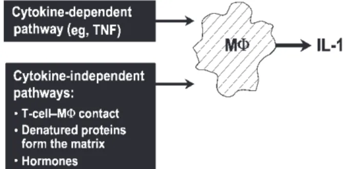
cells producing the prodestructive cytokines IL-1 and TNFα [11, 12]. It is less clear which primary factors control the production of IL-1 and TNFα by macrophages. At least two major pathways exist: (i) the cytokine-dependent pathway—induction of cytokine release by T- or B-lymphocytes, mast cells, or soluble factors (e.g. immune complexes or other cytokines such as TNF) and (ii) the cytokine-independent pathways— induction by direct contact between the macrophage and activated T-lymphocytes, contact with denatured proteins from the matrix and hormonal influences (Fig. 1). Taking
into account our original studies, it appears that the second pathway is more important in activating the production of IL-1 and TNFα in synovial tissue in patients with RA [13–15].
Two major and separate disease processes are characteristic of RA: (i) inflammation and pain and (ii) tissue destruction and lack of tissue repair (Fig. 2; Table 1). A number of mediators are implicated specific-ally in these processes. Therefore, it is unlikely that a single therapeutic agent would have beneficial effects on both pathways. Indeed, one of the key challenges that continues to face rheumatologists is how to both alleviate inflammation and prevent joint destruction.
IL-1 is a key mediator of synovial inflammation and pannus formation [12, 17, 18]. It is involved in the inflammatory processes in RA through activation of monocyte–macrophages and T- and B-lymphocytes. Although most investigations have focused on T-lymphocytes, some recent studies of experimental arthritis suggest that B-lymphocytes and a specific antibody response may drive arthritis pathology also; however, it remains to be seen if these models match human disease [19, 20]. IL-1 also contributes to inflam-mation by inducing the expression of cell-adhesion molecules, other cytokines, chemokines and chemokine receptors, angiogenic factors and small inflammatory mediators (e.g. PGE2 and nitric oxide) through the
stimulation of cyclo-oxygenase type 2 and inducible nitric oxide synthase. Up-regulation of the production of FIG. 1. Different pathways leading to IL-1 production by
mono-cyte–macrophages. Stimulation of monocyte–macrophages (MΦ) to produce IL-1 can be cytokine dependent (e.g. stimulation by TNFα). However, contact with activated T-lymphocytes, denatured proteins and hormones may also stimulate increased IL-1 production, independently of other cytokines.
FIG. 2. IL-1 and TNFα are proinflammatory cytokines within the rheumatoid joint. IL-1 and TNFα are produced by cells within the synovial pannus and act synergistically within the rheumatoid joint to up-regulate the production of small inflammatory mediators, such as IL-6, IL-8 and PGE2. Furthermore, they increase the expression of adhesion molecules on the endothelium of high
endothelial venules, allowing the immigration of inflammatory cells into the joint space. IL-1 and TNFα also activate osteoclasts in bone and stimulate the production of collagenase from chondrocytes [16]. Used with permission from Amgen Inc.
prostaglandins and other proinflammatory mediators by IL-1 thereby accounts for some of the pain, swelling and tenderness typically seen in rheumatoid joint inflammation.
IL-1 is a pivotal cytokine mediating destruction of bone and cartilage in RA and additionally impairs bone and cartilage repair [21–23]. Effects of IL-1 on these parameters appear to be more profound than those of TNFα. IL-1 induces the proliferation of synovial cells and an increase in the production of MMPs by chondrocytes and synovial cells, resulting in cartilage degradation. The cytokine also inhibits cartilage repair through inhibition of matrix protein synthesis [24–26]. In terms of bone erosion, IL-1 causes an increase in expression of the receptor activator of nuclear factor-κB ligand (RANKL), which, in turn, stimulates the differentiation and activation of osteoclasts (the cells responsible for bone resorption), leading to an increase in bone turnover [27].
Relative roles of IL-1 and TNF
α in inflammation
and destruction
Numerous studies based on various animal models of arthritis (including antigen-induced, collagen-induced, immune complex, or streptococcal-cell-wall-induced arthritis) have investigated the relative importance of IL-1 and TNFα in the processes of inflammation and destruction. These studies have been reviewed in detail by Wim van den Berg (Table 2) [28]. This analysis constitutes a semi-quantitative appreciation of the global results obtained from many different experimental conditions; nevertheless, it is evident that, in some models of arthritis, TNFα appears to have a greater inflammatory effect than IL-1, while IL-1 appears to have a greater inflammatory effect in others. What appears to be consistent in animal studies, however, is that IL-1 plays a more important role in the destructive processes of arthritis, presumably due to its extremely potent ability to inhibit the tissue repair process. Synthesis of new matrix proteins in cartilage—such as collagen type II or aggrecans—and proliferation of chondrocytes are necessary for cartilage repair and all of these processes
are inhibited by IL-1. In fact, we found that IL-1 strongly inhibited the new synthesis of glycosaminoglycans in human cartilage [24]. When compared on a molar basis in this system, the inhibition by IL-1 was much stronger than that afforded by TNFα (which had a very small effect) and interferon-γ (which had no effect at all). Of importance, the inhibitory effect of IL-1 on this repair process was restored when IL-1Ra was added at adequate concentrations [29].
Regulation of IL-1
The relative concentrations of agonistic and antagonistic cytokines establish a delicate balance in driving pro- and anti-inflammatory processes. There are currently 10 different gene products identified in the IL-1 superfamily. Three of these have been extensively studied for their role in disease: IL-1α (a predominantly intracellular agonist); IL-1β (a secreted agonist); and IL-1Ra (a secreted antagonist). IL-1 can be antagonized in at least four different ways: (i) by IL-1Ra, which is a true endogenous receptor antagonist; (ii) by soluble receptors that are cleaved on the surface of the cells (IL-1sRII); (iii) by a so-called ‘decoy’ receptor (IL-1RII), which lacks an intracellular signalling domain and, thus, is not capable of signal transduction; and (iv) by natural autoantibodies to IL-1, particularly to IL-1α [30, 31].
Both IL-1α and IL-1β bind to the membrane-bound IL-1 type I receptor (IL-1RI), leading to the recruitment of the IL-1 receptor accessory protein (IL-1RAcP; Fig. 3). This heterotrimeric complex transduces a signal to the cell nucleus, culminating in production of inflammatory and destructive mediators [4].
IL-1Ra is the most important physiological regulator of synovial IL-1 activity [32]. IL-1Ra has a high affinity for the IL-1RI; however, binding of the inhibitory protein to the receptor does not allow the recruitment of the IL-1RAcP, thus there is no signal transduction [33]. The strong binding of IL-1Ra to IL-1RI blocks the access of IL-1α and IL-1β to the receptor [4]. Cleaved fragments of the IL-1RII receptor (IL-1sRII) also inhibit the action of IL-1, by binding to circulating IL-1. Finally, IL-1 can be trapped on the cell surface by the membrane-bound ‘decoy’ IL-1RII [34]. The different inhibitory mechanisms are quite complementary, with in vitro
TABLE1. The involvement of IL-1 in the inflammatory and destructive processes of RA
Inflammation/pain Tissue destruction/inhibition of repair
Monocyte–macrophages and T-and B-lymphocyte activation
Increased synovial cell proliferation Increased expression of cell
adhesion molecules
Increased production of MMPs by chondrocytes and synovial cells Increased expression of cytokine
genes (e.g. TNFα, IL-6)
Increased cartilage degradation (mediated by MMPs) Increased expression of
chemokines and angiogenic factors Increased expression of PGE2, nitric oxide and COX-2
Inhibition of proteoglycan and type II collagen synthesis resulting in impaired cartilage repair Resorption of bone by activation of osteoclasts
TABLE2. Cytokine involvement in inflammation and destruction: studies in TNFα- or IL-1-deficient mice [28]
Murine model of arthritis
Inflammation Destruction TNFα IL-1 TNFα IL-1 Antigen-induced ++ + – +++ Collagen-induced ++ +++ + +++ Immune complex + +++ – +++ Streptococcal-cell-wall-induced ++ + – +++
–, no cytokine involvement; +, minimal cytokine involvement; ++, moderate cytokine involvement; +++ greatest cytokine involvement.
studies on human synoviocytes showing that the simul-taneous addition of both IL-1Ra and soluble IL-1sRII strongly inhibits the IL-1-induced production of MMP and PGE2[35].
Milestones in the discovery of IL-1Ra
The histochemical discovery of IL-1Ra took place at the beginning of the 1980s, when we were undertaking the isolation of large quantities of IL-1 (which had not yet been cloned), using an IL-1 synovial cell bioassay based on the stimulation of collagenase and PGE2.
No immunoassays were available at the time. Our investigations focused on diseases associated with large numbers of monocytes (such as monocytic leukaemia) and chronic debilitating diseases (such as RA and juvenile RA). Our attention also focused on diseases with spontaneous remission of fever, since we suspected the presence of natural inhibitors that reversed the peak of fever. To our surprise, we failed to detect any biological activity of IL-1 in serum or urine of seriously ill patients with one of the above diseases [36–38]. This gave rise to the hypothesis that IL-1 activity may be masked by an inhibitory molecule and that concentrations of such an inhibitory molecule must be considerably elevated during fever remission. This was confirmed when the fever profile of juvenile RA patients was analysed [39]. A protein with a mol. wt of ~17 kDa was partially purified from the urine of afebrile patients; this was found specifically to inhibit the biological activities of IL-1 without affecting those of TNFα [40, 41]. Around the same time, Arend and co-workers [42] made the independent observation of an inhibitor of chondrocyte and thymocyte responsiveness to IL-1 in cultured human monocytes. It should be recognized that at the time the
mechanism of action for this IL-1 inhibitor had not yet been identified. Indeed, the first description of the protein giving rise to the nomenclature of ‘receptor antagonist’ originated from our ligand-binding assay reported in 1987, revealing that a natural, purified molecule was impeding the binding of IL-1 to lympho-cytes [43].
Based on the inhibiting effect of natural IL-1Ra on the binding of IL-1 to lymphocytes, IL-1Ra was fully purified and cloned at Synergen in 1990 [44, 45]. DNA encoding the IL-1Ra protein was obtained from a human monocyte library and the endogenous IL-1Ra partially purified from the urine of patients was found to be similar to recombinant IL-1Ra [41, 46].
An important aspect of the inhibitory effect of IL-1Ra was observed in 1990 during studies conducted in our laboratory in collaboration with Larry Raisz. It was observed that recombinant IL-1Ra blocked IL-1-induced bone resorption in vitro, as determined by calcium release from a model of bone resorption (Fig. 4) [46]. Notably, recombinant IL-1Ra was not found to block the resorption induced by parathyroid hormone, suggesting that this inhibitory protein does not disturb the bone resorptive effects of the parathyroid hormone system or calcitonin homeostasis. Bendele et al. [47] demonstrated subsequently that IL-1Ra markedly decreased bone FIG. 3. Regulation of IL-1 biological activity. (A) IL-1 binds to
IL-1RI, leading to the formation of a heterotrimeric complex with IL-1RAcP and transduction of a signal to the cell nucleus. (B) IL-1Ra binds to IL-1RI; however, IL-1RAcP is unable to bind and, thus, signal transduction does not occur. (C) Circulating IL-1 is trapped by IL-1sRII and is thus unable to bind to IL-1RI. IL-1 can also bind to membrane-associated IL-1RII, known as a ‘decoy’ receptor, as it has no intracellular signalling domain.
FIG. 4. IL-1Ra blocks IL-1-induced bone resorption in vitro. This graph shows the degree of bone resorption associated with IL-1 in neonatal mouse calvariae. Resorption was tested in calvariae from 7-day-old mice and was assessed by measuring the release of previously incorporated45Ca. In calvariae not treated with IL-1Ra (open triangles), an increase in bone resorption was observed with recombinant IL-1 concentrations as low as 0.01 ng/ml. In calvariae treated with recombinant IL-1Ra at 1000 ng/ml (black triangles), the increase in IL-1-mediated bone resorption was blocked. The increase in
45Ca release by parathyroid hormone (light grey triangles) was
not affected by recombinant IL-1Ra (dark grey triangles), suggesting that IL-1Ra does not disturb the bone resorptive effects of parathyroid hormone [46]. Reproduced with per-mission from The Journal of Immunology. ©1990 The American Association of Immunologists Inc.
resorption in rats with collagen-induced arthritis, as substantiated by histopathological results (Fig. 5).
The importance of IL-1Ra in counteracting the destructive effects of IL-1 has been demonstrated in mice deficient in the IL-1Ra gene. Such animals have been found to spontaneously develop arthritis. A specific example is the BALB/cA murine model of arthritis, in which animals developed marked inflammatory poly-arthropathy that closely mimics human RA [48]. The animals also had distinct erosion of the articular bone. These findings suggest that endogenous IL-1Ra down-regulates inflammatory synovitis and joint destruction by inhibiting IL-1, reducing the signs and symptoms of inflammation and preventing bone and cartilage destruction.
Interestingly, the phenotype of IL-1Ra-deficient mice appears to depend on genetic background; mice with certain genetic backgrounds presented with arthritis and bone destruction [48], while mice with a different genetic background developed vasculitis [49]. This observation is likely to have important clinical implications in the future when assessing the response of patients to various RA therapies, which may depend on the patients’ genetic background.
Independent pathways of cytokine production
During recent years, there has been a great deal of contention about the disparate effects of IL-1 and TNFα in arthritic processes. Existing data from in vitroexperiments and animal models indicate that IL-1 production can be induced independently of TNFα and this is also supported by the observation that not all RA patients respond to anti-TNF therapy. Certainly, TNFα can induce macrophages to produce IL-1, but IL-1 can also (at least to some degree) induce macrophages to produce more IL-1 and TNFα. There are also a number of other pathways and factors that may activate IL-1 production from monocytes, independent of TNFα, such as the contact between T-cells, denatured matrix proteins, hormones and possibly neuropeptides (Fig. 1). Strong evidence from in vitro systems suggests that the contact between T-cells and monocyte–macrophages is a major pathway for the induction of IL-1 and TNFα [13–15, 50, 51]. This interaction can be decreased by impeding different ligand–counterligand interactions (e.g. CD69, β2-integrin and apolipoprotein A-I) [14, 52–54]. It follows that, depending on factors such as type of disease, type of stimuli, animal model, or subset of patients, either the IL-1 or TNFα pathway will dominate and lead to different specific manifestations of the disease.
Synergism between IL-1 and TNF
α
Although it is evident that IL-1 and TNFα have inde-pendent roles in mediating some of the pathophysio-logical processes of RA, it is also apparent that these cytokines act in a synergistic manner. This was illustrated in a study of rats with collagen-induced arthritis that were administered IL-1ra alone, anti-TNF alone, or IL-1ra in combination with anti-TNF. Inflammation and bone resorption were both counteracted to some degree with either agent alone, compared with control animals. However, the most striking effects were observed in animals receiving the combination treatment [55]. Clinical studies of combination therapy are in progress to assess safety and efficacy in patients with RA [56, 57]. The goal for the future is to elucidate markers that can discriminate and predict the response to treatment with different biological response modifiers, such as IL-1 and TNF inhibitors. This could include use of genetic polymorphism profiling or genetic expression profiling [58, 59]. However, no definite conclusions can be made for the time being.
Conclusions
In conclusion, the pathogenesis of inflammatory synovitis and rheumatoid joint destruction is mediated by an interdependent network of cytokines. IL-1 and TNFα clearly play central roles in the processes underlying the pathogenesis of RA, as summarized in Fig. 6. It is also evident, however, that there are quantitative differ-ences between these two cytokines. For example, TNFα appears to have a stronger influence in the context of inflammation, while IL-1 is stronger than TNFα in stimulating production of MMPs and in impairing the synthesis of collagen and proteoglycans. These differ-FIG. 5. Effects of IL-1Ra on bone resorption in rats with
arthritis induced by collagen type II. Rats were treated with different concentrations of IL-1Ra by continuous subcutaneous infusion for 7 days. (A) Bone resorption or damage was assessed by measuring trabecular and cortical bone resorption and the number of osteoclasts. (B) Total histopathological assess-ment represents the evaluation of inflammation, pannus formation (caliper measurements, paw weights, inflammatory cell infiltration), cartilage damage (toluidine blue staining) and bone damage. Interleukin-1 receptor antagonist (IL-1Ra) produced dose-dependent reductions in bone resorption in rats with arthritis induced by collagen type II. Reductions were statistically significant compared with vehicle with IL-1Ra 1 and 5 mg/kg/h (P < 0.05). Figure adapted from Bendele et al. [47].
ences have importance when evaluating therapy options for different patients, taking into account their clinical symptoms, stage of disease and genetic background.
The development of agents that specifically target cytokines marks a new therapeutic era for the manage-ment of RA. The respective effects of IL-1 and TNFα in inflammation and tissue destruction require further in-depth comparison and a great deal will be learned from the current trials of combination therapy. However, experimental data support the claim that IL-1 is a pivotal cytokine involved in the pathophysiology of RA and thus is an attractive target, not only for prevention of joint destruction, but also to safeguard repair processes. Such observations indicate that recombinant IL-1ra will be a useful addition to the existing RA management approaches.
References
1. Dayer J-M, Bréard J, Chess L, Krane SM. Participation of monocyte-macrophages and lymphocytes in the production of a factor which stimulates collagenase and prostaglandin release by rheumatoid synovial cells. J Clin Invest 1979; 64:1386–92.
2. Dayer J-M, Goldring SR, Robinson DR, Krane SM. Cell–cell interactions and collagenase production. In: Woolley DE, Evanson JM, eds. Collagenase in normal and pathological connective tissues. New York: John Wiley & Sons, 1980; 83–104.
3. Dayer J-M, Beutler B, Cerami A. Cachectin/tumor necrosis factor stimulates collagenase and prostaglandin E2
production by human synovial cells and dermal fibroblasts. J Exp Med 1985;162:2163–8.
4. Dinarello CA. Biologic basis for interleukin-1 in disease. Blood 1996;87:2095–147.
5. Staite ND, Richard KA, Aspar DG, Franz KA, Galinet LA, Dunn CJ. Induction of an acute erosive monarticular arthritis in mice by interleukin-1 and methylated bovine serum albumin. Arthritis Rheum 1990;33:253–60.
6. Choy EHS, Panayi GS. Cytokine pathways and joint inflammation in rheumatoid arthritis. N Engl J Med 2001;344:907–16.
7. Firestein GS, Alvaro-Gracia JM, Maki R. Quantitative analysis of cytokine gene expression in rheumatoid arthritis. J Immunol 1990;144:3347–53.
8. Chu CQ, Field M, Allard S, Abney E, Feldmann M, Maini RN. Detection of cytokines at the cartilage/pannus junction in patients with rheumatoid arthritis: implications for the role of cytokines in cartilage destruction and repair. Br J Rheumatol 1992;31:653–61.
9. Wood NC, Dickens E, Symons JA, Duff GW. In situ hybridization of interleukin-1 in CD14-positive cells in rheumatoid arthritis. Clin Immunol Immunopathol 1992; 62:295–300.
10. Krane SM, Conca W, Stephenson ML, Amento EP, Goldring MB. Mechanisms of matrix degradation in rheumatoid arthritis. Ann N Y Acad Sci 1990;580:340–54. 11. Dayer J-M, Arend WP. Cytokines and growth factors. In:
Sledge CB, ed. Textbook of rheumatology, 5th edn. Philadelphia: Saunders, 1997; 267–86.
12. Arend WP, Dayer J-M. Cytokines and cytokine inhibitors or antagonists in rheumatoid arthritis. Arthritis Rheum 1990;33:305–15.
13. Vey E, Zhang J-H, Dayer J-M. IFN-γ and 1,25(OH)2D3
induce on THP-1 cells distinct patterns of cell surface antigen expression, cytokine production, and responsiveness to contact with activated T cells. J Immunol 1992; 149:2040–6.
14. Isler P, Vey E, Zhang J-H, Dayer J-M. Cell surface glycoproteins expressed on activated human T cells induce production of interleukin-1 beta by monocytic cells: a possible role of CD69. Eur Cytokine Netw 1993;4:15–23. 15. Lacraz S, Isler P, Vey E, Welgus HG, Dayer J-M. Direct
contact between T lymphocytes and monocytes is a major pathway for induction of metalloproteinase expression. J Biol Chem 1994;269:22027–33.
16. Dinarello CA, Moldawer LL. Proinflammatory and anti-inflammatory cytokines in rheumatoid arthritis: a primer for clinicians, 3rd edn. Thousand Oaks: Amgen, 2001.
17. Harris E Jr. Rheumatoid arthritis. Pathophysiology and implications for therapy. N Engl J Med 1990;322:1277–89. 18. Koch AE, Kunkel SL, Strieter RM. Cytokines in rheumatoid
arthritis. J Investig Med 1995;43:28–38.
19. Matsumoto I, Staub A, Benoist C, Mathis D. Arthritis provoked by linked T and B cell recognition of a glycolytic enzyme. Science 1999;286:1732–5.
20. Matsumoto I, Maccioni M, Lee DM et al. How antibodies to a ubiquitous cytoplasmic enzyme may provoke joint-specific autoimmune disease. Nat Immunol 2002;3:360–5.
21. Joosten LAB, Helsen MMA, Saxne T, van De Loo FAJ, Heinegard D, van Den Berg WB. IL-1αb blockade prevents cartilage and bone destruction in murine type II collagen-induced arthritis, whereas TNF-α blockade only ameliorates joint inflammation. J Immunol 1999;163: 5049–55.
22. van den Berg WB. Arguments for interleukin 1 as a target in chronic arthritis. Ann Rheum Dis 2000;59(Suppl. 1): i81–4.
23. Gravallese EM, Goldring SR. Cellular mechanisms and the role of cytokines in bone erosions in rheumatoid arthritis. Arthritis Rheum 2000;43:2143–51.
24. Yaron I, Meyer FA, Dayer J-M, Bleiberg I, Yaron M. Some recombinant human cytokines stimulate glycosaminoglycan synthesis in human synovial fibroblast cultures and inhibit it in human articular cartilage cultures. Arthritis Rheum 1989;32:173–80.
25. Henderson B, Thompson RC, Hardingham T, Lewthwaite J. FIG. 6. Central roles of IL-1 and TNFα in the pathogenesis
of RA. The thickness of the arrows indicates degrees of involvement.
Inhibition of interleukin-1-induced synovitis and articular cartilage proteoglycan loss in the rabbit knee by recombinant human interleukin-1 receptor antagonist. Cytokine 1991; 3:246–9.
26. van Lent PLEM, van de Loo FAJ, Holthuysen AEM, van den Bersselaar LAM, Vermeer H, van den Berg WB. Major role for interleukin 1 but not for tumor necrosis factor in early cartilage damage in immune complex arthritis in mice. J Rheumatol 1995;22:2250–8.
27. Pettit AR, Ji H, von Stechow D et al. TRANCE/RANKL knockout mice are protected from bone erosion in a serum transfer model of arthritis. Am J Pathol 2001; 159:1689–99.
28. van den Berg WB. Uncoupling of inflammatory and destructive mechanisms in arthritis. Semin Arthritis Rheum 2001;30(5 Suppl. 2):7–16.
29. Seckinger P, Yaron I, Meyer FA, Yaron M, Dayer J-M. Modulation of the effects of interleukin-1 on glycos-aminoglycan synthesis by the urine-derived interleukin-1 inhibitor, but not by interleukin-6. Arthritis Rheum 1990;33:1807–14.
30. Jouvenne P, Fossiez F, Banchereau J, Miossec P. High levels of neutralizing autoantibodies against IL-1α are associated with a better prognosis in chronic polyarthritis: a follow-up study. Scand J Immunol 1997;46:413–18.
31. Forslind K, Svensson B, Svenson M, Bendtzen R. Anti-IL-1α autoantibodies in early rheumatoid arthritis. Scand J Rheumatol 2001;30:167–8.
32. Arend WP, Gabay C. Physiologic role of interleukin-1 receptor antagonist. Arthritis Res 2000;2:245–8.
33. Lang D, Knop J, Wesche H et al. The type II IL-1 receptor interacts with the IL-1 receptor accessory protein: a novel mechanism of regulation of IL-1 responsiveness. J Immunol 1998;161:6871–7.
34. Colotta F, Dower SK, Sims JE, Mantovani A. The type II ‘decoy’ receptor: a novel regulatory pathway for interleukin 1. Immunol Today 1994;15:562–6.
35. Burger D, Chicheportiche R, Giri JG, Dayer J-M. The inhibitory activity of human interleukin-1 receptor antag-onist is enhanced by type II interleukin-1 soluble receptor and hindered by type I interleukin-1 soluble receptor. J Clin Invest 1995;96:38–41.
36. Balavoine J-F, de Rochemonteix B, Cruchaud A, Dayer J-M. Identification of interleukin-1-like activity and inhibitor(s) in urine from a patient with monocytic leukemia. [Abstract] Lymphokine Res 1984;3:233.
37. Balavoine J-F, de Rochemonteix B, Cruchaud A, Dayer J-M. Collegenase- and PGE2-stimulating activity and inhibitor
in urine from a patient with monocytic leukaemia. In: Gahring LC and Daynes RA, eds. The physiological, metabolic, and immunologic actions of interleukin-1. New York: Alan R Liss, 1985; 429–36.
38. Balavoine J-F, de Rochemonteix B, Williamson K, Seckinger P, Cruchaud A, Dayer J-M. Prostaglandin E2
and collagenase production by fibroblasts and synovial cells is regulated by urine-derived human interleukin 1 and inhibitor(s). J Clin Invest 1986;78:1120–4.
39. Prieur A-M, Kaufmann M-T, Griscelli C, Dayer J-M. Specific interleukin-1 inhibitor in serum and urine of children with systemic juvenile chronic arthritis. Lancet 1987;2:1240–2.
40. Seckinger P, Williamson K, Balavoine J-F et al. A urine inhibitor of interleukin 1 activity affects both interleukin 1α and 1β but not tumor necrosis factor α. J Immunol 1987;139:1541–5.
41. Mazzei GJ, Seckinger PL, Dayer J-M, Shaw AR.
Purification and characterization of a 26-kDa competitive inhibitor of interleukin 1. Eur J Immunol 1990;20:683–9. 42. Arend WP, Joslin FG, Massoni RJ. Effects of immune
complexes on production by human monocytes of interleukin 1 or an interleukin 1 inhibitor. J Immunol 1985;134:3868–75.
43. Seckinger P, Lowenthal JW, Williamson K, Dayer J-M, MacDonald HR. A urine inhibitor of interleukin 1 activity that blocks ligand binding. J Immunol 1987;139:1546–9. 44. Hannum CH, Wilcox CJ, Arend WP et al. Interleukin-1
receptor antagonist activity of a human interleukin-1 inhibitor. Nature 1990;343:336–40.
45. Eisenberg SP, Evans RJ, Arend WP et al. Primary struc-ture and functional expression from complementary DNA of a human interleukin-1 receptor antagonist. Nature 1990;343:341–6.
46. Seckinger P, Klein-Nulend J, Alander C, Thompson RC, Dayer J-M, Raisz LG. Natural and recombinant human IL-1 receptor antagonists block the effects of IL-1 on bone resorption and prostaglandin production. J Immunol 1990;145:4181–4.
47. Bendele A, McAbee T, Sennello G, Frazier J, Chlipala E, McCabe D. Efficacy of sustained blood levels of interleukin-1 receptor antagonist in animal models of arthritis: comparison of efficacy in animal models with human clinical data. Arthritis Rheum 1999;42:498–506. 48. Horai R, Saijo S, Tanioka H et al. Development of chronic
inflammatory arthropathy resembling rheumatoid arthritis in interleukin 1 receptor antagonist-deficient mice. J Exp Med 2000;191:313–20.
49. Nicklin MJH, Hughes DE, Barton JL, Ure JM, Duff GW. Arterial inflammation in mice lacking the interleukin 1 receptor antagonist gene. J Exp Med 2000;191:303–12. 50. Vey E, Burger D, Dayer J-M. Expression and cleavage of
tumor necrosis factor-α and tumor necrosis factor receptors by human monocytic cell lines upon direct contact with stimulated T cells. Eur J Immunol 1996;26:2404–9.
51. Chizzolini C, Chicheportiche R, Burger D, Dayer J-M. Human Th1 cells preferentially induce interleukin (IL)-1β while Th2 cells induce IL-1 receptor antagonist production upon cell–cell contact with monocytes. Eur J Immunol 1997;27:171–7.
52. Burger D, Dayer J-M. The role of human T lymphocyte– monocyte contact in inflammation and tissue destruction. Arthritis Res 2002;4(Suppl. 3):S169–76.
53. Rezzonico R, Chicheportiche R, Imbert V, Dayer JM. Engagement of CD11b and CD11cβ2 integrin by antibodies or soluble CD23 induces IL-1β production on primary human monocytes through mitogen-activated protein kinase-dependent pathways. Blood 2000;95:3868–77. 54. Hyka N, Dayer J-M, Modoux C et al. Apolipoprotein A-I
inhibits the production of interleukin-1β and tumor necrosis factor-α by blocking contact-mediated activation of monocytes by T lymphocytes. Blood 2001;97:2381–9. 55. Bendele AM, Chlipala ES, Scherrer J et al. Combination
benefit of treatment with the cytokine inhibitors inter-leukin-1 receptor antagonist and PEGylated soluble tumor necrosis factor receptor type I in animal models of rheumatoid arthritis. Arthritis Rheum 2000;43:2648–59. 56. Rooney T, Madigan A, Dayer J-M et al. Combination
therapy with anakinra and PEGylated soluble tumor necrosis factor receptor type I in rheumatoid arthritis: a study of early clinical and synovial tissue responses. [Abstract] Arthritis Rheum 2002;46(Suppl. 9):S132. 57. Edwards CK III, Williams A, Fitzpatrick VD et al.
PEGylated soluble tumor necrosis factor receptor type I (PEG sTNF-RI) in rheumatoid arthritis (RA): cluster analysis and microarray mRNA expression profiles of patient synovium and peripheral blood mononuclear cells. [Abstract] Arthritis Rheum 2002;46(Suppl. 9):S548. 58. Francis SE, Camp NJ, Dewberry RM et al. Interleukin-1
receptor antagonist gene polymorphism and coronary artery disease. Circulation 1999;99:861–6.
59. Jawaheer D, Seldin MF, Amos CI et al. A genomewide screen in multiplex rheumatoid arthritis families suggests genetic overlap with other autoimmune diseases. Am J Hum Genet 2001;68:927–36.