Introduction
Balloon valvuloplasty has established as an alternative to surgery for the therapy of aortic valve stenosis in childhood [4, 13]. Nevertheless, acute complica-tions after balloon dilation leading to early death (< 30 days) have been described [12] and the devel-opment of aortic insufficiency remains an unwanted
complication during long term follow up [1, 6]. Therefore, the balloon valvuloplasty is stated to be a palliative approach with less morbidity and mortality compared to aortic valve surgery [8], especially in critical ill neonates [12]. But the morbidity in catheter interventional therapy is not fully eliminated [16]. Aortic valve morphology, clinical status, myocardial function at the intervention, and age at intervention Walter Knirsch
Felix Berger Paul Harpes Oliver Kretschmar
Balloon valvuloplasty of aortic valve
stenosis in childhood: early and medium
term results
Received: 19 November 2007 Accepted: 19 February 2008 Published online: 17 March 2008
j Abstract Background Isolated aortic valve stenosis in childhood is treated by balloon valvuloplasty. The role of independent risk factors for the outcome remains unclear. Material and methods We analysed the early and medium term outcome of balloon valvuloplasty in isolated aortic valve stenosis in 44 pediatric patients with isolated, severe aortic valve stenosis at an age younger than 18 years, who received a primary balloon valvuloplasty during the last 5 years in our institution. We evaluated the type of aortic valve morphology, age, clinical status, and myocardial function at the time of the intervention as independent risk factor. Results A significant early relief of the pressure gradient across the aortic valve (P < 0.001) after balloon valvuloplasty was found. This was independent of the aortic valve morphology. Two neonates with a highly stenotic tricuspid aortic valve and severely compromised haemo-dynamics died within the first 30 days after the intervention. During medium term follow up (mean 22.5 months) we observed a functional deterioration for the stenosis as well as for the insufficiency of the aortic valve. ‘‘Symptoms before intervention’’ is an independent risk factors (P < 0.001) for valvuloplasty failure. Patients at an ‘‘age at intervention £ 28 days’’ (P = 0.02) and patients with ‘‘reduced myocardial systolic function’’ (P = 0.01) had a shorter time to reintervention. Conclusions The type of aortic valve morphology only has a weak predictive value for the outcome of balloon valvuloplasty during medium term follow up. Critical ill neonates with an impaired myocardial function are at a higher risk for valvuloplasty failure.
j Key words Aortic valve stenosis – Balloon valvuloplasty – Children
CRC
655
W. Knirsch, MD (&) Æ F. Berger, MD O. Kretschmar, MD
Division of Paediatric Cardiology University Children’s Hospital Zurich Steinwiesstrasse 75 8032 Zu¨rich, Switzerland Tel.: +41-44/2667617 Fax: +41-44/2667981 E-Mail: walter.knirsch@kispi.uzh.ch F. Berger, MD
Department of Congenital Heart Disease Deutsches Herzzentrum Berlin and Charite´ – Universita¨tsmedizin Berlin Augustenburger Platz 1 13353 Berlin, Germany P. Harpes, PhD Department of Biostatistics University Zurich Hirschengraben 84 8001 Zu¨rich, Switzerland
have been assumed to be possible risk factors determining the outcome of balloon valvuloplasty [11,17].
Therefore, the aim of this retrospective study was to analyse the early outcome and medium term follow up results of balloon valvuloplasty of aortic valve stenosis in childhood with special regard to aortic valve morphology and to point out other possible risk factors for valvuloplasty failure.
Material and methods
j Patients
Between October 2001 and September 2006 all con-secutive patients at an age younger than 18 years treated for aortic valve stenosis with balloon val-vuloplasty in our institution were analysed
retro-spectively. Medical records, angiographies,
echocardiographic and haemodynamic data were re-viewed.
All patients had a native aortic valve stenosis and no further congenital heart disease of haemodynamic relevance. Patients with coexisting aortic valve insufficiency more than grade 1 did not receive a valvuloplasty [5].
The clinical status was defined as ‘‘normal’’ in asymptomatic patients, and as ‘‘reduced’’ in patients with symptoms of dyspnoea and other signs of con-gestive heart failure, and ductal dependency in case of neonates.
The myocardial systolic function was assessed by echocardiography and defined as ‘‘normal’’ by a shortening fraction >29%, and as ‘‘reduced’’ by a shortening fraction £29%. The morphology of the aortic valve was classified as functionally bicuspid, anatomically bicuspid, or tricuspid by echocardiog-raphy [18]. The valve morphology itself was no exclusion criteria to perform balloon valvuloplasty for aortic valve stenosis. The aortic valve gradient was measured by continuous wave Doppler technique. The peak pressure gradient was calculated from the peak flow velocity, respectively, the mean gradient from the time-velocity integral of the Doppler curve. Further follow up was obtained by clinical evalu-ation and two-dimensional and Doppler echocardi-ography. Study end points were reintervention either by a second catheter intervention or surgery, an aortic valve insufficiency grade 4, death, and end of follow up.
Valvuloplasty failure was defined as reintervention during medium term follow up, aortic valve insuffi-ciency grade 4, and death. The study end points were tested for the variables ‘‘age at intervention
£28 days’’, ‘‘symptoms before intervention’’, ‘‘re-duced myocardial systolic function (shortening frac-tion £ 29%)’’, and ‘‘aortic valve morphology’’ as possible risk factors.
Ethical approval for the study and data collection were obtained according to the guidelines of the ethical committee of the University of Zu¨rich.
j Technique
As an indication for balloon valvuloplasty a peak-to-peak pressure gradient of more than 50 mm Hg in patients with normal systolic myocardial function (shortening fraction ‡ 30%, ejection fraction ‡ 50%) was mandatory, while patients with an impaired sys-tolic function (shortening fraction £ 29%, ejection fraction £ 49%) were also included with a lower peak-to-peak pressure gradient. All patients underwent the procedure under general anaesthesia and fluoroscopic guidance. Heparin was administered during the pro-cedure with 100 IE/kg. The balloon valvuloplasty was performed retrograde via a femoral access in all cases, as described recently by Mullins [14]. Balloon size was determined by the diameter of the aortic valve annulus in systole measured by two-dimensional echocardiography as well as by angiography in two planes. The initial balloon diameter was 80%–90% of the size of the aortic valve annulus. The procedure was performed in ‘‘temporary cardiac arrest’’ by rapid pacing of the right ventricle in one patient and by an intravenous bolus injection of adenosine in one pa-tient. After balloon valvuloplasty a satisfactory result was defined as a reduction of the peak-to-peak pres-sure gradient of at least 50% or a residual gradient of less than 25 mm Hg. If this pressure gradient reduc-tion could not be achieved, a redilareduc-tion was per-formed with a larger balloon. No further intervention was performed in any case, if a severe aortic valve insufficiency was detected in the following angiogra-phy.
j Statistics
Group data are presented in percentages or relative frequencies for discrete data and as median and range for continuous variables or as means and standard deviations, if the data are approximately normally distributed. Comparisons of continuous and ordinal variables between independent groups were per-formed with the Mann–Whitney U test or t test, if variables were approximately normally distributed. Comparison of continuous and ordinal variables for the same group in two different settings (paired data) was performed with the Wilcoxon signed rank test or
paired t test. Differences in proportions between independent groups were tested by the chi square test or Fisher exact test, as appropriate.
Survival analysis was performed with the Kaplan Meier method and the Log-rank test for differences in time-to-event between independent groups. The combined effect of several risk factors on time-to-event was analysed with a Cox regression.
A P value equal or less than 0.05 was considered statistically significant.
Statistical analysis was conducted with SPSS 11 for Mac OS X.
Results
j Patients and clinical status
Fourty-five patients were treated for isolated severe aortic valve stenosis with balloon valvuloplasty. Complete follow up was available for 44 patients. A functionally bicuspid aortic valve was found in 18 patients, an anatomically bicuspid aortic valve in 12, and a tricuspid aortic valve in 14. The mean age at intervention for all patients was 6.3 ± 5.9 years (range 0–15.9 years). Eleven patients (25%) were neonates at an age of £28 days at the time of inter-vention, seven of them had a tricuspid valve mor-phology (Table1). The majority of the neonates (63.6%) with aortic valve stenosis were symptomatic; ductal dependency with retrograde flow of the aortic arch was present in three patients (27.2%). In con-trast, beyond the neonatal period the majority of the
patients were asymptomatic (P < 0.05). Seven pa-tients (21.2%) beyond the neonatal period showed mild symptoms of congestive heart failure such as dyspnea. The myocardial function was reduced in three neonatal patients (27.3%). Clinical pre- and post-interventional conditions are summarized in Table1.
j Early outcome
The acute haemodynamic results of balloon valvulo-plasty are summarized in Table1. In all patients there was a significant relief of the peak-to-peak pressure gradient after balloon valvuloplasty of aortic valve stenosis (functionally bicuspid, P < 0.001; anatomi-cally bicuspid, P < 0.001; tricuspid, P < 0.001; all pa-tients, P < 0.001). This relief of aortic valve stenosis could also be found with the continuous wave (cw) Doppler measurements before and the day after inter-vention (pre-continuous wave Doppler gradient vs. post-continuous wave Doppler gradient in functionally bicuspid, P < 0.001; anatomically bicuspid, P < 0.001; tricuspid, P = 0.002; all patients, P < 0.001).
In five patients (11.4%) acute complications within 30 days after the intervention occurred: An acute se-vere aortic insufficiency in a 10-year-old boy with previous normal myocardial function could be suc-cessfully treated by an emergency aortic valve reconstruction. The aortic valve was functionally bicuspid, and the used balloon:annulus ratio was 1.0. Two neonatal patients died 3 days and 20 days after the intervention due to persistent congestive heart
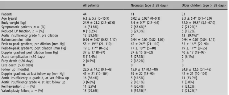
Table 1 Results of aortic balloon valvuloplasty in 44 children
All patients Neonates (age£ 28 days) Older children (age > 28 days)
Patients 44 11 33
Age [years] 6.3 ± 5.9 (0–15.9) 0.02 ± 0.02* (0–0.1) 8.3 ± 5.4* (0.1–15.9)
Body weight [kg] 24.9 ± 21.2 (2.2–67.0) 3.4 ± 0.7* (2.2–4.6) 32.0 ± 19.8* (3.1–67.0)
Symptomatic patients, n = [%] 14 [31.8%] 7 [63.6%]* 7 [21.2%]*
Reduced LV function, n = [%] 8 [18.2%] 3 [27.3%] 5 [15.2%]
Aortic insufficiency grade 1, pre dilation 13 [29.6%] 0* 13 [39.4%]*
Balloon:annulus ratio 0.94 ± 0.07 (0.82–1.17) 0.94 ± 0.09 (0.82–1.07) 0.94 ± 0.07 (0.84–1.17)
Peak-to-peak gradient, pre dilation [mm Hg] 55 ± 19** (21–110) 62 ± 24** (21–110) 52 ± 16** (29–90)
Peak-to-peak gradient, post dilation [mm Hg] 19 ± 11** (0–55) 17 ± 10** (5–40) 19 ± 11** (0–55)
Doppler gradient post dilation [mm Hg] 37 ± 17 (8–97) 27 ± 15 (8–62) 40 ± 17 (18–97)
Acute complication (<30 days) 5 [11.4%] 3 [27.3%] 2 [6.1%]
Early death (£30 days) 2 [4.5%] 2 [18.2%] 0
Late death (>30 days) 0 0 0
Follow up [months] 22.5 ± 14.2 (0.1–48) 15.9 ± 17 (0.1–48) 24.8 ± 12.6 (0.1–48)
Doppler gradient, at last follow up [mm Hg] 41 ± 21 (10–104) 39 ± 22 (18–90) 42 ± 21 (10–104)
Aortic insufficiency < grade 4, at last follow up 16 [36.4%] 5 [45.5%] 11 [33.0%]
Aortic insufficiency grade 4, at last follow up 3 [6.8%] 2 [18.1%] 1 [3.0%]
Reintervention, n = [%] 11 [25%] 4 [36.4%] 7 [21.2%]
Valvuloplasty failure, n = [%] 13 [29.6%] 6 [54.5%]* 7 [21.2%]*
Data are displayed as mean ± SD (range)
* P < 0.05 (Mann–Whitney U test for group comparison) ** P < 0.05 (t test for pre vs. post dilatation comparison)
failure. Both neonates were ductal dependent before, presented signs of endocardial fibroelastosis with systolic as well as diastolic myocardial dysfunction and had a tricuspid valve morphology. Thrombosis of the femoral artery developed in one patient. In one patient surgical reconstruction of the femoral artery was necessary due to technical problems with failure of complete removal of the balloon through the femoral sheath.
j Medium term follow up
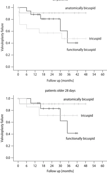
The mean follow up was 22.5 months for all patients. The medium term follow up results of balloon val-vuloplasty for aortic valve stenosis are described in Table1. The mean cw Doppler gradient at last follow up was higher compared to the early value post dilation, but did not reach statistical significance neither for all patients together nor for the different types of aortic valve morphology (functional bicuspid, P = 0.31; anatomic bicuspid, P = 0.89; tricuspid, P = 0.10; all patients, P = 0.07). Patients with tricus-pid aortic valves developed during medium term follow up an increase of the grade of aortic insuffi-ciency, and suffered from a higher reintervention rate, and higher valvuloplasty failure rate compared to patients with functionally or anatomically bicuspid aortic valve, but this trend for the tricuspid aortic valve morphology could not be shown for patients beyond the neonatal period (Fig.1).
Patients with reduced myocardial systolic function (Kaplan Meier survival analysis, P = 0.01), with symptoms before intervention (Kaplan Meier survival analysis, P = 0.01) as well as patients at an age at intervention £28 days (Kaplan Meier survival analy-sis, P = 0.02) had a significantly shorter time to re-intervention compared to patients with a normal systolic function, respectively, no symptoms before interventions as well as patients at older age at intervention (>28 days) (Fig.2).
j Valvuloplasty failure
Valvuloplasty failure was defined as reintervention during follow up, aortic valve insufficiency grade 4, and death. A Cox regression analysis with valvuloplasty failure as primary endpoint was performed for the following explanatory variables ‘‘age at intervention £28 days’’, ‘‘symptoms before intervention’’, ‘‘reduced myocardial systolic function (shortening fraction £ 29%)’’, and ‘‘aortic valve morphology’’. It revealed ‘‘symptoms before intervention’’ as a significant risk factor for valvuloplasty failure (both in a multivariable logistic regression with all variables, P < 0.001, and in a
univariable logistic regression, P < 0.001). The type of aortic valve morphology was not a significant risk factor for valvuloplasty failure (P = 0.08).
Discussion
In our study, we analysed the early and medium term outcome after balloon valvuloplasty of aortic valve stenosis in childhood. We focussed on different risk factors influencing the results. The impact of the type of aortic valve morphology was weak in our study, 60 54 48 42 36 30 24 18 12 6 0 Follow up [months] 1.0 0.8 0.6 0.4 0.2 0.0 Valvuloplasty failure anatomically bicuspid functionally bicuspid tricuspid all patients 60 54 48 42 36 30 24 18 12 6 0 Follow up [months] 1.0 0.8 0.6 0.4 0.2 0.0 Valvuloplasty failure anatomically bicuspid functionally bicuspid tricuspid patients older 28 days
Fig. 1 Kaplan Meier survival analysis comparing the time to reintervention for the three different types of aortic valve morphologies in paediatric patients after balloon valvuloplasty of the aortic valve. A significant difference between the patients with tricuspid aortic valve morphology, which had the poorest outcome, compared to anatomically bicuspid aortic valve morphology (P = 0.047) and the functional bicuspid aortic valve morphology (P = 0.36) regarding all patients (up). In patients older 28 days (down) there was no significant difference between the three types of aortic valve morphology
because during early outcome we found a significant relief of the pressure gradient, which was independent of the type of aortic valve morphology (Table1). Second, during medium term follow up we observed a moderate increase of the residual Doppler gradient, and parallel to this a moderate increase of aortic valve insufficiency, which was also independent of the type of aortic valve morphology. There was a moderate trend towards worse results in medium term follow up for patients with tricuspid aortic valve morphol-ogy. In Kaplan Maier survival analysis the time to reintervention of the patients with tricuspid aortic valve was significantly shorter (P < 0.05) compared with the time to reintervention in patients with
ana-tomically bicuspid aortic valve morphology (Fig. 1, up). But by ruling out the neonates in the Kaplan Meier survival analysis, in the patients at age older 28 days the type of aortic valve morphology was no significant discriminating factor (Fig. 1, down).
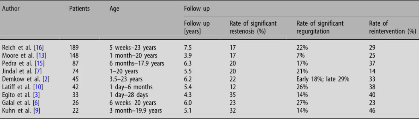
Despite aortic valve morphology, we could focus on other independent risk factors for valvoulplasty failure in medium term follow up. In uni- and multi-variable logistic regression analysis ‘‘symptoms before intervention’’ is a significant risk factor for valvulo-plasty failure. This observation was confirmed by the circumstance that the time of reintervention was significantly shorter in those patients, who had a ‘‘reduced myocardial systolic function (shortening fractions £ 29%)’’ (Fig.2) before intervention, and who were treated early at an ‘‘age at interven-tion £ 28 days’’. These factors of ‘‘young age at intervention’’, ‘‘clinical symptoms’’, and ‘‘myocardial systolic insufficiency’’ incorporates and confirms separate observations of previous investigators [1–3, 6, 7, 9–11, 13, 14] (Table2). Reich et al. clearly showed that functionally bicuspid aortic valve mor-phology is an independent risk factor for aortic valve insufficiency after balloon dilation [16]. In our study, the results of aortic valvuloplasty in patients with functionally bicuspid aortic valve lie in between the more positive results of patients with anatomically bicuspid aortic valve and the more negative results of patients with tricuspid aortic valve morphology (Fig. 1, left). The poor outcome in patients with tri-cuspid aortic valve morphology in medium term fol-low up may be partly explained by the high number (50%) of neonates in the group of patients with tricuspid aortic valve morphology in our study (Table 1). Consecutively a higher number of patients in this group with symptoms before intervention (50%) and reduced myocardial function before intervention lead to a higher rate of acute complica-tions, severe aortic insufficiency, need for reinter-vention or even death (Table 1). Compared to patients with tricuspid aortic valve morphology the patients with anatomically bicuspid aortic valve morphology had the significant longer (P < 0.05) time to reintervention during medium term follow up. The reintervention rate of patients with anatomically bicuspid aortic valve was only 8.3%, and no patient developed aortic insufficiency grade 4 during the follow up period of 2 years (Table1).
In conclusion, this study shows that aortic valve morphology is not a risk factor for the early outcome in balloon valvuloplasty of aortic valve stenosis in children, but is only a weak risk factor during med-ium term follow up. Other independent risk factors as ‘‘symptoms before intervention’’, ‘‘age at interven-tion £ 28 days’’, and ‘‘reduced myocardial systolic function (shortening fraction £ 29%)’’ have to be ta-60 54 48 42 36 30 24 18 12 6 0 Follow up [months] 60 54 48 42 36 30 24 18 12 6 0 Follow up [months] 1.0 0.8 0.6 0.4 0.2 0.0 Valvuloplasty failure
normal myocardial function
reduced myocardial function all patients 1.0 0.8 0.6 0.4 0.2 0.0 Valvuloplasty failure symptomatic patients asymptomatic patients all patients
Fig. 2 Kaplan Meier survival analysis comparing the time to reintervention in paediatric patients after balloon valvuloplasty of the aortic valve. There was a significant difference of the time to reintervention between patients with normal or reduced myocardial systolic function (P = 0.01) (up), and between patients with or without clinical symptoms at the time of interventions (P = 0.02) (down)
ken into account for the risk stratification of balloon valvuloplasty of aortic valve stenosis during medium term follow up.
Our experience on the basis of these data raise the question of interventional vs. surgical relief of aortic valve stenosis, especially for the high risk neonatal group with reduced myocardial function. But the comparison between interventional and surgical
treatment was not the primary aim of our study. Nevertheless, in our institution, we pursue the deci-sion-making process in a multidisciplinary approach together with the complete team of cardiac surgery, interventional cardiology, pediatric intensive care, and anaesthesiology in a highly individualized rela-tion to every single patient.
Table 2 Overview of clinical studies evaluating follow up of balloon valvuloplasty in aortic valve stenosis in children
Author Patients Age Follow up
Follow up [years] Rate of significant restenosis (%) Rate of significant regurgitation Rate of reintervention (%)
Reich et al. [16] 189 5 weeks–23 years 7.5 17 22% 29
Moore et al. [13] 148 1 month–20 years 3.9 17 7% 25
Pedra et al. [15] 87 6 months–17.9 years 6.3 20 17% 37
Jindal et al. [7] 74 1–20 years 5.5 20 21% 14
Demkow et al. [2] 45 3.5–23 years 6.2 22 Early 18%; late 29% 33
Latiff et al. [10] 42 1 day–6 months 5.4 12 26% 38
Egito et al. [3] 33 1 day–28 days 4.3 35 14% 40
Galal et al. [6] 26 6 weeks–20 years 6.0 23 27% 23
Kuhn et al. [9] 22 3 month–19.9 years 5.1 32 14% 46
References
1. Balmer C, Beghetti M, Fasnacht M, Friedli B, Arbenz U (2004) Balloon aortic valvoplasty in paediatric pa-tients: progressive aortic regurgitation is common. Heart 90:77–81
2. Demkow M, Ruzyllo W, Ksiezycka E, Szaroszyk W, Lubiszewska B, Przyluski J, Rozanski J, Wilczynski J, Hoffman P, Rydlewska-Sadowska W (1999) Long-term follow-up of balloon valvuloplasty for congenital aortic stenosis: predic-tors of late outcome. J Invasive Cardiol 11:227–229
3. Egito ES, Moore P, O’Sullivan J, Colan S, Perry SB, Lock JE, Keane JF (1997) Transvascular balloon dilation for neonatal critical aortic stenosis: early and midterm results. J Am Coll Cardiol 29:442–447
4. Ewert P, Bertram H, Breuer J, Da¨hnert I, Dittrich S, Eicken A, Emmel M, Fi-scher G, Gitter R, Gorenflo N, Haas N, Kitzmu¨ller E, Koch A, Kretschmar O, Lindinger A, Michel-Behnke I, Nu¨rn-berg J, Peuster M, Walter K, Zartner P, Uhlemann F (2007) Outcome nach aortaler Ballonvalvuloplastie im Kin-desalter—Analyse von mehr als 1000 Patienten aus 20 Zentren. Clin Res Cardiol 96:661 [abstract]
5. Feigenbaum H (1986) Aortic regurgi-tation. In: Echocardiography. Lea & Febiger, Philadelphia
6. Galal O, Rao PS, Al-Fadley F, Wilson AD (1997) Follow-up results of balloon aortic valvuloplasty in children with special reference to causes of late aortic insufficiency. Am Heart J 133:418–427 7. Jindal RC, Saxena A, Juneja R, Kothari
SS, Shrivastava S (2000) Long-term re-sults of balloon aortic valvulotomy for congenital aortic stenosis in children and adolescents. J Heart Valve Dis 9:623–628
8. Justo RN, McCrindle BW, Benson LN, Williams WG, Freedom RM, Smallhorn JF (1996) Aortic valve regurgitation after surgical versus percutaneous bal-loon valvotomy for congenital aortic valve stenosis. Am J Cardiol 77:1332– 1338
9. Kuhn MA, Latson LA, Cheatham JP, Fletcher SE, Foreman C (1996) Man-agement of pediatric patients with iso-lated valvar aortic stenosis by balloon aortic valvuloplasty. Cathet Cardiovasc Diagn 39:55–61
10. Latiff HA, Sholler GF, Cooper S (2003) Balloon dilatation of aortic stenosis in infants younger than 6 months of age: intermediate outcome. Pediatr Cardiol 24:17–26
11. McCrindle BW, for the Valvoplasty, angioplasty of congenital anomalies (VACA) registry investigators (1996) Independent predictors of immediate results of percutaneous balloon aortic valvotomy in childhood. Am J Cardiol 77:286–293
12. McCrindle BW, Blackstone EH, Wil-liams WG, Sittiwangkul R, Spray TL, Azakie A, Jonas RA (2001) Are out-comes of surgical versus trancatheter balloon valvotomy equivalent in neo-natal critical aortic stenosis? Circula-tion 104:I152–158
13. Moore P, Egito E, Mowrey H, Perry SB, Lock JE, Keane JF (1996) Midterm re-sults of balloon dilation of congenital aortic stenosis: predictors of success. J Am Coll Cardiol 27:1257–1263 14. Mullins CE (2006) Aortic valve dilation.
In: Cardiac catheterization in congeni-tal heart disease: pediatric and adult. Blackwell Publishing, Malden
15. Pedra CAC, Sidhu R, McCrindle BW, Nykanen DG, Justo RN, Freedom RM, Benson LN (2004) Outcomes after bal-loon dilation of congenital aortic ste-nosis in children and adolescents. Cardiol Young 14:315–321
16. Reich O, Tax P, Marek J, Razek V, Gilik J, Tomek V, Chaloupecky V, Bartakova H, Skovranek J (2004) Long term re-sults of percutaneous balloon valvo-plasty of congenital aortic stenosis: independent predictors for outcome. Heart 90:70–76
17. Sholler GF, Keane JF, Perry SB, Sanders SP, Lock JE (1988) Balloon dilation of congenital aortic valve stenosis. Results and influence of technical and mor-phological features on outcome. Cir-culation 78:351–360
18. Van Praagh R, Bano-Rodrigo A, Smo-linsky A, Schuetz TJ, Fyler DX, Van Praagh S (1986) Anatomic variations in congenital valvar, subvalvar, and supravalvar aortic stenosis: a study of 64 postmortem cases. In: Takehashi M (ed) Challenges in the treatment of congenital cardiac anomalies. Futura, New York