HAL Id: hal-02494936
https://hal.archives-ouvertes.fr/hal-02494936
Submitted on 16 Jun 2020HAL is a multi-disciplinary open access archive for the deposit and dissemination of sci-entific research documents, whether they are pub-lished or not. The documents may come from teaching and research institutions in France or abroad, or from public or private research centers.
L’archive ouverte pluridisciplinaire HAL, est destinée au dépôt et à la diffusion de documents scientifiques de niveau recherche, publiés ou non, émanant des établissements d’enseignement et de recherche français ou étrangers, des laboratoires publics ou privés.
pathways and sensitivity to cadmium toxicity in two
marine fish species
Gaël Le Croizier, Camille Lacroix, Sébastien Artigaud, Stéphane Le Floch,
Jean-Marie Munaron, Jean Raffray, Virginie Penicaud, Marie-Laure Rouget,
Raymond Laë, Luis Tito de Morais
To cite this version:
Gaël Le Croizier, Camille Lacroix, Sébastien Artigaud, Stéphane Le Floch, Jean-Marie Mu-naron, et al.. Metal subcellular partitioning determines excretion pathways and sensitivity to cadmium toxicity in two marine fish species. Chemosphere, Elsevier, 2019, 217, pp.754-762. �10.1016/j.chemosphere.2018.10.212�. �hal-02494936�
Please note that this is an author-produced PDF of an article accepted for publication following peer review. The definitive publisher-authenticated version is available on the publisher Web site.
Chemosphere
February 2019, Volume 217, Pages 754-762
https://doi.org/10.1016/j.chemosphere.2018.10.212 https://archimer.ifremer.fr/doc/00464/57606/
Archimer
https://archimer.ifremer.frMetal subcellular partitioning determines excretion
pathways and sensitivity to cadmium toxicity in two marine
fish species.
Le Croizier Gaël 1, *, Lacroix Camille 2, Artigaud Sébastien 1, Le Floch Stéphane 2,
Munaron Jean-Marie 1, Raffray Jean 1, Penicaud Virginie 1, Rouget Marie-Laure 3, Laë Raymond 1, Tito De Morais Luis 4
1
Laboratoire des Sciences de l'Environnement Marin (LEMAR), UMR 6539 CNRS/UBO/IRD/IFREMER, BP 70, 29280 Plouzané, France
2
Centre of Documentation, Research and Experimentation on Accidental Water Pollution (CEDRE), 715 rue Alain Colas, CS 41836, Brest 29218-Cedex 2, France
3
Institut Universitaire Européen de la Mer (IUEM), Université de Bretagne Occidentale (UBO), CNRS UMS 3113, 29280 Plouzané, France
* Corresponding author : Gaël Le Croizier, email address : gael.lecroizier@hotmail.fr
Abstract :
Subcellular cadmium (Cd) partitioning was investigated in the liver of two marine fish species, the European sea bass Dicentrarchus labrax and the Senegalese sole Solea senegalensis, dietary exposed to an environmentally realistic Cd dose for two months followed by a two-month depuration. Cd exposure did not modify Cd cellular partitioning for either species, refuting the spillover hypothesis. Both species contained most of the Cd in the detoxifying fraction but displayed different handling strategies. Cd was largely bound to heat stable proteins (HSP) including metallothioneins (MT) in sea bass while Cd was more linked to metal rich granules (MRG) in sole. Whole liver concentrations and subcellular partitioning were also determined for essential elements. The greatest impairment of essential metal homeostasis due to Cd exposure was found in sole. These elements followed the Cd partitioning pattern, suggesting that they are involved in antioxidant responses against Cd toxicity. Cd consumption diminished sole growth in terms of body weight, probably due to lipid storage impairment. During the depuration period, the two species showed contrasting partitioning patterns, implying different pathways for Cd elimination from the liver. In sea bass, MT-bound Cd would be excreted through bile or released into blood, crossing the cell membrane via a protein transporter. In sole, MRG-bound Cd would be sequestered by organelles before being released into the blood via vesicular exocytosis. These distinct strategies in cellular Cd handling in the liver might account for differential sensitivity to Cd toxicity and differential Cd excretion pathways between the two marine fish species.
Please note that this is an author-produced PDF of an article accepted for publication following peer review. The definitive publisher-authenticated version is available on the publisher Web site.
Graphical abstract
Highlights
► Sea bass and sole displayed different Cd subcellular partitioning in the liver. ► Cd was largely bound to metallothionein-like proteins in the sea bass liver. ► Cd was mainly linked to metal rich granules in the sole liver. ► Essential metals followed the Cd partitioning pattern in hepatic cells. ► Handling strategies might account for sensitivity and Cd excretion.
Keywords : Sub-cellular fractionation ; Dicentrarchus labrax ; Solea senegalensis ; Essential element
M
AN
US
CR
IP
T
AC
CE
PT
ED
KEY WORDS
24Sub-cellular fractionation; Dicentrarchus labrax; Solea senegalensis; essential element
25
distribution; elimination; depuration
26
27
INTRODUCTION
28Aside from natural sources, marine ecosystems can be subjected to metal contamination
29
from urban effluents and industrial activities. Cadmium (Cd) is a common by-product of the
30
mining industry and can reach high levels in some regions (World Health Organization,
31
2010). Coastal regions in West Africa, which belong to the Canary Current Large Marine
32
Ecosystem (CCLME), are thereby particularly subjected to Cd residue due to the direct
33
release of phosphogypsum into water by the phosphate industry (Auger et al., 2015;
34
Cheggour et al., 1999; Gaudry et al., 2007). Marine consumers such as fish mainly
35
accumulate metals through trophic pathway, which can lead to significant Cd levels in the
36
organs of fish from the CCLME. Moreover, fish species from this region display a wide range
37
of Cd concentrations (Afandi et al., 2018; Diop et al., 2016), resulting partially from
38
differences in foraging habitats and dietary habits (Borrell et al., 2016; Goutte et al., 2015; Le
39
Croizier et al., 2016; Metian et al., 2013). In addition to ecological traits, the observed
40
interspecific variability in terms of Cd bioaccumulation results from different physiological
41
characteristics, including the presence of metal binding proteins like metallothioneins (MT)
42
(Le Croizier et al., 2018; Moulis et al., 2014; Zalups and Ahmad, 2003). Cd is a toxic element
43
responsible for numerous impairments in fish, such as oxidative damage, disruption of
44
essential metal homeostasis, endocrine and ionoregulatory disruption, histopathology and
M
AN
US
CR
IP
T
AC
CE
PT
ED
depression of the immune system, which can finally affect growth and survival (McGeer et
46
al., 2011).
47
Recently, attention has been focused on fish species ability to cope with metal toxicity,
48
depending on subcellular partitioning of the element (Eyckmans et al., 2012; Leonard et al.,
49
2014). At the cellular level, Cd can take different toxic chemical forms including: free or
50
complexed ion forms (e.g. Cd2+, CdCl2), bound to enzymes (e.g. cytochromes), bound to
51
organic acids (e.g. citrates), or bound to cellular constituents causing damages (e.g. DNA)
52
(Vijver et al., 2004). Today, only a few forms of Cd speciation are considered as detoxified:
53
Cd complexed to peptides (e.g. glutathione) or functional, transport or sequestration
54
proteins (e.g. metallothioneins), and Cd trapped in vesicles of the lysosomal system or
55
precipitated in mineral granules (Wang and Rainbow, 2006). Very little is however known
56
about the intracellular mechanisms leading to Cd elimination (Moulis et al., 2014; Zalups and
57
Ahmad, 2003).
58
While one of the adverse effects of toxic metals is their interference with essential elements
59
(Martelli et al., 2006; Moulis, 2010a), some of them confer a protective role against metal
60
toxicity, directly through formation of detoxifying complexes (Sasakura and T. Suzuki, 1998)
61
or by indirectly preventing oxidative stress due to their association with antioxidant enzymes
62
(Martínez-Álvarez et al., 2005; Talas et al., 2008). The significance of essential element
63
subcellular partitioning in their protective role against toxic metals has never yet been
64
considered. Moreover, despite evidence of the influence of cellular components like MT in
65
metal accumulation kinetics in marine species (Wang and Rainbow, 2010), most previous
66
studies only emphasize on the influence of metal intracellular handling on sensitivity of
67
organisms to toxic elements (Campbell et al., 2008; Eyckmans et al., 2012; Giguère et al.,
68
2006; Leonard et al., 2014; Wang and Rainbow, 2006). Very few studies have therefore
M
AN
US
CR
IP
T
AC
CE
PT
ED
investigated the link between metal subcellular partitioning and kinetics in fish (Glynn, 1991)
70
and information about the relationships between Cd intracellular handling and excretion
71
pathways is lacking for marine fish.
72
In order to fill these gaps, the present study aimed to investigate Cd intracellular distribution
73
in the liver of two different marine fish species which are naturally present in the CCLME, a
74
region particularly prone to Cd contamination. For this purpose, European sea bass
75
Dicentrarchus labrax and Senegalese sole Solea senegalensis, were exposed for two months
76
to an environmentally realistic dietary cadmium (Cd) dose followed by a depuration period
77
of a further two months. Analyses were conducted at the end of the uptake period and at
78
the end of the depuration period and Cd partitioning was examined in six major subcellular
79
fractions: cellular debris, metal-rich granules, mitochondria, organelles, cytosolic enzymes,
80
cytosolic proteins and peptides, as well as in the storage lipid fraction of the liver when
81
present. Sensitivity of the two species was assessed according to: (1) Cd subcellular
82
partitioning during accumulation and depuration periods, (2) impairments to essential metal
83
subcellular distribution, (3) essential metal concentrations in the whole liver and their
84
partitioning within hepatocytes, (4) growth and hepatosomatic index calculation. Finally,
85
hypotheses were made regarding the influence of Cd cellular speciation in the liver and Cd
86
biliary excretion or transport to other tissues.
87 88
MATERIALS AND METHODS
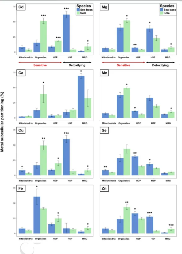
89All animal procedures were in accordance with the French and EU guidelines for animal
90
research (project approval number: 03266.03).
91
Fish and experimental procedures 92
M
AN
US
CR
IP
T
AC
CE
PT
ED
All details about animal procedures and experimental design can be found in Le Croizier et
93
al., 2018. Briefly, immature sea bass Dicentrarchus labrax (length: 14.1 ± 0.7 cm; weight 29.8
94
± 4.5 g) used in this experiment were obtained from a commercial hatchery (Aquastream,
95
Ploemeur, France), whereas immature Senegalese sole Solea senegalensis (length: 14.9 ± 1.1
96
cm; weight 36.4 ± 7.9 g) were provided from a marine farm (Ferme marine de l’Adour,
97
Anglet, France). The fish were transported to the Cedre (Centre of Documentation, Research
98
and Experimentation on Accidental Water Pollution, Brest, France). After receiving
99
anaesthesia by bathing in a 0.05 mL∙L-1 solution of tricaine methanesulfonate (MS-222)
100
(Ackerman et al., 2005), each fish was randomly assigned to one of twelve high density
101
polyethylene tanks that had a 300 L volume (six tanks for each species, 40 sole and 50 sea
102
bass per tank to ensure equivalent biomass) at the Cedre’s marine animal facility. This facility
103
is an independent greenhouse that is submitted to a natural photoperiod and supplied with
104
a continuous seawater flow from the bay of Brest. The temperatures in the experimental
105
tanks followed the outdoor temperature. Fish were first acclimated to the experimental
106
conditions for one month, during which they were fed daily with dried commercial pellets
107
(Turbot label rouge 1.4 mm, Le Gouessant Aquaculture).
108
To obtain an environmental relevant Cd concentration in the fish food, a Cd-enriched diet
109
was prepared in order to reach a Cd level around 25 µg∙g-1, which corresponds to the Cd
110
level reported in potential prey within the natural distribution of the two fish species (Bodin
111
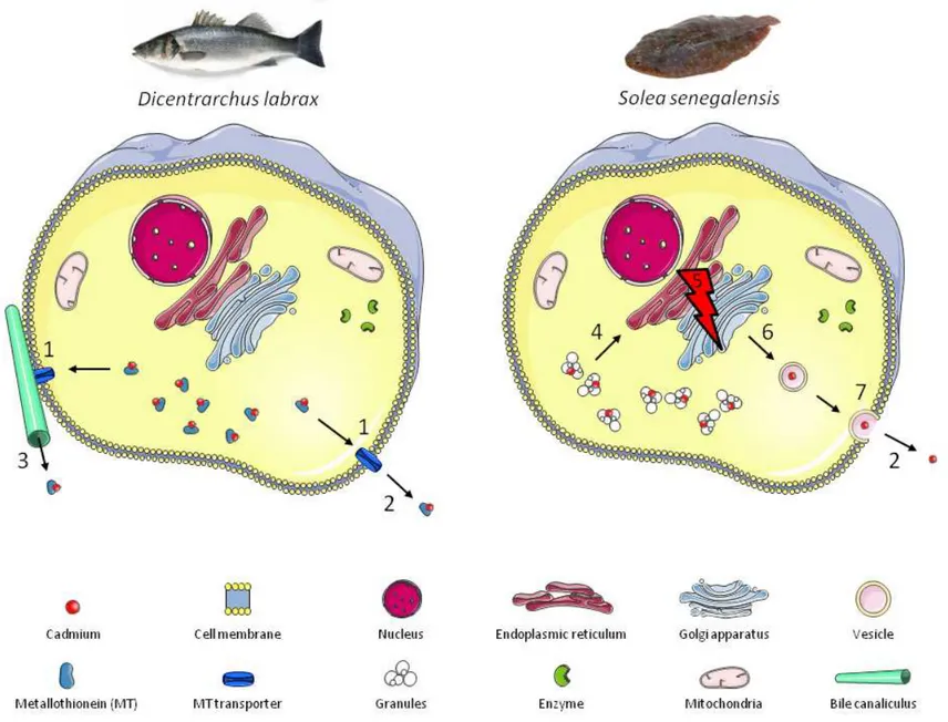
et al., 2013; Maanan, 2008). For this purpose, the commercial pellets were spiked with a 100
112
mg∙L-1 Cd (as CdCl2 in milliQ water) solution for 30 min. The diet was then placed at -20°C,
113
freeze dried and broken into small pellets before usage. This preparation led to a Cd
114
concentration of 22.9 ± 0.3 µg∙g-1 dw in the Cd-enriched food. The control diet was prepared
115
in the same way, but with the addition of milliQ water only, where the measured
M
AN
US
CR
IP
T
AC
CE
PT
ED
background Cd level was 0.71 ± 0.00 µg∙g-1 dw. For each of the four conditions (i.e.,
Cd-117
exposed and control fish, for both species) there were three replicate tanks. Dietary Cd
118
exposure was initiated by randomly assigning three of the six tanks per species to the
Cd-119
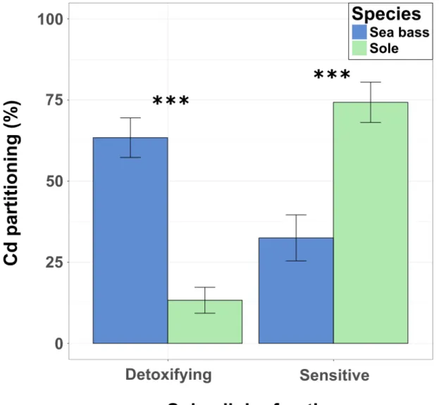
enriched food. The six remaining tanks were fed the control diet. Cd exposure was
120
conducted for 60 days, after which a depuration period was conducted that lasted for 60
121
days. During the depuration period, all of the tanks were fed the control diet. Daily food
122
distribution was performed slowly and continuously over 24 h with a clockwork feeder
123
(COFA, Paris, France) to prevent pellets from remaining in the tank and thus avoid Cd
124
desorption in water. Sea bass were fed at 1.7 % body weight while sole were fed at 1 % body
125
weight per day to meet the physiological requirements of each species (Danion et al., 2011;
126
Salas-Leiton et al., 2010). The bottom of each tank was siphoned every day to avoid Cd
127
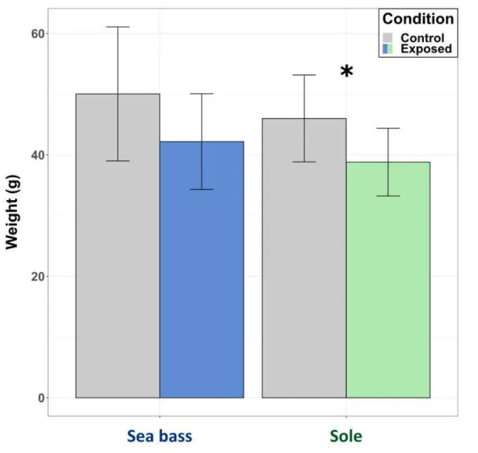
leaching from faeces.
128
Physico-chemical parameters (O2, pH, temperature, salinity) and water quality (nitrates,
129
nitrites) were measured every ten days. During the experiment, the water temperature
130
decreased from 20.7 to 12.8 °C, oxygen saturation increased from 81.2% to 98.2%, pH
131
increased from 8.0 to 8.2 and salinity increased from 35.2 to 35.7. The water was free of
132
nitrate and nitrite (Colorimetric test JBL) in both experiments.
133
Biological sampling 134
After the two months of Cd exposure, three fish per tank (nine for each treatment) were
135
anesthetized by bathing in a 0.05 mL∙L-1 solution of MS-222 before being euthanized by
136
bathing in a 0.2 mL∙L-1 solution of MS-222 (Ackerman et al., 2005). They were weighed,
137
measured and dissected with ceramic tools to avoid metal contamination. The liver was
138
collected, weighed and put in acid-washed (10% HNO3) individual plastic microcentrifuge
M
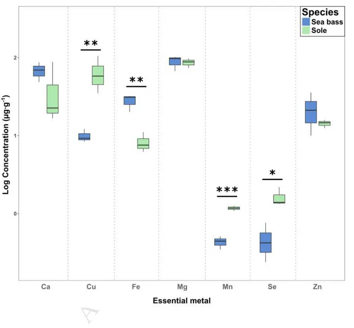
AN
US
CR
IP
T
AC
CE
PT
ED
tubes, flash frozen in liquid nitrogen and stored at -80°C until analysis. Another sampling was
140
made in the same way after a further two months of depuration.
141
Subcellular partitioning procedure 142
This procedure generally followed the protocol of Rosabal et al. (2015), adapted from that of
143
Wallace et al. (2003), which has been validated and extensively used on various fish species
144
(Campbell et al., 2008; Eyckmans et al., 2012; Giguère et al., 2006; Lapointe et al., 2009a).
145
Briefly, liver samples were fractionated into six operationally-defined subcellular fractions:
146
cell membranes (cellular debris), metal-rich granules (MRG); mitochondria, microsomes +
147
lysosomes (organelles), heat-denatured proteins (HDP) including cytosolic enzymes, and
148
heat-stable proteins and peptides (HSP) such as metallothionein (MT) and glutathione (GSH).
149
Three liver samples of fish from the same tank were pooled to obtain approximately 200 mg
150
of wet sample and homogenized in 1.5 mL of a solution containing Tris-HCl 20 mmol∙L-1 pH
151
8.6, 0.01% DTT (dithiothreitol) as a reducing agent and 1 % of an antiproteolytic agent
152
(Protease Inhibitor Mix, GE Healthcare). A 100 µL aliquot was removed from the liver
153
homogenate for determining total trace metal concentrations in the liver and assessing
154
metal recovery from the subcellular fractions. The remainder of the liver homogenate was
155
centrifuged at 1450×g for 15 min at 4°C. The supernatant (S1) was transferred to an
acid-156
washed 1.5-mL polypropylene microcentrifuge tube for further separations. The pellet from
157
this centrifugation was suspended in 0.5 mL of ultrapure water, heated to 100°C for 2 min,
158
digested with an additional 500 µL of 1 N NaOH at 65°C for 60 min. Centrifugation at
159
10,000×g for 10 min at ambient temperature (∼20°C) was performed to separate the
NaOH-160
resistant fraction (referred to as “MRG”) from the cellular debris fraction, which includes cell
161
membranes, unbroken cells and nuclei. The S1 supernatant was centrifuged at 10,000×g for
M
AN
US
CR
IP
T
AC
CE
PT
ED
30 min at 4°C to produce the mitochondrial fraction. The resulting supernatant was
163
ultracentrifuged at 100,000×g for 60 min at 4°C, giving a pellet containing other organelles
164
(microsomes and lysosomes) and the cytosolic fraction in the supernatant. To separate the
165
heat-stable peptides and proteins (HSP) from the heat-denatured proteins (HDP), the
166
cytosolic fraction was held at 80°C for 10 min, left on ice for 1 h and then centrifuged at
167
50,000×g for 10 min at 10°C. The HSP fraction, which includes MT, was collected from the
168
supernatant. Each fraction was finally kept at -20°C until the metal analyses were performed.
169
Metal analyses 170
Subcellular fractions were digested in a mixture of 2 mL 70% HNO3 and 0.5 mL 30% H2O2
171
(both of ultrapure quality) in closed Teflon vessels. Mineralization was performed on a
172
hotplate for 4 h at 100°C. A 100-µL aliquot was removed from the digestateand diluted to 15
173
mL with Milli-Q quality water (Merck Millipore). Cadmium and essential metals (Ca, Cu, Fe,
174
Mg, Mn, Se and Zn) were analysed using an ICP Q-MS (X Series 2, Thermo Scientific) at the
175
Pôle de Spectrométrie Océan (PSO, Plouzané, France) with an internal standard solution of
176
Rhodium (2 µg∙L-1). Reference materials (fish protein DORM-4 and dogfish liver DOLT-5,
177
NRCC) were treated and analysed in the same way as the samples. The results for reference
178
materials displayed mean metal recoveries of 85 ± 6 % for DORM-4 and 91 ± 6 % for DOLT-5.
179
Mean recoveries (± SD) of DORM-4 reference samples (n = 3) were 81 ± 2 % for Cd, 78 ± 3 %
180
for Ca, 95 ± 2 % for Mg, 89 ± 5 % for Mn, 86 ± 5 % for Fe, 81 ± 2 % for Cu, 79 ± 1 % for Zn, 92
181
± 3 % for Se. Mean recoveries (± SD) of DOLT-5 reference samples (n = 3) were 99 ± 2 % for
182
Cd, 83 ± 1 % for Ca, 96 ± 4 % for Mg, 92 ± 4 % for Mn, 98 ± 6 % for Fe, 86 ± 3 % for Cu, 85 ± 2
183
% for Zn, 86 ± 2 % for Se. Blanks were included in each analytical batch. The limits of
184
detection (ng∙g-1 dry wt), corresponding to the mean concentration of the blank solutions,
M
AN
US
CR
IP
T
AC
CE
PT
ED
were 0.2 (Cd), 1.3 (Ca), 0.4 (Cu), 0.1 (Fe), 0.2 (Mg), 0.01 (Mn), 0.1 (Se) and 0.4 (Zn). Total
186
metal concentrations in the liver (µg∙g-1) are given on a dry weight basis (µg∙g-1 dw), based
187
on a previous study involving the same fish and where the liver samples were freeze-dried
188
before analysis (Le Croizier et al., 2018). The proportion of metal in each fraction is
189
expressed as a percentage of the sum of the concentrations of all fractions. As it was not
190
present in all of the liver samples, the storage lipid fraction was not taken into account in the
191
calculation of metal partitioning. Metal proportion in this fraction was expressed as a
192
percentage of the total metal concentration in the liver. “Sensitive fractions” gathered the
193
Cd contained in the sensitive fractions (i.e. mitochondria + HDP + organelles, while the
194
“metal-detoxified fractions” gathered the Cd contained in the detoxifying fractions (i.e. MRG
195
+ HSP) (Eyckmans et al., 2012; Leonard et al., 2014). The total metal concentration recovery
196
in the liver from the sum of the metal concentrations measured in all fractions, including
197
lipids, was 95 ± 11% for sea bass and 96 ± 6% for sole.
198
Data analysis 199
All data tested statistically were first checked for normality (Shapiro–Wilks tests) (Shapiro
200
and Wilk, 1965) and homogeneity of variances (Bartlett tests) (Bartlett, 1937). When these
201
conditions were met, raw data were used and one-way ANOVAs, followed by Tukey’s HSD
202
tests, were performed to test for differences between treatments and species. Otherwise,
203
non-parametric analogues were used, i.e. Kruskal-Wallis tests (KW), followed by
Conover-204
Iman multiple comparison tests with Bonferroni’s adjustment (Conover and Iman, 1979). All
205
of the statistical analyses were performed using the open source software R (version 3.4.3, R
206
Core Team, 2017).
207 208
M
AN
US
CR
IP
T
AC
CE
PT
ED
RESULTS AND DISCUSSION
209Impact of Cd elimination on Cd and essential metal partitioning 210
After being exposed to cadmium, the two-month depuration period led to many changes in
211
metal partitioning (Table 1).
212
First, Cd proportion significantly increased in the organelle fraction (from 37.3 ± 5.9% to 51.5
213
± 4.8%) in sole liver at the end of the depuration period. The organelle fraction includes
214
cellular components involved in the vesicular transport, which is a major pathway for
215
extracellular excretion: Golgi apparatus (GA), endoplasmic reticulum (ER) and lysosomes. ER
216
manages the sequestration of molecules and their binding to excretory proteins, which are
217
transported through the GA and cytosol by vesicles (including lysosomes) and finally
218
excreted across the plasma membrane. Increasing Cd in the organelle fraction during the
219
depuration period may reflect Cd exocytosis via vesicles, which could lead to total Cd
220
elimination from the liver tissue, as described in sole in a previous study (Le Croizier et al.,
221
2018).
222
Second, Cd exposure followed by a depuration period also led to a displacement of essential
223
elements (Fe, Mg, Mn and Zn) from metal rich granules (MRG) to organelles in sole liver (e.g.
224
Fe proportion significantly increased from 30.1 ± 2.4 to 41.2 ± 1.4% in organelles while it
225
significantly decreased from 16.9 ± 3.2% to 9.2 ± 2.6% in MRG). In marine organisms, MRG
226
are inorganic insoluble concretions containing mainly Ca phosphate (George et al., 1980), as
227
reflected by the high proportion of Ca in the MRG fraction in both species (69.3 ± 4.1% and
228
32.6 ± 13.7% for sea bass and sole, respectively, during the depuration period). These
229
structures can play an important role in accumulation and detoxification of metals in fish
230
(Lapointe et al., 2009b; Leonard et al., 2014).
M
AN
US
CR
IP
T
AC
CE
PT
ED
The simultaneous increase in proportion of Cd and essential metals in organelles and
232
decrease of essential metals in MRG may be caused by sequestration of MRG by ER, which is
233
part of the organelle fraction (Table 1; Figure 2). This hypothesis seems reasonable since
234
MRG in our species contained the highest proportion of Ca, and ER is deeply involved in Ca
235
storage (Görlach et al., 2006). Supplementing fish diet has furthermore been shown to
236
increase Cd in MRG while decreasing it in organelles (Ng et al., 2009). Although the authors
237
did not draw this conclusion, our hypothesis is that increasing cellular Ca concentration will
238
reduce the probability for Cd bound to calcium granules to be sequestered by ER during Ca
239
uptake. The changes in essential metal cellular repartition would thus be a side effect of Cd
240
transport from MRG to organelles (i.e. ER, GA and vesicles, in that order) before final
241
exocytosis (Table 1; Figure 2).
242
Conversely, no change in Cd partitioning was observed in sea bass liver despite effective Cd
243
elimination as well as Cd biliary excretion observed in this species during the depuration
244
period (Le Croizier et al., 2018). In mammals, Cd bound to glutathione (GSH) was shown to
245
be excreted from hepatocytes to bile while Cd bound to metallothionein (MT) was released
246
into the blood before reaching other organs like kidney (Ballatori, 1991; Chan et al., 1993;
247
Klaassen, 1978; Nordberg, 1978). In fish, MT were reported in bile (Hauser-Davis et al., 2012)
248
and were shown to transport metal during biliary excretion in Cu- and Se-exposed fish
249
(Hauser-Davis et al., 2016, 2014). As more than half of the Cd (e.g. 61.8 ± 5.6% after the
250
depuration period)was bound to HSP regardless of condition in the sea bass (Table 1), this
251
may suggest that Cd complexed to components of the HSP fraction (i.e. MTLP or GSH) was
252
excreted directly from cytoplasm to bile or blood without passing through another cellular
253
compound (Figure 2).
M
AN
US
CR
IP
T
AC
CE
PT
ED
This elimination of Cd-saturated MT may lead to a new pool of free MT, which would be
255
available for binding other metals. In accordance with the well-identified large Zn-binding
256
capacities of MT and the competition between Cd and Zn (Maret, 2011; Moulis, 2010b), MT
257
turnover during exocytosis of Cd-MT complexes may have enhanced Zn binding to
newly-258
synthesized MT, as suggested by the significant increasing proportion of Zn (from 21.1 ±
259
1.9% to 27.7 ± 2.1%) in the HSP fraction at the end of the depuration period (Table 1).
260
Finally, it should be noted that more disturbances in metal homeostasis were observed in
261
the sole liver following Cd exposure (i.e. changes in Fe, Mg, Mn and Zn partitioning; Table 1),
262
indicating a greater sensitivity to Cd toxicity by interference with essential elements in this
263
species than in sea bass, which only showed a modification in the Zn distribution.
264
Links between subcellular Cd handling and Cd toxicity in the two studied species 265
While more than half of the Cd (61.8 ± 5.6%) wasbound to HSP in the sea bass after the
266
depuration period, Cd partitioning in sole showed a significantly greater Cd pool in the
267
sensitive fractions (74.3 ± 6.2%) compared with the detoxifying fractions (13.3 ± 4.0%). The
268
different storage strategies imply that different supposed paths for Cd exocytosis, discussed
269
above (i.e. direct transport of MT-bound Cd to the membrane in sea bass versus passing of
270
MRG-bound Cd through ER and GA before excretion by vesicles in sole), may thus be
271
responsible for a greater sensitivity of sole to Cd toxicity (Figure 2; Figure 3).
272
When present, the storage lipid fraction accounted for a non-negligible part of the
273
internalized Cd in liver (44.1 ± 9.8% in control sole and 24.9 ± 0.5% in control sea bass after
274
the depuration period) (see Supporting Information), highlighting the need to consider this
275
novel fraction in studies on metal subcellular partitioning. Cd is known to stimulate the lipid
276
peroxidation process, defined as oxidative deterioration of polyunsaturated fatty acids and
M
AN
US
CR
IP
T
AC
CE
PT
ED
resulting in alteration of cell membranes (Roméo et al., 2000; Viarengo et al., 1989). Cd
278
binding to this fraction may thus cause oxidation of neutral lipids, preventing their use in
279
membrane structure. Cd was also shown to reduce lipid storage efficiency, increasing
280
utilisation of triglycerides, which finally led to a lower growth in terms of body weight in
281
exposed fish (Pierron et al., 2007). Although no growth impairment was observed according
282
to length measurement or HSI calculation, a significant weight decrease was found in
Cd-283
exposed sole compared with controls (38.8 ± 5.6 g vs. 46.0 ± 7.2 g, respectively) (Figure 5;
284
Table S2). Moreover, no storage lipid fraction was found in the livers of Cd exposed soles
285
(see Supporting Information). In addition to the disturbances in essential metal homeostasis,
286
the greater sensitivity of the sole to Cd toxicity was thus revealed by an alteration of the
287
whole-body condition due to fat consumption.
288
Essential metal protection against Cd toxicity in the two studied species 289
One of the main mechanisms for Cd cellular toxicity is the induction of oxidative stress by
290
production of oxygen free radicals (Almeida et al., 2002; Roméo et al., 2000). On the other
291
hand, some elements are essential for the activity of antioxidant enzymes like glutathione
292
peroxidases (GPx), catalases (CAT) and superoxide dismutases (SOD), which contain Se, Fe
293
and Mn-Cu-Zn as cofactors, respectively (Vural et al., 2010). These enzymes are highly
294
involved in preventing oxidative stress in fish and a relative higher level of essential metals
295
may thus provide a better protection against Cd toxicity through higher activity of
296
antioxidant enzymes (Basha and Rani, 2003; Janz, 2011; Martínez-Álvarez et al., 2005). For
297
instance, oxidative stress caused by Cd was reduced by Se treatment in the liver of the
298
rainbow trout (Talas et al., 2008), while a Zn-deficiency was responsible for oxidative stress
299
in the same species (Hidalgo et al., 2002). Besides the well documented role of Se in the
M
AN
US
CR
IP
T
AC
CE
PT
ED
detoxification of mercury (Hg) through formation of Hg-Se complexes (Khan and Wang,
301
2009; Pelletier, 1986), it has been suggested that Se could also complex with Cd and
302
subsequently bind to selenoprotein P, thus reducing Cd availability and toxicity (Sasakura
303
and T. Suzuki, 1998; Siscar et al., 2014). The resistance of a species to Cd toxicity may thus
304
depend partially on the level of elements enhancing antioxidant response or able to complex
305
Cd. The two fish species investigated in our study presented some differences in hepatic
306
concentrations of such elements (Figure 3). While sea bass liver contained significantly more
307
Fe (27.8 ± 6.6 µg∙g-1 in sea bass vs. 8.3 ± 2.5 µg∙g-1 in sole), sole showed higher
308
concentrations of Cu, Mn and Se (e.g. 66 ± 35.6 µg∙g-1 of Cu in sole vs. 9.9 ± 2.0 µg∙g-1 of Cu in
309
sea bass) (see Supporting Information and Figure 3). Regarding essential metal
310
concentrations, the two species thus seem to possess contrasting defense capabilities,
311
involving different antioxidant enzymes.
312
Despite significantly higher levels of Cd in two sensitive fractions in sea bass (12 ± 4.2% in
313
mitochondria and 6.3 ± 1.8% in cytosolic HDP) compared with sole (3.8 ± 1.2% in
314
mitochondria and 0.5 ± 0.0% in HDP) after Cd exposure, sea bass also contained more
315
antioxidant metals (Cu, Se and Zn) in these fractions (e.g. 12.3 ± 3.7% vs. 3.2 ± 0.2% of Cu in
316
mitochondria for sea bass and sole, respectively) (Figure 1). Similarly, after depuration, large
317
proportions of Cu, Mg, Mn, Se and Zn were observed alongside the significantly higher Cd
318
proportions in two sensitive fractions (51.5 ± 4.8% in organelles and 18.8 ± 1.1% in cytosolic
319
HDP) in sole compared with sea bass (15.4 ± 4.7% in organelles and 9.0 ± 1.4% in HDP). As
320
the three types of antioxidant enzyme (GPx, CAT and SOD) are all found in cytosol,
321
mitochondria and organelles (Bai et al., 1999; Martínez-Álvarez et al., 2005; Orbea et al.,
322
2000), the fact that essential metals followed Cd distribution in sensitive fractions may
M
AN
US
CR
IP
T
AC
CE
PT
ED
reflect their mobilization to activate antioxidant defences and a role in preventing Cd
324
damage to cellular components.
325
Link between Cd partitioning and hepatic excretion 326
In a previous study, higher MT concentration combined with higher Cd biliary excretion and
327
relocation to muscle were found in sea bass than in sole, suggesting that MT level would
328
enhance Cd excretion from the liver (Le Croizier et al., 2018). These differences between the
329
two species were attributed to metabolism and/or phylogenetic divergences. Indeed,
330
seabass is characterized by a higher metabolism, which can increase the need for essential
331
metals and thus the need for binding sites such as MT. Moreover, the two species exhibited
332
significant variation in the MT sequence, potentially leading to difference in terms of
333
function. Finally, these evolutionary divergences have probably been accentuated by
334
contrasting ecological niches (i.e., demersal for sea bass versus benthic for sole), leading sea
335
bass to be more adapted to manage Cd sequestration in MT compared to sole. Even if Cd
336
was also excreted from sole hepatocytes, Cd elimination seemed higher in sea bass (not
337
significant) since around 60% of the Cd in liver was eliminated in this species after two
338
months of depuration versus around 40% in sole (Le Croizier et al., 2018) (Figure S1,
339
Supporting Information). Regarding subcellular partitioning, two main mechanisms were
340
likely to give sea bass a greater Cd elimination from hepatocytes.
341
First, the large proportion of Cd bound to HSP may facilitate biliary excretion via transport of
342
Cd/GSH complexes through a specific transporter of GSH present in the canalicular
343
membrane (Zalups and Ahmad, 2003), or via transport of Cd/MT complexes as suggested by
344
recent observations of MT in bile of metal-exposed fish (Hauser-Davis et al., 2016, 2014,
345
2012) (Figure 2).
M
AN
US
CR
IP
T
AC
CE
PT
ED
Second, transport of Cd/MT through plasma membrane in the sea bass may be faster than
347
vesicular exocytosis occurring in sole. Although MT release from various organs to the blood
348
has been proven, the mechanisms for epithelial transport of MT (Chan et al., 1993; Moltedo
349
et al., 2000) and Cd/MT complex transport (Moulis et al., 2014; Sabolić et al., 2010; Zalups
350
and Ahmad, 2003) are unknown. FIHUREby the classical vesicular secretory pathway while
351
transport across membranes must occur directly from the cytoplasm to the exterior of the
352
cell through ATP-binding cassette transporters (ABC transporters) (De Lisle et al., 1996)
353
(Figure 2). Conversely, vesicular exocytosis of MRG-derived Cd in sole must involve
354
successive steps, including fusion of vesicle and plasma membranes, which induces a slower
355
removal of Cd compared to direct excretion in sea bass.
356 357
CONCLUSIONS
358The present study identified two different Cd elimination pathways in European sea bass
359
Dicentrarchus labrax and Senegalese sole Solea senegalensis, exposed for two months to an
360
environmentally realistic dietary Cd dose. The species-specific strategies imply differences in
361
metal transport and sensitivity to Cd toxicity. In sea bass, Cd was mainly bound to
362
metallothionein-like proteins and would be excreted through bile or released into the blood.
363
In sole, granules-bound Cd would be sequestered by organelles and may thus be responsible
364
for a greater sensitivity of sole to Cd toxicity, revealed by an alteration of the whole-body
365
condition due to fat consumption. These results moreover bring significant novelties
366
demonstrating that MT and MRG are involved in metal elimination from liver, through either
367
biliary excretion or transport to other tissues, as these cell components were so far
368
considered as long term metal storage fractions (Vijver et al., 2004; Wallace et al., 2003;
369
Wang and Rainbow, 2010). Future studies should therefore focus on the membrane
M
AN
US
CR
IP
T
AC
CE
PT
ED
transport mechanisms of MT and MRG, to fully understand the implications of elimination
371
pathways on metal retention and sensitivity of marine fish species to toxic elements such as
372 Cd. 373 374
Acknowledgements
375The authors thank greatly Jean Raffray for the dissection of the fish. This study benefited of
376
the technical support of the Centre of Documentation, Research and Experimentation on
377
Accidental Water Pollution (CEDRE). This work was financially supported by the French
378
National Research Agency project ANR-11-CEPL-0005 EPURE.
379 380
REFERENCES
381Ackerman, P.A., Morgan, J.D., Iwama, G.K., 2005. Les anésthésiques.
382
Afandi, I., Talba, S., Benhra, A., Benbrahim, S., Chfiri, R., Labonne, M., Masski, H., Laë, R.,
383
Morais, L.T.D., Bekkali, M., Bouthir, F.Z., 2018. Trace metal distribution in pelagic
384
fish species from the north-west African coast (Morocco). Int Aquat Res 10, 191–205.
385
https://doi.org/10.1007/s40071-018-0192-7
386
Almeida, J.A., Diniz, Y.S., Marques, S.F.G., Faine, L.A., Ribas, B.O., Burneiko, R.C.,
387
Novelli, E.L.B., 2002. The use of the oxidative stress responses as biomarkers in Nile
388
tilapia (Oreochromis niloticus) exposed to in vivo cadmium contamination.
389
Environment International 27, 673–679.
https://doi.org/10.1016/S0160-390
4120(01)00127-1
391
Auger, P.A., Machu, E., Gorgues, T., Grima, N., Waeles, M., 2015. Comparative study of
392
potential transfer of natural and anthropogenic cadmium to plankton communities in
393
the North-West African upwelling. Science of The Total Environment 505, 870–888.
394
https://doi.org/10.1016/j.scitotenv.2014.10.045
395
Bai, J., Rodriguez, A.M., Melendez, J.A., Cederbaum, A.I., 1999. Overexpression of Catalase
396
in Cytosolic or Mitochondrial Compartment Protects HepG2 Cells against Oxidative
397
Injury. J. Biol. Chem. 274, 26217–26224. https://doi.org/10.1074/jbc.274.37.26217
398
Baldisserotto, B., Chowdhury, M.J., Wood, C.M., 2005. Effects of dietary calcium and
399
cadmium on cadmium accumulation, calcium and cadmium uptake from the water,
400
and their interactions in juvenile rainbow trout. Aquatic Toxicology, Mechanisms in
401
Metal Toxicology 72, 99–117. https://doi.org/10.1016/j.aquatox.2004.11.019
402
Ballatori, N., 1991. Mechanisms of metal transport across liver cell plasma membranes. Drug
403
Metab. Rev. 23, 83–132. https://doi.org/10.3109/03602539109029757
404
Bartlett, M.S., 1937. Properties of sufficiency and statistical tests. Proc. R. Soc. Lond. A 160,
405
268–282. https://doi.org/10.1098/rspa.1937.0109
M
AN
US
CR
IP
T
AC
CE
PT
ED
Basha, P.S., Rani, A.U., 2003. Cadmium-induced antioxidant defense mechanism in
407
freshwater teleost Oreochromis mossambicus (Tilapia). Ecotoxicology and
408
Environmental Safety 56, 218–221. https://doi.org/10.1016/S0147-6513(03)00028-9
409
Bodin, N., N’Gom-Kâ, R., Kâ, S., Thiaw, O.T., Tito de Morais, L., Le Loc’h, F.,
Rozuel-410
Chartier, E., Auger, D., Chiffoleau, J.-F., 2013. Assessment of trace metal
411
contamination in mangrove ecosystems from Senegal, West Africa. Chemosphere 90,
412
150–157. https://doi.org/10.1016/j.chemosphere.2012.06.019
413
Borrell, A., Tornero, V., Bhattacharjee, D., Aguilar, A., 2016. Trace element accumulation
414
and trophic relationships in aquatic organisms of the Sundarbans mangrove ecosystem
415
(Bangladesh). Science of The Total Environment 545–546, 414–423.
416
https://doi.org/10.1016/j.scitotenv.2015.12.046
417
Campbell, P.G.C., Kraemer, L.D., Giguère, A., Hare, L., Hontela, A., 2008. Subcellular
418
Distribution of Cadmium and Nickel in Chronically Exposed Wild Fish: Inferences
419
Regarding Metal Detoxification Strategies and Implications for Setting Water Quality
420
Guidelines for Dissolved Metals. Human and Ecological Risk Assessment: An
421
International Journal 14, 290–316. https://doi.org/10.1080/10807030801935009
422
Chan, H.M., Zhu, L.F., Zhong, R., Grant, D., Goyer, R.A., Cherian, M.G., 1993.
423
Nephrotoxicity in Rats Following Liver Transplantation from Cadmium-Exposed
424
Rats. Toxicology and Applied Pharmacology 123, 89–96.
425
https://doi.org/10.1006/taap.1993.1225
426
Cheggour, M., Langston, W.J., Chafik, A., Texier, H., Idrissi, H., Boumezzough, A., 1999.
427
Phosphate industry discharges and their impact on metal contamination and intertidal
428
macrobenthos: Jorf Lasfar and Safi coastlines (Morocco). Toxicological &
429
Environmental Chemistry 70, 159–179.
430
Conover, W.J., Iman, R.L., 1979. Multiple-comparisons procedures. Informal report (No.
LA-431
7677-MS). Los Alamos Scientific Lab., NM (USA).
432
Dallinger, R., Egg, M., Köck, G., Hofer, R., 1997. The role of metallothionein in cadmium
433
accumulation of Arctic char (Salvelinus alpinus) from high alpine lakes. Aquatic
434
Toxicology 38, 47–66. https://doi.org/10.1016/S0166-445X(96)00840-5
435
Danion, M., Le Floch, S., Kanan, R., Lamour, F., Quentel, C., 2011. Effects of in vivo chronic
436
hydrocarbons pollution on sanitary status and immune system in sea bass
437
(Dicentrarchus labrax L.). Aquatic Toxicology 105, 300–311.
438
https://doi.org/10.1016/j.aquatox.2011.06.022
439
De Lisle, R.C., Sarras, M.P., Hidalgo, J., Andrews, G.K., 1996. Metallothionein is a
440
component of exocrine pancreas secretion: implications for zinc homeostasis. Am. J.
441
Physiol. 271, C1103-1110.
442
Diop, M., Howsam, M., Diop, C., Cazier, F., Goossens, J.F., Diouf, A., Amara, R., 2016.
443
Spatial and seasonal variations of trace elements concentrations in liver and muscle of
444
round Sardinelle (Sardinella aurita) and Senegalese sole (Solea senegalensis) along the
445
Senegalese coast. Chemosphere 144, 758–766.
446
https://doi.org/10.1016/j.chemosphere.2015.08.085
447
Eyckmans, M., Blust, R., De Boeck, G., 2012. Subcellular differences in handling Cu excess
448
in three freshwater fish species contributes greatly to their differences in sensitivity to
449
Cu. Aquatic Toxicology 118–119, 97–107.
450
https://doi.org/10.1016/j.aquatox.2012.03.019
451
Fan, W., Xu, Z., Wang, W.-X., 2015. Contrasting metal detoxification in polychaetes,
452
bivalves and fish from a contaminated bay. Aquatic Toxicology 159, 62–68.
453
https://doi.org/10.1016/j.aquatox.2014.11.024
454
Gaudry, A., Zeroual, S., Gaie-Levrel, F., Moskura, M., Boujrhal, F.Z., El Moursli, R.C.,
455
Guessous, A., Mouradi, A., Givernaud, T., Delmas, R., 2007. Heavy Metals Pollution
M
AN
US
CR
IP
T
AC
CE
PT
ED
of the Atlantic Marine Environment by the Moroccan Phosphate Industry, as Observed
457
through their Bioaccumulation in Ulva Lactuca. Water, Air, & Soil Pollution 178,
458
267–285.
459
George, S.G., Pirie, B.J.S., Coombs, T.L., 1980. Isolation and elemental analysis of metal-rich
460
granules from the kidney of the scallop, Pecten maximus (L.). Journal of Experimental
461
Marine Biology and Ecology 42, 143–156.
https://doi.org/10.1016/0022-462
0981(80)90172-0
463
Giguère, A., Campbell, P.G.C., Hare, L., Couture, P., 2006. Sub-cellular partitioning of
464
cadmium, copper, nickel and zinc in indigenous yellow perch (Perca flavescens)
465
sampled along a polymetallic gradient. Aquatic Toxicology 77, 178–189.
466
https://doi.org/10.1016/j.aquatox.2005.12.001
467
Glynn, A.W., 1991. Cadmium and Zinc Kinetics in Fish: Studies on Water-Borne 109Cd and
468
65Zn Turnover and Intracellular Distribution in Minnows, Phoxinus phoxinus.
469
Pharmacology & Toxicology 68, 485–491.
https://doi.org/10.1111/j.1600-470
0773.1991.tb01274.x
471
Görlach, A., Klappa, P., Kietzmann, D.T., 2006. The Endoplasmic Reticulum: Folding,
472
Calcium Homeostasis, Signaling, and Redox Control. Antioxidants & Redox
473
Signaling 8, 1391–1418. https://doi.org/10.1089/ars.2006.8.1391
474
Goutte, A., Cherel, Y., Churlaud, C., Ponthus, J.-P., Massé, G., Bustamante, P., 2015. Trace
475
elements in Antarctic fish species and the influence of foraging habitats and dietary
476
habits on mercury levels. Science of The Total Environment 538, 743–749.
477
https://doi.org/10.1016/j.scitotenv.2015.08.103
478
Harrison, S.E., Curtis, P.J., 1992. Comparative accumulation efficiency of 109cadmium from
479
natural food (Hyalella azteca) and artificial diet by rainbow trout (Oncorhynchus
480
mykiss). Bull. Environ. Contam. Toxicol. 49, 757–764.
481
https://doi.org/10.1007/BF00200791
482
Hauser-Davis, R.A., Bastos, F.F., Tuton, B., Chávez Rocha, R., Pierre, T.S., Ziolli, R.L.,
483
Arruda, M.A.Z., 2014. Bile and liver metallothionein behavior in copper-exposed fish.
484
Journal of Trace Elements in Medicine and Biology 28, 70–74.
485
https://doi.org/10.1016/j.jtemb.2013.09.003
486
Hauser-Davis, R.A., Gonçalves, R.A., Ziolli, R.L., Campos, R.C. de, 2012. A novel report of
487
metallothioneins in fish bile: SDS-PAGE analysis, spectrophotometry quantification
488
and metal speciation characterization by liquid chromatography coupled to ICP-MS.
489
Aquatic Toxicology 116–117, 54–60. https://doi.org/10.1016/j.aquatox.2012.03.003
490
Hauser-Davis, R.A., Silva, J.A.N., Rocha, R.C.C., Saint’Pierre, T., Ziolli, R.L., Arruda,
491
M.A.Z., 2016. Acute selenium selenite exposure effects on oxidative stress biomarkers
492
and essential metals and trace-elements in the model organism zebrafish (Danio rerio).
493
Journal of Trace Elements in Medicine and Biology 33, 68–72.
494
https://doi.org/10.1016/j.jtemb.2015.09.001
495
Hidalgo, M.C., Expósito, A., Palma, J.M., Higuera, M. de la, 2002. Oxidative stress generated
496
by dietary Zn-deficiency: studies in rainbow trout (Oncorhynchus mykiss). The
497
International Journal of Biochemistry & Cell Biology 34, 183–193.
498
https://doi.org/10.1016/S1357-2725(01)00105-4
499
Janz, D.M., 2011. 7 - Selenium, in: Chris M. Wood, A.P.F. and C.J.B. (Ed.), Fish Physiology,
500
Homeostasis and Toxicology of Essential Metals. Academic Press, pp. 327–374.
501
https://doi.org/10.1016/S1546-5098(11)31007-2
502
Kamunde, C., MacPhail, R., 2011a. Subcellular interactions of dietary cadmium, copper and
503
zinc in rainbow trout (Oncorhynchus mykiss). Aquatic Toxicology 105, 518–527.
504
https://doi.org/10.1016/j.aquatox.2011.08.005
M
AN
US
CR
IP
T
AC
CE
PT
ED
Kamunde, C., MacPhail, R., 2011b. Metal-metal interactions of dietary cadmium, copper and
506
zinc in rainbow trout, Oncorhynchus mykiss. Ecotoxicol. Environ. Saf. 74, 658–667.
507
https://doi.org/10.1016/j.ecoenv.2010.10.016
508
Khan, M.A.K., Wang, F., 2009. Mercury-selenium compounds and their toxicological
509
significance: Toward a molecular understanding of the mercury-selenium antagonism.
510
Environmental Toxicology and Chemistry 28, 1567–1577.
https://doi.org/10.1897/08-511
375.1
512
Klaassen, C.D., 1978. Effect of metallothionein on hepatic disposition of metals. Am. J.
513
Physiol. 234, E47-53.
514
Kwong, R.W.M., Andrés, J.A., Niyogi, S., 2010. Molecular evidence and physiological
515
characterization of iron absorption in isolated enterocytes of rainbow trout
516
(Oncorhynchus mykiss): Implications for dietary cadmium and lead absorption.
517
Aquatic Toxicology 99, 343–350. https://doi.org/10.1016/j.aquatox.2010.05.012
518
Lapointe, D., Gentès, S., Ponton, D.E., Hare, L., Couture, P., 2009a. Influence of Prey Type
519
on Nickel and Thallium Assimilation, Subcellular Distribution and Effects in Juvenile
520
Fathead Minnows (Pimephales promelas). Environ. Sci. Technol. 43, 8665–8670.
521
https://doi.org/10.1021/es901929m
522
Lapointe, D., Gentès, S., Ponton, D.E., Hare, L., Couture, P., 2009b. Influence of Prey Type
523
on Nickel and Thallium Assimilation, Subcellular Distribution and Effects in Juvenile
524
Fathead Minnows (Pimephales promelas). Environ. Sci. Technol. 43, 8665–8670.
525
https://doi.org/10.1021/es901929m
526
Le Croizier, G., Lacroix, C., Artigaud, S., Le Floch, S., Raffray, J., Penicaud, V., Coquillé,
527
V., Autier, J., Rouget, M.-L., Le Bayon, N., Laë, R., Tito De Morais, L., 2018.
528
Significance of metallothioneins in differential cadmium accumulation kinetics
529
between two marine fish species. Environmental Pollution 236, 462–476.
530
https://doi.org/10.1016/j.envpol.2018.01.002
531
Le Croizier, G., Schaal, G., Gallon, R., Fall, M., Le Grand, F., Munaron, J.-M., Rouget,
M.-532
L., Machu, E., Le Loc’h, F., Laë, R., De Morais, L.T., 2016. Trophic ecology
533
influence on metal bioaccumulation in marine fish: Inference from stable isotope and
534
fatty acid analyses. Science of The Total Environment 573, 83–95.
535
https://doi.org/10.1016/j.scitotenv.2016.08.035
536
Leonard, E.M., Banerjee, U., D’Silva, J.J., Wood, C.M., 2014. Chronic nickel
537
bioaccumulation and sub-cellular fractionation in two freshwater teleosts, the round
538
goby and the rainbow trout, exposed simultaneously to waterborne and dietborne
539
nickel. Aquatic Toxicology 154, 141–153.
540
https://doi.org/10.1016/j.aquatox.2014.04.028
541
Maanan, M., 2008. Heavy metal concentrations in marine molluscs from the Moroccan
542
coastal region. Environmental Pollution 153, 176–183.
543
https://doi.org/10.1016/j.envpol.2007.07.024
544
Maret, W., 2011. Metals on the move: zinc ions in cellular regulation and in the coordination
545
dynamics of zinc proteins. Biometals 24, 411–418.
https://doi.org/10.1007/s10534-546
010-9406-1
547
Martelli, A., Rousselet, E., Dycke, C., Bouron, A., Moulis, J.-M., 2006. Cadmium toxicity
548
in animal cells by interference with essential metals. Biochimie, Facets of
549
Environmental Nuclear Toxicology 88, 1807–1814.
550
https://doi.org/10.1016/j.biochi.2006.05.013
551
Martínez-Álvarez, R.M., Morales, A.E., Sanz, A., 2005. Antioxidant Defenses in Fish: Biotic
552
and Abiotic Factors. Rev Fish Biol Fisheries 15, 75–88.
553
https://doi.org/10.1007/s11160-005-7846-4
M
AN
US
CR
IP
T
AC
CE
PT
ED
McGeer, J.C., Niyogi, S., Scott Smith, D., 2011. 3 - Cadmium, in: Chris M. Wood, A.P.F. and
555
C.J.B. (Ed.), Fish Physiology, Homeostasis and Toxicology of Non-Essential Metals.
556
Academic Press, pp. 125–184. https://doi.org/10.1016/S1546-5098(11)31025-4
557
Metian, M., Warnau, M., Chouvelon, T., Pedraza, F., Rodriguez y Baena, A.M., Bustamante,
558
P., 2013. Trace element bioaccumulation in reef fish from New Caledonia: Influence
559
of trophic groups and risk assessment for consumers. Marine Environmental Research
560
87–88, 26–36. https://doi.org/10.1016/j.marenvres.2013.03.001
561
Moltedo, O., Verde, C., Capasso, A., Parisi, E., Remondelli, P., Bonatti, S.,
Alvarez-562
Hernandez, X., Glass, J., Alvino, C.G., Leone, A., 2000. Zinc Transport and
563
Metallothionein Secretion in the Intestinal Human Cell Line Caco-2. J. Biol. Chem.
564
275, 31819–31825. https://doi.org/10.1074/jbc.M002907200
565
Moulis, J.-M., 2010a. Cellular mechanisms of cadmium toxicity related to the homeostasis of
566
essential metals. Biometals 23, 877–896. https://doi.org/10.1007/s10534-010-9336-y
567
Moulis, J.-M., 2010b. Cellular mechanisms of cadmium toxicity related to the homeostasis of
568
essential metals. Biometals 23, 877–896. https://doi.org/10.1007/s10534-010-9336-y
569
Moulis, J.-M., Bourguignon, J., Catty, P., 2014. CHAPTER 23:Cadmium, in: Binding,
570
Transport and Storage of Metal Ions in Biological Cells. pp. 695–746.
571
https://doi.org/10.1039/9781849739979-00695
572
Ng, T.Y.-T., Klinck, J.S., Wood, C.M., 2009. Does dietary Ca protect against toxicity of a low
573
dietborne Cd exposure to the rainbow trout? Aquatic Toxicology 91, 75–86.
574
https://doi.org/10.1016/j.aquatox.2008.10.008
575
Nordberg, M., 1978. Studies on metallothionein and cadmium. Environmental Research 15,
576
381–404. https://doi.org/10.1016/0013-9351(78)90120-2
577
Orbea, A., Fahimi, H.D., Cajaraville, M.P., 2000. Immunolocalization of four antioxidant
578
enzymes in digestive glands of mollusks and crustaceans and fish liver. Histochem
579
Cell Biol 114, 393–404. https://doi.org/10.1007/s004180000207
580
Pelletier, E., 1986. Mercury-selenium interactions in aquatic organisms: A review. Marine
581
Environmental Research 18, 111–132. https://doi.org/10.1016/0141-1136(86)90003-6
582
Pierron, F., Baudrimont, M., Bossy, A., Bourdineaud, J.-P., Brèthes, D., Elie, P., Massabuau,
583
J.-C., 2007. Impairment of lipid storage by cadmium in the European eel (Anguilla
584
anguilla). Aquatic Toxicology 81, 304–311.
585
https://doi.org/10.1016/j.aquatox.2006.12.014
586
Pouil, S., Bustamante, P., Warnau, M., Metian, M., 2018. Overview of trace element trophic
587
transfer in fish through the concept of assimilation efficiency. Marine Ecology
588
Progress Series 588, 243–254. https://doi.org/10.3354/meps12452
589
Pouil, S., Warnau, M., Oberhänsli, F., Teyssié, J.-L., Metian, M., 2015. Trophic transfer of
590
110mAg in the turbot Scophthalmus maximus through natural prey and compounded
591
feed. Journal of Environmental Radioactivity 150, 189–194.
592
https://doi.org/10.1016/j.jenvrad.2015.08.016
593
Roméo, M., Bennani, N., Gnassia-Barelli, M., Lafaurie, M., Girard, J.P., 2000. Cadmium and
594
copper display different responses towards oxidative stress in the kidney of the sea
595
bass Dicentrarchus labrax. Aquatic Toxicology 48, 185–194.
596
https://doi.org/10.1016/S0166-445X(99)00039-9
597
Rosabal, M., Pierron, F., Couture, P., Baudrimont, M., Hare, L., Campbell, P.G.C., 2015.
598
Subcellular partitioning of non-essential trace metals (Ag, As, Cd, Ni, Pb, and Tl) in
599
livers of American (Anguilla rostrata) and European (Anguilla anguilla) yellow eels.
600
Aquatic Toxicology 160, 128–141. https://doi.org/10.1016/j.aquatox.2015.01.011
601
Sabolić, I., Breljak, D., Škarica, M., Herak-Kramberger, C.M., 2010. Role of metallothionein
602
in cadmium traffic and toxicity in kidneys and other mammalian organs. Biometals 23,
603
897–926. https://doi.org/10.1007/s10534-010-9351-z
M
AN
US
CR
IP
T
AC
CE
PT
ED
Salas-Leiton, E., Anguis, V., Martín-Antonio, B., Crespo, D., Planas, J.V., Infante, C.,
605
Cañavate, J.P., Manchado, M., 2010. Effects of stocking density and feed ration on
606
growth and gene expression in the Senegalese sole (Solea senegalensis): Potential
607
effects on the immune response. Fish & Shellfish Immunology 28, 296–302.
608
https://doi.org/10.1016/j.fsi.2009.11.006
609
Sasakura, C., T. Suzuki, K., 1998. Biological interaction between transition metals (Ag, Cd
610
and Hg), selenide/sulfide and selenoprotein P. Journal of Inorganic Biochemistry 71,
611
159–162. https://doi.org/10.1016/S0162-0134(98)10048-X
612
Shapiro, S.S., Wilk, M.B., 1965. An Analysis of Variance Test for Normality (Complete
613
Samples). Biometrika 52, 591–611. https://doi.org/10.2307/2333709
614
Siscar, R., Koenig, S., Torreblanca, A., Solé, M., 2014. The role of metallothionein and
615
selenium in metal detoxification in the liver of deep-sea fish from the NW
616
Mediterranean Sea. Science of The Total Environment 466–467, 898–905.
617
https://doi.org/10.1016/j.scitotenv.2013.07.081
618
Talas, Z.S., Orun, I., Ozdemir, I., Erdogan, K., Alkan, A., Yılmaz, I., 2008. Antioxidative role
619
of selenium against the toxic effect of heavy metals (Cd+2, Cr+3) on liver of rainbow
620
trout (Oncorhynchus mykiss Walbaum 1792). Fish Physiol Biochem 34, 217–222.
621
https://doi.org/10.1007/s10695-007-9179-9
622
Viarengo, A., Pertica, M., Canesi, L., Accomando, R., Mancinelli, G., Orunesu, M., 1989.
623
Lipid peroxidation and level of antioxidant compounds (GSH, vitamin E) in the
624
digestive glands of mussels of three different age groups exposed to anaerobic and
625
aerobic conditions. Marine Environmental Research, Responses of Marine Organisms
626
to Pollutants 28, 291–295. https://doi.org/10.1016/0141-1136(89)90246-8
627
Vijver, M.G., van Gestel, C.A.M., Lanno, R.P., van Straalen, N.M., Peijnenburg, W.J.G.M.,
628
2004. Internal Metal Sequestration and Its Ecotoxicological Relevance: A Review.
629
Environ. Sci. Technol. 38, 4705–4712. https://doi.org/10.1021/es040354g
630
Vural, H., Demirin, H., Kara, Y., Eren, I., Delibas, N., 2010. Alterations of plasma
631
magnesium, copper, zinc, iron and selenium concentrations and some related
632
erythrocyte antioxidant enzyme activities in patients with Alzheimer’s disease. Journal
633
of Trace Elements in Medicine and Biology 24, 169–173.
634
https://doi.org/10.1016/j.jtemb.2010.02.002
635
Wallace, W.G., Lee, B., Luoma, S.N., 2003. Subcellular compartmentalization of Cd and Zn
636
in two bivalves. I. Significance of metal-sensitive fractions (MSF) and biologically
637
detoxified metal (BDM). Mar Ecol Prog Ser 249, 183–197.
638
https://doi.org/10.3354/meps249183
639
Wang, W., Rainbow, P.S., 2006. Subcellular Partitioning and the Prediction of Cadmium
640
Toxicity to Aquatic Organisms. Environ. Chem. 3, 395–399.
641
Wang, W.-X., Rainbow, P.S., 2010. Significance of metallothioneins in metal accumulation
642
kinetics in marine animals. Comparative Biochemistry and Physiology Part C:
643
Toxicology & Pharmacology 152, 1–8. https://doi.org/10.1016/j.cbpc.2010.02.015
644
World Health Organization, 2010. Exposure to cadmium: A Major public health concern.
645
Zalups, R.K., Ahmad, S., 2003. Molecular handling of cadmium in transporting epithelia.
646
Toxicology and Applied Pharmacology 186, 163–188.
https://doi.org/10.1016/S0041-647
008X(02)00021-2
648
Zhao, W., Liu, W., Chen, X., Zhu, Y., Zhang, Z., Yao, H., Xu, S., 2014. Four Endoplasmic
649
Reticulum Resident Selenoproteins May Be Related to the Protection of Selenium
650
Against Cadmium Toxicity in Chicken Lymphocytes. Biol Trace Elem Res 161, 328–
651
333. https://doi.org/10.1007/s12011-014-0135-0
652 653
M
AN
US
CR
IP
T
AC
CE
PT
ED
ACCEPTED MANUSCRIPT
Subcellular partitioning (%) Organelles MRG MTLPSpecies Element Control Exposed Control Exposed Control Exposed
D. labrax Cd 20.1 ± 0.9 15.4 ± 4.7 1.3 ± 0.3 1.6 ± 0.7 53.7 ± 1.4 61.8 ± 5.6 Ca 18.4 ± 16.3 12.9 ± 3.1 53.8 ± 42.8 69.3 ± 4.1 13.2 ± 12.6 9.6 ± 3.7 Cu 20.2 ± 1.2 16.5 ± 4.8 2.4 ± 0.7 2.5 ± 1.3 53.2 ± 1.2 59.8 ± 5.9 Fe 56.6 ± 1.2 60.6 ± 12.8 2.7 ± 0.5 3.1 ± 1.5 6.7 ± 2 8.3 ± 8.3 Mg 42.1 ± 1 40.3 ± 5.4 3.8 ± 1.3 4.2 ± 1.1 33.7 ± 1.8 38.8 ± 5.7 Mn 38.2 ± 2.3 37.7 ± 4.2 5.8 ± 2.6 6.1 ± 0.7 29.3 ± 1.9 33 ± 4.5 Se 31.7 ± 1.2 28.4 ± 5.5 3.1 ± 0.3 3.9 ± 1 16.6 ± 0.1 18.6 ± 3.1 Zn 28.1 ± 0.8 23.5 ± 5 1.5 ± 0.1 1.5 ± 0.2 21.1 ± 1.9 27.7 ± 2.1 S. senegalensis Cd 37.3 ± 5.9 51.5 ± 4.8 16.4 ± 9 9.2 ± 5.1 3.9 ± 2.4 4.1 ± 2.2 Ca 28.3 ± 10.2 39.5 ± 14.7 37.7 ± 16.9 32.6 ± 13.7 17.5 ± 2 13.5 ± 2.3 Cu 36.6 ± 6.3 49.3 ± 7.6 16.9 ± 9.6 8.1 ± 2.8 3.9 ± 3.3 6.8 ± 3.4 Fe 30.1 ± 2.4 41.2 ± 1.4 16.9 ± 3.2 9.2 ± 2.6 8.9 ± 1.3 6.8 ± 1.3 Mg 36.5 ± 6.4 52.1 ± 3.4 13.6 ± 2.7 7.7 ± 1.3 28.6 ± 2.9 23 ± 4.3 Mn 36.8 ± 3.7 49.4 ± 1.8 16.8 ± 3 10.3 ± 1.5 23 ± 4.2 20.1 ± 2.8 Se 32.8 ± 3.9 43.6 ± 6.8 10.6 ± 6.8 5.8 ± 1.4 11.6 ± 2.9 12.3 ± 1.5 Zn 31.4 ± 3.2 43.3 ± 4.1 10.6 ± 7 7.4 ± 2.2 5.8 ± 0.5 4.6 ± 0.9 Table 1
Total metal concentration (ppm dry weight, mean ± sd) and subcellular partitioning (% of the total metal contained in each fraction, mean ± sd) of metal elements, after 60 days of cadmium depuration, in the liver of sea bass
Dicentrachus labrax and Senegalese sole Solea senegalensis for both treatments (exposed and control fish, n = 3).
Values are shown only for organelles, metal rich granules (MRG) and heat stable proteins (HSP) fractions where metal partitioning significantly differed between control and exposed fish (values in bold; ANOVA, p < 0.05).
M
AN
US
CR
IP
T
AC
CE
PT
ED
Graphical abstractM
AN
US
CR
IP
T
AC
CE
PT
ED
ACCEPTED MANUSCRIPT
Figure 1: Subcellular partitioning of cadmium (Cd) and essential metals (% of the total metal contained in each fraction) in the liver of sea bass Dicentrachus labrax and Senegalese sole Solea senegalensis (n = 3) after 60 days of depuration. The cellular debris fraction is not shown as it is neither a sensitive nor a detoxifying fraction, but was taken into account in the calculation of the partitioning (for Cd proportions in cellular debris, see Supporting Information). Significant difference between species (ANOVA) are indicated by * p < 0.05, ** p < 0.01, *** p < 0.001).