HAL Id: hal-02650491
https://hal.inrae.fr/hal-02650491
Submitted on 29 May 2020
HAL is a multi-disciplinary open access
archive for the deposit and dissemination of
sci-entific research documents, whether they are
pub-lished or not. The documents may come from
teaching and research institutions in France or
abroad, or from public or private research centers.
L’archive ouverte pluridisciplinaire HAL, est
destinée au dépôt et à la diffusion de documents
scientifiques de niveau recherche, publiés ou non,
émanant des établissements d’enseignement et de
recherche français ou étrangers, des laboratoires
publics ou privés.
Both TLR2 and TRIF contribute to interferon-β
production during Listeria infection
Camille Aubry, Sinead C. Corr, Sebastian Wienerroither, Céline Goulard,
Ruth Jones, Amanda M. Jamieson, Thomas Decker, Luke A. J. O’Neill,
Olivier Dussurget, Pascale Cossart
To cite this version:
Camille Aubry, Sinead C. Corr, Sebastian Wienerroither, Céline Goulard, Ruth Jones, et al.. Both
TLR2 and TRIF contribute to interferon-β production during Listeria infection. PLoS ONE, Public
Library of Science, 2012, 7 (3), �10.1371/journal.pone.0033299�. �hal-02650491�
Production during
Listeria
Infection
Camille Aubry1,2,3,4.¤a, Sine´ad C. Corr5., Sebastian Wienerroither6., Ce´line Goulard1,2,3¤b, Ruth Jones5, Amanda M. Jamieson6, Thomas Decker6, Luke A. J. O’Neill5, Olivier Dussurget1,2,3,4*, Pascale Cossart1,2,3*
1 Institut Pasteur, Unite´ des Interactions Bacte´ries-Cellules, Paris, France, 2 Inserm, U604, Paris, France, 3 INRA, USC2020, Paris, France, 4 Universite´ Paris Diderot, Sorbonne Paris Cite´, Cellule Pasteur, Paris, France,5 School of Biochemistry and Immunology, Trinity Biomedical Sciences Institute, Trinity College Dublin, Ireland, 6 Max F. Perutz Laboratories, Department of Genetics, Microbiology and Immunobiology, University of Vienna, Vienna, Austria
Abstract
Synthesis of interferon-b (IFN-b) is an innate response to cytoplasmic infection with bacterial pathogens. Our recent studies showed that Listeria monocytogenes limits immune detection and IFN-b synthesis via deacetylation of its peptidoglycan, which renders the bacterium resistant to lysozyme degradation. Here, we examined signaling requirements for the massive IFN-b production resulting from the infection of murine macrophages with a mutant strain of L. monocytogenes, DpgdA, which is unable to modify its peptidoglycan. We report the identification of unconventional signaling pathways to the IFN-b gene, requiring TLR2 and bacterial internalization. Induction of IFN-b was independent of the Mal/TIRAP adaptor protein but required TRIF and the transcription factors IRF3 and IRF7. These pathways were stimulated to a lesser degree by wild-type L. monocytogenes. They operated in both resident and inflammatory macrophages derived from the peritoneal cavity, but not in bone marrow-derived macrophages. The novelty of our findings thus lies in the first description of TLR2 and TRIF as two critical components leading to the induction of the IFN-b gene and in uncovering that individual macrophage populations adopt different strategies to link pathogen recognition signals to IFN-b gene expression.
Citation: Aubry C, Corr SC, Wienerroither S, Goulard C, Jones R, et al. (2012) Both TLR2 and TRIF Contribute to Interferon-b Production during Listeria Infection. PLoS ONE 7(3): e33299. doi:10.1371/journal.pone.0033299
Editor: Dario S. Zamboni, University of Sa˜o Paulo, Brazil
Received September 16, 2011; Accepted February 7, 2012; Published March 14, 2012
Copyright: ß 2012 Aubry et al. This is an open-access article distributed under the terms of the Creative Commons Attribution License, which permits unrestricted use, distribution, and reproduction in any medium, provided the original author and source are credited.
Funding: Work in PC’s laboratory received financial support from Institut Pasteur (http://www.pasteur.fr), Inserm (http://www.inserm.fr), INRA (http://www.inra. fr), ERC (Advanced Grant 233348, http://erc.europa.eu/), Fondation Pasteur-Weizmann and Fondation le Roch Les Mousquetaires (http://www.fondationleroch-lesmousquetaires.org/). PC is an international research scholar of the Howard Hughes Medical Institute (http://www.hhmi.org/). CA is a doctoral fellow of the Ministe`re de l’Enseignement Supe´rieur et de la Recherche. Work in LO’s laboratory was supported by Science Foundation Ireland (http://www.sfi.ie/) and the Irish Research Council for Science, Engineering and Technology (RS/2005/190, http://www.ircset.ie/). Work in TD’s laboratory was funded by the Austrian Research Foundation (grants SFB-28 and P20522-B05, http://www.fwf.ac.at). The funders had no role in study design, data collection and analysis, decision to publish, or preparation of the manuscript.
Competing Interests: The authors have declared that no competing interests exist. * E-mail: pascale.cossart@pasteur.fr (PC); olivier.dussurget@pasteur.fr (OD)
.These authors contributed equally to this work.
¤a Current address: Interactions des Bacte´ries Commensales et Probiotiques avec l’Hoˆte, INRA-MICALIS, Jouy-en Josas, France
¤b Current address: EA3647, Universite´ de Versailles StQuentinenYvelines et Laboratoire de Microbiologie, Hoˆpital Raymond Poincare´, Assistance Publique -Hoˆpitaux de Paris, Garches, France
Introduction
Detection of microbial pathogens by pattern recognition receptors, such as Toll-like receptors (TLRs) triggers innate immune responses as a first line of defense against infections [1– 3]. Pathogen-associated molecular patterns (PAMPs) such as bacterial cell walls and their structural components induce a vast variety of biological effects in host organisms. The innate response against infection with intracellular pathogens includes the synthesis of type I IFNs (IFN-I). Whereas this cytokine family generally protects against viruses, its impact on bacterial infections can be either detrimental or advantageous for the host organism [4].
Listeria monocytogenes is a bacterial pathogen which replicates in the cytoplasm of infected cells. Cytosolic pattern recognition receptors (PRRs) respond to cytosolic bacterial products and contribute to the induction of the innate immune response [5,6]. Previous studies in bone marrow-derived macrophages (BMM) and epithelial cells show that in these cell types the synthesis of IFN-I in response to infection with L. monocytogenes is independent of TLRs and their
adapters, relying exclusively on signals originating from cytosolic sensors [5–8]. DNA as well as cyclic dinucleotides released from lysed bacteria were suggested to function as the relevant L. monocytogenes PAMPs [9–11]. Several cytosolic proteins with the ability to sense pathogen-derived nucleic acids have recently been described [11–19]. Cytosolic recognition of L. monocytogenes causes the activation of the serine/threonine kinase TBK1 and the phosphorylation of its substrate transcription factors IRF3 and IRF7 [7,8]. Both IRF3 and IRF7 participate in the formation of an enhanceosome at the IFN-b promoter [20].
During uptake by host cells L. monocytogenes is exposed to plasma membrane and endosomal TLRs. Among these, TLR2 which recognizes lipotechoic acids and lipopeptides, contributes to the innate response against infection [21–23]. Reportedly, TLR2 signals through the interacting adapter proteins Mal/TIRAP and MyD88 and does not contribute to the synthesis of type I IFN in Listeria-infected BMM [7–9]. Signaling through TRIF, an adapter protein known to connect TLRs 3 and 4 with the IFN-I genes was similarly ruled out for Listeria-infected BMM [7].
In order to establish a successful infection, pathogens must survive host defense systems or else mitigate the activities of PRRs. Consequently, they have evolved to modify the structural components which normally trigger PRR responses. Bacterial PGN is a hetero-polymer consisting of alternating residues of b-1,4-linked N-acetylglucosamine and N-acetylmuramic acid to which a peptide chain is attached [24]. Interestingly, L. monocytogenes modifies its PGN, with fifty per cent of the muropeptide composition being N-deacetylated [25]. We previ-ously reported that a PGN N-deacetylase gene, pgdA, is responsible for this modification [25]. PGN deacetylation confers resistance to the action of lysozyme, one of the most important and widespread antimicrobial agents of the innate defense system, thus preventing degradation and release of immunostimulants. A strain of L. monocytogenes mutated in its ability to alter its PGN, DpgdA, is sensitive to lysozyme and induces an enhanced IFN-b response in macrophages compared to the isogenic parental strain [25].
The aim of the present study was to decipher the signaling pathways involved in this response to DpgdA infection. We reveal that IFN-b production in peritoneal macrophages requires TLR2 signaling and the TRIF adapter protein.
Results
IFN-b is highly expressed in response to infection with Listeria DpgdA mutant in a TLR2-dependent manner
A L. monocytogenes pgdA mutant induced a much higher IFN-b response than the parental strain [25]. To definitively establish a role for the peptidoglycan deacetylase PgdA in the down-regulation of IFN-b production, we complemented our original pgdA mutant with the wild-type gene and we measured IFN-b secretion of peptone elicited peritoneal macrophages (PEM) infected with wild-type EGDe, DpgdA and a complemented DpgdA strain (Fig. 1). Inactivation of pgdA led to a strong induction of IFN-b secretion in wild-type macrophages. In contrast, the comple-mented strain did not induce any massive IFN-b secretion, similar to wild-type EGDe. Thus, PgdA directly contributes to down-regulation of IFN-b production.
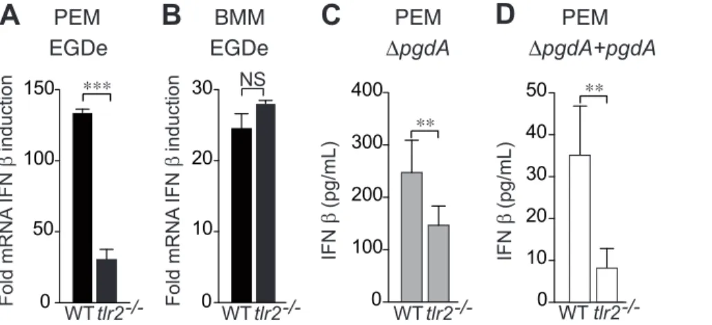
Consistent with our previous report measuring secretion of IFN-b protein in PEM, IFN-IFN-b mRNA synthesis induced IFN-by L. monocytogenes infection of PEM required TLR2 (Fig. 2A), while TLR2-deficient BMM showed no impairment in their synthesis of IFN-b mRNA (Fig. 2B). Moreover, IFN-b secretion was strongly reduced in tlr22/2 PEM infected with both the DpgdA mutant (Fig. 2C) and the complemented DpgdA strain (Fig. 2D), definitively establishing the TLR2 dependence of IFN-b production.
IFN-b induction does not require Mal/TIRAP but depends on TRIF
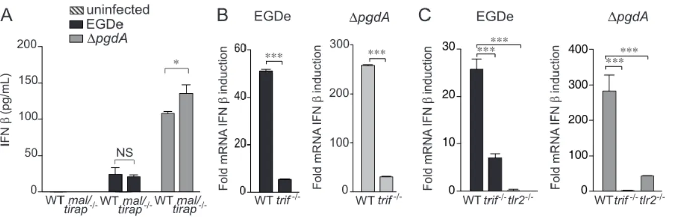
We next analyzed the pathways by which Listeria induces IFN-b. Our previous study and the above results strongly suggested the critical involvement of TLR2 [25]. TLR2 signaling depends on Mal/TIRAP and MyD88 adaptor proteins. We had previously shown that MyD88 contributed to full IFN-b induction by Listeria [25]. We then compared IFN-b production by wild-type and mal/ tirap2/2 macrophages infected with EGDe or DpgdA (Fig. 3A). Surprisingly, production of IFN-b was not decreased in infected macrophages deficient in Mal/TIRAP, indicating that the normal TLR2 adaptor Mal/TIRAP was not required for Listeria-mediated induction of IFN-b.
The adapter TRIF is employed by TLRs 3 and 4 to signal through the TBK1-IRF3/7-IFN-b pathway. There is no previous evidence of an association or functional interaction between TRIF
Figure 1. PgdA-dependent IFN-b response to Listeria in peritoneal macrophages. PEM from WT C57BL/6J mice were infected with the parental EGDe strain (black bars), the DpgdA mutant (grey bars) or the complemented DpgdA strain (white bars). After 7 h of infection, IFN-b levels were measured in supernatants by ELISA. Data are mean 6 SD (***, p,0.0001, n = 5).
doi:10.1371/journal.pone.0033299.g001
Figure 2. TLR2 is required for PgdA-mediated IFN-b response toListeriain peritoneal but not bone-marrow macrophages. (A) PEM from C57BL/6J or tlr22/2mice were infected with the parental EGDe strain. After 4 h of infection, IFN-b induction was measured by qRT-PCR. (B) BMM from C57BL/6J or tlr22/2mice were infected with the parental EGDe strain. After 4 h of infection, IFN-b induction was measured by qRT-PCR. Data are
mean 6 SD (NS, non significant; ***, p,0.0001, n = 3). PEM from WT C57BL/6J or tlr22/2mice were infected with the DpgdA mutant (C) or the complemented DpgdA strain (D). After 7 h of infection, IFN-b levels were measured in supernatants by ELISA. Data are mean 6 SD (**, p,0.01, n = 5). doi:10.1371/journal.pone.0033299.g002
and TLR2. In spite of this, the link between TRIF and the IRF pathway on the one hand, and the unusual employment of TLR2 for signaling to the IFN-b gene in PEM on the other suggested the possibility of a role for TRIF. To test this hypothesis we compared induction of IFN-b expression in wild-type and trif2/2 PEM or BMM infected with EGDe or DpgdA strains. IFN-b induction strongly decreased in TRIF-deficient macrophages infected with any of the two Listeria strains compared to wild-type PEM, showing the requirement for TRIF (Fig. 3B). In contrast, BMM showed a TRIF-independent IFN-b production (Fig. S1).
The PEM used in our studies are recruited to the peritoneal cavity by injection of the sterile irritant proteose peptone. Hence they differ from BMM not only regarding their anatomical location, but also their partially inflammatory character. To distinguish which of these differences was responsible for the TLR2 and TRIF signaling pathways, we examined IFN-b production by resident PEM. Figure 3C demonstrates a requirement for TLR2 and TRIF by the resident macrophage population. Thus, location to the
peritoneal cavity rather than inflammatory character determines the difference in signaling to the IFN-b gene between BMM and PEM.
To examine the role of TLR3, which uses TRIF to trigger IFN-b synthesis, we compared induction of IFN-IFN-b in wild-type and tlr32/2 PEM infected with EGDe or DpgdA strains. IFN-b production was decreased in TLR3-deficient PEM infected with EGDe or DpgdA (Fig. 4A). We also compared induction of IFN-b in wild-type and tlr42/2 PEM infected with EGDe or DpgdA strains, as TLR4 can mediate TRIF-dependent synthesis of IFN-b. In contrast to TLR3-deficient PEM, TLR4-deficient PEM did not show a decrease in IFN-b response to EGDe or DpgdA (Fig. 4B). Thus, IFN-b induction in response to Listeria infection relies in part on TLR3 and does not require TLR4.
IFN-b is induced by intracellular bacteria
Induction of IFN-b via TLR2 is no longer an exception. It has recently been shown that vaccinia virus-induced IFN-b production
Figure 3. TRIF, but not Mal/TIRAP, is necessary for IFN-b response toListeriain peritoneal macrophages. (A) PEM from WT C57BL/6J or mal/tirap2/2mice were infected with the parental EGDe strain (black bars), the DpgdA mutant (grey bars). After 7 h of infection, IFN-b levels were
measured in supernatants by ELISA. (B) PEM from C57BL/6J or trif2/2mice were infected with the parental EGDe strain (black bars) or the DpgdA mutant (grey bars). After 4 h of infection, IFN-b induction was measured by qRT-PCR. (C) Resident peritoneal macrophages from WT C57BL/6J, trif2/2
or tlr22/2mice were infected with the parental EGDe strain (black bars), the DpgdA mutant (grey bars). After 4 h of infection, IFN-b induction was measured by qRT-PCR. Data are mean 6 SD (NS, non significant; *, p,0.05; ***, p,0.0001; n = 3–4).
doi:10.1371/journal.pone.0033299.g003
Figure 4. TLR3, but not TLR4, contributes to IFN-b response toListeriain peritoneal macrophages. (A) PEM from WT C57BL/6J or tlr32/2
mice were infected with the parental EGDe strain (black bars), the DpgdA mutant (grey bars). After 7 h of infection, IFN-b levels were measured in supernatants by ELISA. Data are mean 6 SD (n = 3). (B) PEM from WT C57BL/6J or tlr42/2mice were infected with the parental EGDe strain (black bars), or the DpgdA mutant (grey bars). After 7 h of infection, IFN-b levels were measured in supernatants by ELISA. Data are mean 6 SD (***, p,0.0001; n = 3).
was dependent on TLR2 signaling and it was reported that this was occuring from late endosomes [26]. To investigate if an intracellular localization was also required in the case of Listeria, we pretreated cells with cytochalasin D to prevent internalization and measured IFN-b secretion by macrophages infected with EGDe or the DpgdA mutant (Fig. 5A). In both cases, IFN-b induction was strongly reduced. Thus, internalization is critical for Listeria-mediated IFN-b production. We also used dynasore, a dynamin inhibitor and chloroquine, which inhibits endosome acidification, and measured IFN-b induction in macrophages infected with EGDe or the DpgdA mutant (Fig. 5B–C). IFN-b synthesis was strongly diminished by both dynasore and chloroquine treatments. Together, these results suggest that the TLR2-dependent IFN-b induction is triggered intracellularly.
IRF3 and IRF7 are essential for IFN-b production in response to Listeria infection
In BMM rapid synthesis of IFN-b is entirely dependent on IRF3, but not on IRF7, whereas in bone marrow-derived myeloid DC IFN-b synthesis requires both IRF3 and IRF7 [27]. We investigated the role of IRF3 and IRF7 in the production of IFN-b by PEM. To this end we infected irf32/2and irf72/2macrophages with EGDe or the DpgdA strains. Inactivation of IRF3 totally abrogated IFN-b
mRNA induction in response to both strains (Fig. 6). IFN-b induction in IRF7-deficient macrophages was also strongly affected highlighting the important role of both transcription factors in response to Listeria infection (Fig. 6). PEM thus resemble bone marrow-derived myeloid DC, not BMM, in relation to their IRF requirement for Listeria-mediated IFN-b synthesis.
In addition to IRF3/7, NFkB contributes to the formation of the IFN-b enhanceosome [20,28]. We therefore examined the involvement of the NFkB pathway by measuring induced synthesis of an NFkB-dependent mRNA. IkB is an NFkB-dependent gene and thus a read-out for NFkB activation in response to Listeria infection. We measured the induction of IkB expression in PEM infected with EGDe or the DpgdA mutant. Both strains induced IkB expression and this required internalization as treatment with dynasore reduced the level of IkB induction (Fig. 7A). Degradation of the IkB protein was examined in PEM infected with EGDe by immunoblot using anti-IkB antibodies. IkB level was reduced rapidly after infection of wild-type PEM (Fig. S2A). In contrast, IkB degradation was not observed in tlr22/2PEM infected with Listeria (Fig. S2B). Infection of wild-type, tlr22/2and trif2/2
Figure 5. Internalization of bacteria is required for IFN-b response by peritoneal macrophages. (A) PEM from WT C57BL/6J mice were pretreated with 50 mM of cytochalasin D, and left uninfected (hatched bars) or infected with the parental EGDe strain (black bars) or the DpgdA mutant (grey bars). 7 h post-infection, IFN-b levels were measured in cells supernatants by ELISA. (B) PEM from WT C57BL/6J were treated with 80 mM dynasore. After 4 h of infection with the parental EGDe strain (black bars) or the DpgdA mutant (grey bars), IFN-b induction was measured by qRT-PCR. (C) PEM from WT C57BL/6J mice were treated with 100 mM chloroquine, and left uninfected (hatched bars) or infected with the parental EGDe strain (black bars) or the DpgdA mutant (grey bars). 7 h post-infection, IFN-b concentrations were measured in cells supernatants by ELISA. Data are mean 6 SD (***, p,0.0001, n = 3–4).
doi:10.1371/journal.pone.0033299.g005
Figure 6. IFN-b response toListeria is mediated by IRF3 and IRF7 in peritoneal macrophages. PEM from WT C57BL/6J, irf32/2
and irf72/2mice were infected with the parental EGDe strain (black bars) or the DpgdA mutant (grey bars). 4 h post-infection, mRNA was isolated and the IFN-b induction was measured by qRT-PCR. Data are mean 6 SD (***, p,0.0001, n = 3).
doi:10.1371/journal.pone.0033299.g006
Figure 7. Bacterial internalization and NF-kB are required for TLR2 and TRIF-dependent IFN-b response in peritoneal mac-rophages. (A) PEM from WT C57BL/6J mice and pretreated with dynasore were infected with the parental EGDe strain (black bars) or the DpgdA mutant (grey bars). 4 h post-infection, IkB induction was measured by qRT-PCR. (B) PEM from WT C57BL/6J, tlr22/2and trif2/2 mice were infected with the parental EGDe strain (black bars) or the DpgdA mutant (grey bars). 4 h post-infection, mRNA was isolated and IkB induction was measured by qRT-PCR. Data are mean 6 SD (***, p,0.0001, n = 3).
doi:10.1371/journal.pone.0033299.g007
macrophages with EGDe or DpgdA showed that both TLR2 and the adaptor were required for full induction of IkB mRNA in response to EGDe and DpgdA strains (Fig. 7B). These results suggest that TLR2 and TRIF contribute to NFkB activation. The comparison between EGDe and DpgdA strains showed that both caused similar magnitudes of IkB mRNA synthesis. Thus, the activation of NFkB by Listeria is independent of PgdA, suggesting that the increased IFN-b production after infection with DpgdA relies on activation of other transcription factors such as IRFs.
Nucleic acids released intracellularly are critical for IFN-b induction
TLR2 or TRIF deficiency strongly reduced, but did not completely shut off IFN-b synthesis. This suggested a potential contribution of intracellular, nucleic acid-dependent pathways to IFN-b synthesis, particularly after infection with DpgdA. We therefore examined whether these pathways are able to signal in PEM.
Since inactivation of PgdA increases Listeria sensitivity to peptidoglycan-targeting antimicrobials such as lysozyme, and thus induces bacterial degradation, we measured the DNA and RNA released by EGDe and DpgdA strains following lysozyme exposure. As expected, DpgdA released significantly higher amounts of DNA and RNA than wild-type and complemented DpgdA strains, raising the possibility that both DNA and RNA could be involved in IFN-b production (Fig. 8A). We thus measured IFN-IFN-b induction in THP1 macrophages transfected with Listeria DNA, either undigested or treated with DNase. Intact but not DNase-treated DNA significantly induced IFN-b (Fig. 8B). Macrophages were then transfected with lysozyme-digested EGDe or DpgdA, either untreated or digested with DNase. Treatment with DNase significantly reduced IFN-b production (Fig. 8C). Taken together, these results show that Listeria DNA can induce IFN-b, strongly indicating that destruction of DpgdA bacteria intracellularly activates DNA sensors.
Discussion
We had recently reported that a PGN modification involving a N-deacetylase gene, pgdA, was playing a key role in L. monocytogenes virulence [25]. A DpgdA strain of L. monocytogenes which is unable to
modify its PGN, was shown to be extremely sensitive to the bacteriolytic activity of lysozyme, normally found within macro-phage vacuoles and its virulence was strongly attenuated [25]. Furthermore, this mutant induced a much higher TLR2-dependent IFN-b response than the parental strain [25]. We hypothesised that this unconventional IFN-b response induced by the pgdA mutant was due to an enhanced accessibility of bacterial cell wall components to TLR2. Here we have shown that IFN-b production requires bacterial internalization and is triggered by Mal/TIRAP-independent pathways which involve TLR2, TRIF, IRF3 and IRF7.
It was surprising to see a role for TLR2, as, based on results in BMM and epithelial cells, type I IFNs production is usually not known to result from TLR2 signaling [5–8]. Classical TLR2 signaling leads to NF-kB-dependent production of inflammatory cytokines [21]. However, in support of an unconventional role for TLR2, recent studies reported roles for TLR2-dependent induction of IFN-b in response to vaccinia virus or synthetic ligands [26,29]. In the vaccinia virus study, a specific inflammatory monocyte population -Ly6Chi- was shown to be the source of IFN-b [26]. In the present study we show that TLR2-dependent IFN-IFN-b synthesis is a property of both resident and recruited inflammatory PEM. Furthermore, the two previous studies documented that TLR2 activation of type I IFN responses to TLR ligands occurs within intracellular compartments, and that TLR2 signals from the phagosome in response to viral infection or synthetic TLR2 ligands [26,29]. These results challenged the view that TLR2 signals solely from the plasma membrane. In our experiments, pre-treatment of PEM with either cytochalasin D, an inhibitor of actin polymerization and thus internalization, dynasore, an inhibitor of the endocytic effector dynamin, or chloroquine, which inhibits endosome acidification [30,31], significantly impaired the induc-tion of IFN-b following Listeria infecinduc-tion, strongly suggesting that phagocytosis of L. monocytogenes and intracellular location of TLR2 trigger this response. These observations also correlate with our early hypothesis that the inflammatory response induced by DpgdA is due to an enhanced release or accessibility of bacterial cell wall components to TLR2.
Induction of the IFN-b gene was independent of the TLR adapter Mal/TIRAP, but, unexpectedly required the TLR3/4 adapter TRIF. Francisella tularensis has recently been shown to
Figure 8.Listerianucleic acids trigger IFN-b production. (A) The parental EGDe (black bars) DpgdA (grey bars) and complemented DpgdA strain (hatched bars) were incubated with lysozyme. The amount of DNA and RNA released after treatment was quantified by spectrophotometry. (B) THP-1 macrophages were transfected with DNA from the DpgdA mutant, pretreated or not with DNase, and IFN-b induction was determined using the HEK-blue assay. (C) The parental EGDe strain (black bars) or the DpgdA mutant (grey bars) were incubated with lysozyme. PEM were transfected with bacterial lysates, pretreated with DNase or not treated, and IFN-b production was quantified in cells supernatants 24 h after transfection by ELISA. Data are mean 6 SD (**, p,0.01, n = 3; ***, p,0.0001, n = 3).
signal through TLR2 from the phagosome in a Mal/TIRAP independent manner [32], and it was shown that Mal/TIRAP is dispensable in TLR2 signaling at high concentrations of ligands [33]. Thus our study reinforces the view that TLR2 can act independently from Mal/TIRAP. In addition our report suggests a synergy between a TLR2 pathway and TRIF, an adapter previously known to trigger the synthesis of pro-inflammatory cytokines and type I IFNs upon engagement of TLR3 and TLR4. TLR3 is known to bind viral dsRNA to induce secretion of type I IFN and lead to control of viral infections [3,34,35]. To our knowledge Chlamydia muridarum is the only bacterium reported to induce a TLR3-dependent IFN-b response specifically in murine oviduct epithelial cells [36]. We tested whether the dual requirement for TLR2 and TRIF resulted from a functional or physical interaction between TLR2 and TLR3. In fact, IFN-b production was reduced in TLR3-deficient macrophages, but significantly less so than in trif2/2 PEM. Therefore, there is no evidence for a putative TLR2/TLR3 interaction. Another possibility to incorporate TRIF into the pathway stimulated in PEM by Listeria would be a cooperation of TLR2 and TLR4. This was ruled out by showing that Listeria-infected tlr42/2 PEM produced a similar amount of IFN-b as their wild-type counterparts. TRIF could possibly orchestrate an additional pathway. Along these lines, TRIF has recently been shown to be required for IFN-b synthesis by dendritic cells upon activation of the cytosolic receptor complex DDX1/DDX21/DDX36 by viral RNA [19].
Engagement of TLRs by various microbe-associated molecular patterns induces activation and translocation to the nucleus of NF-kB, IRF3, IRF7 and/or activator protein-1 (AP-1), which collaborate to induce transcription of type I IFNs [37]. We addressed the role of these transcriptional activators in the IFN-b response to wild-type Listeria and DpgdA, and revealed that inactivation of IRF3 totally abrogated this response to both strains while IFN-b induction was significantly but not totally impaired in IRF7-deficient macrophages, indicating that both of these transcription factors are required for induction of IFN-b following infection with L. monocytogenes. We also assessed the involvement of NF-kB in this response using induction of the IkB gene as a readout. We observed an induction of IkB expression in macrophages which was similar after infection with EGDe or DpgdA. Thus, activation of NF-kB by Listeria is independent of PgdA, strongly suggesting that the elevated IFN-b production by the DpgdA mutant mostly relies on IRF3.
The increased IFN-b response to the DpgdA strain probably results from the fact that within the phagosome, its lysozyme-sensitive cell wall is degraded, releasing PAMPs able to interact with TLR2 and other PRRs, including cytoplasmic ones. As recent studies have highlighted novel DNA-sensing pathways in the induction of type I IFNs [9,14–17,38], we thus also investigated the involvement of bacterial nucleic acids in the IFN-b induction, Firstly, we showed that inactivation of PgdA, which confers a higher susceptibility to lysozyme, leads to increased release of DNA. We then showed that DNA from L. monocytogenes can induce IFN-b expression in PEM, suggesting that this macrophage population employs cytoplasmic nucleic acid sensing similar to macrophages or macrophage lines derived from different anatom-ical locations [9,38]. Which -if any- of the recently described nucleic acid sensors are used by PEM for the recognition of Listeria DNA remains subject to future investigation. Nevertheless, other bacterial components could participate in IFN-b production upon infection with the DpgdA mutant. For example, the second messenger molecule cyclic diadenosine monophosphate (c-di-AMP), was shown to be secreted by Listeria multidrug efflux
pumps triggering type I IFN response [10] and could be involved in the process.
In conclusion, this study describes a novel mechanism leading to induction of type I IFNs in which intracellular sensing plays an important role, ultimately showing how these different recognition pathways can synergise to induce innate immune responses which are required to control infection. In this regard cooperation between TLR2 and TRIF may reflect the need for convergence of the NF-kB and IRF pathways at the IFN-b promoter, with TLR2 being responsible mainly for NF-kB activation and TRIF being instrumental for activation of IRF3 and IRF7. By employing the strategy of PGN modification, L. monocytogenes can avoid immune detection by TLR and evade the innate immune response, thus enabling the infectious process to occur. It is important to recall that pgdA orthologs are found in other pathogenic bacteria, such as Streptococcus pneumoniae, Bacillus cereus, Bacillus anthracis and Helico-bacter pylori, strongly suggesting that PGN N-deacetylation is a general mechanism evolved by microbes to escape from pattern recognition receptor-mediated immune recognition [39–42].
Materials and Methods
Bacterial strains and growth conditions
L. monocytogenes EGDe (BUG1600, ATCC BAA-679), L. monocytogenes isogenic mutant DpgdA (BUG2288, [25]) and L. monocytogenes DpgdA complemented strain (BUG2382) were grown in brain heart infusion (BHI, Oxoid), aerobically at 37uC and 200 rpm.
Construction of L. monocytogenes DpgdA complemented strain
A DNA fragment containing the pgdA gene (lmo0415) and its promoter was generated by PCR using oligonucleotides lmo0415-1 (59-AAGGATCCCACAATATGTTAGTTTTCAGGGG-39) and lmo0415-2 (59-AAGGATCCTTATTTCACCATTCTT-GAATCTG-39). The fragment was integrated into pCR-Blunt-II-TOPO (Invitrogen) and the construct was verified by sequencing. After digestion of the construct by BamHI, the fragment was purified on agarose gel and cloned into the integrative vector pPL2 [43], previously digested by BamHI, constructing pOD98. The pOD98 was electroporated into DpgdA at 2,500 V, 200 V and 25mF. Transformants were selected at 37uC on BHI agar containing chloramphenicol (7mg/mL). The presence of the pgdA gene in the complemented strain was confirmed by PCR using oligonucleotides lmo0415-1 and lmo0415-2.
Ethics statement
Mice were used for obtaining peptone-elicited peritoneal macrophages, resident peritoneal macrophages and bone mar-row-derived macrophages. Animal experiments were performed in accordance with protocols approved by the Animal Experimen-tation Ethics Committee of the Institut Pasteur (permit #03-49) and following Austrian law in accordance with protocols approved by the Ethics Committee of the University of Veterinary Medicine, Vienna (#GZ680 205/67-BrGt/2003).
Isolation and culture of murine peptone-elicited peritoneal macrophages (PEM)
PEM were isolated from 7 to 10 week-old C57BL/6J and genetically-matched tlr22/2, tlr32/2, tlr42/2, mal/tirap2/2, trif2/2, irf32/2 and irf72/2 mice as previously described [44]. The percentage of macrophages was determined by flow cytometry
using CD11b (1:100, eBiosciences) and F4/80 (1:100, eBiosciences) antibodies. More than 90% of the cells were macrophages. PEM were seeded onto 6-well plates at a concentration of 26106cells per well in DMEM (PAA) supplemented with 10% FCS, 10% L929 conditioned medium (LCM) and 1% penicillin-streptomycin or RPMI-1640 (Gibco) supplemented with 10% FBS and 1% penicillin-streptomycin.
Isolation of resident peritoneal macrophages
Resident macrophages were isolated from 6 to 8 week-old C57BL/6J and genetically-matched tlr22/2, trif2/2 mice by washing the peritoneum twice with 10 mL DMEM (PAA) supplemented with 10% FBS, 10% LCM and 1% penicillin-streptomycin. Harvested cells were centrifuged at 300 g for 5 minutes and resuspended in complete medium. The percentage of macrophages was determined by flow cytometry analysis as above. Cells were seeded onto 6-well plates (Nunc) at a concentration of 26106cells per well.
Isolation of bone marrow-derived macrophages
Tibia and femur from 6 to 8 week-old C57BL/6J and genetically-matched tlr22/2, trif2/2 mice were collected in ice cold PBS. Bones were sterilized with 70% ethanol and flushed with a 25-G needle using cold DMEM supplemented with 10% FCS, 10% LCM and 1% penicillin-streptomycin. Cells were seeded onto 6-well plates (Nunc) at a concentration of 106cells per well and incubated at 37uC with 5% CO2. After 4 days, complete
medium was added and cells were split at a ratio of 1:2. After 8 days, macrophages were fully differentiated.
Culture of human THP-1-derived macrophages and HEK-blue type I IFN cells
Human acute monocytic leukemia THP-1 cells (ATCC TIB202) were maintained in RPMI-1640 supplemented with 10% FBS and 1% penicillin-streptomycin. Cells were seeded onto a 24-well plate at a concentration of 46105 cells per well in antibiotic-free media supplemented with 12.5 ng/mL phorbol myristate acetate and incubated for 24 h at 37uC with 5% CO2.
Differentiation was determined to be successful upon formation of a confluent adherent monolayer. HEK-blue type I IFN cells (Invivogen) were grown in DMEM supplemented with 10% FBS and 1% penicillin-streptomycin. Cells were seeded at a concen-tration of 5.66104cells per well onto a 96-well plate.
Macrophage infection assays
For cytokine analysis, macrophages were infected with Listeria strains at MOI 10:1, centrifuged at 300 g for 2 min and incubated at 37uC for 15 min. Following phagocytosis, monolayers were washed twice followed by incubation in RPMI-1640 supplemented with 10% fetal bovine serum (FBS) and gentamicin (20mg/mL). Supernatants were collected at various time points, for detection of IFN-b by ELISA. For transcript analysis, macrophages were infected with Listeria strains at MOI 20:1 and incubated at 37uC for 1 h to allow phagocytosis. Monolayers were washed and incubated in DMEM supplemented with 10% FCS and gentamicin (5mg/mL). After 2 h, medium was changed to DMEM supplemented with 10% FCS and gentamicin (1mg/mL). Cells were lysed at various time points and RNA collected for qPCR analysis.
Inhibition assays
For inhibition of bacterial internalization, cell monolayers were pretreated either for 2 h with 100mM cytochalasin-D (Sigma-Aldrich), or 30 min with 80mM dynasore (Sigma-Aldrich) or
30 min with 100mM chloroquine (Sigma-Aldrich) prior to infection assays.
DNA isolation and transfection assays
Listeria were grown overnight in BHI at 37uC and cultures were centrifuged at 8000 g for 5 min. Bacterial pellets were resuspended in 75mg/mL lysozyme and incubated at 37uC for 1 h. DNA was then extracted using the DNeasy blood and tissue kit (Qiagen) and quantified by spectrophotometry (Nanodrop). For transfection assays, THP-1 macrophages were transfected with 200 ng/mL DNA with 2% lipofectamine 2000 (Invitrogen) and incubated for 24 h. Following incubation, supernatants were collected for IFN-b analysis. For pretreatment of DNA with DNase, DNase was added at final concentration of 100mg/mL for 45 min at 37uC.
Lysozyme digestion, quantification of nucleic acids release and identification of Listeria PAMPs
Bacterial cultures were treated with 10mg/mL lysozyme, a concentration leading to lysis of DpgdA but not EGDe, and incubated at 37uC and 200 rpm for 1 h. Following lysozyme treatment, lysed bacterial cultures were centrifuged at 5000 rpm during 10 min. Two types of experiments were performed on supernatants. First, nucleic acid release was quantified. DNA was purified using the Qiagen DNeasy blood and tissue kit omitting lysis steps and quantified by spectrophotometry (Nanodrop). RNA was purified using Qiagen RNeasy kit and quantified by spectrophotometry (Nanodrop). Data shown are representatives of at least three independent experiments. Second, 100mL of each supernatants were treated by DNase during 30 min at 37uC. Enzymes were inactivated and treated- or untreated-supernatants were transfected in PEM. 8 h after transfection, supernatants of cells were recovered and the IFNb was quantified.
Detection of type I IFN by ELISA and HEK-blue type I IFN cell assay
Murine IFN-b production was detected in macrophage supernatants by ELISA according to the manufacturer’s procedure (PBL Biomedical Laboratories). For the HEK-blue type I IFN assay, supernatant from THP-1 macrophage assays was collected and 20mL added onto HEK-blue type I IFN cells plated in 96-well plates, which were incubated at 37uC overnight. Supernatant from HEK-blue cells was collected and 40mL added to 160mL of Quanti-blue reagent (Invivogen) for 20 min at 37uC. The colorimetric reaction was measured at 625 nm on a plate reader. Data was normalised against absorbance for the untreated cells and plotted as relative fold increases. Data shown are represen-tatives of at least three independent experiments.
Detection of IkB by immunoblot
PEM from WT or tlr22/2C57BL/6J mice were infected with EGDe. Cells were lysed 0, 0.5, 1, 1.5, 2, 2.5, or 3 h post-infection. IkB and tubulin were detected in lysates by immunoblotting using anti-IkB (Santa Cruz, 1:100) and anti-a-tubulin (Sigma, 1:5000) antibodies.
RNA isolation for quantitative real-time PCR
RNA preparation was performed using NucleoSpin RNA II kit (Macherey-Nagel) according to the manufacturer’s instructions. Quantitative real-time PCR was performed on a Mastercycler EP realplex S (Eppendorf). Primers for HPRT (housekeeping gene control), IFNb and IkBa mRNA expression were as follows: HPRT forward GTTGGATACAGGCCAGACTTTGTTG, HPRT reverse GAGGGTAGGCTGGCCTATTGGCT, IFNb
forward 59-TCAGAATGAGTGGTGGTTGC-39, IFNb reverse GACCTTTCAAATGCAGTAGATTCA-39; IkBa forward 59-GCAATTTCTGGCTGGTGGG-39, IkBa reverse 59GATCC-GCCAGGTGAAGGG-39. Data shown are representatives of at least three independent experiments.
Statistical analysis
Results are expressed as means of at least three values, with error bars representing standard deviations. Student’s t tests were performed to determine statistical significance where * indicates P,0.05,**indicates P,0.01 and***indicates P,0.0001.
Supporting Information
Figure S1 TRIF is not required for IFN-b response to Listeria in bone marrow macrophages. BMM from C57BL/6J or trif2/2mice were infected with the parental EGDe strain (black bars) or the DpgdA mutant (grey bars). After 4 h of infection, IFN-b induction was measured by qRT-PCR. Data are mean 6 SD (NS, non significant, n = 3).
(EPS)
Figure S2 TLR2 is required for optimal activation of NF-kB. (A) PEM from WT C57BL/6J mice were infected with EGDe. Cells were lysed 0, 0.5, 1, 1.5, 2, 2.5, or 3 h post-infection. Activation of NF-kB was measured by determination of IkB degradation relative to tubulin following immunodetection. (B) PEM from tlr22/2 mice were infected with EGDe. Cells were lysed 0, 0.5, 1, 1.5, 2, 2.5, or 3 h post-infection. Activation of NF-kB was measured by determination of INF-kB degradation relative to tubulin following immunodetection.
(EPS)
Acknowledgments
We thank members of the Cossart laboratory for helpful discussions.
Author Contributions
Conceived and designed the experiments: OD PC LO TD. Performed the experiments: CA SC SW CG RJ AMJ. Analyzed the data: OD PC LO TD CA SC SW. Contributed reagents/materials/analysis tools: OD PC LO TD. Wrote the paper: OD PC LO TD SC CA.
References
1. Creagh EM, O’Neill LAJ (2006) TLRs, NLRs and RLRs: a trinity of pathogen sensors that co-operate in innate immunity. Trends Immunol 27: 352–357. 2. Rasmussen SB, Reinert LS, Paludan SR (2009) Innate recognition of
intracellular pathogens: detection and activation of the first line of defense. APMIS 117: 323–337.
3. Mogensen TH (2009) Pathogen recognition and inflammatory signaling in innate immune defenses. Clin Microbiol Rev 22: 240–273.
4. Decker T, Mu¨ller M, Stockinger S (2005) The Yin and Yang of type I interferon activity in bacterial infection. Nat Rev Immunol 5: 675–687.
5. McCaffrey RL, Fawcett P, O’Riordan M, Lee K-D, Havell EA, et al. (2004) A specific gene expression program triggered by Gram-positive bacteria in the cytosol. Proc Natl Acad Sci USA 101: 11386–11391.
6. O’Riordan M, Yi CH, Gonzales R, Lee K-D, Portnoy DA (2002) Innate recognition of bacteria by a macrophage cytosolic surveillance pathway. Proc Natl Acad Sci USA 99: 13861–13866.
7. Stockinger S, Reutterer B, Schaljo B, Schellack C, Brunner S, et al. (2004) IFN regulatory factor 3-dependent induction of type I IFNs by intracellular bacteria is mediated by a TLR- and Nod2-independent mechanism. J Immunol 173: 7416–7425.
8. O’Connell RM, Vaidya SA, Perry AK, Saha SK, Dempsey PW, et al. (2005) Immune activation of type I IFNs by Listeria monocytogenes occurs independently of TLR4, TLR2, and receptor interacting protein 2 but involves TNFR-associated NF kappa B kinase-binding kinase 1. J Immunol 174: 1602–1607.
9. Stetson D, Medzhitov R (2006) Recognition of cytosolic DNA activates an IRF3-dependent innate immune response. Immunity 24: 93–103.
10. Woodward JJ, Iavarone AT, Portnoy DA (2010) c-di-AMP secreted by intracellular Listeria monocytogenes activates a host type I interferon response. Science 328: 1703–1705.
11. Burdette DL, Monroe KM, Sotelo-Troha K, Iwig JS, Eckert B, et al. (2011) STING is a direct innate immune sensor of cyclic di-GMP. Nature 478: 515–518.
12. Hornung V, Latz E (2010) Intracellular DNA recognition. Nat Rev Immunol 10: 123–130.
13. Rehwinkel J, Reis E, Sousa C (2010) RIGorous detection: exposing virus through RNA sensing. Science 327: 284–286.
14. Ablasser A, Bauernfeind F, Hartmann G, Latz E, Fitzgerald KA, et al. (2009) RIG-I-dependent sensing of poly(dA:dT) through the induction of an RNA polymerase III-transcribed RNA intermediate. Nat Immunol 10: 1065–1072. 15. Chiu Y-H, Macmillan JB, Chen ZJ (2009) RNA polymerase III detects cytosolic
DNA and induces type I interferons through the RIG-I pathway. Cell 138: 576–591.
16. Unterholzner L, Keating SE, Baran M, Horan KA, Jensen SB, et al. (2010) IFI16 is an innate immune sensor for intracellular DNA. Nat Immunol 11: 997–1004.
17. Yang P, An H, Liu X, Wen M, Zheng Y, et al. (2010) The cytosolic nucleic acid sensor LRRFIP1 mediates the production of type I interferon via a b-catenin-dependent pathway. Nat Immunol 11: 487–494.
18. O’Neill LAJ (2009) DNA makes RNA makes innate immunity. Cell 138: 428–430.
19. Zhang Z, Kim T, Bao M, Facchinetti V, Jung SY, et al. (2011) DDX1, DDX21, and DHX36 helicases form a complex with the adaptor molecule TRIF to sense dsRNA in dendritic cells. Immunity 34: 866–878.
20. Panne D, Maniatis T, Harrison SC (2007) An atomic model of the interferon-beta enhanceosome. Cell 129: 1111–1123.
21. Kawai T, Akira S (2010) The role of pattern-recognition receptors in innate immunity: update on Toll-like receptors. Nat Immunol 11: 373–384. 22. Janot L, Secher T, Torres D, Maillet I, Pfeilschifter J, et al. (2008) CD14 works
with Toll-like receptor 2 to contribute to recognition and control of Listeria monocytogenes infection. J Infect Dis 198: 115–124.
23. Torres D, Barrier M, Bihl F, Quesniaux VJF, Maillet I, et al. (2004) Toll-like receptor 2 is required for optimal control of Listeria monocytogenes infection. Infect Immun 72: 2131–2139.
24. Vollmer W (2008) Structural variation in the glycan strands of bacterial peptidoglycan. FEMS Microbiol Rev 32: 287–306.
25. Boneca IG, Dussurget O, Cabanes D, Nahori M-A, Sousa S, et al. (2007) A critical role for peptidoglycan N-deacetylation in Listeria evasion from the host innate immune system. Proc Natl Acad Sci USA 104: 997–1002.
26. Barbalat R, Lau L, Locksley RM, Barton GM (2009) Toll-like receptor 2 on inflammatory monocytes induces type I interferon in response to viral but not bacterial ligands. Nat Immunol 10: 1200–1207.
27. Stockinger S, Kastner R, Kernbauer E, Pilz A, Westermayer S, et al. (2009) Characterization of the interferon-producing cell in mice infected with Listeria monocytogenes. PLoS Pathog 5: e1000355.
28. Panne D (2008) The enhanceosome. Curr Opin Struct Biol 18: 236–242. 29. Dietrich N, Lienenklaus S, Weiss S, Gekara NO (2010) Murine Toll-like
receptor 2 activation induces type I interferon responses from endolysosomal compartments. PLoS ONE 5: e10250.
30. Vincent MJ, Bergeron E, Benjannet S, Erickson BR, Rollin PE, et al. (2005) Chloroquine is a potent inhibitor of SARS coronavirus infection and spread. Virol J 2: 69.
31. Rutz M, Metzger J, Gellert T, Luppa P, Lipford GB, et al. (2004) Toll-like receptor 9 binds single-stranded CpG-DNA in a sequence- and pH-dependent manner. Eur J Immunol 34: 2541–2550.
32. Cole LE, Laird MHW, Seekatz A, Santiago A, Jiang Z, et al. (2010) Phagosomal retention of Francisella tularensis results in TIRAP/Mal-independent TLR2 signaling. J Leukoc Biol 87: 275–281.
33. Kenny EF, Talbot S, Gong M, Golenbock DT, Bryant CE, et al. (2009) MyD88 adaptor-like is not essential for TLR2 signaling and inhibits signaling by TLR3. J Immunol 183: 3642–3651.
34. Liu L, Botos I, Wang Y, Leonard JN, Shiloach J, et al. (2008) Structural basis of Toll-like receptor 3 signaling with double-stranded RNA. Science 320: 379–381. 35. Yamamoto M (2003) Role of adaptor TRIF in the MyD88-independent
Toll-like receptor signaling pathway. Science 301: 640–643.
36. Derbigny WA, Johnson RM, Toomey KS, Ofner S, Jayarapu K (2010) The Chlamydia muridarum-induced IFN-beta response is TLR3-dependent in murine oviduct epithelial cells. J Immunol 185: 6689–6697.
37. Zhong B, Tien P, Shu H-B (2006) Innate immune responses: crosstalk of signaling and regulation of gene transcription. Virology 352: 14–21. 38. Charrel-Dennis M, Latz E, Halmen KA, Trieu-Cuot P, Fitzgerald KA, et al.
(2008) TLR-independent type I interferon induction in response to an extracellular bacterial pathogen via intracellular recognition of its DNA. Cell Host Microbe 4: 543–554.
39. Psylinakis E, Boneca IG, Mavromatis K, Deli A, Hayhurst E, et al. (2005) Peptidoglycan N-acetylglucosamine deacetylases from Bacillus cereus, highly conserved proteins in Bacillus anthracis. J Biol Chem 280: 30856–30863.
40. Vollmer W (2000) The pgdA gene encodes for a peptidoglycan N-acetylgluco-samine deacetylase in Streptococcus pneumoniae. J Biol Chem 275: 20496–20501. 41. Vollmer W, Tomasz A (2002) Peptidoglycan N-acetylglucosamine deacetylase, a
putative virulence factor in Streptococcus pneumoniae. Infect Immun 70: 7176–7178. 42. Wang G, Olczak A, Forsberg LS, Maier RJ (2008) Oxidative stress-induced peptidoglycan deacetylase in Helicobacter pylori. J Biol Chem 284: 6790–6800.
43. Lauer P, Chow MYN, Loessner MJ, Portnoy DA, Calendar R (2002) Construction, characterization, and use of two Listeria monocytogenes site-specific phage integration vectors. J Bacteriol 184: 4177–4186.
44. Alford CE, King TE, Campbell PA (1991) Role of transferrin, transferrin receptors, and iron in macrophage listericidal activity. J Exp Med 174: 459–466.