HAL Id: hal-00377528
https://hal.archives-ouvertes.fr/hal-00377528
Submitted on 22 Apr 2009
HAL is a multi-disciplinary open access
archive for the deposit and dissemination of sci-entific research documents, whether they are pub-lished or not. The documents may come from teaching and research institutions in France or
L’archive ouverte pluridisciplinaire HAL, est destinée au dépôt et à la diffusion de documents scientifiques de niveau recherche, publiés ou non, émanant des établissements d’enseignement et de recherche français ou étrangers, des laboratoires
The catalytic pocket of the ring-hydroxylating
dioxygenase from Sphingomonas CHY-1
Jean Jakoncic, Yves Jouanneau, Christine Meyer, V. Stojanoff
To cite this version:
Jean Jakoncic, Yves Jouanneau, Christine Meyer, V. Stojanoff. The catalytic pocket of the ring-hydroxylating dioxygenase from Sphingomonas CHY-1. Biochemical and Biophysical Research Com-munications, Elsevier, 2007, 352, pp.862-866. �hal-00377528�
The catalytic pocket of the ring-hydroxylating dioxygenase from Sphingomonas CHY-1
Jean Jakoncic1, Yves Jouanneau2, Christine Meyer2, Vivian Stojanoff1
1Brookhaven National Laboratory, National Synchrotron Light Source, Upton, NY 11973,
USA
2Laboratoire de Biochimie et Biophysique des Systèmes Intégrés, CEA, DSV, DRDC and
CNRS UMR 5092, CEA-Grenoble, F-38054 Grenoble Cedex 9, France.
Corresponding author: Vivian Stojanoff
Brookhaven National Laboratory Upton NY 11973 US
Tel. : 1 631 344 8375; Fax : 1 631 344 3238 Email : vivian.stojanoff@gmail.gov
Abstract
Ring-hydroxylating dioxygenases are multicomponent bacterial enzymes that catalyze the first step in the oxidative degradation of aromatic hydrocarbons. The dioxygenase from Sphingomonas CHY-1 is unique in that it can oxidize a wide range of polycyclic aromatic hydrocarbons (PAHs). With a crystal structure similar to that of the seven other known dioxygenases, its catalytic domain features the largest hydrophobic substrate binding cavity characterized so far. Molecular modeling studies indicated that the catalytic cavity is large enough to accommodate a five-ring benzo[a]pyrene molecule. The predicted positions of this and other PAHs in the substrate binding pocket are consistent with the product regio- and stereo-selectivity of the enzyme.
Keywords: dioxygenase; catalytic domain; mononuclear iron; bioremediation; high
The first step in the biodegradation of aromatic hydrocarbons by aerobic bacteria often involves a dihydroxylation on two adjacent carbon atoms of the aromatic ring, catalyzed by a ring-hydroxylating dioxygenase (RHD). RHDs form a large family of enzymes, very diverse in terms of substrate specificity and protein sequence [1]. Their role is crucial in the degradation of many organic pollutants, including polycyclic aromatic hydrocarbons (PAHs), which are notorious for their resistance to biodegradation. Several bacteria were found to degrade PAHs but only a few have been reported to attack four and five ring PAHs [2,3]. In Sphingomonas strain CHY-1 a single RHD has been shown to be responsible for the oxidation of a wide range of PAHs [4]. This dioxygenase consists of three components, a NADH-dependent reductase (PhnA4), a ferredoxin containing a Rieske type [2Fe-2S] cluster (PhnA3), and a terminal oxygenase, PhnI, containing both a mononuclear iron [Fe2+] and a [2Fe-2S] Rieske cluster [5]. Recent biochemical studies showed that the dioxygenase from strain CHY-1 was able to oxidize at least eight PAHs made of 2 to 5 aromatic rings, in contrast to most other dioxygenases, whose selectivity is limited to 2 and 3 ring PAHs [6,7,8]. A positive electron density observed in the PhnI refined three-dimensional structure served as probe in modeling different substrates in the catalytic pocket. This study presents evidence that the broad substrate specificity of the enzyme is primarily due to the large volume and particular shape of its catalytic pocket.
The purification of recombinant His-tagged PhnI was performed as described previously [5]. The protein was further purified by gel filtration chromatography following His-tag removal by thrombin cleavage. Crystals were grown at room temperature using the sitting drop method and a crystallization solution derived from Cryoscreen solution 67 (Nextal Biotechnologies, Montreal, Quebec, CA). Crystals that diffracted up to 1.85 Å resolution (Jakoncic, unpublished work) were obtained by further covering each well with mineral oil [9]. Diffraction data were recorded on beam line X6A at the National Synchrotron Light Source [10]. The 3D structure was determined by molecular replacement, using the α-subunit from naphthalene dioxygenase from Pseudomonas sp. Strain NCIB 9816-4, NDO-O9816-4 [6] and the β-subunit from cumene dioxygenase from P.
fluorescens strain IP01, CDO-OIP01 [11] as templates. The model was refined using
REFMAC [12] and COOT [13] to R and Rfree factors of 19.7 and 23.6 %, respectively (Fig.
1). Most of the amino acids are present in the final model (PDB accession code 2CKF), with the exception of residues in the C- and N- termini regions and a highly flexible region at the entrance of the catalytic pocket LI (residues 221 to 240) and LII (residues 253 to 265) described in the following section.
Results and Discussion
The PhnI catalytic domain. The PhnI quaternary and tertiary structures were found
to be similar to those of other RHDs, featuring a α3β3 arrangement with each αβ
heterodimer (Fig. 1) related to the other by a non-crystallographic three-fold axis [6]. Each
hosting one mononuclear iron. The core region of the catalytic domain is dominated by a nine-stranded anti-parallel β-sheet that divides the domain into two parts: the active site of the enzyme on one side of the β-sheet and the Rieske domain on the other. Strategically located in the vicinity of the catalytic iron atom, and covering one side of the sheet, lies a highly conserved secondary structure amongst RHDs, a α-helix that extends from residues 336 to 373 in PhnI. The mononuclear Fe atom is located in the center of a 35 Å long cavity extending from the solvent to the anti-parallel β-sheet. On one side of the cavity, a hydrophobic pocket extends from the solvent to the mononuclear iron (Fig. 1). Two solvent exposed loops, LI and LII, form the entrance of the pocket. The overall temperature factor of the Cα main chain in this region is relatively higher compared to the rest of the molecule; the B factor being highest for LI. While LI was relatively well defined in one of the α monomers with only five missing amino acids (Fig.1), it was barely detectable in the other two. LII on the other hand was fully defined in the electron density maps, but presented a different conformation for each of the monomers. Highly flexible structures are also observed in the corresponding regions for other RHDs, suggesting that LI might control the access to the catalytic pocket.
In the PhnI crystal structure, an unidentified ligand could be observed in two of the three catalytic pockets. As shown in Fig. 2, the size and shape of the density recalls that of an indole molecule with its C3 carbon located 4.5 Å from the catalytic iron atom. The electron density maps in Fig. 2 clearly show the presence of a residual positive density between the catalytic iron and the modeled indole molecule. Therefore the final PhnI crystal model only includes two water molecules in this region.
The catalytic pocket. The Phn1 catalytic pocket has a trapezoidal shape, wider at the
entrance (close to the solvent) and narrower towards the catalytic Fe atom. The average dimensions are 12 Å in length, 8 Å in height and 5.5 to 6.5 Å in width. The program POCKET [14] was employed to determine the volume of the PhnI substrate binding pocket. The resulting volume corresponds to the unoccupied space determined by a 1.4 Å radius probe following the protein surface. The mononuclear iron and its three ligands, His 207, His 212 and Asp 360, as well as, the coordinated water molecules, were not included for the calculations. For comparison purposes, the volume of the catalytic pocket was also determined for two other dioxygenases in their substrate free form. The smallest pocket size with an estimated volume of 45 Å3 was determined for biphenyl dioxygenase from
Rhodococcus sp. Strain RHA1, BPDO-ORHA1 (PDB access code 1ULI) [15], while the
catalytic pocket volume for naphthalene dioxygenase, NDO-O9816 (PDB access code
1NDO) [6], found to be 65 Å3, is significantly larger. The Phn1 catalytic pocket, with its 91 Å3, is the largest reported so far. Compared to the substrate free form of NDO-O9816 and
BPDO-ORHA1 the PhnI catalytic pocket is at least 2 Å longer, wider and higher at the
entrance in the solvent region, which would likely explain the ability of this enzyme to oxidize larger PAH substrates with up to 5 rings [5]. The structural determination of the BPDO-ORHA1 enzyme in the presence of its substrate showed that the catalytic pocket
becomes 1.5 Å longer compared to the substrate-free enzyme [15]. The α-helix, containing residues Leu 274 and Ile 278 (Leu 223 and Ile260 in Phn1), is translated about 1.4 Å towards the solvent upon binding the biphenyl molecule, thereby increasing the pocket length. A similar translation was observed for 2-Oxoquinoline 8-Monooxygenase from
(equivalent to loop LI in PhnI) underwent a significant conformational change upon substrate binding [16].
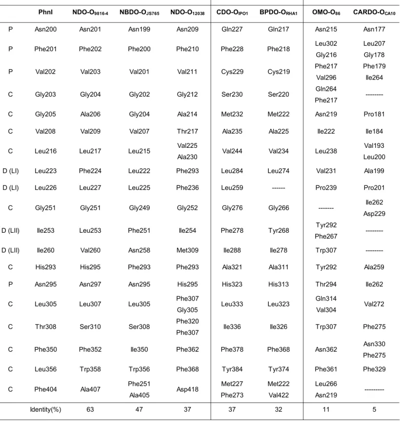
Sequence identity of structurally analogous residues lining the catalytic pocket for different dioxygenases of known crystallographic structure shows that PhnI presents the highest sequence identity with NDO-O9816 and the lowest with carbazole-1-9α- dioxygenase from
P. resinovorans strain CA10 (CARDO-OCA10) [17]. The classification presented in Table
1, based on α-subunit sequence homology and substrate specificity, is consistent with current classifications [18]. The PhnI catalytic pocket represented in Fig. 3 can be divided into three regions, distal, central and proximal, depending on the distance to the mononuclear Fe atom (Table 1). Compared to other dioxygenases, the PhnI pocket is rather uniform in shape and does not present any kinks or torsions as was found for example for BPDO-ORHA1 [15]. Residues Phe 350 and Phe 404 in the central region are expected to
affect the topology of the catalytic pocket and consequently select the shape and form of allowed substrates. Accordingly, the replacement of Phe 352 in NDO-O9816-4 by smaller
amino acids significantly altered the regiospecifity of the reaction products [19]. In NDO-O9816-4 Phe 404 is replaced by Ala 407. Further differences, observed at positions 308 and
356, contribute to enlarge the catalytic pocket of PhnI compared to that of NDO-O9816-4.
Indeed, Leu 356 in PhnI is replaced by a bulky aromatic residue (Trp or Phe) in naphthalene dioxygenases (Table 1).
Residues in the distal region seem to exert a greater influence in selecting the size and shape of allowed substrates. In PhnI, most significant are residues Leu 223 and Leu 226 located on loop LI, and residues Ile 253 and Ile 260 on loop LII. In naphthalene dioxygenases, NDO-O9816 andNDO-O12038, Leu 223, is substituted by a bulkier and less
flexible phenylalanine residue (Table I). The diversity in residues structurally equivalent to Ile 260 in PhnI must relate to the different substrate specificity observed between the members of the naphthalene dioxygenase family.
Substrate specificity. To explore the substrate specificity of PhnI from a structural
point of view, 2, 3, 4 and 5 ring PAHs were modeled into the catalytic pocket of the refined structure (PDB access code 2CKF). The structures of the enzyme-substrate complexes described for NDO-O9816 and BPDO-ORHA1 (PDB access codes 1O7G and
1ULJ), were first used to fit and adjust the position of naphthalene and biphenyl substrates in the PhnI catalytic pocket. As the pocket is narrower in the proximal region (Fig.4), modeling indicated that the two-ring substrates would be locked in a single position consistent with the finding that PhnI hydroxylates naphthalene and biphenyl, respectively, in positions 1,2- and 2,3- [5]. Phenanthrene, a three ring angular molecule, could theoretically be hydroxylated in positions 1,2-, 3,4-, or 9,10-. However, due to steric constraints imposed by the PhnI pocket, only one position, which would bring the C3 and C4 carbon atoms close to the active Fe atom site, is allowed. This analysis corroborates enzymatic assays for which cis-3,4- phenanthrene dihydrodiol was the only product detected [5].
The five ring PAH benzo[a]pyrene can only fit into the PhnI catalytic pocket in a single orientation. Minor conformational changes are assumed in modeling indicating that the side chains of Leu 223 and Phe 350 need to be rotated if the substrate is to fit in the pocket. In this orientation, shown in Fig. 4, benzo[a]pyrene would be hydroxylated in position 9,10-. In fact 9,10-cis-dihydrodiol- benzo[a]pyrene is the only product observed as the
result of enzymatic assays [5]. On the other hand, benzo[a]anthracene (BaA), a four ring PAH, was found to be attacked at positions 1,2-, 8,9- and 10,11- with cis-dihydrodiols 1,2- and 10,11- presenting the highest yields. Substrate orientation leading to an hydroxylation on the 1,2- position would require a minimal rearrangement of residues in the catalytic pocket, involving only side chain conformational changes of Leu 223 in loop LI. Hydroxylation on carbons C10 and C11 would occur for a position of the substrate requiring rearrangements of both Leu 223 and Phe 350 side chains. Conformational changes of residues 223, 253 and 404 should take place to allow hydroxylation of BaA at the less favorable 8,9- position. These predictions are in accordance with the catalytic properties of the enzyme, since it was found to produce much less of the 8,9-dihydrodiol [5].
Phe 350 and Phe 404 in the central region contribute to the regio-specificity and to the PhnI pocket shape. Phe 350 is conserved in NDO-O9816-4 and in the naphthalene
dioxygenase from Rhodococcus sp. strain NCIMB 12038 (NDO-O12038) [7], while Phe 404
is replaced by Ala 407 in NDO-O9816-4 and by Asp 418 in NDO-O12038. In NDO-O9816-4,
mutations of Phe 352 (Phe 350 in PhnI) into a valine and a leucine resulted in altered regio-specificity of the enzyme. These mutations altered the pocket topology, so that other orientations of the biphenyl and phenanthrene substrates were allowed [20]. The pocket size and shape might also influence the effectiveness of the reaction, by locking the substrate in the right position before catalysis. Replacement of Trp 358 by an alanine in NDO-O12038 resulted in inefficient transformation of naphthalene, because the bulky side
interesting to note all RHDs of known structure have an aromatic residue at a position equivalent to Trp 358, except PhnI, which has a leucine in the corresponding position.
In conclusion, PhnI is endowed with a remarkable broad specificity towards high molecular weight PAHs, which might be explained by the shape and size of its substrate binding pocket. The PhnI pocket was found to be at least 2 Å longer and wider at the entrance a unique feature between dioxygenases of known structures certainly allowing the five-ring benzo[a]pyrene to bind to the catalytic Fe. Modeling of various PAHs showed that Phe 350 in the central region of the pocket is essential for the regio- and substrate specificity, while Leu 223 and Ile260 in the distal region contribute to the specificity of high molecular weight PAHs. Further studies involving replacements of specific residues of the substrate-binding pocket by site-directed mutagenesis should bring new insights into the role of these residues in the catalytic activity of the enzyme.
Acknowledgements
We would like to thank the staff of the National Synchrotron Light Source, Brookhaven National Laboratory for their continuous support. The NSLS is supported by the U.S. Department of Energy, Office of Science, Office of Basic Energy Sciences, under Contract No. DE-AC02-98CH10886. The NIGMS East Coast Structural Biology Facility, the X6A beam line, is funded under contract # GM-0080.
References
[1] C.S. Butler, J.R., Mason, Structure–function analysis of the bacterial aromatic ring-hydroxylating dioxygenases, Adv. Microb. Physiol. 38 (1997) 47-84.
[2] A.L. Juhasz, R. Naidu, Bioremediation of high molecular weight polycyclic aromatic hydrocarbons: a review of the microbial degradation of benzo[a]pyrene, Int. Biodet. & Biodegr. 45 (2000) 57-88.
[3] R. Kanaly, S. Harayama, Biodegradation of High-Molecular-Weight Polycyclic Aromatic Hydrocarbons by Bacteria, J. Bacteriol. 182 (2000) 2059-2067.
[4] S. Demaneche, C. Meyer, J. Micoud, M. Louwagie, J.C. Willison, Y. Jouanneau, Identification and functional analysis of two aromatic-ring-hydroxylating dioxygenases from a Sphingomonas strain that degrades various polycyclic aromatic hydrocarbons, App. Environment. Microbiol. 70 (2004) 6714-6725.
[5] Y. Jouanneau, C. Meyer, J. Jakoncic, V. Stojanoff, J. Gaillard, Characterization of a Naphthalene Dioxygenase Endowed with an Exceptionally Broad Substrate Specificity toward Polycyclic Aromatic Hydrocarbons, Biochemistry 45 (2006) 12380-12391.
[6] B. Kauppi, K. Lee, E. Carredano, R.E Parales, D.T. Gibson, H. Eklund, S. Ramaswamy, Structure of an aromatic-ring-hydroxylating dioxygenase-naphthalene 1,2-dioxygenase, Structure 6 (1998) 571–586.
[7] L. Gakhar, Z.A. Malik, C.C.R. Allen, D.A. Lipscomb, M.J. Larkin, S. Ramaswamy, Structure and Increased Thermostability of Rhodococcus sp. Naphthalene 1,2-Dioxygenase, J. Bacteriol. 197 (2005) 7222-7231.
[8] D.J. Ferraro, L. Gakhar, S. Ramaswamy, Rieske business: Structure–function of Rieske non-heme oxygenases, Biochemical and Biophysical Research Communications 338 (2005) 175–190.
[9] N.E. Chayen, A novel technique to control the rate of vapour diffusion, giving larger protein crystals, J. Appl. Cryst. 30 (1997) 198-202.
[10] M. Allaire, M. Aslantas, A. Berntson, L. Bermann, S. Cheung, B. Clay, R. Greene, J. Jakoncic, E. Johnson, C.C. Kao, A. Lenhard, S. Pjerov, D.P. Siddons, W. Stober, V. Venkatagiriyappa, Z. Yin, V. Stojanoff, Synchrotron Radiation News 16 (2003) 20-25.
[11] X. Dong, S. Fushinobu, E. Fukuda, T. Terada, S. Nakamura, K. Shimizu, H. Nojiri, T. Omori, H. Shoun, T. Wakagi, Crystal structure of the terminal oxygenase component of cumene dioxygenase from Pseudomonas fluorescens IP01, J. Bacteriol. 187 (2005) 2483-2490.
[12] G.N. Murshudov, A.A. Vagin, E.J. Dodson, Refinement of Macromolecular Structures by the Maximum-Likelihood Method, Acta Cryst. D53 (1997) 240-255.
[13] P. Emsley, K. Cowtan, Coot: Model-Building Tools for Molecular Graphics, Acta. Cryst. D60 (2004) 2126-2132.
[14] J. Liang, H. Edelsbrunner, C. Woodward (1998) Anatomy of Protein Pockets and Cavities: Measurement of Binding Site Geometry and Implications for Ligand Design, Protein Science, 7, 1884-1897.
[15] Y. Furusawa, V. Nagarajan, M. Tanokura, E. Masai, M. Fukuda, T. Senda, Crystal Structure of the Terminal Oxygenase Component of Biphenyl Dioxygenase Derived from
Rhodococcus sp. Strain RHA1, J. Mol. Biol. 342 (2004) 1041-1052.
[16] B.M. Martins, T. Svetlitchnaia, H. Dobbek, 2-Oxoquinoline 8-Monooxygenase Oxygenase Component: Active Site Modulation by Rieske-[2Fe-2S] Center Oxidation/Reduction, Structure 13 (2005) 817-824.
[17] H. Nojiri, Y. Ashikawa, H. Noguchi, J.W. Nam, M. Urata, Z. Fujimoto, H. Uchimura, T. Terada, S. Nakamura, K. Shimizu, T. Yoshida, H. Habe, T. Omori, Structure of the terminal oxygenase component of angular dioxygenase, carbazole 1,9a-dioxygenase, J. Mol. Biol. 351 (2005) 355-370.
[18] Nam, J.W., Nojiri, H., Yoshida, T., Habe, H., Yamane, H., Omori, T. (2001) New Classification System for Oxygenase Components Involved in Ring-Hydroxylating Oxygenations, Biosci. Biotechnol. Biochem. 65, 254-263
[19] R.E. Parales, The role of active-site residues in naphthalene dioxygenase, J. Ind. Microbiol. Biotechnol. 30 (2003) 271–278.
[20] R.E. Parales, S.M. Resnick, C. Yu, D.R. Boyd, N.D. Sharma, D.T. Gibson, Regioselectivity and Enantioselectivity of Naphthalene Dioxygenase during Arene cis-Dihydroxylation: Control by Phenylalanine 352 in the α−Subunit, J. Bacteriol. 182 (2000) 5495-5504.
[21] R. Friemann, M.M. Ivkovic-Jensen, D.J. Lessner, C. Yu, D.T. Gibson, R.E. Parales, H. Eklund, S. Ramaswamy, Structural Insight Into the Dioxygenation of Nitroarene Compounds: The Crystal Structure of Nitrobenzene Dioxygenase, J. Mol. Biol. 348 (2005) 1139-1151.
[22] W.L. DeLano, DeLano Scientific, San Carlos, CA, USA, 2002.
[23] R.M. Esnouf, Journal of Molecular Graphics 15 (1997) 132-134.
[25] E.A. Merritt, M.E. Murphy, Raster3D Version 2.0: A Program for Photorealistic Molecular Graphics, Acta Cryst. D50 (1994) 869-873.
Table 1. Residues lining the catalytic pocket of dioxygenases of known structure.
(footnote) Relative position to the catalytic Fe: D, distal; C, central and P, proximal. (--) no structurally equivalent residues were observed. Substrate free structures were used in the comparison [6,16,7,12,15,17,18].
Figure Legends
Fig. 1. Structure of the αβheterodimer of PhnI: the α-subunit is shown in grey, catalytic domain, and in pink, Rieske domain, the β-subunit is shown in yellow. The surface of the substrate binding pocket is shown in the foreground; loops LI and LII are indicated. The Rieske domain of the adjacent α-subunit is shown in cyan. This view emphasizes important features of the enzyme, especially the proximity of the 2Fe-2S cluster and the mononuclear Fe at the interface between two adjacent α-subunits. The figure was produced with Pymol [22].
Fig. 2. Close view of the active site showing the residual positive density modeled as an
indole molecule. Four maps are shown: 2fo-fc contoured at the 1σ level, in grey and the fo-fc at the +3σ, ingreen after addition of the indole molecule; and the respective Omit maps, 2fo-fc at 1σ, in cyan and fo-fc at 3σ, in red. fo-fc maps contoured at the -3σ leveldid not present any residual density and are not shown. In this configuration, the C3 atom of indole is located 2 Å from the closest water molecule, in red, which is 2.6 Å from the mononuclear Fe atom shown in green and at 2.2 Å from the second coordinated water molecule. The figure was produced with BOBSCRIPT [23,24] and Raster3D [25].
Fig. 3. The PhnI active site with a BaP substrate modeled in the catalytic pocket. (a)
Residues shown in front, relative to the substrate (in green) are labeled in black and residues in the back are labeled in grey. With the exception of Gly 205, only side chains are represented. The mononuclear Fe is represented as a red ball. (b) Surface plot of the PhnI substrate cavity showing the substrate in the same orientation exposing C9 and C10 towards the mononuclear iron.
Fig. 4. The catalytic pocket for PhnI (upper panel), NDO-O9816-4(central panel)and
BPDO-ORHA1 (lower panel), is shown on the left in its substrate free form and on the right for the
substrate bound enzyme complexes. For each of the dioxygenases represented the comparison between the left and right panels shows the changes in the catalytic pocket upon substrate binding. The structures for NDO-O9816-4 and BPDO-ORHA1 on the right were
obtained using the coordinates of the substrate-bound enzyme complexes (PDB access code 1O7G and 1ULJ, respectively). For PhnI, the cavity shown on the right was obtained
after rotation of side chains of residues, Leu 223 and Phe 350, taking into account steric constrains due to van der Waals contacts. With these minor conformational changes BaP can only fit in the PhnI pocket in the orientation shown. In this orientation BaP would be hydroxylated in position 9,10- consistent with biochemical assays [5]. The figure was produced with Pymol [22].
Table 1. Residues lining the catalytic pocket of dioxygenases of known structure.
GroupI GroupII GroupIII
PhnI NDO-O9816-4 NBDO-OJS765 NDO-O12038 CDO-OIPO1 BPDO-ORHA1 OMO-O86 CARDO-OCA10
P Asn200 Asn201 Asn199 Asn209 Gln227 Gln217 Asn215 Asn177
P Phe201 Phe202 Phe200 Phe210 Phe228 Phe218 Leu302
Gly216
Leu207 Gly178
P Val202 Val203 Val201 Val211 Cys229 Cys219 Phe217
Val296
Phe179 Ile264
C Gly203 Gly204 Gly202 Gly212 Ser230 Ser220 Gln264
Phe217 ---
C Gly205 Ala206 Gly204 Ala214 Met232 Met222 Asn219 Pro181
C Val208 Val209 Val207 Thr217 Ala235 Ala225 Ile222 Ile184
C Leu216 Leu217 Leu215 Val225
Ala230 Val244 Val234 Leu238
Val193 Leu200
D (LI) Leu223 Phe224 Leu222 Phe293 Leu284 Leu274 Val231 Ala199
D (LI) Leu226 Leu227 Leu225 Phe236 Leu259 --- Pro239 Pro201
C Gly251 Gly251 Gly249 Gly252 Gly276 Gly266 --- Ile262
Asp229
D (LII) Ile253 Leu253 Phe251 Ile254 Phe278 Tyr268 Tyr292
Phe267 ---
D (LII) Ile260 Val260 Asn258 Met309 Ile288 Ile278 Trp307 ---
C His293 His295 Phe293 Phe293 Ala321 Ala311 Tyr292 Ala259
P Asn295 Asn297 Asn295 His295 His323 His313 Thr294 Ile262
C Leu305 Leu307 Leu305 Phe307
Gly305 Leu333 Leu323
Gln314
Val304 Val272
C Thr308 Ser310 Ser308 Phe320
Phe307 Ile336 Ile326 Trp307 Phe275
C Phe350 Phe352 Ile350 Phe362 Phe378 Phe368 Asn362 Asn330
Phe275 C Leu356 Trp358 Trp356 Phe368 Tyr384 Tyr374 Phe361 Phe329
C Phe404 Ala407 Phe251
Ala405 Asp418 Met227 Phe273 Met222 Val422 Leu266 Asn219 --- Identity(%) 63 47 37 37 32 11 5 Table 1
(Table 1) Relative position to the catalytic Fe: D, distal; C, central and P, proximal. (--) no structurally equivalent residues were observed. Substrate free structures were used in the comparison [6,16,7,12,15,17,18].