HAL Id: inserm-03124101
https://www.hal.inserm.fr/inserm-03124101
Submitted on 28 Jan 2021
HAL is a multi-disciplinary open access
archive for the deposit and dissemination of
sci-entific research documents, whether they are
pub-lished or not. The documents may come from
teaching and research institutions in France or
abroad, or from public or private research centers.
L’archive ouverte pluridisciplinaire HAL, est
destinée au dépôt et à la diffusion de documents
scientifiques de niveau recherche, publiés ou non,
émanant des établissements d’enseignement et de
recherche français ou étrangers, des laboratoires
publics ou privés.
Human Hemoglobin: Interaction with α-Hemoglobin
Stabilizing Protein (AHSP) and Heme Molecule
Elisa Domingues-Hamdi, Corinne Vasseur, Jean-Baptiste Fournier, Michael
Marden, Henri Wajcman, Véronique Baudin-Creuza
To cite this version:
Elisa Domingues-Hamdi, Corinne Vasseur, Jean-Baptiste Fournier, Michael Marden, Henri Wajcman,
et al.. Role of α-Globin H Helix in the Building of Tetrameric Human Hemoglobin: Interaction with
α-Hemoglobin Stabilizing Protein (AHSP) and Heme Molecule. PLoS ONE, Public Library of Science,
2014, 9 (11), pp.e111395. �10.1371/journal.pone.0111395�. �inserm-03124101�
Role of
a-Globin H Helix in the Building of Tetrameric
Human Hemoglobin: Interaction with
a-Hemoglobin
Stabilizing Protein (AHSP) and Heme Molecule
Elisa Domingues-Hamdi1, Corinne Vasseur1, Jean-Baptiste Fournier1¤, Michael C Marden1, Henri Wajcman2, Ve´ronique Baudin-Creuza1*
1 Institut National de la Sante´ et de la Recherche Me´dicale (Inserm) U779, Universite´ Paris XI, Paris, France, 2 INSERM U955, Universite´ Paris Est Cre´teil (UPEC), Hoˆpital Henri Mondor, Paris, France
Abstract
Alpha-Hemoglobin Stabilizing Protein (AHSP) binds to a-hemoglobin (a-Hb) or a-globin and maintains it in a soluble state until its association with the b-Hb chain partner to form Hb tetramers. AHSP specifically recognizes the G and H helices of a-Hb. To investigate the degree of interaction of the various regions of the a-globin H helix with AHSP, this interface was studied by stepwise elimination of regions of the a-globin H helix: five truncated a-Hbs a-Hb1-138, a-Hb1-134, a-Hb1-126, a-Hb1-123, a-Hb1-117 were co-expressed with AHSP as two glutathione-S-transferase (GST) fusion proteins. SDS-PAGE and Western Blot analysis revealed that the level of expression of each truncated a-Hb was similar to that of the wild type a-Hb except the shortest protein a-Hb1-117 which displayed a decreased expression. While truncated Hb1-138 and GST-a-Hb1-134 were normally soluble; the shorter globins GST-a-Hb1-126 and GST-a-Hb1-117 were obtained in very low quantities, and the truncated GST-a-Hb1-123 provided the least material. Absorbance and fluorescence studies of complexes showed that the truncated a-Hb1-134 and shorter forms led to modified absorption spectra together with an increased fluorescence emission. This attests that shortening the H helix leads to a lower affinity of the a-globin for the heme. Upon addition of b-Hb, the increase in fluorescence indicates the replacement of AHSP by b-Hb. The CO binding kinetics of different truncated AHSPWT/a-Hb complexes showed that these Hbs were not functionally normal in terms of the
allosteric transition. The N-terminal part of the H helix is primordial for interaction with AHSP and C-terminal part for interaction with heme, both features being required for stability of a-globin chain.
Citation: Domingues-Hamdi E, Vasseur C, Fournier J-B, Marden MC, Wajcman H, et al. (2014) Role of a-Globin H Helix in the Building of Tetrameric Human Hemoglobin: Interaction with a-Hemoglobin Stabilizing Protein (AHSP) and Heme Molecule. PLoS ONE 9(11): e111395. doi:10.1371/journal.pone.0111395 Editor: Maria Gasset, Consejo Superior de Investigaciones Cientificas, Spain
Received July 2, 2014; Accepted September 23, 2014; Published November 4, 2014
Copyright: ß 2014 Domingues-Hamdi et al. This is an open-access article distributed under the terms of the Creative Commons Attribution License, which permits unrestricted use, distribution, and reproduction in any medium, provided the original author and source are credited.
Data Availability: The authors confirm that all data underlying the findings are fully available without restriction. All relevant data are within the paper and its Supporting Information files.
Funding: This work was supported by the Institut National de la Sante´ et de la Recherche Me´dicale and the University Paris XI. The funders had no role in study design, data collection and analysis, decision to publish, or preparation of the manuscript.
Competing Interests: The authors have declared that no competing interests exist. * Email: veronique.baudin-creuza@inserm.fr
¤ Current address: CNRS, UMR 8227, Integrative Biology of Marine Models, Station Biologique de Roscoff, Roscoff, France
Introduction
The human adult hemoglobin (Hb A) is a tetrameric protein containing two a- and two b-chains, each associated with a heme molecule. It is now admitted that the a and b subunits do not associate spontaneously to form a2b2 Hb. The scheme of Hb
biosynthesis is the following: firstly Alpha hemoglobin stabilizing protein (AHSP), the a-Hb chaperone, binds to the a-globin (without heme molecule) or a-Hb (with heme), maintaining it in a soluble state [1], secondly AHSP is replaced by b-hemoglobin chains (b-Hb) to form an ab dimer, and tetramers in a further step [2,3]. Unlike the free b-Hb which are soluble and form homologous tetramers, freshly synthesized a-Hb chains are unstable molecules. Spontaneously they form hemichromes, which precipitate and generate reactive oxygen species (ROS) within the erythrocyte precursors of the bone marrow leading to apoptosis and ineffective erythropoiesis [4]. AHSP is a 102 amino-acid protein, synthesized at a high concentration in the erythroid
precursors, which forms a stable soluble heterodimer with a-Hb [1]. This a chaperone specifically recognizes the G and H helices of a-Hb, a region which also binds to the b-Hb subunit [5,6].In vitro, it has been demonstrated that AHSP inhibits ROS production by a-Hb and it was hypothesized that AHSP might protect erythroid cells from a-Hb toxicity by maintaining this subunit in a stable state prior to its incorporation into Hb A [7]. At leastin vitro, AHSP facilitates folding of newly formed a-chains and promotes Hb A assembly [7]. More recently we have shown that redox properties and stability of a-Hb are modified by AHSP [8]. The presence of AHSP facilitates reduction of the oxidized a-Hb chain which could trigger its release from AHSP toward its final Hb b-chain partner producing functional ferrous Hb-tetramers [8]. These different data emphasize the important role of AHSP in Hb biosynthesis.
A defect in the production of one of the globin chains in the red blood cell (RBC) precursors results in various degrees of anemia and thalassemia syndromes, for example an a-thalassemia in the
case of reduced a-globin production [9]. In human, the b-globin chain is encoded by a single gene localized on each of the chromosomes 11 and the a-globin chain by two genes carried by each copy of chromosome 16. Studies in mice model have shown that AHSP could play a role in Hb disorders. In mice, the loss of AHSP (AHSP2/2) led to reticulocytosis indicating a shortened erythrocyte half-life, abnormal erythrocyte morphology with intracellular denatured Hb inclusion and increased ROS with subsequent cellular oxidative damage as observed in thalassemias [1]. In addition, the loss of AHSP in b-thalassemic mice exacerbated thalassemic syndrome [10] and its loss in a-thalassemia silent carrier a-/aa, (3 functional a-genes) led to more severe erythroid pathologies (less hemoglobin, more reticulocytes) than in only a-thalassemia silent carrier mice.
In human a-thalassemias, the reduced availability of a-globin chains may be due to several molecular mechanisms, the most common being gene deletion [11]. However, in the past few years, the number of a chain Hb variants discovered with point mutations, short deletions, or insertions which resulted in a-thalassemia phenotypes, has increased [11]. Variants in which the contact region between a and b-subunits is altered, were often considered as the cause of these a-thalassemia phenotypes. The discovery of AHSP allows to consider an alternate explanation of some non-deletional a-thalassemias by impairing interaction between a-Hb and AHSP (Figure 1A). In a first study, it has been reported that two elongated a-Hb variants, Hb Constant Spring and Pakse´ associated with an a-thalassemia phenotype led to an impaired binding to AHSP [12]. Beside the elongated chains, point mutations with a single amino acid change were reported also to cause this clinical presentation. Our group has shown that Hb Groene Hart [a119(H2)ProRSer], a variant common in North Africa and in Southern Italy, causes a defect in the association with AHSP and behaves as an a+-thalassemia with two functional a-genes [13]. In a similar way, Lacerraet al described Hb Foggia [a117(GH5)Phe RSer] an a-2 gene variant that led to an a-thalassemia phenotype in the carriers [14]. The mutated mRNA a-globin of this variant was present at normal level in the reticulocytes but no abnormal chains were detected suggesting that it was synthesized but very rapidly degraded likely due to impaired interaction with AHSP [14]. Recent studies, from our group and from others, demonstrated a similar defect in AHSP binding for an increasing number of a-Hb variants [15,16]. The region 95–137 of a-Hb, composed by G and H helices, plays a predominant role in the interaction between a-Hb and AHSP [6]. In addition, the region 103–124 allows together the binding to AHSP and to b-Hb. Some amino-acids interact with AHSP, others only with b-Hb within an a1b1 dimer [17] and three (103, 117, 119) are involved in both contacts as shown in the figure 1A.
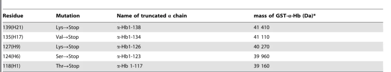
In this work, we investigated the minimal length of the a-chain helix H allowing in the first step the formation of AHSPWT/a-Hb complex and in the second one that of the stable tetramer. For this purpose, we engineered wild type a-Hb1-141 with a N-terminal Gly-Pro-Leu-Gly-Ser peptide (a-HbWT) and five truncated a-Hbs in which the H helix and the C-terminal were altered (Table 1). These truncated Hbs (Hb1-138, Hb1-134, Hb1-126, a-Hb1-123, a-Hb1-117) were selected from the list of a mutants already described (Table 2) [18]. They extended from the complete removal of helix H (a-Hb1-117) to the simple deletion of the three C-terminal residues located at the end of the helical structure (a-Hb1-138) (Table 1, Figure 1B). These molecules were engineered in the presence of wild type AHSP with a N-terminal Gly-Pro-Leu-Gly-Ser peptide (AHSPWT) using the co-expression vector that allows one to produce at high yield a-Hb as a complex with AHSPWT [19]. We studied their stability during the three
steps of the process: i) co-expression with AHSPWTin bacteria, ii) solubilization of the products and iii) their purification. We studied the consequences of the different truncations on the interaction between the resulting a-Hbs and AHSPWTor heme molecule by absorbance spectroscopy, circular dichroism (CD) and dynamic light scattering (DLS). The formation of a2b2 tetramers was
investigated by fluorescence studies and by kinetics of CO recombination after flash photolysis.
Results
Expression and purification of the truncated a-Hbs
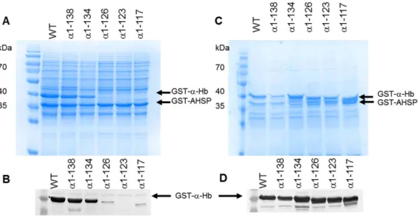
Five a-Hbs with shortened H helix were co-expressed with AHSPWT. The presence of the truncated a-Hbs was analyzed at three steps of protein production by SDS-polyacrylamide gel electrophoresis (SDS-PAGE) and Western blot analysis (Figure 2, Table 3). After 4 h induction, the whole cell lysate of the different truncated a-Hbs showed two major bands, 42 kDa and 38.6 kDa, corresponding to the GST-a-Hbs and GST-AHSPWT proteins, respectively. This pattern is similar to that obtained with the WT
Figure 1. H helix of human a-globin. (A) H helix sequence of the different truncated a-globins. The symbols indicate the amino-acids interacting with b-globin within the same ab dimer (+) and those interacting with the heme molecule (
N
) [14]. The (x) symbol indicates the amino-acids interacting with AHSP. (B) Three-dimensional view of a-Hb. The representation was obtained from crystallographic data of oxyHb (2HHB) using the Pymol software. The truncated regions of the H helix investigated in this work are represented in blue (a-Hb1-138), purple Hb1-134), red Hb1-126), orange Hb1-123) and grey (a-Hb1-117). The GH corner is shown in yellow.construct (Figure 2A). The identity of the various truncated GST-a-Hbs was attested by Western blotting using anti-a globin antibodies (Figure 2B). The protein expression level of these GST-a-Hbs was similar to that of the WT construct except for the truncated GST-a-Hb1-117 protein for which a clearly decreased intensity was observed (Table 3). The expected decreased size of the proteins (Table 1) for the GST-a-Hb1-126, GST-a-Hb1-123 and GST-a-Hb1-117 mutant was observed (Figure 2B).
After solubilization, only the 138 and GST-a-Hb1-134 were observed in the supernatant by SDS-PAGE analysis (Figure 3A) indicating their good solubility. These results were confirmed by Western blotting using anti-a globin antibodies (Figure 3B). To evaluate the amount of truncated GST-a-Hbs remaining in the pellet, the insoluble fraction was also analyzed by SDS-PAGE (Figure 3C). The GST-a-Hbs are clearly present but less abundant in the case of the truncated GST-a-Hb1-138, which was mostly present in the supernatant (Table 3). The same trend was observed by Western blotting analysis (Figure 3D), indicating that the truncated a-Hb1-126, a-Hb1-123 and a-Hb1-117 are expressed but are poorly soluble.
Finally, SDS-PAGE analysis after purification of the different soluble fractions is shown in Figure 4A. Two major bands, 42 kDa
and 38.6 kDa, were observed corresponding to the different GST-a-Hbs and GST-AHSPWT
proteins for the normal construct as well as for the truncated a-Hb1-138 and a-Hb1-134. In contrast, for the truncated a-Hb1-126, a-Hb1-123 and a-Hb1-117, no band corresponding to GST-a-Hb was visible. However, the Western blot analysis using anti-a antibodies revealed the presence of a band corresponding to GST-a-Hb (Figure 4B). Nevertheless, the intensity of the bands for a-Hb1-126 and a-Hb1-117 were drastically decreased and that of a-Hb1-123 was at the limit of detection. Thus, while the truncated a-Hb1-138 and a-Hb1-134 were purified in quantity, the truncated 126 and Hb1-117 were obtained in very low quantities and for the truncated a-Hb1-123 no further study was possible.
Spectroscopic characteristics of the complexes
Native Hb A and free a-Hb exhibit characteristic spectra attesting the presence of the heme molecule with Soret c band (415–420 nm), a and b absorbance peaks (540 and 571 nm) and d-band (344 nm) [20]. The spectra of the AHSPWT/truncated a-Hb complexes are shown in Figure 5. The relative contributions at 280 nm, a wavelength which is sensitive to the aromatic amino acids and heme, and at visible wavelengths which are essentially Table 1. Different truncated a-Hbs produced in E. coli with AHSP.
Residue Mutation Name of truncated a chain mass of GST-a-Hb (Da)* 139(H21) LysRStop a-Hb1-138 41 410
135(H17) ValRStop a-Hb1-134 41 110
127(H9) LysRStop a-Hb1-126 40 270
124(H6) SerRStop a-Hb1-123 39 960
118(H1) ThrRStop a-Hb 1-117 39 160 *Masses are equal to 42 602 Da and 38 665 Da for GST-a-HbWT
and GST-AHSPWT
, respectively. doi:10.1371/journal.pone.0111395.t001
Table 2. Natural variants in the G and H helices and C terminal extremity altering the sequence length of a-Hb.
Position Hb Name Affected a Mutation Length** G Helix
14 a2 a107 delT 132
H helix
14 Fez a1 a1-131 132
17 Senlis a1 a134 delT 136
15 Pak or Num Po a1 a132 insT TAARTAT 175 C-term Constant Spring a2 a142RGln TAAR CAA 172
Icaria a2 a142RLys TAARAAA 172
Koya Dora a2 a142RSer TAARTGA 172
Seal Rock a2 a142RGlu TAARGAA 172
Pakse´ a2 a142RTyr TAARTAT 172
Natal a2 a140R0 TACR TAA 139
Wayne a2 or a1 a139 delA 146
The data were collected from HbVar, the globin gene server (http://globin.cse.psu.edu/globin/hbvar/). These different mutations led to an a-thalassemic phenotype more or less pronounced.
*the production of a-globin is under the control of two genes a1 and a2-globin genes (HBA1 and HBA2 respectively) **Length expressed as number of amino-acid residues;
del, deletion; ins, insertion;
due to the heme contribution can provide information on the heminization state of truncated a-globin. The spectra of the AHSPWT/a-HbWT(solid black line in Figure 5) and AHSPWT/ native a-Hb complexes are similar [21]. The spectrum observed for AHSPWT/a-Hb1-138 (solid red line in Figure 5) is similar to that observed for the AHSPWT/a-HbWT complex, while the spectrum of the AHSPWT/a-Hb1-134 complex exhibits a decreased amplitude of the Soret band and an asymmetry of the a and b bands (black dashed line in Figure 5). These differences are more marked for AHSPWT/a-Hb1-126 complex with a Soret band decreased by a factor 2.7 and an asymmetry of the a and b bands (solid grey line in Figure 5). In the case of the AHSPWT /a-Hb1-117 complex, an abnormal UV-visible spectrum is observed with an absence of characteristic bands of the heme molecule showing that most of globin has not incorporated the heme molecule. These results indicate that the more H helix is truncated, the less heme molecule incorporates into the a-globin (Table 4).
Effects of truncation on secondary structure and thermal stability of AHSPWT/a-Hb1-138 and AHSPWT/a-Hb1-134
To assess the consequences of end extremity truncation on the structure and stability of a-Hb chain, we have first analyzed by CD spectroscopy the AHSPWT/a-Hb1-138 and AHSPWT /a-Hb1-134 complexes in the CO form. The far UV CD revealed that the AHSPWT/a-Hb1-138 and AHSPWT/a-Hb1-134 com-plexes display an a-helical content of 74% and 71%, respectively. These a-helical contents are slightly higher than for AHSPWT/ native a-Hb (67%) or AHSPWT/a-HbWT(65%) (Table 4) and can
Figure 2. Co-expression of truncated a-Hbs and AHSPWT in E.coliBL21(DE3) cells. After 4 h induction by 0.2 mM IPTG, the E. coli cells containing the different pGEX-a-AHSP were lysed and total cell lysates were subjected to SDS-PAGE (12%) analysis (A) and Western Blotting using anti-a globin antibodies (B). All samples were prepared by boiling in SDS loading buffer. In all gels, GST-a-HbWT and
GST-AHSPWTare indicated by arrows. PAGE rulerTMprestained protein ladder
(Fermentas Thermo Fisher Scientific, Waltham, MA, USA) was analyzed in the first lane.
doi:10.1371/journal.pone.0111395.g002 Table 3. Presence of different truncated a -Hbs observed by SDS-PAGE or by Western blot. SDS-PAGE Western Blot truncated a chain Expression Solubilization Purification Expression Solubilization Purification soluble fraction insoluble fraction s oluble fraction insoluble fraction a -Hb WT * + + + + + + + + + + + + + + a -Hb1-138 + + + + + + + + + + + + + + a -Hb1-134 + + + + + + + + + + + + + + a -Hb1-126 + + 2 + + 2 + + + + + + a -Hb1-123 + + 2 + + 2 + + 2 + + 2 a -Hb1-117 + 2 + + 2 + + + + + ++ : normal p resence; + : d ecreased presence; 2 : a bsence or highly d ecreased presence. *a -Hb WT is recombinant w ild type a -Hb1-141 with a N -terminal Gly-Pro-Leu-Gly-Ser peptide. doi:10.1371/journal.pone. 0111395.t003
be explained by the absence of three C-terminal residues following the helical structure.
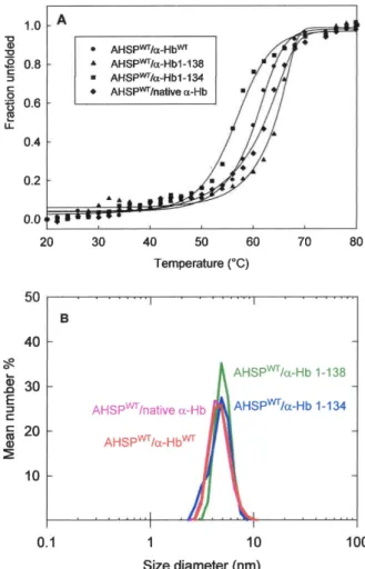
The instability of the protein was measured by thermal denaturation using the CD and by determination of the melting temperature (Tm) for which 50% of molecules are unfolded. The AHSPWT/a-HbWTand AHSPWT/native a-Hb complexes exhibit a Tm of 60uC and 63uC, respectively (Figure 6A). For AHSPWT
/ a-Hb1-138 complex, the Tm (63uC) is close to that observed for
WT recombinant complex or for isolated native a-Hb (64uC). In contrast, a decreased Tm value for AHSPWT/a-Hb1-134 complex (56uC) was found (Table 4).
Secondly, we investigated the effect of truncation on the hydrodynamic diameter of complexes based on DLS. In the same experimental conditions, AHSPWT, small protein consisting of an elongated a-helix bundle [5] exhibits a hydrodynamic diameter about 3.5 nm, while that for the more globular tetrameric Hb is about 6 (data not shown). It is important to note that AHSPWT/ native a-Hb and AHSPWT/a-HbWTcomplexes exhibit a similar size
of hydrodynamic diameter, 4.3 and 4.5 nm respectively (Figure 6B) although there are 5 additional amino acids (Gly-Pro-Leu-Gly-Ser) for each recombinant protein. For AHSPWT/a-Hb1-138 and
Figure 3. Effect of truncation on the solubility of various truncated a-Hbs. The induced E.coli BL21(DE3) cells containing different pGEX-a-AHSP plasmids were disrupted. After centrifugation, soluble fractions were analyzed by (A) SDS-PAGE (12%) and by (B) Western Blotting using anti-a globin antibodies. The insoluble fractions were analyzed by (C) SDS-PAGE (12%) and by (D) Western Blotting using anti-a globin antibodies. Page rulerTMprestained protein ladder (Fermentas Thermo Fisher Scientific) was in the first lane.
doi:10.1371/journal.pone.0111395.g003
Figure 4. Yield of various truncated a-Hbs after purification. The different soluble fractions obtained after disruption of induced cells was applied on Glutathione Sepharose 4B in PBS (150 mM NaCl, 10 mM Na2HPO4, pH 7.4). After washing, various truncated GST-AHSP
WT
/GST-a-Hb complexes were eluted (elution buffer: 50 mM Tris-HCl, 20 mM reduced glutathione pH 8.0) and analyzed by (A) SDS-PAGE (12%) and (B) Western Blotting using anti-a globin antibodies.
doi:10.1371/journal.pone.0111395.g004
Figure 5. Absorption spectra of truncated AHSPWT/a-Hbs complexes. The spectra are represented in black for AHSPWT/a-HbWT
complex, in red for of AHSP/a-Hb1-138 complex, in black dashed line for AHSP/a-Hb1-134 complex and in grey for AHSP/a-Hb 1-126 complex. The measurements were performed in PBS pH 7.4 at 25uC. doi:10.1371/journal.pone.0111395.g005
AHSPWT/a-Hb1-134 complexes, one observes a slight increase of the size of hydrodynamic diameter indicating that the truncation of the C-terminal end does not modify the size of these complexes compared to those observed for WT complex.
Truncation effects on fluorescence of various complexes
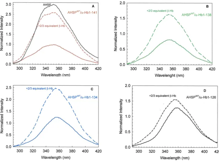
The fluorescence energy transfer technique was used to investigate the interaction between AHSP and truncated a-Hb within the AHSPWT/a-Hb complexes. AHSP has a single Trp (position 44) and exhibits a fluorescence spectrum typical of an exposed Trp (solide black line in Figure 7A). a-Hb has also a single Trp at position 14 (A2 in the A helix) but the a-Hb fluorescence is highly quenched by its heme group. It is important to note that there is no difference in fluorescence emission spectra between AHSPWT/native a-Hb and AHSPWT/a-HbWT complexes as illustrated in the Figure S1. In Figure 7 are illustrated the different emission spectra obtained for different complexes after normali-zation with respect to the emission maxima obtained with the WT
Table 4. Characteristics of different truncated AHSP WT /a -Hb complexes. Spectroscopy CO binding kinetics CD Spectroscopy Quenching of Fluorescence** UV Visible + b -Hb* a -helix (%) T m + b -Hb Controls AHSP WT ++ 22 2 60 55.6–58 no no native a Hb AHSP WT /native a -Hb AHSP WT /a -Hb WT ++ ++ ++ +++ +++ +++ 100% R 1 00% I 100% IR and T state R and T state 56% R , 3 2% T 71 67 65 64 63 60 ++++ +++ +++ ++++ + + AHSP WT /a -Hb1-138 ++ +++ 97% R 95% R , 5 % I 74 63 +++ + AHSP WT /a -Hb1-134 ++ ++ 10% I, 8 7% R 8 4% R% 12% I 7 1 5 6 ++ + AHSP WT /a -Hb1-126 ++ + 19% I, 8 1% R 8 0% R, 20% I n d n d ++ ++ AHSP WT /a -Hb1-117 +++ no 20% I, 8 0% R 8 8% R, 12% I n d n d n d n d *The C O recombination kinetics were measured in the p resence of IHP. **The fluorescence signal o f g lobin a nd AHSP WT /a -Hb WT complex is highly quenched by the h eme molecule demonstrating the interaction between AHSP WT and a -Hb WT . doi:10.1371/journal.pone. 0111395.t004
Figure 6. Conformational Stability of truncated AHSPWT/a-Hb complexes. (A) Thermal unfolding curve measured by CD experi-ments. Fraction unfolded (fu) is versus temperature for AHSPWT/native a-Hb (¤), AHSPWT/a-HbWT(
N
), AHSPWT/a-Hb1-138, (m) and AHSPWT/a-Hb1-134 (&) complexes. The solid lines are a smooth curve (sigmoidal
representation) to help estimate Tm. Protein concentration was around 20 mM (on a heme basis) in 2.5 mM Na2HPO4, 37.5 mM NaCl buffer at
pH 7.4 in the presence of sodium dithionite at 1 mM. The change in ellipticity was recorded at 222.6 nm from 20 to 80uC with a heat rate of 1uC.min21
. (B) Hydrodynamic diameter for the complexes of AHSPWT with native a-Hb, a-HbWT, a-Hb1-138 and a-Hb1-134. Protein
concen-tration was around 20 mM (on a heme basis) in PBS. doi:10.1371/journal.pone.0111395.g006
complex. The emission spectrum of AHSPWT/a-HbWTcomplex is highly decreased compared to AHSPWT(solid red line and solid black line respectively, in Figure 7A), the quenching of AHSP fluorescence intensity demonstrating the interaction between AHSP and a-Hb [21]. Only the AHSPWT/a-Hb1-138 complex exhibits an emission spectrum with a slightly decreased fluores-cence signal compared to that AHSPWT/a-HbWTcomplex (solid line in Figure 7B). The AHSPWT/a-Hb1-134 and AHSPWT /a-Hb1-126 complexes (solid lines in Figure 7C and 7D) have emission spectra with an increased amplitude, due to less heme molecule and therefore less quenching. These data are in agreement with absorbance spectra results (Figure 5). For all truncated complexes, one observes a red shift of fluorescence indicating that the environment of Trp is more polar.
Addition of b-Hb subunits to the AHSPWT/a-HbWTcomplex (dashed line in Figure 7A) leads to an increase in fluorescence intensity demonstrating the release of the part of AHSPWTand the formation of dimeric (ab) Hb [21]. Addition of b-Hb to the AHSPWT/truncated a-Hb complexes leads to an increase in fluorescence intensity in all cases, as observed for WT complex (dashed lines in Figure 7 B,C,D). These results indicate that all
truncated a-Hb into the AHSPWT/a-Hb complexes are able to release AHSP for replacement by the b-Hb chains.
Effect of truncation on CO binding kinetics of various complexes
Tetrameric Hb presents CO binding kinetics including a rapid phase (CO association rate of 6mM21s21) and a slow phase (CO association rate of 0.2mM21s21) corresponding to the R and T states, respectively. Unlike isolated a- and b-Hb subunits, which display CO binding kinetics typical of the R-state, a-Hb in the presence of AHSP exhibits a rate of about 2mM21s21, interme-diate to the R and T states of tetrameric Hb, and was thus denoted as the I phase [21]. We measured the influence of a-Hb chain truncation on CO recombination kinetic of different complexes (Figure 8A). These measurements were made directly on GST-AHSPWT/GST-a-Hb complexes because the GST moiety does not interfere with the CO recombination kinetics [19,21]. Unlike AHSPWT/a-HbWTcomplex, the CO binding kinetic of different truncated complexes did not show mainly the intermediate phase but a rapid phase typical of R-state with a CO association rate of 6.5mM21s21. The a-Hb1-138 complex exhibits nearly 100% of
Figure 7. Fluorescence emission spectra of AHSPWT/a-Hb complexes before and after addition of b-Hb. (A). Fluorescence emission
spectra of AHSPWT/a-HbWT complex; (B). Fluorescence emission spectra of AHSPWT/a-Hb1-138 complex; (C). Fluorescence emission spectra of AHSPWT/a-Hb1-134 complex; (D). Fluorescence emission spectra of AHSPWT/a-Hb1-126 complex. The fluorescence emission spectrum of AHSPWTis
shown in the figure 7A (solid black line). The concentrations are around 3 mM (on a heme basis) in PBS. Each different emission spectrum of AHSPWT/ truncated a-Hb complex is normalized with respect to the emission maxima obtained for normal complex. The dashed lines illustrate the fluorescence emission spectra of different AHSPWT/a-Hb complexes after addition of b-Hb chains (2/3 equivalent).
the rapid phase. The other truncated a-Hb complexes display between 10% and 20% of the I phase (Figure 8A and Table 4).
After addition of native b-Hb and inositol hexaphosphate (IHP), an allosteric effector stronger than 2,3 diphosphoglycerate that increases the fraction of slow (T-state) phase, the kinetics for the WT complex become biphasic demonstrating the formation of functional Hb tetramers [21] with both R and T phases, corresponding to the two allosteric states. In contrast, for truncated a-Hb complexes, mainly the R phase was observed indicating the loss of allostery (Figure 8 B, Table 4).
Discussion
It is now admitted that normally a-Hb first binds AHSP, this chaperone maintaining the a-Hb in a soluble state until association with the b-Hb chain partner to form a2b2 tetramer [1,2,3].
Different abnormal a-Hbs are described as being unstable because leading to a low abundance of the abnormal Hbs in the RBCs [18]. The amino acid changes, deletions or insertions of some of these unstable Hbs are located in the G or H helices which are involved in the interaction with protein partners, AHSP or b-Hb. Different molecular mechanisms have been proposed to explain the instability of these mutated a-chains [22]. In the case of the
mutants with elongated chains (Table 2), it has been proposed that the mutated a-globin mRNA is unstable with a shortened life span contrasting with the normal globin mRNAs considered as being highly stable [23]. Another possibility is that the abnormal a chains are unable to properly bind AHSP by disruption of hydrophobic interactions between helices.
In this study, we investigated the interaction of the H helix of a-Hb with AHSP using as probe different natural a-a-Hb variants known as being unstable in the RBCs (Table 2). Instability of native a-Hb is well known [24] and in a previous study, we have shown that it is possible to express normal a-Hb alone as GST fusion protein, but it is not recovered after the solubilization step despite the presence of the GST moiety that can help to stabilize the folding of recombinant proteins [19]. So the presence of the protein partner is required to produce soluble recombinant a-Hb. Its co-expression with AHSP permits one to obtain the GST-a-Hb associated with AHSP in the heterodimer form (GST-AHSPWT/ GST-a-Hb) [19]. Thus the five a-Hb variants with shortened H helix were engineered in association with AHSP.
The SDS-PAGE results showed that the truncation of the a-globin chain did not alter the expression of the GST-a-Hb protein except in the case of GST-a-Hb1-117 where it was decreased (Figure 2). This normal expression of different GST-a-globins
Figure 8. Ligand binding studies of the various truncated GST-AHSPWT/GST-a-Hb complexes (A) and after addition of b-Hb in the
presence of IHP (B). The CO recombination kinetics reveal whether the protein complexes display the known rates for the classical R and T states, or the reference intermediate rate for the AHSPWT/a-HbWTcomplex. The truncated forms of a-Hbs complexed with AHSP show less of the I state,
indicating a modified interaction. Usually, the addition of b-Hb in the presence of IHP provokes a replacement of the AHSP by the b-Hb and leads to formation of Hb dimers and tetramers which display the usual slow T-state kinetics; however, the truncated forms displayed little of this allosteric form. Experimental conditions were 50 mM Bis-Tris buffer at pH 7.0, 100 mM CO at 25uC in absence or in presence of IHP at a final concentration of 1 mM. The concentration of different complexes on heme basis varies between 3.4 and 10.6 mM depending on the truncation.
suggests that their mRNAs are stable in BL21 (DE3) cells. An instability of mRNA for GST-a-Hb1-117 could be considered, but a further study would be required to confirm such a hypothesis.
After the step of solubilization, the truncated a-globin chains exhibit variable solubilities. When the globin has a length of 134 amino acids residue, the protein is mainly in the soluble form. Conversely when shortened to a length of 126 amino acids a-globin it can be does not permit to maintain soluble the truncated a-globin chains. Thus, SDS-PAGE and Western blotting analysis showed that when the H helix was truncated, less complex was formed. This reveals an impaired attachment or interaction between truncated a-Hb and AHSP. In the case of GST-a-Hb1-123, the absence of this protein after solubilization may be partially explained by the absence of residue 129 (H17) which interacts with AHSP (Figure 1A). Indeed we showed in a previous study that the substitution of Leu129 by Pro residue perturbs the interaction with AHSP and explains the absence of abnormal Hb in the RBCs and the a+-phenotype of the patient [16].
Another parameter that can enhance the stability, and facilitate the correct folding of the a apoglobin, is the presence of exogenous heme during expression. Indeed it has been demonstrated that the co-expression of bacterial membrane heme transport increase the rate and extent of the heme capture through the bacterial membranes [25]. It has been also shown in a wheat germ cell-free translation system that nascent a-globin having 86 amino acids can interact with hemin [26]. Consequently, in our expression conditions where hemin molecule is added in the same time than the inductor of globin synthesis, it is reasonable to assume that the truncated a-globins have enough amino acids to bind heme. Indeed the absorbance spectra reveal well the presence of heme for truncated a-Hb1-138, a-Hb1-134 and a-Hb1-126 complexes (Figure 5). While the AHSPWT/a-Hb1-138 exhibits a similar spectrum than this of WT complex, the AHSPWT/a-Hb1-134 exhibits some spectral modification and the AHSPWT/a-Hb1-126 complex presents a highly decreased amplitude of the Soret band indicating a low heminization. The spectrum of the truncated a-Hb1-117 does not exhibit the characteristic bands of the heme molecule (data not shown). Three amino acid residues in the H helix are in contact with the heme; the 129(H12) residue at the distal region and 132(H15) and 136(H19) residues at the proximal region (Figure 1A). In the truncated a-Hb1-126 and a-Hb1-123 where these three residues are deleted, the different results reflect the decreased interaction with heme. In addition for the two truncated a-Hbs, there is the loss of one contact (position 129 (H17)) with AHSP can explain the decreased quantity of purified proteins. However these different results do not explain why the shorter truncated a-Hb1-117 is obtained in sufficient quantity for some studies while for the truncated a-Hb1-123, we do not obtain this a-Hb after purification. One could hypothesize that in this last truncated a-Hb, the presence of only 6 residues of H helix including a proline residue is more destabilizing than the total absence of the H helix.
Study of the fluorescence signal is a good method to evidence modifications in the interactions between the different truncated a-Hbs, AHSP and heme. In a previous study, we have shown that quenching of the fluorescence signal of the single Trp residue of AHSP may be used as a probe for its binding to a-Hb chain.The heme group being an excellent energy acceptor, strongly quenches the globin fluorescence as well as that of interacting partner proteins [21]. The fluorescence studies clearly show an increase in fluorescence intensity emission for the AHSPWT/a-Hb1-134 and AHSPWT/a-Hb1-126 complexes compared to that observed for the WT complex (Figure 7C,D) and indicate that a fraction of the truncated a-Hb is not associated to the heme group and is present
in a apo-globin form. The studies of fluorescence properties of a-globin have shown that its fluorescence emission spectrum exhibited emission maxima at 33562 nm which is characteristic of an apolar environment [27]. This data is in agreement with crystallographic studies which showed that the Trp 14(A12) of the a-chain is an internal residue [28,29]. For AHSPWT
/a-Hb1-134 and AHSPWT/a-Hb1-126 complexes, a red shift is observed that may be due to the presence of apo a-globin but with a different structure of that observed for WT a-globin in particular the Trp 14(A12) will be in the more polar environment and this may be related to intramolecular structural rearrangements. These mod-ifications of the structure in AHSPWT/a-Hb1-134 can explain the
diminution of stability found by CD results. In addition, the loss of heme molecule may also have consequences in the fluorescence emission of the AHSP Trp. Increase of the fluorescence signal upon addition of b-Hb to the truncated a-Hbs demonstrates the release of AHSP and the further formation of dimers and tetramers. Finally results of the CO binding kinetics show that the truncated AHSPWT/a-Hb complexes do not exhibit the intermediate phase as observed for the AHSPWT/a-HbWT
(Figure 8A). The CO binding kinetics of the Hb tetramers incorporating the truncated a-Hbs revealed that these molecules were functionally abnormal (Figure 8B).
In summary, the deletion of the C-terminal extremity including the last three amino acids has no effect on the stability of the molecule. The end of helix H (134–138) is a region that does not interact with either AHSP or b-Hb subunits, so its deletion does not greatly influence the stability of a-globin. In contrast, the region 129-137 is involved in the interaction with heme; in particular the absence of the three amino acids (129, 132 and 136) as observed in the truncated 126, 123 and a-Hb1-117 leads to a decrease of heme incorporation and abnormal function of these a-globin chains. Finally the N-terminal extremity of the H helix is primordial to the interaction with AHSP. The data presented here clearly show that the stability of a-globin chain requires the interaction between AHSP and heme molecule at different sites of the H helix.
Materials and Methods Site-directed mutagenesis
The different stop codons were introduced at thead hoc position of a-globin by site-directed mutagenesis (Quick change Lightning Site Directed Mutagenesis kit, Stratagene, Agilent technologies, Santa Clara, CA, USA) using the pGEX6P-a-AHSP vector as template [19]. Primers were purchased from Eurofins MWG Operon (Ebersberg, Germany) and are described in Table 5. Once the different stop codons were introduced, the integrity of the human a-globin and AHSP cDNA coding regions were checked by DNA sequencing (Eurofins MWG Operon).
Expression and purification of AHSPWT/a-Hb complexes
WT and truncated recombinant a-Hbs were co-expressed with AHSPWTinE.coli BL21(DE3) cells (Lucigen, Middleton, USA) as two fusion proteins with GST, GST-a-Hb and GST-AHSP [19]. Briefly, after induction by 0.2 mM isopropyl b-thiogalactipyrano-side (IPTG) at 37uC and supplementation with hemin (30mg/mL), the growth was continued for 4 hours. Bacteria were then harvested by centrifugation. Pellets were dissolved in Phosphate Buffered Saline (PBS) (150 mM NaCl, 10 mM Na2HPO4, pH 7.4)
in the ratio of 1 mL PBS for 5 mL of culture and then frozen at 280uC. After thawing, 5 mM dithiothreitol (DTT) and 1 g lysozyme/Liter were added to the resuspended pellets and incubated on ice for 30-min. The lysis was completed by brief
ultrasonic pulses using a Sonifier II disrupter (Branson Ultrasonic, Carouge-Geneva, Switzerland). The obtained solution was incu-bated in the presence of 1% Triton X-100 for 1 h at 4uC. The homogenate was then centrifuged at 14 000 rpm for 30 min at 4uC. The different supernatants containing soluble GST-AHSPWT/GST-a-Hb complexes were recovered, gazed with CO and purified by a single step of affinity chromatography on Glutathione Sepharose 4B (GE Healthcare Lifesciences, Uppsala, Sweden) [19]. The insoluble fraction remaining after solubilization was resuspended in one volume of PBS containing 10% sodium dodecyl sulfate (SDS), sonicated and analyzed by SDS-PAGE and western blotting.
For AHSPWT/a-Hb complex studies, direct protease cleavage of GST proteins bound to glutathione beads was achieved by the addition of the Prescission Protease (80 U/mL glutathione beads) (GE Healthcare Lifesciences) in PBS containing 1 mM DTT overnight at 4uC under gentle agitation. The released recombinant proteins were recovered in the supernatant after centrifugation, while GST moiety and Prescission Protease remained bound to the matrix. The recombinant proteins were concentrated by ultracentrifugation (Amicon Ultra 10 kDa) and gazed with CO. Then AHSPWT/a-Hb complexes were purified in PBS, on SuperoseTM12 HR 10/300 GL column (GE Healthcare Life-sciences) adapted to Akta Purifier 10 (GE Healthcare LifeLife-sciences) FPLC-Fast Protein liquid chromatographic system.
Preparation of AHSPWT/native a-Hb complex
The GST-AHSPWT/native a-Hb complex was prepared as previously described [21]. Briefly, after reaction of human Hb with p-hydroxymercuribenzoic acid, the isolated a-Hb chains were purified by ion exchange chromatography, then saturated with CO and stored at -80uC. AHSPWT
was expressed as a GST fusion protein and purified as described above. The GST-AHSPWT/ native a-Hb complex was obtained after adding an excess of carbonmonoxy a-Hb chains to GST-AHSPWTfixed to glutathi-one-Sepharose 4B beads (GE Healthcare Lifesciences).
Gel electrophoresis and western blotting
Ten microliters of aliquots at each step of the recombinant protein production were analyzed by SDS-PAGE as described by Laemmli [30] using 12% polyacrylamide gels (acrylamide:bisa-crylamide 37.5:1). Protein bands were stained by Coomassie Brilliant Blue R, scanned with a GeldocXR+system (Bio-Rad Life
Science, Hercules, CA USA) and analyzed by Image LabTM software version 3.0 (Bio-Rad Life Science). After SDS-PAGE, the proteins were transferred to hydrophobic polyvinylidene difluoride (PVDF) membrane (Hybond-P, GE Healthcare, Life Sciences). Membranes were rinsed briefly in Tris-buffered saline (10 mM Tris HCl pH 7.5, 150 mM NaCl) containing 0.05%Tween 20 (TBST). PVDF membranes were blocked with TBST containing 2% bovine serum albumin for 3 h and then incubated at 4uC over-night with primary monoclonal mouse anti-a-Hb antibodies (Abnova, Taipei, Taiwan) diluted 1:2000 in TBST.
After primary antibody incubations, PVDF membranes were washed 3 times with TBST and incubated with secondary antibody conjugated with alkaline phosphatase at a dilution of 1:1500 for 2 h at room temperature. Binding proteins were visualized using a tablet of 5-Bromo-4-Chloro-39-Indolyphosphate p-Toluidine/Nitro-Blue Tetrazolium (SIGMAFAST BCIP/NBT, Sigma-Aldrich, Saint Louis, MO, USA) following the manufac-turer’s instructions.
All experiments were repeated at least three times.
UV-visible spectroscopic measurements
UV-visible spectra of different truncated GST-AHSPWT /GST-a-Hb or AHSPWT
/a-Hb complexes were carried out with an Hewlett-Packard 8453 spectrophotometer. Spectra of complexes in CO form were recorded between 200 to 700 nm at room temperature using 1 cm path-length quartz cuvettes. All spectra were duplicated on two different preparations.
CD measurements and thermal denaturation
CD spectra were collected from 190 to 260 nm on a Jasco spectropolarimeter (model J-810) (Tokyo, Japan) equipped with a thermo-stated cell holder at 20uC under a constant nitrogen flow. Cuvettes with 0.5 mm path length were employed. Each spectrum was the average of five scans, each normalized against the 2.5 mM Na2HPO4, 37.5 mM NaCl buffer at pH 7.4. Far-UV CD spectra
were collected with a step resolution of 0.1 nm, a scan speed of 100 nm per minute and a bandwidth of 2 nm. The CD spectra were deconvoluted using CDNN software (Bohm, Halle, Ger-many, http://bioinformatik.biochemtech.uni-halle.de/cdnn). The different AHSPWT/a-Hb complexes were equilibrated under 100% CO at a concentration of 18–20mM (on a heme basis) in 2.5 mM Na2HPO4, 37.5 mM NaCl buffer at pH 7.4 in the
presence of sodium dithionite at 1 mM. To determine thermal Table 5. Primer sequences for site directed-mutagenesis.
Name of truncated a chain primer sequences (59-39)
a-Hb1-138 Forward CCGTGCTGACCTCCTAATACCGTTAACTC a-Hb1-138 Reverse GAGTTAACGGTATTAGGAGGTCAGCACGG a-Hb1-134 Forward GGCTTCTGTGAGCACCTAGCTGACCTCCAAATACC a-Hb1-134 Reverse GGTATTTGGAGGTCAGCTAGGTGCTCACAGAAGCC a-Hb1-126 Forward GCACGCCTCCCTGGACTAATTCCTGGCTTCTGTGA a-Hb1-126 Reverse CTCACAGAAGCCAGGAATTAGTCCAGGGAGGCGT a-Hb1-123 Forward CTGCGGTGCACGCCTAACTGGACAAGTTCCTG a-Hb1-123 Reverse CAGGAACTTGTCCAGTTAGGCGTGCACCGCAG a-Hb1-117 Forward CTCCCCGCCGAGTTCTAACCTGCGGTGCACGCC a-Hb1-117 Reverse GGCGTGCACCGCAGGTTAGAACTCGGCGGGGAG
Couples primers containing the mutations are designed in silico using the PrimerX software (http://www.bioinformatics.org/primerx/) following the manufacturer’s recommendations. The primers were synthesized in vitro and purified by HPSF (High Purity Salt Free or Highly purified salt-free, Eurofins MWG Operon). doi:10.1371/journal.pone.0111395.t005
denaturation curves, the ellipticity at 222.6 nm was monitored over a temperature range of 20–80uC, using a bandwidth of 1 nm, and a temperature gradient of 1uC per min. The CD signal was normalized to obtain the unfolded fraction: fu = (yN–yobs) / (yN– yu), where yobs is the observed CD signal and yN and yu the CD signal of the native and unfolded protein, respectively.
DLS
Size distribution of different AHSPWT/a-Hb complexes was determined by DLS in a Zetasizer Nano-ZS (Malvern Instruments Ltd, Worcestershire, UK) with determination of the hydrodynamic diameter from the standard Stokes Einstein equation for spherical particle. Measurements were performed in PBS at 25uC and determined from the average of at least five measurements.
Fluorescence measurements
All the fluorescence measurements were made on a Perkin Elmer fluorescence spectrophotometer (LS-55) (Waltham, MA, USA) with a 4610 mm path length quartz cuvette. Both the excitation and emission band passes were kept at 2.5 nm and 5 nm, respectively. In all experiments, the fluorescence was followed using excitation at 280 nm and measuring the emission fluorescence spectra between 290 and 420 nm. The AHSPWT
/a-Hb complexes were prepared in 100% CO and at a concentration of 3mM (on a heme basis) in PBS containing 10 mM DTT and 1 mM sodium dithionite. The b-Hb equilibrated with 100% CO was added and a 10 minute incubation was made before the measurement.
CO recombination kinetics after flash photolysis
The kinetics of CO recombination after flash photolysis on the different GST-AHSPWT/GST-a-Hb complexes were measured
using 10 ns YAG laser pulses (Quantel, Les Ulis, France) at 532 nm [31,32]. Samples at 5mM on a heme basis, were analyzed in 363 mm quartz cuvettes with observation at 436 nm. Measurements were done at 25uC in 50 mM Bis-Tris at pH 7.0. Samples were equilibrated under 10% CO corresponding to about 100mM CO. The different complexes were first studied alone. In the second series of experiments, b-Hb was added in the presence of 1 mM IHP.
Supporting Information
Figure S1 Fluorescence emission spectra of AHSPWT /a-HbWT complex compared to those obtained for AHSPWT/native a-Hb complex before and after addition different quantities of b-Hb. The fluorescence emission spectrum of AHSP is shown in the solid black line. The concentrations are around 3mM (on a heme basis) in PBS. The solid lines and dashed lines illustrate the fluorescence emission spectra of different AHSPWT/native a-Hb and AHSPWT/a-HbWT complexes, respectively.
(TIF)
Acknowledgments
We thank L Kiger for expert advice in the spectroscopy data.
Author Contributions
Conceived and designed the experiments: VBC. Performed the exper-iments: EDH CV JBF. Analyzed the data: EDH CV JBF MCM VBC. Contributed reagents/materials/analysis tools: EDH CV JBF MCM VBC. Wrote the paper: VBC HW. Edited and corrected manuscript: VBC HW CV MCM EDH JBF.
References
1. Kihm AJ, Kong Y, Hong W, Russel JE, Rouda S, et al (2002) An abundant erythroid protein that stabilizes free a-haemoglobin. Nature 417: 758–763. 2. Weiss MJ, Dos Santos CO (2009) Chaperoning erythropoiesis. Blood 113: 2136–
2144.
3. Khandros E, Mollan TL, Yu X, Wang X, Yao Y, et al (2012) Insights into Hemoglobin Assembly through in Vivo Mutagenesis of a-Hemoglobin Stabilizing Protein. J Biol Chem 287: 11325–11337.
4. Bunn FH, Forget BG (1986). Hemoglobin: Molecular, Genetic and Clinical Aspects. WB Sanders Company.
5. Santiveri CM, Perez-Canadillas JM, Vadivelu MK, Allen MD, Rutherford TJ, et al (2004) NMR structure of the alpha-hemoglobin stabilizing protein: insights into conformational heterogeneity and binding. J Biol Chem 279: 34963–34970. 6. Feng L, Gell DA, Zhou S, Gu L, Kong Y, et al (2004) Molecular mechanism of
AHSP-mediated stabilization of alpha-hemoglobin. Cell 119: 629–640. 7. Yu X, Kong Y, Dore LC, Abdulmalik O, Katein AM, et al (2007) An erythroid
chaperone that facilitates folding of alpha-globin subunits for hemoglobin synthesis. J Clin Invest 117: 1856–1865.
8. Kiger L, Vasseur C, Domingues-Hamdi E, Truan G, Marden MC, et al (2014) Dynamics of a-Hb chain binding to its chaperone AHSP depends on heme coordination and redox state. Biochim Biophysi Acta 1840: 277–287. 9. Weatherall DJ (2004) Thalassaemia: the long road from bedside to genome. Nat
Rev Genet 5: 625–631.
10. Kong Y, Zhou S, Kihm AJ, Katein AM, Yu X, et al (2004) Loss of alpha-hemoglobin-stabilizing protein impairs erythropoiesis and exacerbates beta-thalassemia. J Clin Invest 114: 1457–1466.
11. Harteveld CL, Traeger-Synodinos J, Ragusa A, Fichera M, Kanavakis E, et al (2001) Different geographic origins of Hb Constant Spring [a2 codon 142 TAARCAA]. Haematologica 86: 36–38.
12. Turbpaiboon C, Limjindaporn T, Wongwiwat W, U-Pratya Y, Siritanaratkul N, et al (2006) Impaired interaction of alpha-haemoglobin-stabilising protein with alpha-globin termination mutant in a yeast two-hybrid system. Br J Haematol 132: 370–373.
13. Vasseur-Godbillon C, Marden MC, Giordano P, Wajcman H, Baudin-Creuza V (2006) Impaired binding of AHSP to alpha chain variants: Hb Groene Hart illustrates a mechanism leading to unstable hemoglobins with alpha thalassemic like syndrome. Blood Cells Mol Dis 37: 173–179.
14. Lacerra G, Scarano C, Musollino G, Flagiello A, Pucci P, et al (2008) Hb Foggia or alpha 117aGH5)Phe -. Ser: a new alpha 2 globin allele affecting the alpha Hb-AHSP interaction. Haematologica 93: 141–142.
15. Yu X, Mollan TL, Butler A, Gow AJ, Olson JS, et al (2009) Analysis of human globin gene mutations that impair binding to the hemoglobin stabilizing protein Blood 113: 5961–5969.
16. Vasseur C, Domingues-Hamdi E, Brillet T, Marden MC, Baudin-Creuza V (2009) The alpha-hemoglobin stabilizing protein and expression of unstable alpha-Hb variants. Clin Biochem 42: 1818–1823.
17. Sack JS, Andrews LC, Magnus KA, Hanson JC, Rubin J, et al (1978) Location of amino acid residues in human deoxy hemoglobin. Hemoglobin 2: 153–169. 18. Patrinos GP, Giardine B, Riemer C, Miller W, Chui DH, et al (2004) Improvements in the HbVar database of human hemoglobin variants and thalassemia mutations for population and sequence variation studies. Nucleic Acids Res 32: (Database issue) D537–541; (http://globin.cse.psu.edu/globin/ hbvar/).
19. Vasseur-Godbillon C, Hamdane D, Marden MC, Baudin-Creuza V (2006) High-yield expression in Escherichia coli of soluble human {alpha}-hemoglobin complexed with its molecular chaperone. Protein Eng Des Sel 19: 91–97. 20. Antonini E, Brunori M (1971) Hemoglobin and myoglobin in their reactions
with ligands. Frontiers of biology, In Neuberger A, Tatum EL, editors pp13– 37(Chapter 2: The derivatives of ferrous hemoglobin and myoglobin). 21. Baudin-Creuza V, Vasseur-Godbillon C, Pato C, Prehu C, Wajcman H, et al
(2004) Transfer of human alpha- to beta-hemoglobin via its chaperone protein: evidence for a new state. J Biol Chem 279: 36530–365333.
22. Wajcman H, Traeger-Synodinos J, Papassotiriou I, Giordano PC, Harteveld CL, et al (2008) Unstable and thalassemic alpha chain hemoglobin variants: a cause of Hb H disease and thalassemia intermedia. Hemoglobin 32: 327–349. 23. Weiss IM, Liebhaber SA (1994) Erythroid cell-specific determinants of
alpha-globin mRNA stability. Mol Cell Biol 14(12): 8123–8132.
24. Rachmilewitz EA (1976) The role of intracellular hemoglobin precipitation, low MCHC, and iron overload on red blood cell membrane peroxidation in thalassemia. Birth Defects Orig Artic Ser 12(8): 123–133.
25. Graves PE, Henderson DP, Horstman MJ, Solomon BJ, Olson JS (2008) Enhancing stability and expression of recombinant human hemoglobin in E. coli: Progress in the development of a recombinant HBOC source. Biochim Biophys Acta 1784: 1471–1479.
26. Komar AA, Kommer A, Krasheninnikov IA, Spirin AS (1997) Cotranslational folding of globin. J Biol Chem 272: 10646–10651.
27. Chiu F, Vasudevan G, Morris A, McDonald MJ (1998) Fluorescence studies of human semi-beta-hemoglobin assembly. Biochem Biophys Res Commun 242: 365–368.
28. Fermi G, Perutz M (1981) in Haemoglobin and Myoglobin (Phillips DC and Richards FM Eds) Clarendon Press, Oxford.
29. Shaanan B (1983) Structure of human oxyhaemoglobin at 2.1 A resolution. J Mol Biol 171(1): 31–59.
30. Laemmli UK (1970) Cleavage of structural proteins during the assembly of the head of bacteriophage T4. Nature 227: 680–685.
31. Marden MC, Kister J, Bohn B, Poyart C (1988) T-state hemoglobin with four ligands bound. Biochemistry 27: 1659–1664.
32. Uzan J, Dewilde S, Burmester T, Hankeln T, Moens L, et al (2004) Neuroglobin and other hexacoordinated hemoglobins show a weak temperature dependence of oxygen binding. Biophysical J 87: 1196–1204.