HAL Id: inserm-02296599
https://www.hal.inserm.fr/inserm-02296599
Submitted on 25 Sep 2019
HAL is a multi-disciplinary open access archive for the deposit and dissemination of sci-entific research documents, whether they are pub-lished or not. The documents may come from teaching and research institutions in France or abroad, or from public or private research centers.
L’archive ouverte pluridisciplinaire HAL, est destinée au dépôt et à la diffusion de documents scientifiques de niveau recherche, publiés ou non, émanant des établissements d’enseignement et de recherche français ou étrangers, des laboratoires publics ou privés.
Energy of Hydrogen Bonds Probed by the Adhesion of
Functionalized Lipid Layers
David Tareste, Frédéric Pincet, Eric Perez, Stéphane Rickling, Charles
Mioskowski, Luc Lebeau
To cite this version:
David Tareste, Frédéric Pincet, Eric Perez, Stéphane Rickling, Charles Mioskowski, et al.. Energy of Hydrogen Bonds Probed by the Adhesion of Functionalized Lipid Layers. Biophysical Journal, Biophysical Society, 2002, 83 (6), pp.3675-3681. �10.1016/S0006-3495(02)75367-8�. �inserm-02296599�
Energy of Hydrogen Bonds Probed by the Adhesion of Functionalized
Lipid Layers
David Tareste,* Fre´de´ric Pincet,* Eric Perez,* Ste´phane Rickling,†Charles Mioskowski,†and Luc Lebeau†
*Laboratoire de Physique Statistique de l’Ecole Normale Supe´rieure, Unite´ de Recherche Associe´e Centre National de la Recherche Scientifique, 1306 Associe´e aux Universite´s Paris VI et Paris VII, 75231 Paris cedex 05, France; and†Laboratoire de Synthe`se Bio-Organique, Unite´ de Recherche Associe´e Centre National de la Recherche Scientifique, 1386, Faculte´ de Pharmacie, 67401 Illkirch, France
ABSTRACT It is now well admitted that hydrophobic interactions and hydrogen bonds are the main forces driving protein folding and stability. However, because of the complex structure of a protein, it is still difficult to separate the different energetic contributions and have a reliable estimate of the hydrogen bond part. This energy can be quantified on simpler systems such as surfaces bearing hydrogen-bonding groups. Using the surface force apparatus, we have directly measured the interaction energy between monolayers of lipids whose headgroups can establish hydrogen bonds in water: nitrilotriac-etate, adenosine, thymidine, and methylated thymidine lipids. From the adhesion energy between the surfaces, we have deduced the energy of a single hydrogen bond in water. We found in each case an energy of 0.5 kcal/mol. This result is in good agreement with recent experimental and theoretical studies made on protein systems showing that intramolecular hydrogen bonds make a positive contribution to protein stabilization.
INTRODUCTION
To recognize other proteins and achieve their biological functions, most proteins fold to globular conformations. For many years, it has been considered that the hydrophobic effect was the main driving force involved in protein folding (Kauzmann, 1959; Tanford, 1962). Hydrogen bonds also play a role during the folding process, although it is not clear whether they make a favorable contribution to protein stability (Baker and Hubbard, 1984; McDonald and Thorn-ton, 1994). When a protein folds, most of the intermolecular hydrogen bonds between polar groups and water molecules are broken and replaced by intramolecular hydrogen bonds (Fig. 1). A key question is whether it is energetically more favorable for the polar groups to make hydrogen bonds with water molecules in an unfolded protein or to make hydrogen bonds with each other in a folded one. The aim of this study is to bring new information on this question. Recent studies indicate that hydrogen bonds increase the stability of pro-teins and are as strong as the hydrophobic effect (Pace et al., 1996; Myers and Pace, 1996; Pace et al., 2001; Pokkuluri et al., 2002). It has been estimated that the average net stabi-lization is⬃1 kcal/mol per intramolecular hydrogen bond. However, because of the complex structure of a protein, it appears that both experimentally and theoretically it is still difficult to obtain a reliable estimate of the contribution of the hydrogen bonds in the protein folding process. One way for probing hydrogen bonds is the measurement of interac-tion energies between surfaces bearing hydrogen-bonding
groups. In such systems, the pure effect of the hydrogen bonds are measured, and the energy can be deduced from the adhesion energy between the surfaces. Very few studies have so far been reported on such measurements (Pincet et al., 1994; Berndt et al., 1995; Schneider et al., 1998). This study presents forces measurements as a function of their separation distance between surfaces functionalized with groups that are able to establish hydrogen bonds in water: nitrilotriacetate (NTA), adenosine (A), thymidine (T), and methylated thymidine (MeT). The anchoring of these groups on the surfaces is obtained by coating mica sheets with monolayers of lipids having the appropriate head-groups. This method has the advantage of providing a controlled orientation of the headgroups, and it has proved so far that the strength of the anchoring is higher than the interactions between the studied groups. We will show that the headgroup/headgroup hydrogen bonds in our systems are energetically more favorable than the headgroup/water ones. The energetic contribution per hydrogen bond will be estimated for each type of group studied. It will also be shown that the adhesion is several orders of magnitude higher than the one of phospholipids that do not make hydrogen bonds.
MATERIALS AND METHODS Surface force apparatus
The force measurements are made between two lipid bilayers in water by using a surface force apparatus (SFA). This technique gives the force between two surfaces (⫾0.1N) versus the distance that separates them (⫾0.1 nm). The force is measured with a cantilever spring, and the distance is obtained interferometrically (Israelachvili and Adams, 1978). Lipid bilayers are deposited on mica surfaces using the Langmuir-Blodgett technique (Gaines, 1966): a first monolayer of dimyristoyl-phosphatidyl-ethanolamine (DMPE) with its headgroups on the mica surface and a second monolayer of the lipid to be studied with its headgroup facing the
Submitted March 15, 2002, and accepted for publication August 16, 2002.
Address reprint requests to Eric Perez, Laboratoire de Physique Statistique de l’Ecole Normale Supe´rieure, URA CNRS 1306 Associe´e aux Univer-site´s Paris VI et Paris VII, 24 rue Lhomond, 75231 Paris cedex, 05 France. Tel.: 33-1-44323419; Fax: 33-1-44323433; E-mail: perez@lps.ens.fr. © 2002 by the Biophysical Society
The two surfaces are in a crossed cylinders geometry (radius R). The ratio F/R is proportional to the interaction energy between two plane surfaces following the Derjaguin approximation: F/R⫽ 2E. Therefore, the SFA curves are shown as F/R as a function of the separation distance
D between the two surfaces.
The forces are measured by cycles of approaching and then separating the surfaces. For each case, at least two different experiments and three cycles per experiment are performed.
Functionalized lipids
We have synthesized functionalized lipids made of two unsaturated C18 alkyl chains covalently bound to the functional headgroup via a flexible spacer (Lebeau et al., 1992). By adding disorder, the unsaturations allow the lipids to be in a liquid expanded state even at high surface pressures. This configuration allows the functional headgroups to have translational and rotational freedom so they can adopt the more favorable configuration to be accessible to the ones of the other surface. The headgroups are NTA,
A, T, and MeT (Fig. 2). The A and T lipids have been studied in a previous article (Pincet et al., 1994). Both A and T groups are able to form two hydrogen bonds. Besides the A-T link that matches perfectly, A-A and T-T links are also possible. In MeT, one hydrogen atom of the thymidine molecule is replaced by a methyl group therefore suppressing one hydro-gen bond. The NTA molecule contains three carboxyl groups that can form hydrogen bonds. These carboxyl groups can chelate a metal ion such as nickel, in which case the hydrogen bonds are hindered. A, T, and NTA lipids, thus, offer the possibility of probing the strength of the hydrogen bonds they can form. As for the MeT and NTA-Ni lipids, they are used to measure the effect of reducing the number of potential hydrogen bonds on the forces.
RESULTS
Lipids isotherm and stability
Each lipid was first studied as a monolayer spread at the water/air interface in a Langmuir trough to obtain its
com-FIGURE 1 Schematic illustration of the formation of intramolecular hydrogen bonds during the protein folding process: two protein/water hydrogen bonds in the unfolded state (state 1) break and one protein/ protein bond can form along with one water/water bond (state 2). The energetic balance is unclear because the number of hydrogen bonds re-mains constant.
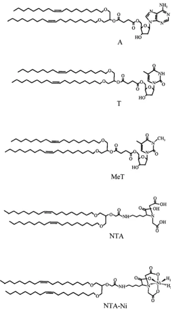
FIGURE 2 Chemical structures of the functionalized lipids: A, T, MeT, NTA, and NTA-Ni lipids.
3676 Tareste et al.
pression isotherm and verify its stability to desorption. They all presented a similar behavior. Their compression iso-therm showed no phase transition plateau and thus was characteristic of a fluid monolayer (for the case of the NTA lipid, see Fig. 3). By keeping the monolayer under a con-stant surface pressure, the one at which the lipids will be deposited on the mica surfaces, and measuring the decrease with time of its area, we can estimate the quantity of lipid that leaves the water/air interface to go in solution. These pressures were 39.5 mN/m for the A and T lipids, 37.5 mN/m for the MeT lipid, and 35 mN/m for the NTA and NTA-Ni lipids. All the lipids revealed a very stable behavior with an average desorption of 1%/h.
When deposited as bilayers on a solid surface, the lipids may have different desorption kinetics. To verify the sta-bility of the coated surfaces to desorption, we have depos-ited on a large mica sheet (⬃10 cm2), a first monolayer of
DMPE, and a second monolayer of the lipid to be studied. The sample stayed under water (in a 1-l dish) up to 24 h, which is more than the time scale for an SFA experiment. Then the lipid was redeposited at the water/air interface, and the number of molecules were recounted. We found again that on average 1% of the lipid had desorbed per hour. So the lipids can be considered as being irreversibly bound to the DMPE surfaces, which ensures that the layers are close-packed on the time scale of the experiments.
SFA experiments
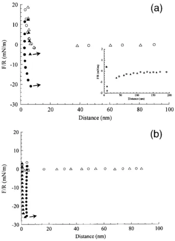
The NTA/NTA, MeT/T, and MeT/A systems display simi-lar features. On approaching the surfaces, the force-distance profiles display no double layer repulsion. The resolution of the SFA being less than one charge per thousand lipids (Pincet et al., 1999), this establishes that the surfaces are electrostatically neutral. From a distance of ⬃100 nm, an attractive force sets in (see inset in Fig. 4 a for the case of the NTA/NTA system). Such an attraction has already been
observed with the A and T lipids (Pincet et al., 1994), but its origin is still unknown. When the force gradient is higher than the spring constant, the surfaces jump into contact. The resulting distance between the two mica surfaces right after the jump corresponds to the thickness of two close-packed monolayers (D0). While the surfaces remain in contact (⬃15
min), the distance decreases, indicating a loss of lipid in the outer monolayers and leading to a flat contact between the two surfaces. This is generally not observed with phospho-lipids layers, whereas it has been noticed with hydrogen bonding lipids (Pincet et al., 1996). Upon pulling to separate the surfaces, the distance remains approximately constant until a sufficient force is applied. Then the distance in-creases back to D0(the contact is still flat), and the surfaces
jump apart. In this article, we focus mainly on the pull off force F0corresponding to this detachment. The interaction
profiles for the three systems are given in Fig. 4.
FIGURE 3 Compression isotherm of the NTA lipid on pure water subphase.
FIGURE 4 (a) Force-distance profiles in the NTA/NTA case (circles) and the NTA-Ni/NTA-Ni case (triangles). Open symbols correspond to the approaching data, filled symbols correspond to the separation data. During the separation phase, when a sufficient pulling force is applied, the surfaces jump apart (the jumps are symbolized by the arrows). (Inset) Same points are shown on a different force scale to reveal the attraction. (b) Force-distance profiles in the MeT/T case (circles) and the MeT/A case
(trian-gles). Open symbols correspond to the approaching data, filled symbols
In contrast to the NTA/NTA case, the NTA-Ni/NTA-Ni system presents a behavior similar to the one of neutral phospholipids: no double layer repulsion, no significant variation of the contact distance, and a weak pull off force (Fig. 4 a). One can also notice that the long-range attractive force is still present (inset in Fig. 4 a).
The macroscopic adhesion energy E0 between the two
surfaces can be deduced from the measured pull off force F0. Two main theories have been developed on this subject.
First, the Johnson, Kendal, and Roberts theory (JKR theory; Johnson et al., 1971) considers that the adhesive forces between two surfaces cause an increase in contact area, so that the surfaces are flattened just before separation. It predicts that F0is related to E0by:
F0⫽ 3RE0/2 (1)
The second theory, the Derjaguin, Muller, and Toporov theory (DMT theory; Derjaguin et al., 1975), accounts for the effects of the forces between the two surfaces just outside the contact area, so that this contact area falls down to zero just before separation. F0is related to E0by:
F0⫽ 2RE0 (2)
Maugis reconciled both theories by considering each of them as a special case (Maugis, 1992). In the case of two surfaces having a strong adhesion energy in comparison to their elasticity, the contact area is flattened and the relation between F0and E0is given by Eq. 1 (JKR theory). This is
the case for all the systems but the NTA-Ni/NTA-Ni sys-tem. In the opposite case, the contact area falls down to zero, and the relation between F0and E0is given by Eq. 2
(DMT theory). This is the case for the NTA-Ni/NTA-Ni system.
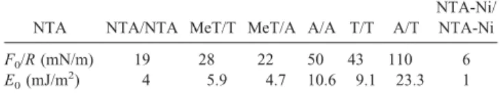
The values of F0 and E0 for the different systems are
displayed in Table 1. Except for the NTA-Ni/NTA-Ni sys-tem, the surface adhesion energies are much higher than the ones of phospholipids that do not make hydrogen bonds (Marra and Israelachvili, 1985). When the NTA group is functionalized with a nickel atom, the three OH endings are replaced by bonds between the oxygen atoms and the nickel ion and the measured energy E0decreases from 4 mJ/m
2
to 1 mJ/m2. This confirms that the adhesion energy measured in the NTA/NTA system is due to hydrogen bonds, which are blocked in the NTA-Ni/NTA-Ni case. From previous measurements, we also know than the bonds involved in the
experiments with nucleosides are hydrogen bonds (Pincet et al., 1994). In the systems involving MeT groups for which one hydrogen atom has been replaced by a methyl group, therefore blocking one hydrogen bond, one can also notice that the adhesion energy is substantially decreased.
Therefore, it seems that the headgroup/headgroup hydro-gen bonds are, in all the cases, energetically more favorable than the headgroup/water ones. This is coherent with pre-vious results obtained on glycine lipids (Schneider et al., 1998).
DISCUSSION
Many of the force-distance profiles show characteristic fea-tures: an attraction from 100 nm, a strong adhesion, and a decrease of the distance when the bilayers are in contact. Such features have been previously observed on deliber-ately depleted lipid layers (Helm et al., 1989, 1992; Helm and Israelachvili, 1993) and were rightly interpreted as hydrophobic attractions and adhesion, which are now well documented. One could be tempted to give the same inter-pretation to the present data obtained on hydrogen bonding lipids. This interpretation would be right if the distance decrease was irreversible or if the layers were already depleted by lipid desorption before going into contact. However it was checked by desorption measurements from water/air interface and also from mica supported bilayers that no significant lipid desorption takes place on the time scale of the experiments. Moreover, the decreases seen in the contact distance were in our cases fully reversible: by pulling the surfaces apart the bilayers recovered their initial thickness. It was at this point, i.e., with full bilayers, that the adhesion energy was measured. These features have already been observed with A and T lipid layers for which hydrogen bonds accounted for the adhesion energies (Pincet et al., 1994). The molecular binding energies were indeed the ones known from the literature and these values were also ob-tained by two other methods (Pincet et al., 1997, 2001). These adhesion energies can therefore be interpreted in terms of hydrogen bonds between close-packed lipid layers.
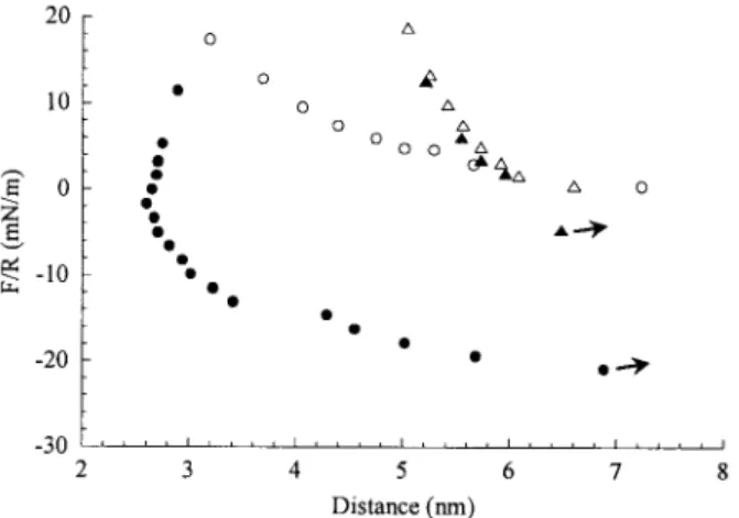
Force– distance profiles in contact
The unusual force– distance profiles when the surfaces are in contact in the NTA/NTA, MeT/T, and MeT/A cases have already been observed in similar experiments involving the A and T lipids (A/T, A/A, and T/T experiments). The decrease of the separation distance below D0 indicates a
rearrangement of the molecules in the contact area (“sticky fluid” behavior; Pincet et al., 1996). In the NTA-Ni/NTA-Ni case, where the hydrogen bonds are hindered, the “sticky fluid” phenomenon is not observed (Fig. 5). Therefore, the main new result about this behavior is that it appears to be
TABLE 1 Values of F0/R ( F0is the pull off force, accuracy
⬃10%) and of the adhesion energy E0as deduced from Eq. 1
(Eq. 2 in the NTA-Ni/NTA-Ni case)
NTA NTA/NTA MeT/T MeT/A A/A T/T A/T
NTA-Ni/ NTA-Ni F0/R (mN/m) 19 28 22 50 43 110 6 E0(mJ/m 2 ) 4 5.9 4.7 10.6 9.1 23.3 1 3678 Tareste et al. Biophysical Journal 83(6) 3675–3681
very common between surfaces bearing close-packed hy-drogen bonding groups.
Surface charges
All the interaction profiles presented here show no double layer repulsion and, thus, indicate that the surfaces are neutral. This is surprising because, given their pKa(see Fig.
6 for the cases of the A and T groups), all the systems but the T/T one are supposed to bear at least one charge per hundred lipids in pure water (up to one charge per lipid for the NTA-Ni/NTA-Ni system). This can be explained by the fact that, even in pure water, a considerable amount of counter-ions can bind to the surfaces (Marra, 1986). These ions may come from detachment from different parts of the SFA trough (stainless steel, teflon, glass) or from the dis-solution of carbon dioxide. Marra found that more than 90
percent of the surface charges can be neutralized. In our case, this effect seems even stronger.
Hydrogen bonding energy
As the NTA group has the highest number of available hydrogen bonding sites, one should expect a higher adhe-sion energy in the NTA/NTA case. Each NTA group con-tains indeed three carboxyl groups corresponding to six possible hydrogen bonds. However, the relatively small adhesion observed between two NTA surfaces is under-standable for two reasons. First, a high proportion of car-boxyl groups have been ionized, and the hydrogen has probably been replaced by another cation from the solution. Second, hydrogen bonds can be formed not only between carboxyl groups from opposite surfaces but also within the same molecule or within the same layer. From previous experiments made on weakly acidic and basic monolayers (Payens, 1955; Betts and Pethica, 1956; Zhao et al., 1993; Vezenov et al., 1997), one can estimate the surface pKaof
the carboxylic groups of NTA to 5.8. Our experiments were performed in pure water whose pH is comprised between 5.3 and 5.7. Thus, we can consider that one-half of the carboxyl groups have been ionized. The layers being close-packed, it can be assumed that only one-third of the hydro-gened carboxyl groups (which is the proportion for an hexagonal lattice) make interlayer bonds. Therefore, we estimate that only one-sixth of all the hydrogen bonding sites contribute to the adhesion energy, that is to say one hydrogen bonding site per NTA group.
In the case of A, T, and MeT lipids, hydrogen bonds within the same layer can be neglected. Indeed, it has been shown by x-ray diffraction performed on monolayers func-tionalized with nucleosides that the headgroups have a tendency to associate by setting their planes parallel to each other (Perez et al., 1998). This phenomenon, called “stack-ing,” strongly hinders the formation of hydrogen bonds within the same layer. The A and T groups can thus make two interlayer hydrogen bonds and the MeT group one interlayer hydrogen bond (Fig. 7).
All hydrogen bonds in the A/A system are identical (NH哹N) as well as for the T/T (NH哹O) and for the NTA/NTA (OH哹O) systems. Therefore, the molecular binding energy for each of these cases can be considered to be equal to the energy of one hydrogen bond times the number of such bonds between the molecules. In principle, one cannot do the same for the A/T case because it involves two different hydrogen bonds. However, as a first approx-imation, we will do it and discuss it further below.
From the adhesion energy E0 and the number nH of
hydrogen bonding sites involved in each single molecular bond, we can deduce the energy eH of a single hydrogen
bond. Assuming that each molecule has nHhydrogen
bond-ing sites that can be either bound or unbound, Boltzmann statistics leads to the relation:
FIGURE 5 Force-distance profile in contact between two NTA surfaces (circles, “sticky fluid” behavior) and between two NTA-Ni surfaces
(tri-angles, “classical” behavior).
E0⫽共nH/兲 ⫻ eH/共1 ⫹ exp共⫺eH兲兲 (3)
in which E0and eHare given in kBT units and in which is the average area occupied by a lipid inside the outer mono-layer. The molecular areas are 0.56 nm2for NTA, 0.62 nm2
for NTA-Ni, 0.63 nm2for A, 0.56 nm2for T, and 0.51 nm2
for MeT.
nHand eHvalues are reported in Table 2. For each case, except the A/T one, we found a hydrogen bonding energy of the order of 0.5 kcal/mol. The unexpected similar values of the different hydrogen bonds justify a posteriori the approx-imation made in the case of A/T. However, one can notice that the hydrogen bond energy for A/T is larger than for the other cases. These stronger hydrogen bonds may have two origins: their geometry (better angle and length) gives them a more optimal strength, and a third hydrogen bond might partially be formed between the A and T groups (represent-ed by the small dots in Fig. 7).
The values that we obtained for the different cases are in good agreement with previous studies on proteins stability through simulations on model compounds and site directed mutational techniques (Pace et al., 1996, 2001; Myers and Pace, 1996; Pokkuluri et al., 2002).
CONCLUSION
Force measurements between surfaces functionalized with lipids having hydrogen bonding headgroups (NTA, A, T, and MeT lipids) lead to a reproducible value of the energy of a single hydrogen bond in pure water:⬃0.5 kcal/mol. It shows that it is energetically more favorable for the head-groups to make hydrogen bonds with each other than to make hydrogen bonds with water molecules. This is coher-ent with past studies made on proteins stability, which showed that intramolecular hydrogen bonds in a folded protein are energetically more favorable than bonds with water molecules in an unfolded protein with an average stabilization of ⬃1 kcal/mol per intramolecular hydrogen bond. Moreover, in our systems, the hydrogen bonds can be perfectly isolated and probed, which is not the case in protein systems that are much more complex. These new results can thus give a more precise estimate of the energetic contribution of the hydrogen bonds in the protein folding process. This approach could be complementary to the usual
FIGURE 7 Hydrogen bonds involved in each studied system. Each NTA group contains three carboxyl groups that can form two hydrogen bonds. This leads to six possible hydrogen bonds per NTA group. However, on average, only one hydrogen bond is formed between two NTA groups (see Discussion).
TABLE 2 Values of the adhesion energy E0, of the estimated
number nHof hydrogen bonds per lipid and of the hydrogen
bonding energy eHas deduced from Eq. 3
NTA/NTA MeT/T MeT/A A/A T/T A/T
E0(mJ/m 2 ) 4 5.9 4.7 10.6 9.1 23.3 nH 1 1 1 2 2 2 eH (kBT/bond) 0.8 1.1 1 1.1 0.9 2.0 (kcal/mol) 0.5 0.6 0.6 0.6 0.5 1.2 3680 Tareste et al. Biophysical Journal 83(6) 3675–3681
ones, i.e., site-directed mutational studies and simulations on model compounds. It could be used for any other systems that involve hydrogen bonds such as nucleic acids or col-loidal systems.
REFERENCES
Baker, E. N., and R. E. Hubbard. 1984. Hydrogen bonding in globular proteins. Prog. Biophys. Mol. Biol. 44:97–179.
Berndt, P., K. Kurihara, and T. Kunitake. 1995. Measurement of forces between surfaces composed of two-dimensionally organized, comple-mentary and noncomplecomple-mentary nucleobases. Langmuir. 11:3083–3091. Betts, J. J., and B. A. Pethica. 1956. The ionization characteristics of monolayers of weak acids and bases. Trans. Faraday Soc. 52: 1581–1589.
Derjaguin, B. V., V. M. Muller, and Y. P. Toporov. 1975. Effect of contact deformations on the adhesion of particles. J. Colloid Interf. Sci. 53: 314 –326.
Gaines, G. L. 1966. Insoluble Monolayers at Liquid-Gas Interfaces. Inter-science Publishers, New York.
Helm, C. A., and J. N. Israelachvili. 1993. Forces between phospholipid bilayers and their relationship to membrane fusion. Method. Enzymol. 220:130 –143.
Helm, C. A., J. N. Israelachvili, and P. M. McGuiggan. 1989. Molecular mechanisms and forces involved in the adhesion and fusion of amphiphi-lic bilayers. Science. 246:919 –922.
Helm, C. A., J. N. Israelachvili, and P. M. McGuiggan. 1992. Role of hydrophobic forces in bilayer adhesion and fusion. Biochemistry. 31: 1794 –1805.
Israelachvili, J. N., and G. E. Adams. 1978. Measurement of forces between two mica surfaces in aqueous electrolyte solutions in the range 0 –100 nm. J. Chem. Soc. Faraday Trans. I. 74:975–1001.
Johnson, K. L., K. Kendall, and A. D. Roberts. 1971. Surface energy and the contact of elastic solids. Proc. R. Soc. London Ser. A. 324:301–313. Kauzmann, W. 1959. Some factors in the interpretation of protein
dena-turation. Adv. Protein Chem. 14:1– 63.
Lebeau, L., S. Olland, P. Oudet, and C. Mioskowski. 1992. Rational design and synthesis of phospholipids for the two-dimensional crystallization of DNA gyrase, a key element in chromosome organization. Chem. Phys.
Lipids. 62:93–103.
Marra, J. 1986. Effects of counterion specificity on the interactions be-tween quaternary ammonium surfactants in monolayers and bilayers.
J. Phys. Chem. 90:2145–2150.
Marra, J., and J. N. Israelachvili. 1985. Direct measurement of forces between phosphatidylcholine and phosphatidylethanolamine bilayers in aqueous solutions. Biochemistry. 24:4608 – 4618.
Maugis, D. 1992. The JKR-DMT transition using a dugdale model. J.
Col-loid Interf. Sci. 150:243–269.
McDonald, I. K., and J. M. Thornton. 1994. Satisfying hydrogen bonding potential in proteins. J. Mol. Biol. 238:777–793.
Myers, J. K., and C. N. Pace. 1996. Hydrogen bonding stabilizes globular proteins. Biophys. J. 71:2033–2039.
Pace, C. N., G. Horn, E. J. Hebert, J. Bechert, K. Shaw, L. Urbanikova, J. M. Scholtz, and J. Sevcik. 2001. Tyrosine hydrogen bonds make a large contribution to protein stability. J. Mol. Biol. 312:393– 404. Pace, C. N., B. A. Shirley, M. McNutt, and K. Gajiwala. 1996. Forces
contributing to the conformational stability of proteins. FASEB J. 10: 75– 83.
Payens, T. A. J. 1955. Ionized monolayers. Philips Res. Rep. 10:425– 481. Perez, E., F. Pincet, M. Goldmann, C. Mioskowski, and L. Lebeau. 1998. Translational order in liquid-expanded lipid monolayers functionalized with nucleosides. Eur. Phys. J. B. 6:1– 4.
Pincet, F., S. Cribier, and E. Perez. 1999. Bilayers of neutral lipids bear a small but significant charge. Eur. Phys. J. B. 11:127–130.
Pincet, F., E. Perez, G. Bryant, L. Lebeau, and C. Mioskowski. 1994. Long-range attraction between nucleosides with short-range specificity: direct measurements. Phys. Rev. Lett. 73:2780 –2783.
Pincet, F., E. Perez, G. Bryant, L. Lebeau, and C. Mioskowski. 1996. Specific forces between DNA bases. Mod. Phys. Lett. B. 10:81–99. Pincet, F., E. Perez, J. C. Loudet, and L. Lebeau. 2001. From macroscopic
adhesion energy to molecular bonds: a test of the theory. Phys. Rev. Lett. 87:178101–178104.
Pincet, F., W. Rawicz, E. Perez, L. Lebeau, C. Mioskowski, and E. Evans. 1997. Electrostatic nanotitration of weak biochemical bonds. Phys. Rev.
Lett. 79:1949 –1952.
Pokkuluri, P. R., R. Raffen, L. Dieckman, C. Boogaard, F. J. Stevens, and M. Schiffer. 2002. Increasing protein stability by polar surface residues: domain-wide consequences of interactions within a loop. Biophys. J. 82:391–398.
Schneider, J., Y. Dori, M. Tirrell, and R. Sharma. 1998. Effect of substrate anchoring on the mechanical strength of Langmuir-Blodgett bilayers.
Thin Solid Films. 327–329:772–777.
Tanford, C. 1962. Contribution of hydrophobic interactions to the stability of the globular conformation of proteins. J. Am. Chem. Soc. 84: 4240 – 4247.
Vezenov, D. V., A. Noy, L. F. Rozsnyai, and C. M. Lieber. 1997. Force titrations and ionization state sensitive imaging of functional groups in aqueous solutions by chemical force microscopy. J. Am. Chem. Soc. 119:2006 –2015.
Zhao, X., S. Ong, H. Wang, and K. B. Eisenthal. 1993. New method for determination of surface pKausing second harmonic generation. Chem.