HAL Id: hal-02533762
https://hal.sorbonne-universite.fr/hal-02533762
Submitted on 6 Apr 2020
HAL is a multi-disciplinary open access
archive for the deposit and dissemination of
sci-entific research documents, whether they are
pub-lished or not. The documents may come from
teaching and research institutions in France or
abroad, or from public or private research centers.
L’archive ouverte pluridisciplinaire HAL, est
destinée au dépôt et à la diffusion de documents
scientifiques de niveau recherche, publiés ou non,
émanant des établissements d’enseignement et de
recherche français ou étrangers, des laboratoires
publics ou privés.
subconjunctival triamcinolone injection and intravitreal
dexamethasone implant: medical and economic
importance of this randomized controlled trial
Chloé Couret, Alexandra Poinas, Christelle Volteau, Valéry-Pierre Riche,
Marie-Laure Le Lez, Marie-Hélène Errera, Catherine Creuzot-Garcher,
Stephanie Baillif, Laurent Kodjikian, Catherine Ivan, et al.
To cite this version:
Chloé Couret, Alexandra Poinas, Christelle Volteau, Valéry-Pierre Riche, Marie-Laure Le Lez, et
al.. Comparison of two techniques used in routine care for the treatment of inflammatory macular
oedema, subconjunctival triamcinolone injection and intravitreal dexamethasone implant: medical
and economic importance of this randomized controlled trial. Trials, BioMed Central, 2020, 21 (1),
pp.159. �10.1186/s13063-020-4066-0�. �hal-02533762�
S T U D Y P R O T O C O L
Open Access
Comparison of two techniques used in
routine care for the treatment of
inflammatory macular oedema,
subconjunctival triamcinolone injection and
intravitreal dexamethasone implant:
medical and economic importance of this
randomized controlled trial
Chloé Couret
1†, Alexandra Poinas
2*†, Christelle Volteau
3, Valery-Pierre Riche
4, Marie-Laure Le Lez
5,
Marie-Hélène Errera
6, Catherine Creuzot-Garcher
7, Stéphanie Baillif
8, Laurent Kodjikian
9, Catherine Ivan
1,
Laurence Mathilde Le Jumeau de Kergaradec
1, Anne Chiffoleau
10, Alexandra Jobert
10, Julie Jaulin
10,
Laetitia Biron
10, Elisabeth Hervouet
10and Michel Weber
1Abstract
Background: Whether they are injected peri- or intraocularly, corticosteroids are still essential tools in the therapeutic arsenal for treating inflammatory macular oedema. A few years ago, however, only triamcinolone acetonide was available to ophthalmologists. While this compound was initially developed for rheumatological or dermatological use, it has been increasingly deployed in ophthalmology, despite still being off-label. In 2011, the system for delivery of dexamethasone from a biodegradable, injectable implant into the vitreous cavity obtained approval for use in inflammatory macular oedema. While the efficacy and safety of triamcinolone in macular oedema, including inflammatory oedema, have already been studied, there are currently no publications on subconjunctival triamcinolone injections, which are simple, effective and well tolerated. To date, the dexamethasone 700μg implant has been authorized for the treatment of noninfectious intermediate and posterior uveitis, but there have been no studies to evaluate the efficacy and safety of the different peri- and intraocular strategies, including the treatment of inflammatory macular oedema.
Methods: This protocol is therefore designed to compare the efficacy and safety of peri- and intraocular corticosteroid injections in the treatment of inflammatory macular oedema. In this ongoing study, 142 patients will be included, and the oedematous eye will be randomised to treatment with either subconjunctival triamcinolone injection or an intravitreal implant containing 700μg dexamethasone. Follow-up is planned for 6 months with monthly visits. Each visit will include visual acuity measurement, a slit lamp examination, fundoscopy, intraocular pressure measurement,
(Continued on next page)
© The Author(s). 2020 Open Access This article is distributed under the terms of the Creative Commons Attribution 4.0 International License (http://creativecommons.org/licenses/by/4.0/), which permits unrestricted use, distribution, and reproduction in any medium, provided you give appropriate credit to the original author(s) and the source, provide a link to the Creative Commons license, and indicate if changes were made. The Creative Commons Public Domain Dedication waiver (http://creativecommons.org/publicdomain/zero/1.0/) applies to the data made available in this article, unless otherwise stated.
* Correspondence:alexandra.poinas@chu-nantes.fr
†Chloé Couret and Alexandra Poinas contributed equally to this work. 2Clinical Investigation Centre CIC1413, INSERM and CHU Nantes, Nantes,
France
(Continued from previous page)
laser flare measurement (if available) and spectral domain optical coherence tomography.
Discussion: The results of this trial will have a real impact on public health if it is shown that a Kenacort retard® (i.e. triamcinolone) injection costing just€2.84 and performed in the physician’s office (with no additional overhead costs) is at least as effective as the dexamethasone 700μg implant (Ozurdex®; costing approximately €960 with the injection performed in a dedicated room), with no increased side effects.
Trial registration: ClinicalTrials.gov,NCT02556424. Registered on 22 September 2015.
Keywords: Macular oedema, Corticoids, Periocular injection, Intraocular injection, Medical cost–benefit analyses
Background
Any ocular inflammation may be complicated by inflam-matory macular oedema. All cases of uveitis may poten-tially lead to macular oedema, with varying frequency according to location [1].
Epidemiological studies offer little information about blindness from uveitis, yet uveitis is the fifth leading cause of legal blindness in adults aged 20–65 years in the western world and is responsible for 10–15% of cases of total blindness in the United States [2]. Macular oedema is present in one-third of cases of uveitis in the Larde-noye series [1], and is the leading complication causing blindness in uveitis. It is responsible for 26.8% to 42% of acute visual loss [1,3], comparable to the older Rothova series, which pointed to inflammatory macular oedema as being responsible for 29% of legal blindness and 41% of decreases in visual acuity [4].
The mechanisms of disruption to the blood–retinal barrier that cause inflammatory macular oedema are many and varied. Disruption to the blood–retinal barrier causes exudation of plasma proteins and lipids which have oncotic properties that result in retention of the li-quid mainly in the extracellular space; this is the macu-lar oedema. The disruption to homeostasis and retinal detoxification also causes macular cell death, which ex-plains the absence of ad integrum recovery of visual acu-ity in cases of extended inflammatory macular oedema.
Depending on its severity, the macular oedema may re-main asymptomatic, or it may cause macular degeneration of varying complexity—decreased near and far visual acu-ity, metamorphopsia, central scotoma, central phosphenes and micropsia. Symptoms of the inflammatory condition responsible for macular oedema may be present and mask the signs associated with macular oedema, hence the im-portance of systematic testing in uveitis.
There is no consensus on the therapeutic management of inflammatory macular oedema [5]. Similarly, there is no clear definition of “clinically significant” inflamma-tory macular oedema, as there is in diabetes, to provide a basis for comparison of the different studies.
Many hypotheses have been advanced to explain how various conditions can cause macular oedema. The in-flammatory hypothesis is based on the increase in
proinflammatory cytokines and in vascular endothelial growth factor expression which is central to the dynam-ics of inflammatory macular oedema. However, the mechanisms underlying inflammatory macular oedema are varied and may involve mechanical, toxic or inflam-matory factors.
Oral steroids are better suited to the treatment of bi-lateral inflammatory attacks or when the use of a topical treatment is not possible. They are usually administered in the form of prednisone on account of its enhanced bioavailability compared to prednisolone.
Peri- and intraocular routes are used to limit the sys-temic side effects of anti-inflammatory steroids. Weijtens et al. showed that the intravitreal route enabled a max-imum concentration of vitreous corticosteroid to be ob-tained, followed by the subconjunctival and peribulbar routes, for which the concentrations achieved were 120 and 13 times higher, respectively, than after oral admin-istration [6–9].
The justification for the subconjunctival route is that the intravitreal corticosteroid concentration is 15 times higher after a subconjunctival injection of 2.5 mg dexa-methasone 700μg than after taking 50 mg of oral pred-nisone for several days [6]. Regarding peribulbar injections, some results provide evidence of intraocular penetration through the lamina rather than through the sclera, with the trans-scleral passage of molecules being disrupted by the choroidal blood flow as well as by the low permeability of the pigmented epithelial layer of the retina; this could be an argument in favour of subcon-junctival injections instead of peribulbar or potentially sub-Tenon’s injections [6]. It is essential to remember that such periocular treatment (subconjunctival or peri-bulbar) is not a purely local treatment, to the extent that the blood corticosteroid concentration is found to be comparable to that of oral treatment.
In practical terms, subconjunctival injections are easily achievable in routine care. In case of complica-tions, crystals can be easily removed under local an-aesthesia in the operating room. Kalina et al. showed that removal of subconjunctival triamcinolone crystals is effective in normalising intraocular pressure [10] (Fig. 1).
The arrival on the market of the dexamethasone 700μg intravitreal device has changed current practice. Its use is now authorized for first-line treatment of macular oedema secondary to occlusion of the central retinal vein and venous branches. It is also indicated for the treatment of noninfectious posterior uveitis and is being investigated for diabetic macular oedema.
Intravitreal injections require greater material, human and financial resources than subconjunctival injections since they must be performed in the operating room or in a dedicated room that meets specific criteria for strictly aseptic conditions.
Complications can occur and may be transient or per-manent and may require medical or surgical treatment. The most common complication, subconjunctival haem-orrhage, is trivial. In rare cases, endophthalmitis, ele-vated intraocular pressure (IOP) requiring medical or surgical treatment, damage to the lens causing a cata-ract, intravitreal haemorrhage, or retinal detachment may occur.
The ocular complications of corticosteroids are sum-marized in Additional file1.
The efficacy and safety of triamcinolone in macular oedema, including inflammatory oedema, have already been studied [12–14]. For many years, the Department of Ophthalmology of CHU Nantes (University Hospital of Nantes), and other institutions, used subconjunctival triamcinolone injections. Since they are easy to perform extemporaneously on the day of consultation, they ap-pear to be very effective in inflammatory macular oedema, both anatomically and functionally, with few complications. Unfortunately, there are currently no publications on these simple, effective and well-tolerated
injections. Similarly, the sub-Tenon’s route is a possibil-ity, but is more complex to execute.
Finally, the dexamethasone 700μg implant has been authorized for the treatment of noninfectious intermedi-ate and posterior uveitis, but there have been no studies to evaluate the efficacy and safety of the different peri-and intraocular strategies, including the treatment of in-flammatory macular oedema.
Ozurdex® (i.e. dexamethasone 700μg) consists of a biodegradable copolymer of glycolic acid and lactic acid and 700μg dexamethasone which is gradually released into the eye [15, 16]. The clinical safety of Ozurdex® in patients with inflammation of the posterior segment of the eye presenting as noninfectious uveitis was assessed in a single, multicentre, masked, randomised study known as Huron [17]. The most frequently reported ad-verse reactions in the eye of patients who received Ozur-dex® were conjunctival haemorrhage (30.3%), increased IOP (25.0%) and cataract (11.8%).
Pivotal studies and real-life studies have confirmed that the safety profile of Ozurdex® is good, with the same complications of cataract progression in the range of 29.8% [18] to 67.9% [19], closely related to the number of implants received, and an increase in IOP >10 mmHg from baseline reported in 15.4% to 27.7% of cases [19].
Furthermore, in real life, it has been shown that shorter interval retreatment is required because the drug is effective for less than 6 months, with a reported range that varies from 4 to 5.9 months [20, 21]. Indeed, drug release peaks at 2 months and there is then a steady de-cline that prolongs its effects for up to 6 months [16].
Our department was one of the first and only teams to perform a retrospective study of patients treated with
Ozurdex® versus subconjunctival triamcinolone versus sub-Tenon’s triamcinolone [22]. This study of 88 pa-tients demonstrated neither superiority nor any differ-ence in the efficacy and safety of the three treatments.
Moreover, we must emphasize the potential impact on public health of a randomised prospective trial if sub-conjunctival injections of triamcinolone (a Kenacort re-tard® bulb costs €2.84 and the injection is performed in the physician’s office, with no additional overhead costs) were to prove at least as effective as injection of the dexamethasone 700μg implant (Ozurdex® costs €962.65 and each injection must be performed in a dedicated room).
Our question is, how do intravitreal injections of a dexamethasone 700μg implant and subconjunctival tri-amcinolone injections compare in terms of efficacy and safety? The arrival on the market of the dexamethasone 700μg implant with authorisation for the treatment of posterior and intermediate uveitis tends to eliminate subconjunctival triamcinolone injections. However, these are simple, effective and well tolerated; they have the ad-vantage that they are not delivered intraocularly, and they cost less.
Methods/design
Study design
The TRIOZ clinical trial is an open-label, prospective, randomised study. For technical and ethical reasons, it is not possible to inject two products (drug versus placebo) in two different injections to maintain the masking; fur-thermore, the corticosteroids are visible to the investiga-tor during control examinations (subconjunctival crystals, Fig. 1; intravitreal implant, Fig. 2). However, it was planned that visual acuity and central macular
thickness (CMT) would be assessed by an ophthalmolo-gist unacquainted with the trial who had not attended the patient’s surgery. The choice of these end points with a masked team allows the primary outcome and one of the secondary outcomes to be evaluated masked. No other assessments can be made on a masked basis, since the examination itself may reveal to the ophthal-mologist which treatment the patient has received. Lo-gistically, however, and especially due to the number of people in charge of clinical research in every centre, it was very difficult to create two teams, one masked and the other open, so the data have therefore been ex-tracted on an open basis.
Study population
Description of the population
Recruitment is planned over a period of 60 months in 14 French study centres: the Ophthalmology Departments in the University Hospitals of Nantes, Lille, Lyon, Tours, Brest, Paris (Pitié Salpêtrière), Paris (Kremlin Bicêtre), Paris (Fondation Rothschild), Bordeaux, Nancy, Gre-noble, Nice, Montpellier and Dijon.
Inflammatory macular oedema is a common pathology found in each centre at the rate of 10 cases per centre per month, making these recruitment targets achievable.
Recruitment for the trial
Patients of both sexes, aged over 18 years with inflam-matory macular oedema (meaning that they have a CMT >320μm), will be recruited by the Ophthalmology Departments of the 14 French centres participating in the trial. For patients with bilateral asymmetric inflam-matory macular oedema, the eye most affected will be treated. These patients should also be healthy and
should not present a progressive disease. Patients who are HIV positive or who have hepatitis B or C virus, syphilis (TPHA-VDRL) or tuberculosis (Quantiferon) will not be included. Additional file2presents all the in-clusion and exin-clusion criteria.
Study schedule
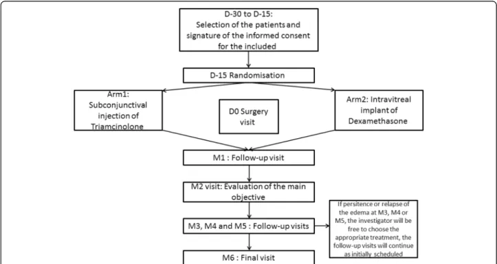
The plan for the study described in this section is pre-sented in Fig. 3, and Additional file 3 shows the time points at which the assessments are to be made.
Before proceeding with any examination related to the research, the investigator obtains the patient’s freely given, informed consent, in writing.
At the inclusion visit, the investigating physician pro-vides the patient with information on and answers any questions about the purpose of the research, the de-mands of the study, the foreseeable risks and the ex-pected benefits. They also specify the patient’s rights when taking part in biomedical research and verify the eligibility criteria. The investigating physician then gives the patient a copy of the information form and consent form. Patients who do not consent to the trial will be treated according to standard care.
After the briefing, the patient has a reasonable period of reflection between receiving the information and con-sent forms and making a decision.
The investigative physician is responsible for obtaining the patient’s written informed consent. The consent form must be signed prior to any clinical or paraclinical examination required for the research. If the patient
cannot read the information letter and informed consent form, their companion shall read their documents and countersign the consent.
The different copies of the information form and con-sent form are then distributed as follows: 1) the patient receives a copy of the information form, a signed con-sent form (see Additional file4) and a patient card; and 2) the investigating physician keeps the original copy (even when the patient is moved for the duration of the research) in the investigator’s file.
The investigative physician will check the inclusion and exclusion criteria again at the inclusion visit and after signing the consent form, and will note the pa-tient’s medical history and concomitant medications.
Screening visit
The following screening examinations will be conducted before the surgery visit (days (D)–30 to D0): 1) measure-ment of CMT using optical coherence tomography (OCT); 2) measurement of visual acuity (using the Early Treatment Diabetic Retinopathy Study (ETDRS) scale), a basic visual function parameter; 3) measurement of IOP, as hypertension is a major complication of peri- and in-traocular corticosteroid injection; 4) examination of the anterior segment by slit lamp (SL) to quantify the flare and assess the clarity of the lens since the second most common complication of peri- and intraocular cortico-steroids is development of a cataract; 5) examination of the posterior segment (fundus) and nonmydriatic fundus photography to assess vitreous haze; 6) examination of
fluorescein angiography to detect vasculitis, an associ-ated papillitis; 7) automassoci-ated quantitative and objective measurement of the flare when available using a laser flare meter (LFM), which enables a more accurate as-sessment of the status and previous inflammatory condi-tion of the blood–aqueous barrier (reference value); 8) measurement of blood pressure, as systemic corticoste-roids can induce hypertension and the pathways around the eyes especially mean that treatment is not purely local (exclusion criterion); and 9) blood tests for fasting plasma glucose and glycohaemoglobin (because systemic corticosteroids can induce diabetes and, more import-antly, the pathways around the eyes mean that treatment is not purely local; and exclusion criterion). In addition, a pregnancy test (beta human chorionic gonadotropin) will be conducted for women of childbearing age; preg-nant women are excluded from the protocol even though peri- and intraocular corticosteroids are permit-ted during pregnancy. Finally, a serology test will be made for HIV, hepatitis B and C virus, TPHA-VDRL and Quantiferon if the status is unknown to eliminate macular oedema due to infection and systemic infectious diseases at risk of aggravation by treatment with cortico-steroids, with the exception of obvious postoperative inflammation.
Treatment visit
At the beginning of the treatment visit (D0; injection of the product), the patient will answer their first EuroQol five dimensions (EQ-5D) questionnaire.
Before either the dexamethasone 700μg implant or tri-amcinolone is injected, anaesthetic and antiseptic eye drops will be given according to the centre’s practices. Analgesics are permitted. Initiation of hypotensive eye drops for curative or preventive purposes is allowed. The investigator must report this in the case report form (CRF). According to the exclusion criteria, general anti-inflammatory treatments or systemic immunosuppres-sive or immunomodulatory treatments at unstable doses are not authorised during the trial. From D0, acetazol-amide cannot be continued or introduced later.
For the subconjunctival triamcinolone injection (Kena-cort retard®), the patient is positioned comfortably. The injection is performed after the instillation of anaesthetic drops. Triamcinolone injections are carried out using a 25-gauge needle. A volume of 0.4 ml is injected under the inferior bulbar conjunctiva so that the eyelid covers the visible crystals without discomfort.
For Ozurdex®, the injection is performed in the supine or semi-seated position. The eye is numbed with the in-stillation of anaesthetic eye drops. The eyelids and ocular surface are disinfected with antiseptic to reduce the risk of infection. The face is covered with a sterile drape and a sterile eyelid retractor is positioned. The injection
device for the dexamethasone 700μg implant is sterile and ready to use; it is inserted through the sclera 3.5 mm (pseudophakic eye) or 4 mm (phakic eye) from the lamina after moving the conjunctiva a few millimetres (Fig. 4). Following the injection, antibiotic eyedrops are instilled. The treated eye remains painless in the vast majority of cases. A spot may appear in the visual field, corresponding to the presence of the implant in the vit-reous cavity (Fig.2).
After treatment, the investigator will give the patient a tray with a 10-cm slider for scoring the injection pain from 0 (no pain) to 10 (unbearable pain). The pointer will provide physicians with a measurement of pain in centimetres.
The investigator will then ask the patient to rate the injection, choosing between “tolerable”, “uncomfortable” and“very unpleasant”.
The physician will record the patient’s concomitant medications and any adverse events (AEs) and serious adverse events (SAEs). All such events will be reported to the person responsible for pharmacovigilance in clin-ical trials at CHU Nantes.
Please note that as the treatment takes place at hos-pital there is no need to monitor adherence.
Follow-up visits
The monthly follow-up visits (M1–M6) allow for early detection of efficacy and complications (including ocular hypertonia) beginning within approximately 1 month [12,23]. In addition, they provide reassurance for the pa-tient and reduce protocol deviations and papa-tients lost to follow-up.
The investigating physician will record any AEs/SAEs and any concomitant medications.
They will include the following in their examinations of the patient during visits M1, M2, M3, M4, M5 and M6 (visits M1 and M4 do not form part of the usual care): 1) measurement of CMT (by OCT); 2) measure-ment of visual acuity (using the ETDRS scale); 3) meas-urement of IOP; 4) examination of the anterior segment (by SL) for flare and crystalline lens clarity; 5) examin-ation of the posterior segment (fundus) and nonmydria-tic fundus photography for posterior vitreous haze; and 6) automated measurement of the flare (by LFM), if available. In addition, the patient will complete the EQ-5D questionnaire.
At M3 and M6, the patient’s blood pressure will be taken. A fluorescein angiograph is conducted at M2 and M6 if anomalies were detected at the inclusion visit. In addition, a blood sample will be taken at M6 to evaluate haemoglycaemia and fasting plasma glucose.
In cases of inadequate response or relapse between the M3 visit and the M6 visit, the investigator will be free to
choose the appropriate treatment on the day that the in-adequate response/relapse is detected, or later (± 30 days), at their discretion; follow-up visits will continue according to the initial schedule, i.e. monthly visits.
The research may be discontinued as a result of: 1) the patient withdrawing their consent; 2) necessity as de-cided by the investigator in case of SAEs that prevent the patient from continuing with the protocol; or 3) a decision by the authorities or the suspension/withdrawal of the drugs from the market.
In case of premature discontinuation from the study, the patient will be referred immediately to the investiga-tor for a consultation to provide care for their disease. The premature end of study visit will be completed dur-ing this consultation.
Objectives and statistics Objectives
The main objective of our study is to evaluate the efficacy of subconjunctival injection of triamcinolone in reducing CMT measured by OCT, the most objective, relevant and noninvasive criterion, compared with intravitreal injection of a dexamethasone 700μg implant which has been ap-proved for this indication, between the inclusion visit and 2 months after treatment (M2 visit).
The timeframe was chosen based on data from the Huron study of Ozurdex® [17,24,25] and data from our retrospective study on triamcinolone in the department at CHU Nantes [22].
The secondary objectives are: 1) evaluation of the ex-perience of the injection; 2) evaluation of the effective-ness of the injection studied at each visit, measured by the gain in visual acuity (by the EDTRS scale; a key par-ameter of visual function), the reduction in flare and vit-reous haze which are quantifiable ocular inflammatory parameters measured using an SL and LFM in centres equipped with this technology, the CMT measured using OCT (enabling evaluation of the duration of action of a subconjunctival triamcinolone injection compared with an intravitreal injection of a dexamethasone 700μg im-plant; the duration of action of the injection is deter-mined by the reappearance of oedema), the local and general tolerance of the two methods (collecting details of all AEs/SAEs) and the patients’ quality of life; and 3) economic evaluation of the efficiency (cost–utility ana-lysis) of the subconjunctival triamcinolone injection compared with intravitreal injection of a dexamethasone implant from a societal perspective and over a 6-month period.
Primary outcome
The primary outcome is the difference in CMT in the treated eye, measured using spectral-domain OCT in both groups between selection and M2.
The micrometric CMT was converted into a logarith-mic CMT (logSD-OCT) for statistical analysis, consider-ing that the normal CMT was 250μm.
The formula used in the trial is: logarithmic CMT = log10 (micrometric CMT/250).
Use of a logarithmic scale for analysing changes in CMT gives a more normal distribution for CMTs that coincides with the distribution of logarithmic visual acu-ity. Some studies have shown that the logarithmic trans-formation of the CMT provides a better picture of visual acuity [26,27].
Our CMT results were expressed in microns and logSD-OCT so they could be compared with those in the literature.
A retrospective study in the department at CHU de Nantes on the effectiveness of triamcinolone injections in reducing CMT converted into logSD-OCT obtained the following results: between M0 and M1, −0.12logSD-OCT (p < 0.001) and between M0 and M3, −0.09logSD-OCT (p = 0.002), introducing the value of M2 as the main criterion.
Secondary outcomes
Secondary outcomes of the study are: 1) the scoring of “the moment” of injection on the day of injection (toler-able, uncomfort(toler-able, very unpleasant) and the rating on a visual analogue scale (from 0 cm = no pain to 10 cm = extreme pain); 2) visual acuity (ETDRS scale) at every visit to determine the gain between the inclusion visit and the follow-up visits (the mean scores for each arm will be compared at each follow-up visit); 3) the flare (using SL and LFM if available) at every visit to deter-mine the reduction between the inclusion visit and the follow-up visit (the mean scores for each arm will be compared at each follow-up visit); 4) the vitreous haze at every visit to determine the reduction between the inclu-sion visit and the follow-up visit (the mean scores for each arm will be compared at each follow-up visit); 5) the thickness of the central macula of the treated eye to determine the duration of action at every visit (as stated in the objectives, the duration of action of the injection is determined by the reappearance of oedema; the mean scores for each arm will be compared at each follow-up visit); 6) AEs/SAEs including intermittent ocular hyper-tension, cataract, endophthalmitis, glycaemic and blood pressure imbalances at every visit; and 7) EQ-5D ques-tionnaire on patient quality of life at every visit.
Efficiency outcomes
The incremental cost-effectiveness ratio (ICER; cost per quality-adjusted life-year (QALY)) will be calculated for the comparison between subconjunctival triamcinolone injection and intravitreal injection of a dexamethasone implant from a societal perspective and over a 6-month time period.
Measures used to determine the outcomes
OCT is used to accurately visualise the different layers of the retina, including the macula, using an infrared laser. This is a contactless examination which is nonin-vasive, painless and brief.
An LFM is a device used to measure the protein con-centration in the anterior chamber using a helium–neon laser. This is a contactless examination that is non-invasive, painless and brief.
All the ophthalmic examinations performed at inclu-sion and during follow-up are common practice in oph-thalmology. The visual acuity, IOP, examination using the SL and fundus examination form the basis of all clin-ical ophthalmic examinations. OCT is the test of choice for characterising and quantifying macular oedema, irre-spective of type. This testing is contactless and is nonin-vasive, painless and brief. The values considered to be normal are: visual acuity, ETDRS 100; IOP, 12–21 mmHg; LFM, no proteinic flare or cellular Tyndall; fun-dus, no cellular Tyndall or vitreous haze; macula as de-scribed, normally LFM <10 ph/ms, CMT <300μm and >250μm.
The patient will be given the EQ-5D questionnaire val-idated in France [28, 29] at each visit to measure their quality of life. The EQ-5D consists of a questionnaire and a visual analogue scale. The questionnaire focuses on five areas: mobility, personal autonomy, current activ-ities, pain/discomfort and anxiety/depression. There are three possible answers for each of these dimensions (EQ-5D-3 L), thus allowing for 243 health states. QALYs will be calculated for each arm using area under the curve methodology and the weighting coefficients avail-able in France for the EQ-5D-3 L [29,30].
Statistical methods
The data will be reviewed at the end of the study, prior to statistical analysis. The aim will be to review the pro-gress of the study, identify potential problems and clas-sify any minor or major deviations.
The variables measured at baseline will be described for all patients in both groups in terms of numbers and percentages for each category for categorical variables and minimum, maximum, average, standard deviation and quartile values for quantitative variables.
The primary endpoint is the difference in CMT in the treated eye between D0 and M2. CMT measurements will be converted into logarithmic CMT: logarithmic CMT = log10(CMT/250).
The main objective is to demonstrate the noninferior-ity of the group with subconjunctival triamcinolone in-jection compared with the group with intravitreal injection of a dexamethasone implant. The noninferior-ity margin was set at 0.06 (equivalent to CMT = 287). To demonstrate the noninferiority of the triamcinolone
group versus the dexamethasone group, the 95% bilateral confidence interval (CI) of the difference between the two groups (dexamethasone– triamcinolone) will be es-timated using a mixed linear regression model. This model will reflect the stratification factor of randomisa-tion to the centre (the centre will be considered as a ran-dom effect) and will be adjusted according to the measurement at D0. The estimated upper boundary of the CI will be compared to the predefined noninferiority margin. If the upper boundary is less than 0.06, the non-inferiority of the triamcinolone group as compared with the dexamethasone group will be demonstrated. For missing data, the worst observed value will be imputed for triamcinolone patients and the best value for dexa-methasone patients. A sensitivity analysis will be per-formed with multiple imputation.
For secondary end points, the mean visual analogue scale score evaluating the moment of injection will be compared between groups using a linear mixed model; the score for the moment (tolerable, uncomfortable or very unpleasant) will be compared using the Mantel– Haenszel stratified Chi-squared test. A linear mixed model will be used to compare the change in ETDRS from D0 to 6 months between the groups. SL and LFM between D0 and 2 months will be compared between the groups using nonparametric Van Elteren tests (semi-quantitative outcome). Duration of injection efficacy (duration is determined by the reappearance of oedema) will be compared between the groups using the Van Elteren test (semiquantitative outcome). Descriptions of AEs/SAEs will be reported in the two groups. Compari-sons between the groups will be performed using Chi-squared or Fisher tests for intermittent ocular hyperten-sion, cataract, endophthalmitis, glycaemic and blood pressure imbalances in both arms. There will be no im-putation for missing data for these secondary end points. For the economic assessment, mean costs and their corresponding 95% CIs will be presented. The ICERs will be estimated along with their corresponding acceptabil-ity curves, i.e. the curves indicating the probabilacceptabil-ity that an intervention is cost effective conditional on society’s willingness to pay for an additional unit of effectiveness (i.e. an additional QALY gained) and considering the sampling uncertainty around the estimated ICERs.
As costs and ICERs are not normally distributed, the 95% CI and the cost-effectiveness acceptability curves will be determined using the nonparametric bootstrap resampling technique.
Sensitivity analyses will be performed for all end points, adjusted for the duration of macular oedema. All statistical tests will be bilateral. For secondary endpoints, a P value less than 0.05 will be considered statistically significant. Analyses will be performed using SAS statis-tical software (SAS Institute Inc., Cary, NC, USA).
As this is a noninferiority study, the analyses will be carried out on the intent-to-treat population and on the per-protocol population. The intent-to-treat population consists of all randomised patients in the study. The per-protocol population includes the most compliant pa-tients, based on compliance with the inclusion and ex-clusion criteria, absence of major deviations from the protocol and availability of the main criterion.
Sample size
This TRIOZ trial aims to demonstrate the noninferiority of subconjunctival triamcinolone injections compared to intravitreal dexamethasone implants. Noninferiority will be evaluated in terms of the difference in macular thick-ness in the treated eye between D0 and M2. The dead-line of M2 was chosen based on the plan for the Huron study on dexamethasone (NCT000333814) and a retro-spective study on triamcinolone at CHU Nantes. Prelim-inary data observed retrospectively in Nantes between 2011 and 2013 in 25 patients who received triamcino-lone injections showed a decrease of 0.12 ± 0.12 log OCT at M1 (D0, 0.27 ± 0.11; M1, 0.15 ± 0.08) and of 0.09 at M3 (0.18 ± 0.11). The difference in macular thickness between D0 and M2 is assumed to be the same in the two groups and the common standard devi-ation is set at 0.12. The noninferiority margin was set at 0.06, the power at 80% and the type I error rate at 2.5%. Based on these assumptions, 128 patients are needed to demonstrate the noninferiority of triamcinolone com-pared to dexamethasone. A maximum rate of 10% for missing data is taken into consideration for M2 and 142 patients, or 71 patients per group, will therefore be ran-domised in the study.
Randomisation
Randomisation will be conducted openly and stratified by centre. It will be performed according to a 1:1 ratio and balanced by blocks. The random numbers will be generated by computer. Subjects are randomised into blocks as the allocation progresses, a block being a sub-group of predetermined size within which there is a ran-dom allocation of patients. The software used for the randomisation is SAS version 9.4. The randomisation key is known only to the biostatistician and the data managers to make it impossible for the investigator to assign a particular treatment.
As mentioned above, for logistical reasons the trial is now open-label. As Karanicolas et al. pointed out in their article from 2010, the study should have been masked to the biostatisticians until analyses were per-formed to reduce the study bias [31].
Logistically, as Ozurdex® requires an operating room, randomisation will be performed 15 days prior to the date of surgery. After confirming the inclusion/exclusion
criteria on the electronic CRF, the investigator will per-form the randomisation without the patient present; the patient will only know to what treatment they have been assigned at the treatment visit.
Upon activation, each study centre will receive two batches, each containing triamcinolone and Ozurdex® for the treatment of the first two patients. Based on the randomisation, the batch used will be replaced by the pharmacy of CHU Nantes. The treatments will be kept at the pharmacy in each study centre for issue on the prescription of the investigator during the treatment visit.
Adverse event management
There are no pharmacokinetic data in the literature for triamcinolone administered subconjunctivally. The clin-ical experience of the various centres performing these injections shows a duration of action of approximately 3 to 4 months for 0.3 and 0.4 ml, respectively. The dur-ation of action of an intravitreal injection of a dexa-methasone 700μg implant is approximately 3 to 6 months.
Inflammatory macular oedema, a chronic and recur-rent pathology, requires regular ophthalmological moni-toring at least every 6 months, regardless of the type of treatment administered.
The most frequently occurring adverse reactions iden-tified based on the summary of product characteristics for Ozurdex® and the experience of the University Hos-pital of Nantes for triamcinolone are: 1) corticosteroid-induced hypertension (eye tone will be monitored monthly until the effectiveness of any injected cortico-steroid has been exhausted and at longer intervals there-after based on blood pressure control); and 2) cataract (the visual acuity and appearance of the lens will be monitored using an SL during the usual follow-up con-sultations for inflammatory macular oedema until cata-ract surgery is performed).
All the AEs encountered that are observed by the in-vestigator or reported by the subject during the study, whether or not they are expected (see the summary of product characteristics for Ozurdex®), should be docu-mented in the AE section of the CRF.
SAE reporting
All SAEs, whether expected or unexpected, require the completion of an SAE report. The investigator must en-sure that the information entered in this report is accur-ate and clear. The SAE should be reported immediaccur-ately (within 24 h of being highlighted by the investigator) to the sponsor. After receiving an unexpected SAE report, the sponsor notifies the authorities. Once a year, the sponsor prepares an annual safety report.
Furthermore, a Data and Safety Monitoring Commit-tee (DSMC) has been set up. This is a consultative com-mittee responsible for reviewing the safety of a study on behalf of the sponsor and the coordinator/principal in-vestigator of the study. Members of the committee who are competent in the field of clinical trials (pathology and methodology) are not involved in the study.
The DSMC receives the annual safety reports and is a point of referral for pharmacovigilance if a suspected un-expected serious adverse reaction or an SAE poses par-ticular analytical difficulty or if a doubt arises in the study about the risk/benefit.
In the event of early termination of the study by a de-cision of the DSMC or the study sponsor, the regulatory authorities and the Ethical Review Board will be in-formed by post within a maximum of 15 days.
In any event, written confirmation will be sent to the coordinating investigator for the study (specifying the reasons for early termination) and to the principal inves-tigator of each centre, if applicable. All patients in the study will be informed and will be required to complete their early discharge visit.
Ethical, regulatory and dissemination aspects
The clinical study will be conducted in accordance with the relevant versions of the French Public Health Code, national and international Good Clinical Practice guide-lines, and the Declaration of Helsinki, each in the applic-able version.
In compliance with French law, the study protocol was submitted to the French regulatory authority (ANSM) and was approved on 31 August 2015.
This clinical study was submitted to and approved by the Ethical Review Board of Angers (Comité de Protec-tion des Personnes – CPP OUEST II - Angers) on 24 August, 2015 (see Additional file 4 for French informed consent). Requests for substantial modifications should be addressed by the sponsor for approval or notification to ANSM and/or the Ethical Review Board concerned in compliance with Law 2004–806 of 9 August 2004 and its implementing decrees.
The clinical protocol has been writeen according to Spirit check-list (see Additionnal file 5). The amended protocol should be a dated, updated version. If neces-sary, the information form and consent form should be amended.
The updated protocol is at version 10 on 7 July 2018. All the submissions/declarations were made by the Sponsor Department at CHU Nantes which manages the quality of the data collected. The data collected during the study will be processed electronically in accordance with the requirements of the CNIL, the French Data Protection Authority (in compliance with the French Reference Methodology MR001).
As required, the sponsor has provided an insurance policy to cover the financial consequences of its civil li-ability in accordance with the regulations.
It has been possible to carry out the protocol and the trial thanks to an Executive Committee which includes a Scientific Committee and a Steering Committee. The Sci-entific Committee was created by M. Weber and its mem-bership comprises external experts in this pathology, biostatisticians and methodologists, the medical economist and the project manager of the clinical investigation centre (CIC1413). It is coordinated by Dr. Couret. The Steering Committee is composed of the members of the Scientific Committee, except the external experts, and with the addition of the data management team, the nurse study coordinator from the Ophthalmology Department of CHU Nantes who coordinates assistance for patient inclusion in the other centres, and the monitoring Clinical Research Assistant (CRA). The sponsor project manager coordinates this committee and drafts the“TRIOZ newsletter” which provides, among other things, the latest news on patient inclusion, amendments to the protocol, and so forth.
An inspection or audit may take place as part of this study, performed by the sponsor and/or by the regula-tory authorities. Inspectors will check the documents, lo-gistics, records and any other resources that the authorities consider to be associated with the clinical trial and that may be located at the trial site itself.
The trial results will be published in international oph-thalmological, medical and scientific journals and pre-sented at national and international conferences. The investigators, who will share the entirety of the final trial dataset, will follow the rules and guidelines of the Inter-national Committee for Medical Journal Editors (ICMJE) [32]. In practice, the Scientific Committee will be among the authors of the publication, as will the investigators who have included the most patients in the trial. The trial sponsor and the French Ministry of Health, which provided the grant, must be cited in the publication.
Discussion
Demonstration of the efficacy and safety of subconjunc-tival triamcinolone injections will enable their continued use at a time when the marketing authorisation for an intravitreal device requires the intraocular delivery of a unique, expensive compound, exposing the patient to a rare risk of endophthalmitis and retinal detachment, while the relative efficacy and safety of these approaches have never been compared.
The European Union has established a tight safety net. No medicinal product may be marketed in a member state unless the competent authorities of that state have issued a marketing authorisation [33]. Recently, a control framework for medically justified off-label prescriptions has been implemented in France. Act no. 2011–2012 of
29 December 2011 reinforcing the safety of medicines and health products [34] addresses the ambitious goal of controlling off-label drug use. The Act explicitly recog-nizes the right of physicians to prescribe drugs off-label for use by an individual patient, under their direct per-sonal responsibility and in the patient’s interest. It also introduces a second, unique derogating provision into French law pursuant to Article L.5121-12-1 of the French Public Health Code, a regulatory process called “Temporary Recommendations for Use” (Recommanda-tions Temporaires d’Utilisation (RTUs)) [35].
The French health authorities have amended the RTU framework in order to authorise the reimbursement of a drug used off-label, despite the existence of licensed thera-peutic alternatives, because of the burden of licensed drugs on the health care system. To date, bevacizumab (Avastin®)/ranibizumab (Lucentis®) have provided an illus-tration of this“economic RTU”. Both drugs are antivascu-lar endothelial growth factor agents and are made by the same parent company but only one, Lucentis®, has market-ing authorisation for age-related macular degeneration (AMD). The other significant difference is the striking dis-parity in the cost of the two drugs. The new price of Avastin® is now €100 including VAT, compared to €10 initially; a monthly injection of Lucentis® is €738. Avastin® is registered for the treatment of systemic cancer but is used off-label to treat AMD. One study published in 2012 using data from Medicare 2010 surveyed physicians on why they chose Avastin® ver-sus Lucentis®. Cost was reported as the primary factor for choosing bevacizumab (70%) [36]. In France, Avastin® has been indicated for the treatment of the neovascular form of AMD since 2015 under the eco-nomic RTU.
We hope, therefore, that if this clinical trial proves the efficiency of subconjunctival triamcinolone injections the French Health Authorities will authorise the reim-bursement of this drug.
Trial status
This trial is still ongoing; patient inclusion is not yet complete. The updated protocol is at version 10 on 7 July 2018. The first patient was included on 13 January 2016. Recruitment by the investigating centres is planned to continue until 13 October 2020 and the study period will end in March 2021.
Supplementary information
Supplementary information accompanies this paper athttps://doi.org/10. 1186/s13063-020-4066-0.
Additional file 1. Major ocular complications of steroids by route of administration (from Turpin et al. [11]).
Additional file 2. Inclusion and exclusion criteria. Additional file 3. TRIOZ study schedule. Additional file 4. TRIOZ informed consent.
Additional file 5. SPIRIT 2013 checklist: recommended items to address in a clinical trial protocol and related documents.
Abbreviations
AE:Adverse event; AMD: Age-related macular degeneration; CI: Confidence interval; CMT: Central macular thickness; CRF: Case report form; DSMC: Data and Safety Monitoring Committee; EQ-5D: EuroQol five dimensions; ETDRS: Early Treatment Diabetic Retinopathy Study; ICER: Incremental cost effectiveness; IOP: Intraocular pressure; LFM: Laser flare meter; OCT: Optical coherence tomography; QALY: Quality-adjusted life year; SAE: Serious adverse event; SL: Slit lamp
Acknowledgements Not applicable. Authors’ contributions
CC and AP contributed equally to the drafting of the manuscript. CV, EH, AC and MW assisted with the drafting of the manuscript. CC, AP, CV, V-PR and MW designed the trial. CC, AP, CV and MW wrote the protocol and/or the file for the experimental drug and assisted with the drafting of the manuscript. EH, JJ and LB coordinated the submission of the protocol and the follow-up of the Health Ministry’s tender and the regulatory authorities and coordinated the trial. CV wrote the methodological/statistical analyses in the protocol. V-PR wrote the medical economic analyses. M-LLL, M-HE, CC-G, SB, LK, CI and LMLJdK participated in patient enrolment and follow-up. AC and AJ assisted with pharmacovigilance for the trial. All authors read and approved the final manuscript.
Funding
This study was supported by a grant from the French Ministry of Health awarded in 2014 (under the Hospital Clinical Research Programme), no. 14– 0365, for TRIOZ“Comparison of efficacy and safety between subconjunctival triamcinolone injection of and intravitreal dexamethasone (700μg) implant for the treatment of inflammatory macular oedema”. This grant has funded the TRIOZ clinical trial, for which MW is the Coordinating Investigator. This grant is allocated following peer review. The research projects selected by this call for tenders must contribute to medical progress and the improvement of the health care system. The experts’ comments have been taken into account in the final protocol submitted to the regulatory authorities. The funding body will be mentioned in the acknowledgements as having funded the research but does not get involved in the study, analysis or interpretation of the data.
Availability of data and materials
Data sharing is not applicable to this paper as no datasets were generated or analysed during the current study. The data from the completed trial will not be shared and will only be transmitted to the sponsor. Data collected during the test may be processed electronically, in accordance with the requirements of the CNIL (compliance with reference methodology MR001). Ethics approval and consent to participate
All patients participating in the study will be given oral and written information about this trial and will sign the informed consent form. An independent ethical review board, the Comité de Protection des Personnes d’Angers, CPP OUEST II, issued a favourable opinion for this clinical trial and gave its informed consent on 24 August 2015.
Consent for publication Not applicable. Competing interests
The authors declare that they have no competing interests. This study is considered to be externally funded as MW has been awarded government funding (via a funding body).
Author details
1Department of Ophthalmology, CHU Nantes, Nantes, France.2Clinical
Investigation Centre CIC1413, INSERM and CHU Nantes, Nantes, France.
3
Sponsor Department, Methodology and Biostatistics Platform, CHU Nantes, Nantes, France.4Research Department, Innovation Unit, CHU Nantes, Nantes,
France.5Department of Ophthalmology, CHU Tours, Tours, France. 6Quinze-Vingts National Ophthalmology Hospital, DHU Sight Restore, CIC
1423, Sorbonne-Universités, UPMC Université, Paris, France.7Department of Ophthalmology, CHU Dijon, Dijon, France.8Department of Ophthalmology,
CHU Nice, Nice, France.9Department of Ophthalmology, Hospices Civils de
Lyon, Lyon, France.10Sponsor Department, CHU Nantes, Nantes, France.
Received: 18 October 2019 Accepted: 11 January 2020
References
1. Lardenoye CWTA, van Kooij B, Rothova A. Impact of macular edema on visual acuity in uveitis. Ophthalmology. 2006;113:1446–9.
2. Suttorp-Schulten MS, Rothova A. The possible impact of uveitis in blindness: a literature survey. Br J Ophthalmol. 1996;80:844–8.
3. Durrani OM, Tehrani NN, Marr JE, Moradi P, Stavrou P, Murray PI. Degree, duration, and causes of visual loss in uveitis. Br J Ophthalmol. 2004;88:1159– 62.
4. Rothova A, Suttorp-van Schulten MS, Frits Treffers W, Kijlstra A. Causes and frequency of blindness in patients with intraocular inflammatory disease. Br J Ophthalmol. 1996;80:332–6.
5. Rothova A. Inflammatory cystoid macular edema. Curr Opin Ophthalmol. 2007;18:487–92.
6. Weijtens O, Schoemaker RC, Cohen AF, Romijn FP, Lentjes EG, van Rooij J, et al. Dexamethasone concentration in vitreous and serum after oral administration. Am J Ophthalmol. 1998;125:673–9.
7. Weijtens O, Schoemaker RC, Romijn FPHTM, Cohen AF, Lentjes EGWM, van Meurs JC. Intraocular penetration and systemic absorption after topical application of dexamethasone disodium phosphate. Ophthalmology. 2002; 109:1887–91.
8. Weijtens O, van der Sluijs FA, Schoemaker RC, Lentjes EG, Cohen AF, Romijn FP, et al. Peribulbar corticosteroid injection: vitreal and serum
concentrations after dexamethasone disodium phosphate injection. Am J Ophthalmol. 1997;123:358–63.
9. Weijtens O, Feron EJ, Schoemaker RC, Cohen AF, Lentjes EG, Romijn FP, et al. High concentration of dexamethasone in aqueous and vitreous after subconjunctival injection. Am J Ophthalmol. 1999;128:192–7.
10. Kalina PH, Erie JC, Rosenbaum L. Biochemical quantification of triamcinolone in subconjunctival depots. Arch Ophthalmol Chic Ill 1960. 1995;113:867–9.
11. Turpin C. Comment manier la corticothérapie locale et générale en cabinet ? Réal Ophtalmol. 2013;203:36–43.
12. Helm CJ, Holland GN. The effects of posterior subtenon injection of triamcinolone acetonide in patients with intermediate uveitis. Am J Ophthalmol. 1995;120:55–64.
13. Byun YS, Park Y-H. Complications and safety profile of posterior subtenon injection of triamcinolone acetonide. J Ocul Pharmacol Ther Off J Assoc Ocul Pharmacol Ther. 2009;25:159–62.
14. Sakamoto T, Hida T, Tano Y, Negi A, Takeuchi S, Ishibashi T, et al. Survey of triamcinolone acetonide for ocular diseases in Japan. Nippon Ganka Gakkai Zasshi. 2007;111:936–45.
15. Haller JA, Dugel P, Weinberg DV, Chou C, Whitcup SM. Evaluation of the safety and performance of an applicator for a novel intravitreal
dexamethasone drug delivery system for the treatment of macular edema. Retina Phila Pa. 2009;29:46–51.
16. London NJS, Chiang A, Haller JA. The dexamethasone drug delivery system: indications and evidence. Adv Ther. 2011;28:351–66.
17. Lowder C, Belfort R, Lightman S, Foster CS, Robinson MR, Schiffman RM, et al. Dexamethasone intravitreal implant for noninfectious intermediate or posterior uveitis. Arch Ophthalmol Chic Ill 1960. 2011;129:545–53. 18. Haller JA, Bandello F, Belfort R, Blumenkranz MS, Gillies M, Heier J, et al.
Dexamethasone intravitreal implant in patients with macular edema related to branch or central retinal vein occlusion twelve-month study results. Ophthalmology. 2011;118:2453–60.
19. Boyer DS, Yoon YH, Belfort R, Bandello F, Maturi RK, Augustin AJ, et al. Three-year, randomized, sham-controlled trial of dexamethasone intravitreal
implant in patients with diabetic macular edema. Ophthalmology. 2014;121: 1904–14.
20. Bezatis A, Spital G, Höhn F, Maier M, Clemens CR, Wachtlin J, et al. Functional and anatomical results after a single intravitreal Ozurdex injection in retinal vein occlusion: a 6-month follow-up— the SOLO study. Acta Ophthalmol. 2013;91:e340–7.
21. Coscas G, Augustin A, Bandello F, de Smet MD, Lanzetta P, Staurenghi G, et al. Retreatment with Ozurdex for macular edema secondary to retinal vein occlusion. Eur J Ophthalmol. 2014;24:1–9.
22. Carbonnière C, Couret C, Blériot A, Lebreton O, Massé H, Le Meur G, et al. Treatment of macular edema: comparison of efficacy and tolerability of subconjunctival triamcinolone injections, sub-tenon’s triamcinolone injections and intravitreal dexamethasone implant. J Fr Ophtalmol. 2017;40: 177–86.
23. Kok H, Lau C, Maycock N, McCluskey P, Lightman S. Outcome of intravitreal triamcinolone in uveitis. Ophthalmology. 2005;112:1916.e1–7.
24. Lightman S, Belfort R, Naik RK, Lowder C, Foster CS, Rentz AM, et al. Vision-related functioning outcomes of dexamethasone intravitreal implant in noninfectious intermediate or posterior uveitis. Invest Ophthalmol Vis Sci. 2013;54:4864–70.
25. Naik RK, Rentz AM, Foster CS, Lightman S, Belfort R, Lowder C, et al. Normative comparison of patient-reported outcomes in patients with noninfectious uveitis. JAMA Ophthalmol. 2013;131:219–25.
26. Payne JF, Bruce BB, Lee LBK, Yeh S. Logarithmic transformation of spectral-domain optical coherence tomography data in uveitis-associated macular edema. Invest Ophthalmol Vis Sci. 2011;52:8939–43.
27. Ferris FL, Miller KM, Glassman AR, Beck RW, Diabetic Retinopathy Clinical Research Network. A proposed method of logarithmic transformation of optical coherence tomography data for use in clinical research. Ophthalmology. 2010;117:1512–6.
28. Chevalier J. Mesure de l’utilité attachée aux états de santé. Valorisation de l’index d’utilité EQ-5D et évolution de l’échelle actuelle en France. France: Paris IX Dauphine; 2010.https://basepub.dauphine.fr/bitstream/
handle/123456789/5598/These_JulieChevalier.pdf?sequence=1&isAllowed=y. 29. Chevalier J, de Pouvourville G. Valuing EQ-5D using time trade-off in France.
Eur J Health Econ HEPAC Health Econ Prev Care. 2013;14:57–66. 30. Manca A, Hawkins N, Sculpher MJ. Estimating mean QALYs in trial-based
cost-effectiveness analysis: the importance of controlling for baseline utility. Health Econ. 2005;14:487–96.
31. Karanicolas PJ, Farrokhyar F, Bhandari M. Practical tips for surgical research: blinding: who, what, when, why, how? Can J Surg J Can Chir. 2010;53:345–8. 32. ICMJE | Recommendations | Defining the Role of Authors and Contributors.
Available from: http://www.icmje.org/recommendations/browse/roles-and-responsibilities/defining-the-role-of-authors-and-contributors.html. Cited 28 Sept 2018.
33. Directive 2001/83/EC of the European Parliament and of the Council of 6 November 2001 on the Community code relating to medicinal products for human use Official Journal L 311. 2001. p. 0067–128.
34. Journal Officiel de la république Française. LOI n° 2011-2012 du 29 décembre 2011 relative au renforcement de la sécurité sanitaire du médicament et des produits de santé [Internet]. 2011-2012. 2011. Available from:https://www.legifrance.gouv.fr/affichTexte.do?cidTexte=
JORFTEXT000025053440&categorieLien=id.
35. Emmerich J, Dumarcet N, Lorence A. France’s new framework for regulating off-label drug use. N Engl J Med. 2012;367:1279–81.
36. Department of Health and Human Services. Medicare payments for drugs used to treat wet age-related macular degeneration. Washington, DC: Office of Inspector General; 2012.
Publisher’s Note
Springer Nature remains neutral with regard to jurisdictional claims in published maps and institutional affiliations.
![Fig. 1 Subconjunctival triamcinolone crystals (from Turpin et al. [11])](https://thumb-eu.123doks.com/thumbv2/123doknet/14462491.520740/4.892.85.808.130.468/fig-subconjunctival-triamcinolone-crystals-turpin-et-al.webp)
![Fig. 2 An intravitreal dexamethasone 700 μ g implant (from Turpin et al. [11])](https://thumb-eu.123doks.com/thumbv2/123doknet/14462491.520740/5.892.86.809.767.1070/fig-intravitreal-dexamethasone-μ-implant-turpin-et-al.webp)
![Fig. 4 Intravitreal injection (from Turpin et al. [11])](https://thumb-eu.123doks.com/thumbv2/123doknet/14462491.520740/8.892.87.807.127.541/fig-intravitreal-injection-turpin-et-al.webp)
