Lactobacillus johnsonii La1 shares carbohydrate-binding specificities with several enteropathogenic bacteria
8
0
0
Texte intégral
(2) J.-R.Neeser et al.. specificities of L.johnsonii La1 by testing soluble oligosaccharides and glycoconjugates as adhesion inhibitors. As Caco-2 cells display typical features of an enterocytic differentiation (Pinto et al., 1983), bacterial binding to these cells was studied in the presence of such potential inhibitors. It should be noted that the adhesion properties of L.johnsonii La1 onto Caco-2 intestinal cells have previously been shown to be dependent of the state of enterocytic differentiation of the cells (Bernet et al., 1994). Thus, these cells were cultured accordingly, in order to assure the expression of the required receptor(s) for La1. In parallel, we examined the attachment of L.johnsonii La1 to glycolipids extracted from different sources and compared them to those already known to be specifically recognized by enteropathogens (Karlsson, 1989; Sporsem Oro et al., 1990; Jagannatha et al., 1991; Wenneras et al., 1995). As a result, we identified here two major carbohydrate-binding specificities of L.johnsonii La1, a first one mannose-specific, and a second one revealed by a strong affinity for the gangliotetraosylceramide (GA1 = asialo-GM1) glycolipid. These findings put a new light on the way by which probiotic bacteria can provide protection to the gut against microbial pathogens, since both these specificities are equally shared by several enteropathogens, the latter using them for adhering to the host gut mucosa.. in the absence of potential inhibitors gave adhesion scores around 10% (namely ∼107 adhering bacterial cells per well plate). At confluence, well plates contained between 106 and 5 × 10⁶ Caco-2 cells. Thus, it can be deduced that a typical adhesion experiment retained between 2 and 10 bacteria per intestinal cell, a number which excludes saturation. Moreover, it has been previously established that at pH 5, artifacts due to microaggregation are minimized (Granato et al., 1999). A typical microscopical examination of the La1 bacteria bound to Caco-2 cells under such conditions is shown on Figure 1. Mean values of adhesion inhibition scores are reported on Table I, for all experiments. First, the inhibition obtained with the mixture of newborn meconium glycolipids was significantly (p < 0.05) different and clearly more potent than inhibition scores obtained with all other probes (except the Endo-H-treated mnn9 yeast mannoprotein). In addition, this effect was evidently dose-dependent (Table II). The potent inhibitory potential of this glycolipid mixture together with its extreme structural complexity prompted us to study further the La1 binding affinities to known glycolipids separated and immobilized on. Results and discussion Inhibition of the adhesion of L.johnsonnii La1 to Caco-2 cell monolayers Recent studies on the nature of the receptor molecules responsible for the attachment of bacteria to mammalian epithelial cells indicate that host cells expose binding structures belonging to various molecular families. In some cases, binding determinants may be carried by (glyco)proteins, whilst in other cases, bacteria attach to glycolipids (Finley and Cossart, 1997). Consequently, we first tested the possible effect of different glycoproteins, glycolipids and oligosaccharides at the concentration of 3 mg/ml, on the adhesion of metabolically labeled La1 to Caco-2 cells. In the conditions used here, experiments. Fig. 1. Light microscopy of Lactobacillus johnsonnii (formerly acidophilus) La1 adhering onto Caco-2 intestinal cell monolayers.. Table I. Effect of various carbohydrates and glycoconjugates (3mg/ml) on the adhesion of Lactobacillus johnsonnii (formerly acidophilus) La1 to Caco-2 cell monolayers Adhesion Inhibition (%) Ma. MEDb. Logit c) M′ M′ – 2SEM′ M′ + 2SEM′. 48.6 / 74.8 / 50.4. 57.9. 50.4. 58.6d. 40.4. 74.8. Endo-H treated mannoprotein from S.cerevisiae mnn9 mutant 34.3 / 69.1 / 67.7. 57.0. 67.7. 57.4d. 34.3. 77.7. α-Methyl-mannoside. 13.4 / 15.0 / 4.4. 10.9. 13.4. 9.7. 4.4. 20.2. BSA. ≤3e / 19.2. 11.1. 11.1. 7.9. 1.10. 39.7. New born human meconium glycolipids. Fetuin. 21.7 / ≤3. 12.4. 12.4. 8.5. 1.02. 45.3. Lactose. 21.0 / ≤3 / 10.0. 11.3. 10.0. 8.8. 2.71. 25.3. N-Acetyllactosamine. 4.5 / ≤3. 3.8. 3.8. 3.7. 2.44. 5.5. Fucosyllactose. ≤3 / 29.2 / ≤3. 11.7. 3.0. 6.8. 1.29. 29.2. N-Acetylneuraminyl(α2–3)lactose. 17.9 / 28. 23.0. 23.0. 22.6. 14.03. 34.2. aM. = mean (average). = median. cLogit transformation: y = logit (p) = log (p / (1 - p)); p = antilog (y) / (1 + antilog(y)) . dBoth these M′ values are significantly different from all others (not contained in their 95% confidence interval (p < 0.05)). eValues between 0 and 3 cannot be distinguished. The value of 3 was retained for the logit transformation. bMED. 1194.
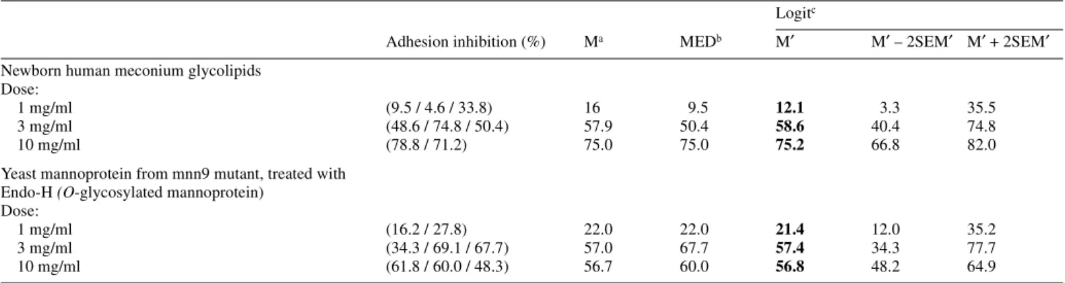
(3) Carbohydrate receptors for Lactobacillus adhesion in the gut. TLC plates (Karlsson, 1989; see Binding of L.johnsonii La 1 on thin-layer chromatograms). Binding studies done in the presence of a mannose glycoside, and of a glycoprotein bearing oligomannosides led to the following data (Table I): Methyl-α-D-mannoside, a specific monosaccharide glycoside able to inhibit E.coli adhesion mediated by type 1 pili (Firon et al., 1983; Neeser et al., 1986), was unable to significantly inhibit the adhesion of La1 to Caco-2 cells. Then, the yeast cell wall mannoprotein from the mnn9 mutant of Saccharomyces cerevisiae was tested, after treatment with Endo-H. Indeed, structural studies indicated that N-linked oligomannosides from this mnn9 mannoprotein expose mainly Manα1–3Man terminal sequences, whereas O-linked sugar chains expose mainly Manα1–2Man terminal sequences (Ballou, 1982). Since Manα1–3Man precisely corresponds to the “natural” high affinity ligand of E.coli type 1 pili (Neeser et al., 1986), we were prompted to selectively remove the sugar chains containing such sequences. Thus, after treatment with Endo-H which cleaves N-linked chains only, this mannoprotein can be considered as a probe carrying O-linked oligomannosides, terminated mainly by the Manα1–2Man carbohydrate sequence. The convincing adhesion inhibition data obtained with this “modified” mannoprotein can be compared with the negative results yielded by methyl-α-D-mannoside (see Table I). However, an increase from 3 to 10 mg/ml of this glycoprotein concentration did not result in an increase of its inhibitory potency (Table II). This last finding pinpoints the presence of another adhesion mechanism which is not inhibited by the mannoprotein. Together, these data suggest for La1, a mannose-sensitive adhesion mechanism of a fine carbohydrate specificity different from that of E.coli type 1 pili. These findings may be compared with those of Adlerberth et al. (1996) who identified a mannose specific adhesin in L.plantarum 299 and 299v strains, and could show that these bacteria were able to agglutinate cells of Saccharomyces cerevisiae and bind to the human colonic cell line HT- 29 in an unknown mannosesensitive manner. Similarly, our results showed an inhibition of adhesion in the presence of a O-glycosylated yeast mannoprotein, whose binding determinant was confirmed to be different from that of E.coli type 1 pili. Interestingly, Salmonella species are known to express different types of fimbriae, among which mannose-sensitive type 1 fimbriae seem to be. the most common (Glegg and Swenson, 1994). Here also, it has long been known that the fine carbohydrate specificity of the Salmonella type 1 fimbriae is different from that of the E.coli type 1 pili (Firon et al., 1983). All other compounds tested (see Table I) were ineffective, which suggested a binding of La1 to Caco-2 cells inhibited either by specific glycolipids or by selected oligomannosides. Binding of L.johnsonii La1 on thin-layer chromatograms In a second step, we focused our attention on the binding of metabolically labeled L.johnsonii La1 to glycolipids extracted from different sources and separated on TLC plates according to the method of Karlsson (1987). As seen in Table III, there was a consistent binding to gangliotriosyl- and gangliotetraosylceramide (also fucosylated gangliotetraosylceramide) and to lactotetraosylceramide (occurring in human meconium) and lactosylceramide. Some glycolipids ending in N-acetyllactosamine showed occasional binding and a number of glycolipids were consistently negative. Binding to asialo-GM1 (gangliotetraosylceramide) was very strong and has also been observed for a rat L.casei strain, another lactic acid bacteria (Yamamoto et al., 1996), and for some species of Bifidobacteria by Fujiwara et al. (1997). When dealing with pathogenic strains, Sporsem Oro et al. (1990) showed by immuno-thin layer chromatography that asialo-GM1 was a binding structure for E.coli colonization factor antigens CFA/II and CFA/IV. Later, Wenneras et al. (1995) demonstrated by blotting experiments that the coli surface 3 (CS3) subcomponent of the colonization factor antigen II of enterotoxigenic E.coli was binding to electrophoretically separated and transblotted rabbit intestinal proteins, but that the binding could be inhibited by asialo-GM1 as well as by GM1 and GM2, but not GM3. This observation implied that the critical CS3 epitope consisted of the carbohydrate sequence GalNAcβ1–4Gal and that this sequence or a sequence having the same tertiary configuration was found in the blotted protein recognized by CS3. This sequence has also been implicated as a binding structure for enteropathogenic E.coli (Jagannatha et al., 1991). In the case of La1, the highest binding to TLC plates was found with this sequence, but was not restrictive as can be seen with the lactosylceramides.. Table II. Dose/effect of two glycoconjugates mixtures on the adhesion of Lactobacillus johnsonnii (formerly acidophilus) La1 to Caco-2 cell monolayers Logitc Adhesion inhibition (%). Ma. MEDb. M′. M′ – 2SEM′ M′ + 2SEM′. Newborn human meconium glycolipids Dose: 1 mg/ml 3 mg/ml 10 mg/ml. (9.5 / 4.6 / 33.8) (48.6 / 74.8 / 50.4) (78.8 / 71.2). 16 57.9 75.0. 9.5 50.4 75.0. 12.1 58.6 75.2. 3.3 40.4 66.8. 35.5 74.8 82.0. Yeast mannoprotein from mnn9 mutant, treated with Endo-H (O-glycosylated mannoprotein) Dose: 1 mg/ml 3 mg/ml 10 mg/ml. (16.2 / 27.8) (34.3 / 69.1 / 67.7) (61.8 / 60.0 / 48.3). 22.0 57.0 56.7. 22.0 67.7 60.0. 21.4 57.4 56.8. 12.0 34.3 48.2. 35.2 77.7 64.9. a, b, c M,. MED, and Logit transformation (as in Table I).. 1195.
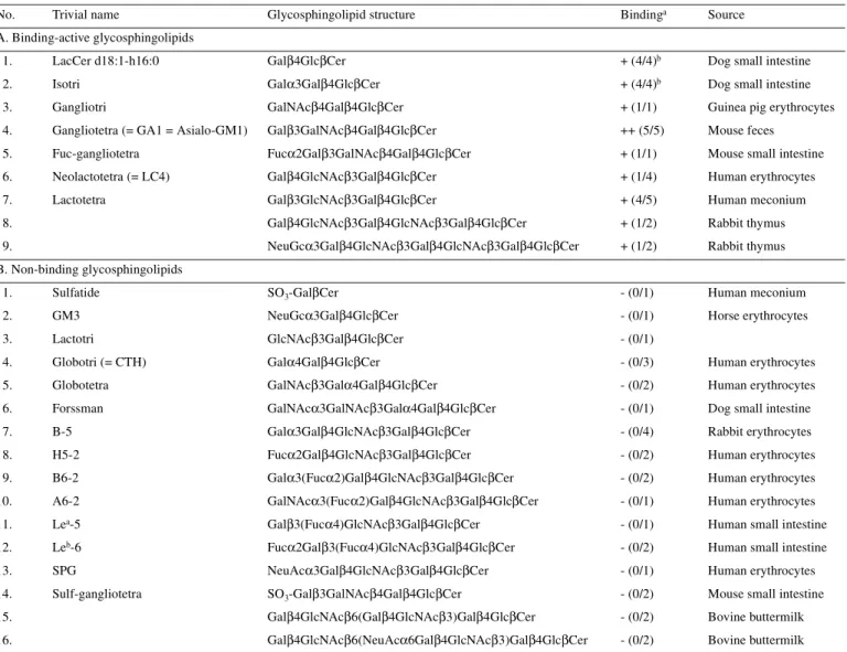
(4) J.-R.Neeser et al.. Table III. Binding of Lactobacillus johnsonii La1 to pure glycosphingolipids on thin-layer chromatograms Glycosphingolipid structure. Bindinga. Source. LacCer d18:1-h16:0. Galβ4GlcβCer. + (4/4)b. Dog small intestine. 2.. Isotri. Galα3Galβ4GlcβCer. +. 3.. Gangliotri. GalNAcβ4Galβ4GlcβCer. + (1/1). 4.. Gangliotetra (= GA1 = Asialo-GM1). Galβ3GalNAcβ4Galβ4GlcβCer. ++ (5/5). Mouse feces. 5.. Fuc-gangliotetra. Fucα2Galβ3GalNAcβ4Galβ4GlcβCer. + (1/1). Mouse small intestine. 6.. Neolactotetra (= LC4). Galβ4GlcNAcβ3Galβ4GlcβCer. + (1/4). Human erythrocytes. 7.. Lactotetra. Galβ3GlcNAcβ3Galβ4GlcβCer. + (4/5). Human meconium. No.. Trivial name. A. Binding-active glycosphingolipids 1.. (4/4)b. Dog small intestine Guinea pig erythrocytes. 8.. Galβ4GlcNAcβ3Galβ4GlcNAcβ3Galβ4GlcβCer. + (1/2). Rabbit thymus. 9.. NeuGcα3Galβ4GlcNAcβ3Galβ4GlcNAcβ3Galβ4GlcβCer. + (1/2). Rabbit thymus. Sulfatide. SO3-GalβCer. - (0/1). Human meconium. 2.. GM3. NeuGcα3Galβ4GlcβCer. - (0/1). Horse erythrocytes. 3.. Lactotri. GlcNAcβ3Galβ4GlcβCer. - (0/1). 4.. Globotri (= CTH). Galα4Galβ4GlcβCer. - (0/3). Human erythrocytes. 5.. Globotetra. GalNAcβ3Galα4Galβ4GlcβCer. - (0/2). Human erythrocytes. 6.. Forssman. GalNAcα3GalNAcβ3Galα4Galβ4GlcβCer. - (0/1). Dog small intestine. 7.. B-5. Galα3Galβ4GlcNAcβ3Galβ4GlcβCer. - (0/4). Rabbit erythrocytes. B. Non-binding glycosphingolipids 1.. 8.. H5-2. Fucα2Galβ4GlcNAcβ3Galβ4GlcβCer. - (0/2). Human erythrocytes. 9.. B6-2. Galα3(Fucα2)Galβ4GlcNAcβ3Galβ4GlcβCer. - (0/2). Human erythrocytes. 10.. A6-2. GalNAcα3(Fucα2)Galβ4GlcNAcβ3Galβ4GlcβCer. - (0/1). Human erythrocytes. 11.. Lea-5. Galβ3(Fucα4)GlcNAcβ3Galβ4GlcβCer. - (0/1). Human small intestine. 12.. Leb-6. Fucα2Galβ3(Fucα4)GlcNAcβ3Galβ4GlcβCer. - (0/2). Human small intestine. 13.. SPG. NeuAcα3Galβ4GlcNAcβ3Galβ4GlcβCer. - (0/1). Human erythrocytes. 14.. Sulf-gangliotetra. SO3-Galβ3GalNAcβ4Galβ4GlcβCer. - (0/2). Mouse small intestine. 15.. Galβ4GlcNAcβ6(Galβ4GlcNAcβ3)Galβ4GlcβCer. - (0/2). Bovine buttermilk. 16.. Galβ4GlcNAcβ6(NeuAcα6Galβ4GlcNAcβ3)Galβ4GlcβCer. - (0/2). Bovine buttermilk. marks a significant darkening on the autoradiogram when 4 µg of the glycosphingolipid was applied on the thin-layer plate, + marks a weak darkening, while – marks no darkening. The numbers in parenthesis give the number of positive binding/times tested.bGlycosphingolipid mixture utilized for testing.. a++. In the present work, we have been able to identify two major carbohydrate-binding specificities of L.johnsonii La1. The first one (mannose-specific) is slightly different from that of E.coli type 1 pili (Firon et al., 1983; Neeser et al., 1986), as are those exhibited by Salmonella type 1 fimbriae (Firon et al., 1983) and L.plantarum 299 and 299v adhesin (Adlerberth et al., 1996). The second one mediates a strong affinity for the asialoGM1 glycolipid, which has already been demonstrated for ETEC and EPEC strains (Sporsem Oro et al., 1990; Jagannatha et al., 1991; Wenneras et al., 1995), as well as for a rat L.casei strain (Yamamoto et al., 1996) and several bifidobacterial strains (Fujiwara et al., 1997). To date, the presence of asialoGM1 in human intestine has never been established but the presence of microorganisms from the normal flora able to desialylate GM1 may be assumed. However, the inhibitory activity of the human meconium glycolipid mixture observed earlier in this study is unlikely due to asialo-GM1, since newborn meconium is not exposed to a bacterial microflora during fetal life. 1196. Hence, the positive binding of La1 to other structures occurring in human meconium such as lactotetraosylceramide (see Table III), even if less strong, is probably a better explanation for the activity of such a glycolipid mixture. On the other hand, asialo-GM1 is abundantly found in the mouse intestine where it should act as a receptor for La1, enabling an already observed important colonization of the mouse gut (F.Rochat, unpublished observations) which in turn may protect the animal against infectious challenge (Bernet-Camard et al., 1997). Thus, we have shown that L.johnsonii La1 can bind cell membrane molecules through mechanisms which are shared by other pathogenic and nonpathogenic bacteria. This observation could explain, at least partly, the La1 competitivity for pathogen adhesion onto Caco-2 cells (Bernet et al., 1994). The asialo-GM1 glycolipid has also been implicated as a binding structure for other bacteria with tropism for non-intestinal tissues such as pulmonary tissue (Krivan et al., 1988). This is true in particular for Pseudomonas aeruginosa whose binding to.
(5) Carbohydrate receptors for Lactobacillus adhesion in the gut. asialo-GM1 is a tip-associated event mediated by the Cterminal region of its structural pilin subunit (Lee et al., 1994). Thus, asialo-GM1 seems to be important for the colonizing capacity of many pathogenic and nonpathogenic bacteria at different human and animal mucosal surfaces. Finally, asialoGM1 has been recognized as a specific marker of NK cells (Yu et al., 1992). The interaction of La1 with this cell population may also be one of the molecular basis of the immunomodulatory activities of this probiotic bacterial strain (Haller et al., 2000). Further studies are now needed to identify the receptor or receptors for La1 on the Caco-2 epithelial cells, and to assess the precise involvement of the La1-LTA in the cell–cell recognition mechanism or mechanisms. Materials and methods Inhibition of the adhesion of L.johnsonnii La1 to Caco-2 cell monolayers Adhesion of L.johnsonii La1 onto Caco-2 cells was studied as follows: L.johnsonii La1 bacteria were grown in anaerobic conditions in Man, Rogosa, Sharpe (MRS) broth (Difco) overnight at 37°C in the presence of 10 µCi of tritiated adenine/ml (21 Ci/mmol, Amersham). Caco-2 cells (American Type Culture Collection, USA) were routinely grown in Dulbecco modified Eagle’s minimal essential medium (Gibco, BRL) supplemented with 20% heat-inactivated fetal serum (Boehringer) and 1% nonessential amino acids (Gibco, BRL) as described previously (Bernet et al., 1994). Cells were used at post-confluence after 20 days of culture (fully differentiated state; Pinto et al., 1983). For adhesion assays, metabolically labeled bacteria were washed three times with PBS and resuspended at a concentration of 108 cfu/ml in 0.05 M acetate buffer pH 5 containing 0.15 M NaCl (a solution 0.05 M sodium acetate and 0.15 M sodium chloride was adjusted to pH 5.0 with glacial acetic acid); 1 ml of the bacterial suspension was incubated at 37°C for 1 h onto Caco-2 cells (6 wells plates) which had been previously washed with acetate buffer. Afterwards, supernatants were removed and cells were washed three times with the same buffer. Cell monolayers were disrupted by addition of 1 ml of 1 M NaOH. The lysate and a 0.5-ml wash were transferred in a counting vial containing 1 ml of benzethonium hydroxide (Sigma). Vials were put in an incubator at 60°C for 1 h, cooled, and counted after the addition of 10 ml of Ultima Gold (Packard). Each experiment was performed in triplicate and mean values were calculated. For adhesion inhibition assays, the compounds to be tested were dissolved in acetate buffer, then mixed with the bacterial suspension and incubated on Caco-2 cells as described above. Experiments were repeated twice or three times (always involving triplicates) for each test compound. The logit transformation was chosen as the best tool for statistical calculations (better than the use of a mean or a median), since it takes into account the limitation of the inhibition rate between 0 and 100% only. Human newborn meconium glycolipids and glycoproteins were separated as described by Hounsell et al. (1985). The preparation of the samples to be used for testing concentrations of 1 mg/ml and 3 mg/ml of this glycolipid mixture in acetate. buffer yielded clear solutions, whereas the most concentrated suspensions (10 mg/ml) required a centrifugation step before mixing with the bacteria. Saccharomyces cerevisiae mnn 9 mutant was a kind gift of C.E.Ballou (Department of Biochemistry, University of California, Berkeley). The isolation of the cell wall mannoprotein was done according to Frevert and Ballou (1985). Briefly, anion-exchange chromatography on DEAESephadex A-50 using a 0.3 M sodium chloride solution as an eluent yielded a large retained fraction which was collected and further homogenized to purity on a hydroxyapatite gel. The release of the N-linked sugar chains was performed by a treatment with endo-β-N-acetylglucosaminidase-H (Endo-H), as described by Tsai et al. (1986). This enzymatic treatment was repeated twice and the removal of N-glycans was followed by high-performance thin layer chromatography: under such conditions, no more N-glycan chains were released at the end of the second incubation period. The resulting mannoprotein was finally purified by liquid chromatography on a Sephadex G-50 column (Pharmacia, Sweden). The other tested compounds were from Sigma Chemical Co. (St. Louis, MO). Binding of L.johnsonii La1 on thin-layer chromatograms Briefly, total acid and nonacid glycolipid fractions, from the sources given in Table III, were isolated as described (Karlsson, 1987). The pure glycolipids used in the binding studies were isolated by repeated chromatography of native glycolipids, or acetylated derivatives, on silicic acid columns (Iatrobeads 6RS-8060, Iatron Laboratories Inc., Tokyo, Japan) or by HPLC on silicic acid columns. The isolated glycolipids were characterized by mass spectrometry (Samuelsson et al., 1990), proton NMR spectroscopy (Falk et al., 1979a–c; Koerner et al., 1983), and degradation studies (Yang and Hakomori, 1971; Stellner et al., 1973). Binding of 35S-labeled L.johnsonii La1 to glycolipids on thin-layer chromatograms was examined as follows: L.johnsonii bacteria were cultured in MRA broth supplemented with 50 µCi 35S-methionine/10 ml (Amersham, UK) at 37°C overnight under anaerobic conditions. The bacteria were collected by centrifugation, and washed three times with phosphatebuffered saline (PBS), pH 7.3. The bacteria were thereafter suspended in PBS to approximately 1 × 10⁸ CFU/ml. The specific activities of the suspensions were approximately 1 c.p.m. per 100 bacterial cells. The chromatogram binding assay was done as described previously (Hansson et al., 1985). Glycolipid mixtures (20–40 µg/lane), or pure glycolipids (1–4 µg/lane), were separated on glass- or aluminum-backed silica gel 60 HPTLC plates (Merck, Darmstadt, Germany), using chloroform/ methanol/water (60:35:8, by volume) as solvent system. The dried chromatograms were dipped in 0.3–0.5% (w/v) polyisobutylmethacrylate (Plexigum P28, Röhm, GmbH, Darmstadt, Germany) in diethylether/n-hexane (1:5, by volume) for 1 min, and air-dried. Blocking of unspecific binding sites was done by immersing the plates in PBS containing 2% bovine serum albumin (w/v) and 0.1% Tween 20 (w/v) for 2 h at room temperature. Thereafter, suspensions of radiolabeled bacteria (diluted in PBS to 1 × 10⁸ CFU/ml and 1–5 × 10⁶ c.p.m./ml) were gently sprinkled over the chromatograms, and incubated for 2 h at room temperature. After washing six times with PBS, and drying, the thin-layer plates were autoradiographed for 12–48 h using XAR-5 x-ray films (Eastman Kodak, Rochester, NY). 1197.
(6) J.-R.Neeser et al.. Acknowledgments We thank M.Golliard for skillful technical assistance in the preparation of the Endo-H treated yeast cell wall mannoprotein, J.-M.Aeschimann for statistical analysis, I.Corthesy and E.Pasche for helpful discussions, and F.Stingele for careful reading and improvement of the manuscript. References Adlerberth,I., Ahrné,S., Johansson, M-L., Molin,G., Hanson,L.A. and Wold,A.E. (1996) A mannose-specific adherence mechanism in Lactobacillus plantarum conferring binding to the human colonic cell line HT-29. Appl. Environ. Microbiol., 62, 2244–2251. Ballou,C.E. (1982) Yeast cell wall and cell surface, In Strathern,J.N., Jones,E.W. and Broach,J.R. (eds.), The Molecular Biology of the Yeast Saccharomyces. Metabolism and Gene Expression. Cold Spring Harbor Laboratory Press, Cold Spring Harbor, NY, pp. 335–360. Beachey,E. and Ofek,I. (1976) Epithelial cell binding of group A Streptococci by lipoteichoic acid on fimbriae denuded M protein. J. Exp. Med., 143, 759–771. Berg,R.D. (1996) The indigenous gastrointestinal microflora. Trends Microbiol., 4, 430–435. Bernet,M-F., Brassart,D., Neeser,J.-R. and Servin,A.L. (1994) Lactobacillus acidophilus LA 1 binds to cultured human intestinal cell lines and inhibits cell attachment and cell invasion by enterovirulent bacteria. Gut, 35, 483–489. Bernet-Camard,M-F., Liévin,V., Brassart,D., Neeser,J.-R., Servin,A.L. and Hudault,S. (1997) The human Lactobacillus acidophilus strain La1 secretes a non bacteriocin antibacterial substance active in vitro and in vivo. Appl. Environ. Microbiol., 63, 2747–2753. Brassart,D. and Schiffrin,E.J. (1997) Probiotics for improved gut health. Trends Food Sci. Technol., 8, 321–326. Canil,C., Rosenshine,I., Ruschkowski,S., Donnenberg,M.S., Kaper,J.B. and Finlay,B.B. (1993) Enteropathogenic Escherichia coli decreases the transepithelial electrical resistance of polarized epithelial monolayers. Infect. Immun., 61, 2755–2762. Falk,K.-E., Karlsson,K.-A. and Samuelsson,B.E. (1979a) Proton nuclear magnetic resonance analysis of anomeric structure of glycolipids. The globoseries (one to five sugars). Arch. Biochem. Biophys., 192, 164–176. Falk,K.-E., Karlsson,K.-A. and Samuelsson,B.E. (1979b) Proton nuclear magnetic resonance analysis of anomeric structure of glycolipids. Blood group ABH-active substances. Arch. Biochem. Biophys., 192, 177–190. Falk,K.-E., Karlsson,K.-A. and Samuelsson,B.E. (1979c) Proton nuclear magnetic resonance analysis of anomeric structure of glycolipids. Lewis-active and Lewis-like substances. Arch. Biochem. Biophys., 192, 91–202. Finley,B.B. and Cossart,P. (1997) Exploitation of mammalian host cell functions by bacterial pathogens. Science, 276, 718–725. Finlay,B.B. and Falkow,S. (1990) Salmonella interactions with polarized human intestinal Caco-2 epithelial cells. J. Infect. Dis., 162, 1096–1106. Finlay,B.B. and Siebers,A. (1995) Mechanisms of mucosal colonization and penetration by bacterial pathogens. In Roth,J.A., Bolin,C.A., Brogden,K.A., Minion,F.C. and Wannemuhler,M.O. (eds.), Virulence Mechanisms of Bacterial Pathogens, 2nd ed. American Society for Microbiology, Washington, DC, pp. 33–45. Firon,N., Ofek,I. and Sharon,N. (1983) Carbohydrate specificity of the surface lectins of Escherichia coli, Klebsiella pneumoniae and Salmonella typhimurium. Carbohydr. Res., 120, 235–249. Frevert,J. and Ballou,C.E. (1985) Saccharomyces cerevisiae structural cell wall mannoprotein. Biochemistry, 24, 753–759. Fujiwara,S., Hashiba,H., Hirota,T. and Forsner,J.F. (1997) Proteinaceous factor (s) in culture supernatant fluids of Bifidobacteria which prevents the binding of enterotoxigenic Escherichia coli to gangliotetraosylceramide. Appl. Environ. Microbiol., 63, 506–512. Glegg,S. and Swenson,D.L. (1994) Salmonella fimbriae. In Klemm,P. (ed.), Fimbriae: Adhesion, Genetics, Biogenesis and Vaccines. CRC Press, Boca Raton, FL, pp. 105–112. Granato,D., Perotti,F., Masserey,I., Rouvet,M., Golliard,M., Servin,A. and Brassart,D. (1999) Cell surface-associated lipoteichoic acid acts as an adhesion factor for attachment of Lactobacillus johnsonii La1 to human enterocyte-like Caco-2 cells. Appl. Environ. Microbiol., 65, 1071–1077.. 1198. Haller,D., Blum,S., Bode, Ch., Hammes,W.P. and Schiffrin,E.J. (2000) Activation of human PBMC by non-pathogenic bacteria in vitro: evidence of NK cells as primary targets. Infect. Immun., 68, 752–759. Hansson,G.C., Karlsson,K.-A., Larson,G., Strömberg,N. and Thurin,J. (1985) Carbohydrate-specific adhesion of bacteria to thin-layer chromatograms: a rationalized approach to the study of host cell glycolipid receptors. Anal. Biochem., 146, 158–163. Hounsell,E.F., Lawson,A.M., Feeney,J., Gooi,H.C., Pickering,N.J., Stoll,M.S., Lui,S.C. and Feizi,T. (1985) Structural analysis of the O-glycosidically linked core-region oligosaccharides of human meconium glycoproteins which express oncofetal antigens. Eur. J. Biochem., 148, 367–377. Jagannatha,H.M., Sharma,U.K., Ramaseshan,T., Surolia,A. and Balganesh,T.S. (1991) Identification of carbohydrate structures as receptors for localized adherent enteropathogenic Escherichia coli. Microbial Pathogenesis, 11, 259–268. Karlsson,K.-A. (1987) Preparation of total non-acid glycolipids for overlay analysis of receptors for bacteria and viruses and for other studies. Methods Enzymol., 138, 212–220. Karlsson,K-A., (1989) Animal glycosphingolipids as membrane attachment sites for bacteria. Annu. Rev. Biochem., 58, 309–350. Koerner,T.A.W.,Jr., Prestegard,J.H., Demou,P.C. and Yu,R.K. (1983) Highresolution proton NMR studies of gangliosides. 1. Use of homonuclear spin-echo J-correlated spectroscopy for determination of residue composition and anomeric configurations. Biochemistry, 22, 2676–2687. Krivan,H.C., Roberts D.D. and Ginsburg V. (1988) Many pulmonary pathogenic bacteria bind specifically to the carbohydrate sequence GalNAcβ1–4Gal found in some glycolipids. Proc. Natl. Acad. Sci. USA, 85, 6157–6161. Lee,K.K., Sheth,H.B., Wong,W.Y., Sherburne,R., Paranchych,W., Hodges,R.S., Lingwood,C.A., Krivan,H. and Irvin,R.T. (1994) The binding of Pseudomonas aeruginosa pili to glycosphingolipids is a tip-associated event involving the C-terminal region of the structural pilin subunit. Mol. Microbiol., 11, 705–713. Lee,Y.-K. and Salminen,S. (1995) The coming of age of probiotics. Trends Food Sci. Technol., 6, 241–245. Link-Amster,H., Rochat,F., Saudan,K.Y., Mignot,O. and Aeschlimann,J.-M. (1994) Modulation of a specific humoral immune response and changes in intestinal flora mediated through fermented milk intake. FEMS Immunol. Med. Microbiol., 10, 55–64. Neeser,J.-R., Koellreutter,B. and Wuersch,P. (1986) Oligomannoside-type glycopeptides inhibiting adhesion of Escherichia coli strains mediated by type 1 pili: preparation of potent inhibitors from plant glycoproteins. Infect. Immun., 52, 428–436. Neeser,J.-R., Chambaz,A., Golliard,M., Link-Amster,H., Fryder,V. and Kolodziejczyk,E. (1989) Adhesion of colonization factor antigen II-positive enterotoxigenic Escherichia coli strains to human enterocyte-like differentiated HT-29 cells: a basis for host–pathogen interactions in the gut. Infect. Immun. 57, 3727–34. Ofek,I., Beachey,E.H., Jefferson,W. and Campbell,G.L. (1975) Cell membrane binding properties of group A streptococcal lipoteichoic acid. J. Exp. Med. 141, 990–1003. Pinto,M., Robine-Leon,S., Appay,M.D., Kedinger,M., Triadou,N., Dussaulx,E., Lacroix,B., Simon-Assmann,P., Haffen,K., Fogh,J. and Zweibaum,A. (1983) Enterocyte-like differentiation and polarization of the human colon carcinoma cell line Caco-2 in culture. Biol. Cell., 47, 323–330. Samuelsson,B.E., Pimlott,W. and Karlsson,K.-A. (1990) Mass spectrometry of mixtures of intact glycosphingolipids. Methods Enzymol., 193, 623–646 Schiffrin,E.J., Rochat,F., Link-Amster,H., Aeschlimann,J.-M. and DonnetHughes,A. (1995) Immunomodulation of human blood cells following the ingestion of lactic acid bacteria. J. Dairy Sci., 78, 491–497. Sherman,L.A. and Savage,D.C. (1986) Lipoteichoic acids in Lactobacillus strains that colonize the mouse gastric epithelium. Appl. Environ. Microbiol., 52, 302–304. Sporsem Oro,H., Kolsto, A-B., Wenneras,C. and Svennerholm, A-M. (1990) Identification of asialo GM1 as a binding structure for Escherichia coli colonization factor antigens. FEMS Microbiol. Lett., 72, 289–292. Stellner,K., Saito,H. and Hakomori,S.-I. (1973) Determination of aminosugar linkages in glycolipids by methylation. Aminosugar linkages of ceramide pentasaccharides of rabbit erythrocytes and of Forssman antigen. Arch. Biochem. Biophys., 155, 464–472. Tancrede,C. (1992) Role of human microflora in health and disease. Eur. J. Clin. Microbiol. Infect. Dis., 11, 1012–1015..
(7) Carbohydrate receptors for Lactobacillus adhesion in the gut. Teti,G., Chiofalo,M.S., Tomasello,F., Orefici,G. and Mastroeni,P. (1987a) Mediation of Staphylococcus saprophytus adherence to uroepithelial cells by lipoteichoic acid. Infect. Immun., 55, 839–842. Teti,G., Tomasello,F., Chiofalo,M.S., Orefici,G. and Mastroeni,P. (1987b) Adherence of group B Streptococci to adult and neonatal epithelial cells is mediated by lipoteichoic acid. Infect. Immun., 55, 3057–3064. Toba,T., Virkola,R., Westerlund,B., Björkman,Y., Sillanpää,J., Vartio,T., Kalkkinen,N. and Korhonen,T.K. (1995) A collagen-binding S-layer protein n Lactobacillus crispatus. Appl. Environ. Microbiol., 61, 2467–2471. Tsai,P-K., Dell,A. and Ballou,C.E. (1986) Characterization of acetylated and acetolyzed glycoprotein high-mannose core oligosaccharides by fast-atombombardment mass spectrometry. Proc. Natl. Acad. Sci. USA, 82, 4119–4123.. Wenneras,C., Neeser,J.-R. and Svennerholm,A.-M. (1995) Binding of the fibrillar CS3 adhesin of enterotoxigenic Escherichia coli to rabbit intestinal glycoproteins is competitively prevented by GalNAcβ1–4Gal-containing glycoconjugates. Infect. Immun., 63, 640–646. Yamamoto,K., Miwa,T., Taniguchi,H., Nagano,T., Shimamura,K., Tanaka,T. and Kumagai,H. (1996) Binding specificity of Lactobacillus to glycolipids. Biochem. Biophys. Res. Commun., 228, 148–152. Yang,H.-J. and Hakomori,S.-I. (1971) A sphingolipid having a novel ceramide and lacto-N-fucopentose III. J. Biol. Chem., 246, 1192–1200. Yu,Y.Y., Kummar,V. and Bennett,M. (1992) Murine natural killer and marrow graft rejection. Annu. Rev. Immunol., 10, 189–213.. 1199.
(8) J.-R.Neeser et al.. 1200.
(9)
Figure

Documents relatifs