Thierry A. G. M. Huisman
Received: 12 July 2004 Accepted: 29 November 2004 Published online: 5 February 2005 # Springer-Verlag 2005
Intracranial hemorrhage: ultrasound, CT
and MRI findings
Abstract Intracranial hemorrhage is one of the most common causes of acute focal neurologic deficit in children and adults. Neuroimaging including ultrasonography (US), computer tomography (CT) and magnetic resonance imaging (MRI) is essential in the diagnosis of intracra-nial hemorrhage. Imaging findings should guide treatment. The highly variable appearance of an intracranial hemorrhage can be challenging. A thorough knowledge of hematoma
evolution and US, CT and MR hematoma characteristics is manda-tory for adequate interpretation of findings. The purpose of this review is (1) to summarize the imaging characteristics of intracranial hemor-rhage on various imaging techniques and (2) to review the various types of intracranial hemorrhage, and their causes.
Keywords Intracranial hemorrhage . Ultrasonography . CT . MRI
Introduction
Intracranial hemorrhage is one of the most common causes of acute focal neurologic deficit in children and adults [1,2]. Acute morbidity and mortality, as well as final outcome, is influenced by an early and accurate diagnosis [1]. Imaging findings guide acute treatment and should prevent therapy-induced secondary complications. Computer tomography (CT), magnetic resonance imaging (MRI) and ultrasonog-raphy (US), mainly used in neonates, are well established imaging modalities in the diagnosis of intracranial hemor-rhage [1–8]. The highly variable appearance of intracranial hemorrhage can, however, be challenging. Imaging char-acteristics depend on multiple factors including size and location of the hemorrhage, as well as biological factors such as the patient’s hematocrit, tissue pO2 and pH and
protein concentration (mainly hemoglobin, Hb). In addi-tion, the physiological breakdown of the hematoma with formation of various Hb derivatives (intracellular oxy-Hb, intracellular deoxy-Hb, intracellular met-Hb, extracellular met-Hb and hemosiderin) will change the hematoma imaging characteristics over time [1–7]. Moreover, while ultrasonography and CT imaging characteristics of an in-tracranial hematoma are straightforward, the MRI
charac-teristics are variable due to the complex interaction between the different hematoma breakdown products and the MRI used sequences [1–7]. Consequently, a thorough knowl-edge of hematoma evolution and MR signal characteristics is mandatory for adequate interpretation of MR findings.
Finally, US, CT and MRI findings can guide treat-ment by narrowing the differential diagnosis of intracra-nial hemorrhages.
The purpose of this review is (1) to summarize the imaging characteristics of intracranial hemorrhage on var-ious imaging techniques and (2) to review the varvar-ious types of intracranial hemorrhage, and their causes.
Imaging characteristics of intracranial hemorrhage
A basic knowledge of the pathophysiology and evolu-tion of intracranial hemorrhages is essential for the under-standing of the sequential imaging changes of intracranial hemorraghes.
Intracranial hemorhages are typically defined in five distinct stages: hyperacute (<12 h), acute (12 h to 2 days), early subacute (2–7 days), late subacute (8 days to 1 month) and chronic (>1 month to years) [3].
T. A. G. M. Huisman (*) Department of Diagnostic Imaging, University Children’s Hospital Zurich, Steinwiesstrasse 75,
8032 Zurich, Switzerland
e-mail: thierry.huisman@kispi.unizh.ch Tel.: +41-1-2667110
A hyperacute intracranial hemorrhage is initially liquid, composed of up to 98% oxygen saturated hemoglobin. Within the first hours, a complex heterogeneous mass consisting of a matrix of red blood cells (RBC), white blood cells (WBC) and platelet thrombi intermixed with protein-rich serum develops. Within the next few hours, the clot retracts. The expelled serum as well as a reactive vasogenic edema within the adjacent brain tissue, sur-rounds the retracting blood clot with densely packed RBCs. During the hyperacute stage, the trapped RBCs keep their normal biconcave shape and the incorporated hemoglobin remains oxygenated (intracellular oxy-Hb). With evolution to the acute stage, progressive RBC energy depletion re-sults in dehydration and shrinking of the RBCs. At the same time, the intracellular hemoglobin becomes progres-sively deoxygenated (intracellular deoxy-Hb). The cell membranes of the RBCs remain intact. In the subsequent early subacute phase, a centripetal oxidative denaturation of hemoglobin within the intact RBCs occur and deoxy-Hb is consequently gradually converted to methemoglobin (intracellular meth-Hb). During the following late sub-acute stage, progressing RBCs lysis result in a release of meth-Hb in the extracellular space (extracellular meth-Hb). At the same time, the surrounding vasogenic edema slowly subsides. In the final chronic stage, macrophages and astroglial cells will surround the hematoma and slowly phagocytize the hematoma. Extracellular meth-Hb is con-verted by and stored within the macrophages as hemosid-erin and ferritin. Eventually, the hematoma will resolve completely and a cystic fluid filled or collapsed brain de-fect will ensue. In adults, the hemosiderin/ferritin loaded macrophages can persist for years, while in children the macrophages may disappear with time [9].
Ultrasonography has traditionally been used in neo-nates, in whom the anterior fontanelle is used as an acoustic window to the cranial vault. The posterior and mastoid (posterolateral) fontanelles can be used for examining the posterior fossa, including cerebellum and brainstem [8]. This approach allows examination of the intracranial anat-omy and disease processes at the bedside, and is cost-effi-cient. In addition, because of the lack of ionizing radiation and the fact that the examination can be performed with-out the need for sedation, neurosonography is exquisitely well suited for the detection of intracranial hemorrhages in neonates. One disadvantage is that ultrasonography is limited to neonates and small infants because the fonta-nelles typically close by 12 months of age [8]. Ultrasonog-raphy including color coded duplex sonogUltrasonog-raphy is done with phased-array, multiple-foci 5.0- or 7.0-MHz trans-ducers. The most frequent indication for neonatal ultraso-nography is to rule out germinal matrix hemorrhage. In premature infants, intracranial hemorrhage is usually seen within the germinal matrix, the area along the ventricular wall from which the cortical neurones as well as glial cells originate. The germinal matrix is highly perfused because the neuronal proliferation is a very active metabolic
pro-cess. Consequently, hemorrhages in premature infants typ-ically occur within the germinal matrix (Fig. 1a, b, c). Ultrasonography has proven its value in the identification of germinal matrix hemorrhages and their complications (hydrocephalus). Germinal matrix hemorrhages are divided into four grades, depending upon their severity. Grade I is defined as a germinal matrix hemorrhage or subependymal hemorrhage with no or minimal intraventricluar extension. Grade II is a germinal matrix hemorrhage that extends into the normal sized ventricular system. Grade III is a germinal matrix hemorrhage that extends into the enlarged ventric-ular system. Grade IV is a germinal matrix hemorrhage that extends into the adjacent cerebral hemispheres. Hemi-spheric hemorrhage most probably results from a hemor-rhagic venous ischemia (Fig.1c) [12,13]. Ultrasonography findings of germinal matrix hemorrhages parallel the pre-viously described stages of intracranial hemorrhage; an exact differentiation between the stages is, however, lim-ited. Ultrasonography shows germinal matrix hemorrhages as focal hyperechogenic lesions. On follow-up the hemor-raghic clot retracts and becomes well defined and less echogenic. On late follow-up, the hematoma can dissolve completely with development of a cystic, fluid-filled brain defect. An intraventricular hemorrhage is usually easily delineated with a blood clot that is surrounded by hypo-echogenic cerebrospinal fluid. On follow up a hyperecho-genic lining of the ventricular system can persist for several weeks.
Subarachnoid blood is notoriously difficult to detect by ultrasonography, due to the wide normal variation in the ultrasound appearance of subarachnoid cisterns [8]. The identification of hematomas near the skull (epidural and subdural hematomas) is also limited because the bony edges of the fontanelles obscure the subdural or epidural spaces along the brain’s surface.
Comparative studies have shown that CT and MRI have a better interobserver agreement for germinal matrix and intraventricular hemorrhage and reveal more instances of intraparenchymal hemorrhages compared with ultrasonog-raphy [14]. Nevertheless, ultrasonography remains an easy to perform, bed side, first line imaging tool in neonates.
Computer tomography represents the most frequently used imaging modality in the evaluation of hemorrhagic lesions in the acute setting [1, 2]. CT is widely available and allows rapid and reliable visualization of the hemor-rhage and of its possible complications [1,2]. Moreover, in most instances, CT provides important information about the etiology of the hemorrhage. Finally, the use of intra-venous contrast agents offers the possibility of an angiog-raphic display of the intracranial vasculature in the same examination.
CT imaging characteristics are determined by the degree of X-ray attenuation by the hemorrhage (Fig. 2a). The degree of blood clot retraction, the hematocrit, the hemo-globin fraction and protein content (hemo-globin) determine the density of the lesion on CT [9]. The appearance of an
intracranial hemorrhage varies on CT parallel to the tem-poral evolution of the hemorrhage. In the hyperacute phase, the matrix mass of RBCs, WBCs, platelets and interspersed serum is equally dense as the adjacent normal brain pa-renchyma. Consequently, in the hyperacute phase it can be difficult to delineate the hemorrhage. In the acute and early subacute phase, progressive blood clot retraction with con-comitant extrusion of the serum component will rapidly increase the density of the hemorrhage. In particular, the increasing concentration of the protein component of hemoglobin (globin) will appear hyperdense on CT. Simul-taneously a hypodense rim develops around the
hemor-rhage due to serum extrusion and reactive perifocal vaso-genic edema. During the subsequent late subacute phase, progressive RBC lysis and proteolysis of the globin protein result in decreasing density of the hematoma. In the late subacute phase the hematoma may become isodense to the adjacent brain parenchyma (Fig. 2b). The injection of contrast media can be helpful in the delineation of these isodense hematomas because the periphery of the hemato-ma hemato-may show enhancement due to a disrupted blood–brain barrier [1]. During the chronic phase, the hypodense he-matoma will be progressively phagocytized, ultimately re-sulting in a resolution of the hemorrhage. Typically a small Fig. 1 a Cranial US in a 4-week-old premature (31 weeks gestation)
girl demonstrating bilateral hyperechogenic subependymal hemor-rhage grade I located at the foramen of Monroe. b Serial cranial US in a premature boy, gestational age: 9 weeks. The first US 3 days after birth, demonstrates a bilateral hyperechogenic subependymal hemorrhage grade I. On follow-up, the hemorrhage had progressed to a grade IV hemorrhage. Follow-up imaging (last two images) 13 days after birth, shows a retracting blood clot with a central hypo-echogenic signal. In addition, a severe secondary hydrocephalus de-veloped with hyperechogenic lining of the ventricular system due intraventricular extension of the hemorhage. c Serial US and MRI images of a premature boy, gestational age: 29 weeks.
Ultrasonog-raphy 5 days after birth (a) shows a grade I hemorrhage at the right side and a grade IV hemorrhage at the left side with extensive venous ischemia (hyperechogenic) within the periventricular white matter. Follow-up sagittal and axial T2-weighted MRI (b, c) 42 days after birth shows an extensive CSF filled porencephalic cyst at the left side as well as severe hydrocephalus. T2-hypointense blood products are seen subependymal at the basal ganglia. On T1-weighted MRI (d), the blood breakdown products are partially hyperintense. On T2*-GRE MRI (e), the hemosiderin is seen best as hypointense signal alterations. On DWI and apparent diffusion coefficient (ADC) maps (f), the blood clots show a DWI-hyperintense and ADC-hypointense signal due to the blood clot retraction with restricted diffusion
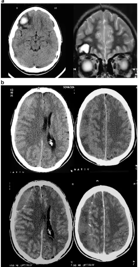
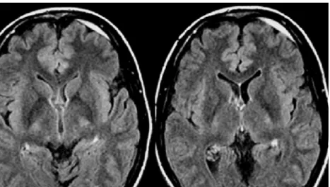
Fig. 2 a Serial axial CT and coronal T2-weighted MRI in a 22-year-old male with a fronto-basal contusion. CT in the early subacute phase shows a cen-trally hyperdense blood clot surrounded by a hypodense rim of vasogenic edema and ex-trused serum. Follow-up coronal MRI in the chronic phase shows a T2-hyperintense CSF filled brain defect lined by a T2-hypointense rim of hemosiderin. b Axial pre- and post-contrast CT in a 54-year-old male with a chronic right SDH. The SDH is nearly isodense to the adjacent cortex. After injection of trast agent, the displaced con-trast enhancing cortical vessels facilitate lesion detection
hypodense brain defect remains. Occasionally a focal en-largement of neighbouring sulci or a focal atrophy as well as residual hyperdense calcifications remain [1,9].
In cases of severe anemia (low hematocrit), due to the reduced concentration of the globin protein, the acute hematoma will be less hyperdense than expected. In ad-dition, rebleeding within a chronic hematoma will increase the overall density of the hematoma, which can be mis-leading in the estimation of the age of the hemorrhage.
Extra-axial hemorrhages (epidural and subdural hema-tomas) parallel the intraparenchymal hematomas in their CT appearance. Acute subarachnoid hemorrhages (SAH) are also hyperdense. The density of the SAH decreases rapidly, however, due to the dilution by cerebrospinal fluid. Magnetic resonance imaging is widely accepted as the most sensitive imaging modality for intracranial hemor-rhage and its sequelae. The temporal evolution of an intracranial hematoma can be studied in detail, and follows the previously described sequence. The selective sensitiv-ity of the different MRI sequences combined with MRI sequence related varying signal intensities of an intracra-nial hemorrhage increase diagnostic accuracy significantly. MR signal characteristics are determined by the para-magnetic effects of the different breakdown products of hemoglobin, the magnetic field strength, the used pulse se-quence and many other technical factors [1,2]. Intracellular oxy-Hb, intracellular deoxy-Hb, intracellular met-Hb, ex-tracellular met-Hb and hemosiderin have different mag-netic properties due to the different oxidation state of iron as it is incorporated or released from the hemoglobin mac-romolecule. Depending on the number of unpaired elec-trons in the outer orbit of the iron atom, these hematoma breakdown products will be diamagnetic (oxy-Hb, no un-paired electrons), paramagnetic (deoxy-Hb and met-Hb, few unpaired electrons) or superparamagnetic (hemosid-erin, many unpaired electrons). Consequently in the hy-peracute stage the hemorrhage is T1-iso or hypointense
and T2 hyperintense; in the acute stage T1-iso or hypo-intense and T2-hypohypo-intense, in the early subacute stage T1-hyperintense and T2-hypointense, in the late subacute stage T1 and T2-hyperintense and finally in the chronic phase T1-hypointense and T2-centrally hyperintense, surrounded by a rim of hypointensity (hemosiderin) (Fig. 2a). T1- and T2-weighted MR sequences are classically used for de-lineation of the extent and estimation of the age of an in-tracranial hemorrhage. The age of a lesion is of diagnostic importance because it determines the management of the patient. Acute and chronic hemorrhages require different treatment plans. Many other MR sequences have been de-veloped to increase the diagnostic accuracy of MR imag-ing. Fluid attenuated inversion recovery (FLAIR) and T2*-gradient and echo (T2*-GRE) MR sequences have been applied in the depiction of intracranial hemorrhages. The high sensitivity of T2*-GRE MR sequences (Fig.1c) for the susceptibility effects of paramagnetic and super-paramagnetic substances increases the number of hemor-rhagic lesions that can be identified [3, 6, 7]. A major limitation of the T2*-GRE sequence is that this sequence cannot help in estimating the age of the hematomas [3]. FLAIR sequences are favourable in the detection of SAH because the suppression of the cerebrospinal fluid signal in combination with the strong T2-weighting enhances the appearance of small amounts of blood within the cere-brospinal fluid of the ventricles or extra-axial spaces (Fig.3) [1, 7]. A recent study showed, however, that FLAIR se-quences do not provide any additional information than T2-weighted sequences for intraparenchymal hemorrhages [3]. The value of diffusion weighted imaging (DWI) in the evaluation of intracranial hemorrhage is still under active investigation [4,6,7]. DWI seems to be promising in dif-ferentiating primary intracerebral hemorrhage from hem-orrhagic cerebral ischemia [4].
Fig 3 Axial FLAIR images in a 24-year-old male with an acute small SDH. The suppression of the CSF signal enhances detec-tion of the frontal FLAIR-hy-perintense SDH
Various types of intracranial hemorrhage and their cause
Intracranial hemorrhage can be classified according to their location and etiology. Location and etiology are tightly coupled. An epidural hematoma results, for exam-ple, most frequently from a traumatic skull fracture, while a subarachnoid hemorrhage usually results from a ruptured aneurysm. Consequently, the neuroradiologist should ana-lyze and locate the hemorrhage in detail to get information about the possible etiology of the hemorrhage to guide treatment.
Intracranial hemorrhages can be (1) epidural, (2) sub-dural, (3) subarachnoidal, (4) intraventricular, (5) intra-cerebral or (6) may involve a combination of multiple compartments.
The etiologic factors for intracranial hemorrhage are numerous. Among the main categories are hemorrhagic stroke, arterial hypertension, head trauma, aneurysms and vascular malformations, intracranial neoplasms, hemato-logic disorders, drugs, cerebral amyloid angiopathy, an-giitis and miscellaneous angiopathies [10,11].
Epidural hematomas (EDH) usually occur in the setting of acute head traumas. Clinical presentation may be de-layed (symptom free or “lucid” interval). Arterial EDH usually results from a laceration of branches of the menin-geal arteries within the skull, while venous EDH result from a tear of dural venous sinuses. Venous EDH are more frequently encountered in children. Venous EDH may de-velop more slowly and the clinical symptoms are less acute than arterial EDH. A rapidly enlarging arterial EDH can result in a significant midline shift with different de-grees of secondary herniation and possible ischemia. EDH are typically well-defined biconvex extracerebral lesions that may displace the adjacent brain. EDH are located between the skull and the periostal layer of the dura mater. The tight adherence between the dura mater and the skull prevents extension of an EDH across skull sutures [15]. CT attenuation and MR signal intensities follow the pre-viously described temporal evolution. No dilution occurs with cerebrospinal fluid because an EDH is separated from the CSF by the dura mater.
Subdural hematomas (SDH) can be of traumatic or non-traumatic origin. In SDH, blood is accumulated between the dura mater and the arachnoidea. SDH are usually of venous origin and result from lacerations of cortical veins that cross the subarachnoid space before entering the dural sinuses. A concomitant arachnoidal laceration with leak-age of CSF from the subarachnoid space into the subdural space results in a mixture of CSF and venous blood. SDH may occur spontaneously in elderly patients. They usually enlarge slowly with initially minimal neurologic symp-toms, and may evolve to larger chronic hematomas that compress the adjacent brain. In children, SDHs are com-monly seen in cases of child abuse. SDHs are typically crescent-shaped along the surface of the brain or along dural duplications such as the falx cerebri and tentorium
cerebelli. Because they are located between the dura mater and the arachnoidea, SDHs may cross skull sutures. The CT attenuation or MR signal intensities also follow the previously described temporal evolution. In cases of a significant mixture with CSF the progression of blood clot resorption can be accelerated [1, 2].
Subarachnoid hemorrhage (SAH) can be of traumatic and non-traumatic origin. In SAH, blood is accumulated within the subarachnoid space, that is the space between the arachnoidea and the pia mater. SAH can occur in head injury due to laceration of cortical veins or arteries that course in the subarachnoid space but may also occur due to cortical contusions and lacerations with extravasation of blood into the subarachnoid space. A typical location for SAH after head trauma is within the interpeduncular cistern or in the sylvian fissure [15]. Spontaneous SAH usually result from ruptured aneursyms (older patients) or arterio-venous vascular malformations (younger patients). CT is the preferred method of imaging SAH because it is very sensitive in detecting blood in the CSF space and is usually more easily available in the acute setting of a SAH. Typ-ically, in acute SAH hyperdense blood is seen within the basal cisterns and subarachnoid spaces. The location and distribution of the SAH can indicate the site of the aneu-rysm. In addition, contrast enhanced CT angiography can display the exact location and shape of the aneurysm. On MRI, the rapid dilution of the hemorrhage by CSF can hamper the detection of a SAH. FLAIR sequences have shown to be very sensitive in the detection of acute SAH because the suppression of the CSF signal enhances the conspicuity of SAH [1, 2]. Identification of SAH by neonatal ultrasonography is very limited due to the wide normal variation in the normal ultrasound appearance of subarachnoid cisterns [8].
Intraventricular hemorrhage (IVH) can result from the secondary extension of a neonatal germinal matrix hem-orrhage into the ventricular system as previously described or from the reflux of blood from the subarachnoid spaces into the ventricular system after spontaneous or traumatic SAH. In addition, an intraparenchymal hemorrhage may rupture into the ventricular system. Traumatic IVH is rare and usually associated with other manifestations of intra-cranial posttraumatic lesions [9]. In analogy to SAH, the blood is rapidly diluted by the CSF. Ultrasonography in neonates and CT in adults are the primary, acute imaging modalities. MRI, especially FLAIR sequences, can also be helpful in the detection of IVH. Fluid sedimentation levels are frequently seen within the dependant parts of the ven-tricular system. On late follow up MRI, a T2-hypointense lining of the ventricular system can persist due to hemo-siderin depositions.
Intracerebral hemorrhages (ICH) has many different etiologies which can roughly be divided into traumatic and spontaneous reasons. Post-traumatic ICH are relatively straightforward and result from the direct impact of forces to the skull and underlying brain with laceration and
rup-ture of intraparenchymal vessels. Arterial hypertension, hemorrhagic stroke and coagulation disorders including anticoagulant drugs are among the most frequent reasons for spontaneous ICH. In addition, spontaneous ICH can occur due to the rupture of intracranial aneurysms and vascular malformations as well as due to hemorrhages within intracerebral neoplasms. The shape and imaging characteristics of an ICH follow the previously described evolution characteristics of a hematoma. US can easily identify intraparenchymal hemorrhages in neonates, CT and MRI are both very sensitive in the detection of ICH. The multiplanar imaging capability and the different MR imaging sequences (T1, T2, T2*, FLAIR, DWI) can increase the diagnostic accuracy of MRI compared to CT. One disadvantage is that the monitoring of a critically diseased patient is somewhat limited in MRI compared with CT.
Combinations of the different kinds of intracranial hem-orrhages are usually encountered in critically injured trau-ma patients. Prognosis is usually limited.
Conclusion
A proper knowledge of the imaging characteristics of intracranial hemorrhages and their evolution over time as well as an insight into the different etiologies of intracra-nial hemorrhages are mandatory to guide treatment. US, CT and MRI rely on different techniques. The neuro-radiologist should be aware of the advantages and dis-advantages of each individual imaging technique. US is almost exclusively limited to neonatal hemorrhages, while CT is most frequently used in the acute setting. MRI is used when the CT imaging findings do not explain the neurologic symptoms or if a discrepancy exists between the CT imaging findings and neurology.
References
1. Parizel PM, Makkat S, Van Miert E, van Goethem JW, van den Hauwe L, De Schepper AM (2001) Intracranial hemorrhage: principles of CT and MRI interpretation. Eur Radiol 11:1770– 1783
2. Anzalone N, Scotti R, Riva R (2004) Neuroradiologic differential diagnosis of cerebral intraparenchymal hemor-rhage. Neurol Sci 24:S3–S5 3. Alemany Ripoll M, Stenborg A,
Sonninen P, Terent A, Raininko R (2004) Detection and appearance of intraparencymal haematomas of the brain at 1.5 T with spin–echo, FLAIR and GE sequences: poor relationship to the age of the haematoma. Neuroradi-ology 46:435–443
4. Zaheer A, Ozsunar Y, Schaefer PW (2000) Magnetic resonance imaging of cerebral hemorrhagic stroke. Top Magn Reson Imaging 11(5):288–299
5. Wasenko JJ, Lieberman KA, Rodziewicz GS, Holsapple (2002) Magnetic resonance imaging charac-teristics of hyperacute hemorrhage in the brain and spine. Clin Imaging 26:330–337
6. Wiesmann M, Mayer TE, Yousry I, Hamann GF, Brückmann H (2001) detection of hyperacute parenchyma hemorrhage of the brain using echo-planar T2*-weighted and diffusion-weighted MRI. Eur Radiol 11:849–853 7. Lin DDM, Filippi CG, Steever AB,
Zimmerman RD (2001) Detection of intracranial hemorrhage: comparison between gradient-echo images and b0
images obtained from diffusion-weighted echo-planar sequences. Am J Neuroradiol 22:1275–1281
8. Di Salvo DN (2001) A new view of the neonatal brain: clinical utility of sup-plemental neurologic US imaging win-dows. Radiographics 21:943–955 9. Osborn AG (1994) Intracranial
hemor-rhage. In: Diagnostic neuroradiology. Mosby, St Louis, pp 154–198 10. Garcia JH (1997) Vascular diseases. In:
Neuropathology the diagnostic ap-proach. Mosby, St Louis, pp 263–320
11. Hillemans A, Kortmann RD, Herrlinger U, Skalej M, Krapf H (2003) Recurrent delayed brain hemorrhage over years after irradiation and chemotherapy for astrocytoma. Eur Radiol 13(8):1891– 1894
12. Volpe JJ (2000) Intracranial haemor-rhage: subdural, primary subarachnoid, intracerebellar, intraventricular (term infant), and miscellaneous. In: Volpe JJ (ed) Neurology of the newborn. Intra-cranial hemorrhage, 4th edn. Saunders, Philadelphia, pp 397–429
13. Barkovich AJ (1995) Destructive brain disorders of childhood. In: Barkovich AJ (ed) Pediatric neuroimaging. 2nd edn. Raven Press, New York, pp 107– 175
14. Blankenberg FG, Loh NN, Bracci P et al (2000) Sonography, CT, and MR imaging: a prospective comparison of neonates with suspected intracranial ischemia and hemorrhage. Am J Neu-roradiol 21:213–218
15. Gean AD (1994) Extra-axial collec-tions. In: Imaging of head trauma. Raven Press, New York, pp 75–145