Structure and Dynamics of the Acyl Chains in the Membrane
Tra
fficking and Enzymatic Processing of Lipids
Stefano Vanni,
*
,†,‡Laura Riccardi,
§Giulia Palermo,
∥and Marco De Vivo
*
,§ †Department of Biology, University of Fribourg, Chemin du Musée 10, 1700 Fribourg, Switzerland ‡Université Côte d’Azur, CNRS, IPMC, 06560 Valbonne, France§Laboratory of Molecular Modeling and Drug Discovery, Istituto Italiano di Tecnologia, Via Morego 30, 16163 Genoa, Italy ∥Department of Bioengineering, University of California Riverside, Riverside, California 92521, United States
CONSPECTUS:The regulatory chemical mechanisms of lipid trafficking and degradation are involved in many pathophysio-logical processes, being implicated in severe pain, in flamma-tion, and cancer. In addiflamma-tion, the processing of lipids is also relevant for industrial and environmental applications. However, there is poor understanding of the chemical features that control lipid membrane trafficking and allow lipid-degrading enzymes to efficiently select and hydrolyze specific fatty acids from a complex cellular milieu of bioactive lipids. This is particularly true for lipid acyl chains, which have diverse structures that can critically affect the many complex reactions needed to elongate, desaturate, or transport fatty acids.
Building upon our own contributions in this field, we will
discuss how molecular simulations, integrated with experimental evidence, have revealed that the structure and dynamics of the lipid tail are actively involved in modulating membrane trafficking at cellular organelles, and enzymatic reactions at cell membranes. Further evidence comes from recent crystal structures of lipid receptors and remodeling enzymes. Taken together, these recent works have identified those structural features of the lipid acyl chain that are crucial for the regioselectivity and stereospecificity of essential desaturation reactions. In this context, we will first illustrate how atomistic and coarse-grained simulations have elucidated the structure−function relationships between the chemical composition of the lipid’s acyl chains and the molecular properties of lipid bilayers. Particular emphasis will be given to the prominent chemical role of the number of double carbon−carbon bonds along the lipid acyl chain, that is, discriminating between saturated, monounsaturated, and polyunsaturated lipids. Different levels of saturation in fatty acid molecules dramatically influence the biophysical properties of lipid assemblies and their interaction with proteins. We will then discuss the processing of lipids by membrane-bound enzymes. Our focus will be on lipids such as anandamide and 2-arachidonoylglycerol. These are the main molecules that act as neurotransmitters in the endocannabinoid system. Specifically, recent findings indicate a crucial interplay between the level of saturation of the lipid tail, its energetically and sterically favored conformations, and the hydrophobic accessory cavities in lipid-degrading enzymes, which help form catalytically active conformations of the selected substrate.
This Account will emphasize how the specific chemical structure of acyl chains affects the molecular mechanisms for modulating membrane trafficking and selective hydrolysis. The results examined here show that, by using molecular simulations to investigate lipid plasticity and substrateflexibility, researchers can enrich their interpretation of experimental results about the structure−function relationships of lipids. This could positively impact chemical and biological studies in the field and ultimately support protein engineering studies and structure-based drug discovery to target lipid-processing enzymes.
■
INTRODUCTIONLipids are one of the major components of the cell and are essential for several processes that are necessary for life and health.1 They are the building blocks of cellular membranes, defining the boundaries and intracellular compartmentalization within the cell. In addition, they play a major role as signaling molecules in a wide variety of cellular pathways.2 For these reasons, lipid trafficking and processing within the cell are often implicated in pain control and in severe in flammation-related diseases such as cancer.3 In addition, lipid-degrading
enzymes have relevant biotechnological and environmental applications such as the degradation of lipid-rich wastewaters.4 Yet there has been little exploration of how the structural and chemical diversity of lipids affects their trafficking and degradation.
The vast majority of endogenous lipids share a common chemical architecture, characterized by a polar head that is
http://doc.rero.ch
Published in "Accounts of Chemical Research 52(11): 3087–3096, 2019"
which should be cited to refer to this work.
linked by a distinct structural backbone to a tail made of one or more acyl chains.2,5 Amazingly, they have diverse chemical structures, since the existing variety of possible heads and tails generates countless combinatorial chemical possibilities to form lipids with specific structures and chemistry (Figure 1). As a consequence, lipid nomenclature and conventions combine the chemical structure of the polar head (e.g., phosphoinositides, PI; phosphatidylcholine, PC; phosphatidy-lethanolamine, PE; phosphatidylserine, PS) with that of the acyl chains (e.g., dipalmitoyl, DP; dioleoyl, DO; palmitoyl-oleoyl, PO, etc.), giving rise to a 4-letter acronym that represents different lipid species, like DPPC, POPE, DOPS, etc.
Despite this chemical diversity in both the head and tail, the vast majority of biological studies have focused specifically on the role of the lipid polar head,1,2,6,7andin vitro reconstitutions of lipid assemblies are often prepared using lipid mixtures with mixed acyl chain content, such as egg-PC, the commercial cost of which is typically cheaper than that of lipids with defined acyl chains. This reflects the wide recognition of the head as the lipid structure’s crucial functional component, possibly as a consequence of the observation that several membrane binding proteins show a marked specificity for polar head groups of lipids.6,8While pioneering studies in the late 1990s had already proposed that lipid tails could be important in membrane recognition and trafficking,9−12 recent findings have revived the general interest in the critical role of lipid acyl chains in numerous important cellular processes.13−18In particular, two main properties of acyl chains have been identified as key determinants in lipid-mediated mechanisms: (i) the hydro-phobic length of the acyl chain and (ii) the ratio between saturated and unsaturated bonds along the acyl chains (Figure
1). These structural properties play important roles in establishing specific interactions of individual lipid molecules with proteins and, at high concentrations, in shaping the overall properties of membranes.13−18
Researchers have investigated and largely clarified how structurally diverse polar heads affect interactions with, for example, partner proteins.19 However, there is still poor understanding of how the chemical composition of the lipid’s acyl chains affects cellular mechanisms within the cell. Indeed, it is only recently that researchers have asked how subtle structural differences between acyl tails may affect trafficking and enzymatic processing. How does the presence of two additional carbon atoms (e.g., 16 vs 18) influence protein recognition and binding? And, in the case of unsaturated lipids, how does the presence of a CC double bond (basically characterized only by a shorter interatomic distance and two fewer hydrogen atoms) generate such a dramatic impact on membrane properties?
An obvious consideration is that lipid acyl chains are intrinsicallyflexible. Thus, entropic effects are expected to play a role in determining how lipids interact with the biological components of the surrounding environment. Yet this observation is not sufficient to envision and comprehend molecular mechanisms that are modulated by the specific chemical structure of acyl chains. An atomic-level under-standing of the conformationalflexibility of acyl chains is thus critical to obtain an accurate representation of their behavior and functional roles.
In this regard, molecular dynamics (MD) simulations are a powerful approach for investigating lipid function and examining the dynamics of molecular systems at atomic resolution.20−22Indeed, in the past few years, MD studies have
Figure 1.Schematic representation of the combinatorial possibilities giving rise to the diversity of lipid chemistry. Only some of the major species of polar heads and acyl chains are shown for clarity. PC, phosphatidylcholine; PE, phosphatidylethanolamine; PS, phosphatidylserine; PI, phosphatidylethanolamine; PA, phosphatidic acid.
combination of modeling with structural and kinetic experimental data has furthered our knowledge of the structure−function relationships of lipids in the context of membrane biology and biochemistry.
In this Account, we present and discuss results from our recent MD-based studies on membrane trafficking and selective hydrolysis, where we consider the structural and dynamical origins of several acyl chain-mediated cellular processes, such as membrane targeting and lipid remodeling. From quantum-mechanical to atomistic and coarse-grained simulations, our computational investigations highlight the functional role of selected acyl chains. With these representa-tive examples, we wish to emphasize that the specific chemical structure of acyl chains is emerging as a major factor in the modulation of biological processes involved in the membrane trafficking and enzymatic processing of lipids.
■
LIPID ACYL CHAINS IN MEMBRANE TRAFFICKINGLipid membranes are the entry and exit point of intracellular compartments and of the entire cell. As such, they can be considered the “border control” of the cell, regulating the trafficking in and out of the various organelles. While part of this regulation is done by proteins present at the cellular surface, lipid diversity also contributes to modulating these processes.2,19 A typical example of the role of lipids in membrane trafficking is the case of phosphoinositides for intracellular transport.19 These lipids are characterized by having a phosphoinositol group as their polar head (phosphatidylinositol, PI), which can then be phosphorylated by kinases at positions 3, 4, and 5, either individually or at multiple positions (named PI(n)P, where n indicates the phosphorylated position, see ref 19). This combinatorial probability generates 8 different phosphoinositides. Impor-tantly, these lipids are preferentially found at specific membranes (e.g., PI(4)P at the Golgi and PI(3)P in endosomes), thus acting as bona fide markers of specific intracellular compartments, where they regulate the binding of proteins to precise cellular locations.2,5
However, in the past few years, numerous experiments have highlighted that, in addition to the polar headgroup, the acyl chain length and its level of saturation play key roles in membrane trafficking processes. For example, regarding the role of chain length, p24-dependent coat protein complex (COPI) export (i.e., retrograde vesicular transport from the Golgi complex to the endoplasmic reticulum) has been shown to depend on specific interactions between the transmembrane (TM) domain of the p24 protein and one sphingomyelin species, SM18, which is characterized by having exactly 18 carbon atoms in its acyl chains.13In another notable example, it has been shown that a lateral diffusion barrier characterized by the presence of long-chain sphingolipids is required to confine misfolded proteins of the endoplasmic reticulum (ER) into the mother compartment of budding yeast cells.14
Analogously, it has been shown that numerous trafficking mechanisms, including sensing ER stress,15 COPI disassem-bly,16 vesicle capture by the Golgi apparatus,17 and endocytosis,18 depend on the capability of proteins to specifically recognize the ratio of saturated to unsaturated (or polyunsaturated) lipids. Notably, chain length and saturation level are correlated. That is, for a given amount of
membranes become more saturated and thicker. Remarkably, this change in membrane properties is reflected in changes in the hydrophobic length and amino acid composition of TM proteins along the secretory pathway and can thus be used to directly predict the localization of TM proteins solely from their amino acid sequence.24
Notably, some of these discoveries have been made by integrating MD simulations with information from experi-ments, allowing for a detailed molecular comprehension of the structural and dynamical behavior of lipids in membranes and trafficking processes. These advances will be discussed in the next sections.
■
ATOMISTIC VIEW OF LIPID MEMBRANES FROMTHE CYTOSOLIC SPACE
Lipid membranes can be considered as a thin (approximately 4−5 nm) material with comparatively much longer (micro-meter) lateral (x,y) dimensions. As such, unlike other nanometer-sized objects (e.g., proteins), lipid bilayers are often characterized by their mechanical properties.
It has long been known that acyl chain properties have a dramatic effect in regulating these properties. A prototypical example is the well-known relationship between acyl chain length and bending rigidity: the thicker the membrane, the higher the bending rigidity, that is, the resistance to deformations along the normal to the membrane plane.25 However, while these mechanical properties have important consequences for membrane deformation and remodeling, these phenomena are generally more relevant at scales that are beyond those of individual interactions between proteins and membranes.
On the other hand, the impact of acyl chain diversity is also extremely relevant at protein-like scales. For example, acyl chain length determines the hydrophobic thickness of membranes. This, in turn, is a major determinant of the stability of integral membrane proteins inside the bilayer, where a good match between membrane and protein hydrophobic thickness is often a driving force for membrane protein localization. In addition, numerous lipids have recently been found to bind specifically to protein cavities located in the hydrophobic core of the bilayers.26
Integral membrane proteins are fully immersed in the bilayer. Thus, it is quite obvious that lipid acyl chains of the bilayer play a role in the interactions between these proteins and their environment. This is not necessarily the case for cytosolic proteins. These proteins, also called peripheral proteins, are soluble in most circumstances but bind to membranes upon specific signaling events. Given that lipid polar heads form a dense layer that shields acyl chains from the aqueous environment of the cytosol or the intraorganellar lumen, a key challenge has been to understand how these proteins recognize variations in membrane properties driven by variations in acyl chains.
Atomistic simulations of explicitly solvated lipid bilayers have helped address this question. Lipid bilayers are usually analyzed from the cross-sectional point of view (Figure 2A). By looking instead from the point of view of a peripheral membrane protein, we recognized that, to some extent, there were inaccuracies in the common representation of a well-packed layer of lipids that prevents the exposure of the
hydrophobic chains to the aqueous environment. From our simulations,27we observed that, even for small (approximately 10 × 10 nm2) membrane patches, in each membrane leaflet numerous hydrophobic regions were water-accessible. Thus, we hypothesized that those hydrophobic regions could be seen and“sensed” by peripheral proteins (Figure 2).
To further investigate this observation, we developed a tool to map and measure those water-exposed hydrophobic patches in individual membrane leaflets, generally termed “lipid-packing defects”.28We opted for a Cartesian approach based on mapping the bilayer surface to a grid and a perpendicular scanning procedure to detect those exposed aliphatic patches. This effectively provided a 3D topography of the membrane surface (Figure 2). Notably, this method is similar to that
based on image analysis.
Importantly, we found that variations in acyl chain saturation lead directly to variations in the number and size of these “defects” but, interestingly, not their lifetime.16,27
We also found that certain peripheral proteins take advantage of the presence of these defects to bind to membranes (Figure 2). Specifically, we discovered that protein partitioning is driven by the insertion of hydrophobic residues into large packing defects that are preformed in the bilayer.31The same binding mechanism has been shown to be valid for hydrophobic nanoparticles,32 disaccharides,33 and numerous other pro-teins.30,34−36
■
ROLE OF MEMBRANE SATURATION INMEMBRANE TRAFFICKING
Once it was established that proteins could specifically recognize these interfacial voids, it became clear that MD simulations could help reveal the relationship between variations in lipid composition and the size and distribution of these membrane defects.
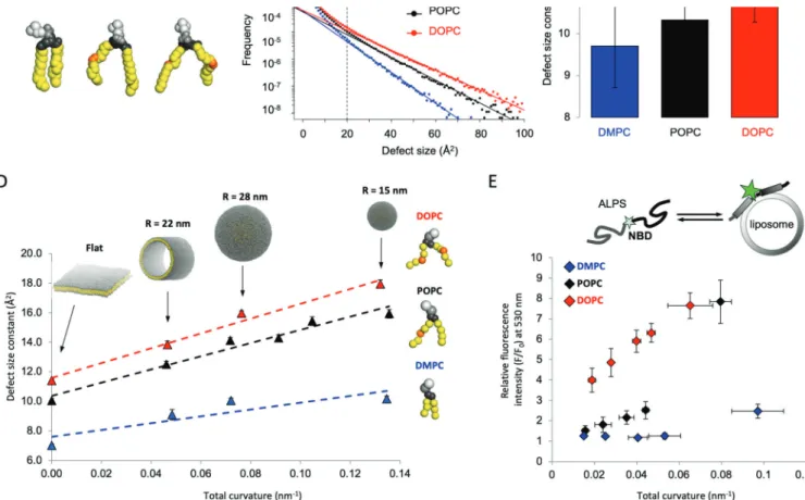
Using all-atom simulations, we found that by increasing the amount of lipids containing monounsaturated chains relative to those containing fully saturated ones, we significantly increased the amount of lipid packing defects and, according to our model, protein binding too (Figure 3). We confirmed these predictions by performing membrane binding assays of different proteins and peptides belonging to the ALPS (amphipathic packing lipid sensor) family, thus corroborating that membranes enriched in monounsaturated lipids are “stickier” than saturated ones.16,31
We also clarified the relationship between membrane curvature and membrane composition in the formation of these membrane binding pockets. Indeed, it has long been known that membrane curvature is a key property in determining protein binding to membranes. One of the proposed mechanisms was through an increase of these interfacial voids due to imbalances in the lipid density of the outer leaflet of curved bilayers.
Our first step was to model lipids using a coarse-grained (CG) representation, where multiple heavy atoms are described by individual beads (Figure 3). Different CG models were selected in order to always maintain the necessary chemical resolution, for example, to distinguish between saturated and monounsaturated fatty acids. Propitiously, we found that using appropriate thresholds for the identification of lipid-packing defects resulted in an acceptable accuracy for various CG models for lipids, such as MARTINI16and SDK.37 Thus, we could set up various curved membrane systems, such as tubules and vesicles of different radii, and investigate to what extent curvature and acyl chain saturation played a role in shaping the interfacial properties of these lipidic assemblies. Notably, we found that, while these two properties have a synergistic effect on membrane binding, acyl chain saturation is by far the most relevant parameter. This is because highly curved bilayers comprising 100% saturated chains have far fewer defects (and consistently show much lower binding) than flat bilayers comprising 100% monounsaturated lipids (Figure 3D,E).
While our in silico results and the experimental binding assays showed near perfect agreement, we next tested whether our results and evidence were valid in a more challenging
Figure 2.An atomistic view of the membrane−water interface. (A) “Classical” side view of a lipid bilayer. Polar heads are in gray and hydrophobic tails are in yellow. (B) Top view of a lipid bilayer. On the left, yellow patches exposed to the solvent are visible. On the right, lipid-packing defects found by our algorithm are in blue. They perfectly overlap with the exposed hydrophobic surface patches. (C) Structure of an ALPS peptide bound to the lipid bilayer. Lipid packing defects are in cyan, and the peptide insertions in the membrane perfectly colocalize with the detected packing defects. Panel B adapted with permission from ref27. Copyright 2013 Elsevier.
Figure 3. Role of acyl chain saturation in the generation of lipid-packing defects. (A) Atomistic representation of saturated (DMPC), monounsaturated (POPC), and doubly monounsaturated (DOPC) lipid molecules. (B) Size distribution of lipid-packing defects in DMPC, POPC, and DOPC membranes. (C) Exponentialfits corresponding to the plots presented in panel B. Increasing the number of monounsaturated chains increases the size and number of lipid-packing defects. (D) Exponentialfits of lipid-packing defects as a function of total curvature for lipids with a different number of monounsaturated acyl chains. (E) Experimental validation of the predictions in panel D using NBD-fluorescence microscopy of an ALPS peptide binding to DMPC, POPC, and DOPC lipids of different curvatures. Adapted with permission from ref16. Copyright 2014 Nature.
Figure 4.Flexibility of polyunsaturated acyl chains. (A) Schematic representation of monounsaturated and polyunsaturated PC lipids in a CG representation. The angleθ between ester linkage, middle, and terminal atom is explicitly shown. (B) Angular distribution of the angle θ between ester linkage, middle, and terminal atom of the mono- (black) or polyunsaturated (red) acyl chains. The polyunsaturated chain can adopt multiple conformations, while monounsaturated lipids are mostly restricted to the cis-conformation at 140°. (C) Schematic representation of the mechanism by which polyunsaturated lipids prevent the formation of deep lipid-packing defects while increasing surface-exposed hydrophobic patches. Reproduced with permission from ref18. Copyright 2014 Science AAS.
cellular context.17 To do so, we took advantage of a peptide that was sensitive to lipid-packing defects from our in vitro experiments. This peptide is at the N-terminus of a golgin protein, GMAP210, in the cis-Golgi network. One of the main functions of golgins is to capture trafficking vesicles to facilitate their fusion with the Golgi apparatus. We therefore speculated that this protein might be able to sense differences in lipid-packing in incoming vesicles according to their acyl chain composition. To test this, we usedfluorescence microscopy to follow the trafficking of exogenous vesicles made of various ratios of saturated to monounsaturated lipids. Only vesicles made of monounsaturated lipids were redirected toward the Golgi apparatus, and this targeting was not observed in the absence of the membrane-binding peptide of the golgin protein. This confirmed that recognition of lipid-packing
defects is one of the mechanisms used in the cell to modulate protein targeting to lipid membranes.17
■
CONFORMATIONAL FLEXIBILITY OFPOLYUNSATURATED LIPIDS
Lipids carrying saturated and monounsaturated acyl chains are extremely abundant in cellular membranes. However, unlike in plant seeds or in animals such as fishes, in humans it is relatively uncommon to have lipids with more than one double carbon−carbon bond, the hallmark of polyunsaturated lipids.7 Such acyl chains are either specifically found in some lipid classes that are present in cellular membranes at very low concentrations (e.g., phosphoinositides) or are abundant in specific tissues and organs, such as neurons.7Our simulations also clarified the impact of multiple double bonds in the acyl chains on interfacial packing defects.18Intriguingly, we found a
Figure 5.(A) FAAH enzyme embedded in the membrane. Its natural ligand anandamide (chemical structure shown on the bottom, with the parent lipids oleamide and palmytoyletanolamide). The close-up view shows the substrate anandamide (yellow) within the FAAH catalytic site. The membrane access (MA, red) and the acyl-chain binding (AB, orange) accommodate the lipid tail of anandamide, whose movement is modulated by the key gating resides Phe432 (green) and Trp531 (magenta). The interface region between the MA and AB channels is indicated as transition region (T). (B) Statistical distribution (% over the total equilibrated conformations, reported as bar graphs) of anandamide (4 unsaturated bonds) and palmitoylethanolamide (all saturated bonds) conformations when located in the MA, T, and AB binding regions of the FAAH binding site. One representative conformation of the most populated state is shown. (C) Competition assays performed for the wt and F432A FAAH enzyme in the presence of both anandamide (black) and palmitoylethanolamide (red) confirm the role of F432 for anandamide recognition. Adapted with permission from ref44. Copyright 2015 Public Library of Science.
presence of polyunsaturated lipids, interfacial voids at the surface of the membrane were extremely abundant, while defects extending deep below the membrane surface were scarcer than in the case of lipid-carrying monounsaturated chains. This behavior has interesting implications for the binding of peripheral proteins, with some proteins displaying enhanced binding in the presence of polyunsaturated lipids and others displaying lower (or even absent) binding.18
Our simulations showed that the structural origin of this behavior is indeed the conformational flexibility of polyunsa-turated chains (Figure 4), which is responsible for this unique response. That is, polyunsaturated chains can adopt various tilted conformations in order to adapt to membrane stresses and“fill” the voids in the lipid bilayer. We also found that this behavior was responsible for some of the remarkably different mechanical properties of polyunsaturated membranes, such as their reduced bending rigidity (i.e., the energy that is necessary to achieve a specific remodeling in its shape) and facilitated fission propensity.18
Remarkably, the degree of saturation and the specific conformations adopted by these differently saturated lipid tails can also modulate specificity in lipid recognition and hydrolysis, as discussed in the following section.
■
SATURATION IN FATTY ACID MOLECULES ANDENZYMATIC PROCESSING OF LIPIDS
This flexibility of polyunsaturated chains is also relevant for recognition and catalysis in lipid-processing enzymes. Intrigu-ingly, by inspecting recent structural data, we discovered that these lipid-processing enzymes are often characterized by a peculiar architecture of the catalytic site. These catalytic sites comprise a series of cavities that are tailored to accommodate a long andflexible fatty acid substrate, to efficiently cleave it, and to subsequently release the products. Demonstrated examples include structures of the cyclooxygenases COX-1 and COX-2, the human fatty acid synthase (FAS), and the lipid transporters Osh4 and Osh6, each one cocrystallized in complex with the respective lipid substrate.38−40 We and others have analyzed additional examples, such as the fatty acid amide hydrolase (FAAH) enzyme. FAAH is a pharmaceutically important membrane-bound serine hydrolase, which is responsible for the deactivating hydrolysis of a family of naturally occurring fatty acid amides.41,42FAAH regulates the endocannabinoid system by cleaving primarily the lipid messenger anandamide, with consequent generation of arachidonic acid. However, other lipid substrates, such as oleamide or palmitoylethanolamide, which contain a lower degree of unsaturation, are hydrolyzed at significantly slower rates (∼50 to 100 times slower than anandamide).43
In FAAH, the substrates reach the catalytic site via a membrane access (MA) channel, where two charged residues, Asp403 and Arg486, are suggested to favor the entrance of the polar head groups of fatty acid molecules. The catalytic action of FAAH occurs at the very core of the binding site, where the catalytic triad Ser241−Ser217−Lys142 allows proton transfers and nucleophilic attack for the hydrolysis of the substrate, which is kept properly oriented for catalysis by the oxyanion hole. Tightly connected to the catalytic region, a cytosolic port (CP) opens a cavity toward the bulk for the leaving group release after substrate cleavage. A third acyl chain binding
Our MD simulations have shown that this intricate yet functional architecture of the FAAH active site, with its multiple cavities, has evolved to properly select and accommodate specific lipid tails. We investigated this using multi-microsecond classical MD simulations and hybrid quantum−classical approaches applied to a model of FAAH embedded in a realistic membrane/water environment. We thus showed that the endogenous ligand anandamide does not lock itself into the FAAH active site but rather assumes catalytically significant conformations for hydrolysis by moving its flexible arachidonoyl tail between the MA and AB cavities (Figure 5).44 A conserved phenylalanine residue, Phe432, which is located at the boundary between these two cavities, acts as a“dynamic paddle”, orienting the substrates within the active site for hydrolysis.45,46
In this context, we clarified the role of lipid flexibility and its dependence on the acyl chain saturation. We did this using MD simulations to comparatively analyze the binding mechanism of fatty acid substrates with different hydrolysis rates (anandamide > oleamide > palmitoylethanolamide). We found that the different flexibility and preferred conformations of these lipids allow FAAH to preferentially accommodate the fastest hydrolyzed lipid (anandamide) into its multipocket binding site.44 Conversely, oleamide and palmitoylethanola-mide lipids, which display a higher degree of saturation than anandamide, assume restricted conformations within the FAAH active site (Figure 5). We performed experimental competition assays for FAAH in the presence of both anandamide and palmitoylethanolamide. These assays showed a clear selectivity for the former substrate. When the dynamic paddle residue was mutated, however, the selectivity toward different lipid chains was lost (Figure 5). This suggests that the protein framework has been tailored by evolution to preferentially recognize, select, and bind the conformation of the lipid acyl chain that is more prone to attaining hydrolysis. These molecular simulations have thus proposed an essential role for substrate dynamics in facilitating the formation of catalytically active conformations in FAAH for catalysis. This dynamic scenario complements and enriches the static picture for lipid processing in FAAH, which is provided by a wealth of X-ray structures containing substrate analogues or inhibitors.41 In addition, we also described, at the hybrid quantum−classical level, the energetics and detailed mechanisms of anandamide hydrolysis,47which were in agreement with previous computa-tional investigations.48−50 Our investigation detailed how the proper location of the acyl chain between the MA and AB cavities is crucial for efficient catalysis and nitrogen inversion for amide hydrolysis.47 These results have been substantiated by studies on the binding modes of alternative lipids44 and covalent FAAH inhibitors.50,51
As mentioned above, this strategy for substrate selectivity through conformational selection of specific lipid tails seems to be shared by other lipid-processing enzymes with similar multipocket enzymatic architectures. The COX-1 and COX-2 enzymes transform the product of FAAH catalysis (the arachidonic acid) into prostaglandin H2. In the active site of the COX enzymes, the arachidonoyl chain specifically locates within a multiple-cavity active site for catalysis, differing from other fatty acids that are processed with lower catalytic efficiency.52,53 Remarkably, the similarity of the active site
A final example is the endocannabinoid enzyme mono acylglycerol lipase (MAGL), a membrane-interacting enzyme involved in fatty acid metabolism. MAGL is the main enzyme responsible for the hydrolysis of the endocannabinoid lipid 2-arachidonoylglycerol (2AG) into glycerol and arachidonic acid, which favors the biosynthesis of proinflammatory eicosa-noids.55 Interestingly, MAGL can hydrolyze several mono-acylglycerols, all bearing the same glycerol headgroup as 2AG but with different chains, characterized by their number of carbon atoms and unsaturated bonds. Fluorescence-based glycerol assays revealed a relationship between MAGL’s rate of hydrolysis and the chemical nature of the chain. That is, the hydrolysis rate decreases for monoacylglycerols with shorter and more saturated chains, such as 2-arachidonoylglycerol (C20:4, 2AG) > 2-linoleoylglycerol (C18:2) > 2-palmitoylgly-cerol (C16:0, 2PG).56 Furthermore, activity assays in human embryonic kidney cells showed that the rate of hydrolysis of 2AG is almost two times faster than that of 2PG.57
Through multiple and extended MD and free energy (metadynamics) simulations of wild-type and mutated forms of MAGL, either alone or in complex with the preferred substrate 2AG or the less reactive 2PG, we determined that the specific chemical structure of the acyl chain affects substrate selection in enzymatic lipid-processing.58 While 2PG mainly adopted an elongated form of its acyl chain, 2AG was more flexible thanks to the rotation of the two adjacent dihedral angles located between the double bonds (Figure 6). Lipid flexibility was found to be linked to enzymatic plasticity. In this specific case, MAGL acts via the so-called lid domain, a 75-amino-acid-long cap domain located at the entrance to the binding site, which anchors the enzyme to the membrane in order to recruit the substrate. Lid domain plasticity, which we found to be enhanced only in the presence of the preferred 2AG substrate, modulates the free-energy barrier for lipid binding to the catalytic pocket in MAGL. Our simulations pointed to two highly flexible residues in the lid domain: Ile179, the gatekeeper responsible for the closing of the lid domain, and Phe159, which interacts with the substrate’s unsaturated bonds via hydrophobic interactions. These bonds are properly positioned along the 2AG chain, while they are missing in 2PG (Figure 6). The strategic location of these unsaturated bonds for substrate recognition and enzymatic catalysis was confirmed by kinetic experiments of single-point mutations of Ile179 and Phe159. Thus, our simulations and experimental results support the idea that substrate selection and binding is regulated by lid domain plasticity, combined with lipid flexibility modulated by acyl chain (un)saturation.
■
CONCLUSIONS AND OUTLOOKIn this Account, we have discussed the specific chemical structure of the acyl tail of lipid molecules as an emerging key factor in the trafficking and enzymatic processing of lipids. The polar head is widely recognized as relevant to lipid function. However, the level of saturation and the number and specific position of double carbon−carbon bonds along the lipid acyl chain (i.e., differentiating between saturated, monounsaturated, and polyunsaturated lipids) has only recently emerged as an equally important factor in lipid metabolism. Our recent computational studies, integrated with experimental data, have highlighted the importance of the specific chemical structure of
the lipid acyl tail for membrane packing and for selective binding to lipid-processing enzymes. We hope that these examples will prompt further computational and experimental studies of this structural and functional aspect of lipid metabolism. A key goal will be to further clarify and exploit the general principles by which similar but different tails and chemical structures of lipids were optimized during evolution. This would likely have important implications for protein engineering and drug design efforts.
■
AUTHOR INFORMATIONCorresponding Authors
*M.D.V. E-mail:[email protected]. *S.V. E-mail: [email protected].
Figure 6.(A) Homodimeric MAGL simulated in a water/membrane environment (explicit solvent not shown). The lid domain, shown in orange, recruits the lipidic substrate 2AG, shown as red licorice. (B) 2AG conformations are defined based on the three pairs of contiguous dihedral angles between the unsaturated bonds: when a pair has the same sign (I), then that part of the chain is more elongated than when the dihedral angles have opposite signs (X). The size of the node is proportional to the population of that state, whereas the size and color of the links are proportional to the transition probability between them. The networks on the left and the right refer to the dynamics of 2AG when the lid domain is open and closed, respectively. (C) Probability distribution of the distance between the center of mass of the phenyl ring of residue Phe159 and the carbon atoms of the acyl chain of 2AG or 2PG. Adapted with permission from ref 58. Copyright 2017 Elsevier.
Marco De Vivo: 0000-0003-4022-5661
Notes
The authors declare no competingfinancial interest.
Biographies
Stefano Vanniis a tenured researcher at CNRS in France and Swiss National Science Foundation Associate Professor in the Department of Biology at the University of Fribourg, Switzerland. His main research focus is understanding, at a molecular level, how the physicochemical properties of membranes and lipids affect intra-cellular trafficking and lipid metabolism.
Laura Riccardiis a research associate in the Molecular Modeling and Drug Discovery Lab at the Italian Institute of Technology (IIT). She uses MD simulations, free-energy calculations, and molecular docking to investigate the structure and dynamics of pharmaceutically relevant enzymes, such as the monoacylglycerol lipase enzyme, and function-alized nanoparticles.
Giulia Palermo is an Assistant Professor at the University of California, Riverside. Her lab applies and develops computational methods to unravel the function of key biomolecular systems and to devise novel biotechnological applications. She was a Ph.D. student in De Vivo’s lab, doing her postdoctoral training in the groups of U. Rothlisberger (EPFL) and J. A. McCammon (UCSD). She received the 2017 HPC wire“Best use of HPC in Life Sciences” award. She has been featured in the 2018 Young Investigators Virtual Issue of the Journal of the American Chemical Society.
Marco De Vivo is Director of the Molecular Modeling and Drug Discovery Lab at the Italian Institute of Technology (IIT). In 2017, he received the ACS COMP Outstanding Junior Faculty Award. His research interests include molecular simulations to rationalize enzymatic catalysis, nanoparticle functionalization, and drug design. He has worked extensively on key membrane-bound enzymes that hydrolyze lipids.
■
ACKNOWLEDGMENTSWe acknowledge Valeria Zoni for help in preparingFigure 1,
and for critical reading of the manuscript. S.V. acknowledges support by the Swiss National Science Foundation (No. 163966). M.D.V. thanks the Italian Association for Cancer Research (AIRC) for financial support (IG 18883). We acknowledge Guillaume Drin for preparing Figure 2, panel C.
■
REFERENCES(1) Wymann, M. P.; Schneiter, R. Lipid signalling in disease.Nat. Rev. Mol. Cell Biol. 2008, 9, 162−176.
(2) Harayama, T.; Riezman, H. Understanding the diversity of membrane lipid composition.Nat. Rev. Mol. Cell Biol. 2018, 19, 281− 296.
(3) Ahn, K.; McKinney, M. K.; Cravatt, B. F. Enzymatic pathways that regulate endocannabinoid signaling in the nervous system.Chem. Rev. 2008, 108, 1687−1707.
(4) Dors, G.; Mendes, A. A.; Pereira, E. B.; de Castro, H. F.; Furigo, A. Simultaneous enzymatic hydrolysis and anaerobic biodegradation of lipid-rich wastewater from poultry industry.Appl. Water Sci. 2013, 3, 343−349.
(5) van Meer, G.; Voelker, D. R.; Feigenson, G. W. Membrane lipids: where they are and how they behave.Nat. Rev. Mol. Cell Biol. 2008,9, 112−124.
Trends Cell Biol. 2015, 25, 427−436.
(8) Lemmon, M. A. Membrane recognition by phospholipid-binding domains.Nat. Rev. Mol. Cell Biol. 2008, 9, 99−111.
(9) Antonny, B.; Huber, I.; Paris, S.; Chabre, M.; Cassel, D. Activation of ADP-ribosylation Factor 1 GTPase-Activating Protein by Phosphatidylcholine-derived Diacylglycerols. J. Biol. Chem. 1997, 272, 30848−30851.
(10) Matsuoka, K.; Orci, L.; Amherdt, M.; Bednarek, S. Y.; Hamamoto, S.; Schekman, R.; Yeung, T. COPII-Coated Vesicle Formation Reconstituted with Purified Coat Proteins and Chemically Defined Liposomes.Cell 1998, 93, 263−275.
(11) Schneiter, R.; Brügger, B.; Sandhoff, R.; Zellnig, G.; Leber, A.; Lampl, M.; Athenstaedt, K.; Hrastnik, C.; Eder, S.; Daum, G.; Paltauf, F.; Wieland, F. T.; Kohlwein, S. D. Electrospray Ionization Tandem Mass Spectrometry (Esi-Ms/Ms) Analysis of the Lipid Molecular Species Composition of Yeast Subcellular Membranes Reveals Acyl Chain-Based Sorting/Remodeling of Distinct Molecular Species En Route to the Plasma Membrane.J. Cell Biol. 1999, 146, 741−754.
(12) Mukherjee, S.; Soe, T. T.; Maxfield, F. R. Endocytic Sorting of Lipid Analogues Differing Solely in the Chemistry of Their Hydrophobic Tails.J. Cell Biol. 1999, 144, 1271−1284.
(13) Contreras, F.-X.; Ernst, A. M.; Haberkant, P.; Björkholm, P.; Lindahl, E.; Gönen, B.; Tischer, C.; Elofsson, A.; von Heijne, G.; Thiele, C.; Pepperkok, R.; Wieland, F.; Brügger, B. Molecular recognition of a single sphingolipid species by a protein’s trans-membrane domain.Nature 2012, 481, 525−529.
(14) Clay, L.; Caudron, F.; Denoth-Lippuner, A.; Boettcher, B.; Buvelot Frei, S.; Snapp, E. L.; Barral, Y. A sphingolipid-dependent diffusion barrier confines ER stress to the yeast mother cell. eLife 2014,3, No. e01883.
(15) Covino, R.; Ballweg, S.; Stordeur, C.; Michaelis, J. B.; Puth, K.; Wernig, F.; Bahrami, A.; Ernst, A. M.; Hummer, G.; Ernst, R. A eukaryotic sensor for membrane lipid saturation.Mol. Cell 2016, 63, 49−59.
(16) Vanni, S.; Hirose, H.; Barelli, H.; Antonny, B.; Gautier, R. A sub-nanometre view of how membrane curvature and composition modulate lipid packing and protein recruitment.Nat. Commun. 2014, 5, 4916.
(17) Magdeleine, M.; Gautier, R.; Gounon, P.; Barelli, H.; Vanni, S.; Antonny, B. A filter at the entrance of the Golgi that selects vesicles according to size and bulk lipid composition. eLife 2016, 5, No. e16988.
(18) Pinot, M.; Vanni, S.; Pagnotta, S.; Lacas-Gervais, S.; Payet, L.-A.; Ferreira, T.; Gautier, R.; Goud, B.; Antonny, B.; Barelli, H. Polyunsaturated phospholipids facilitate membrane deformation and fission by endocytic proteins.Science 2014, 345, 693−697.
(19) Kutateladze, T. G. Translation of the phosphoinositide code by PI effectors.Nat. Chem. Biol. 2010, 6, 507−513.
(20) Marrink, S. J.; Corradi, V.; Souza, P. C. T.; Ingólfsson, H. I.; Tieleman, D. P.; Sansom, M. S. P. Computational Modeling of Realistic Cell Membranes.Chem. Rev. 2019, 119, 6184−6226.
(21) Wen, P.-C.; Mahinthichaichan, P.; Trebesch, N.; Jiang, T.; Zhao, Z.; Shinn, E.; Wang, Y.; Shekhar, M.; Kapoor, K.; Chan, C. K.; Tajkhorshid, E. Microscopic view of lipids and their diverse biological functions.Curr. Opin. Struct. Biol. 2018, 51, 177−186.
(22) Leonard, A. N.; Wang, E.; Monje-Galvan, V.; Klauda, J. B. Developing and Testing of Lipid Force Fields with Applications to Modeling Cellular Membranes.Chem. Rev. 2019, 119, 6227−6269.
(23) Bigay, J.; Antonny, B. Curvature, Lipid Packing, and Electrostatics of Membrane Organelles: Defining Cellular Territories in Determining Specificity.Dev. Cell 2012, 23, 886−895.
(24) Sharpe, H. J.; Stevens, T. J.; Munro, S. A Comprehensive Comparison of Transmembrane Domains Reveals Organelle-Specific Properties.Cell 2010, 142, 158−169.
Noskov, S. Y.; Marrink, S. J.; Tieleman, D. P. Emerging Diversity in Lipid−Protein Interactions. Chem. Rev. 2019, 119, 5775.
(27) Vamparys, L.; Gautier, R.; Vanni, S.; Bennett, W. F. D.; Tieleman, D. P.; Antonny, B.; Etchebest, C.; Fuchs, P. F. J. Conical Lipids in Flat Bilayers Induce Packing Defects Similar to that Induced by Positive Curvature.Biophys. J. 2013, 104, 585−593.
(28) Gautier, R.; Bacle, A.; Tiberti, M. L.; Fuchs, P. F.; Vanni, S.; Antonny, B. PackMem: A Versatile Tool to Compute and Visualize Interfacial Packing Defects in Lipid Bilayers. Biophys. J. 2018, 115, 436−444.
(29) Cui, H.; Lyman, E.; Voth, G. A. Mechanism of Membrane Curvature Sensing by Amphipathic Helix Containing Proteins. Biophys. J. 2011, 100, 1271−1279.
(30) Wildermuth, K. D.; Monje-Galvan, V.; Warburton, L. M.; Klauda, J. B. Effect of Membrane Lipid Packing on Stable Binding of the ALPS Peptide.J. Chem. Theory Comput. 2019, 15, 1418−1429.
(31) Vanni, S.; Vamparys, L.; Gautier, R.; Drin, G.; Etchebest, C.; Fuchs, P. F. J.; Antonny, B. Amphipathic Lipid Packing Sensor Motifs: Probing Bilayer Defects with Hydrophobic Residues.Biophys. J. 2013, 104, 575−584.
(32) Van Lehn, R. C.; Ricci, M.; Silva, P. H. J.; Andreozzi, P.; Reguera, J.; Voïtchovsky, K.; Stellacci, F.; Alexander-Katz, A. Lipid tail protrusions mediate the insertion of nanoparticles into model cell membranes.Nat. Commun. 2014, 5, 4482.
(33) Moiset, G.; López, C. A.; Bartelds, R.; Syga, L.; Rijpkema, E.; Cukkemane, A.; Baldus, M.; Poolman, B.; Marrink, S. J. Disaccharides Impact the Lateral Organization of Lipid Membranes.J. Am. Chem. Soc. 2014, 136, 16167−16175.
(34) Rowe, E. R.; Mimmack, M. L.; Barbosa, A. D.; Haider, A.; Isaac, I.; Ouberai, M. M.; Thiam, A. R.; Patel, S.; Saudek, V.; Siniossoglou, S.; Savage, D. B. Conserved Amphipathic Helices Mediate Lipid Droplet Targeting of Perilipins 1−3. J. Biol. Chem. 2016, 291, 6664− 6678.
(35) Garten, M.; Prévost, C.; Cadart, C.; Gautier, R.; Bousset, L.; Melki, R.; Bassereau, P.; Vanni, S. Methyl-branched lipids promote the membrane adsorption ofα-synuclein by enhancing shallow lipid-packing defects.Phys. Chem. Chem. Phys. 2015, 17, 15589−15597.
(36) Prévost, C.; Sharp, M. E.; Kory, N.; Lin, Q.; Voth, G. A.; Farese, R. V.; Walther, T. C. Mechanism and Determinants of Amphipathic Helix-Containing Protein Targeting to Lipid Droplets. Dev. Cell 2018, 44, 73−86.
(37) Bacle, A.; Gautier, R.; Jackson, C. L.; Fuchs, P. F. J.; Vanni, S. Interdigitation between Triglycerides and Lipids Modulates Surface Properties of Lipid Droplets.Biophys. J. 2017, 112, 1417−1430.
(38) von Filseck, J. M.; Vanni, S.; Mesmin, B.; Antonny, B.; Drin, G. A phosphatidylinositol-4-phosphate powered exchange mechanism to create a lipid gradient between membranes. Nat. Commun. 2015, 6, 6671.
(39) Palermo, G.; Favia, A. D.; Convertino, M.; De Vivo, M. The Molecular Basis for Dual Fatty Acid Amide Hydrolase (FAAH)/ Cyclooxygenase (COX) Inhibition.ChemMedChem 2016, 11, 1252− 1258.
(40) Moser von Filseck, J.; Opi, A.; Delfosse, V.; Vanni, S.; Jackson, C. L.; Bourguet, W.; Drin, G. Phosphatidylserine transport by ORP/ Osh proteins is driven by phosphatidylinositol 4-phosphate. Science 2015,349, 432−436.
(41) Bracey, M. H.; Hanson, M. A.; Masuda, K. R.; Stevens, R. C.; Cravatt, B. F. Structural Adaptations in a Membrane Enzyme That Terminates Endocannabinoid Signaling. Science 2002, 298, 1793− 1796.
(42) Cravatt, B. F.; Giang, D. K.; Mayfield, S. P.; Boger, D. L.; Lerner, R. A.; Gilula, N. B. Molecular characterization of an enzyme that degrades neuromodulatory fatty-acid amides.Nature 1996, 384, 83−87.
A.; Girotto, S.; Rothlisberger, U.; De Vivo, M. Keys to lipid selection in fatty acid amide hydrolase catalysis: structural flexibility, gating residues and multiple binding pockets.PLoS Comput. Biol. 2015, 11, No. e1004231.
(45) Mileni, M.; Johnson, D. S.; Wang, Z.; Everdeen, D. S.; Liimatta, M.; Pabst, B.; Bhattacharya, K.; Nugent, R. A.; Kamtekar, S.; Cravatt, B. F.; Ahn, K.; Stevens, R. C. Structure-guided inhibitor design for human FAAH by interspecies active site conversion.Proc. Natl. Acad. Sci. U. S. A. 2008, 105, 12820−12824.
(46) Palermo, G.; Campomanes, P.; Neri, M.; Piomelli, D.; Cavalli, A.; Rothlisberger, U.; De Vivo, M. Wagging the Tail: Essential Role of Substrate Flexibility in FAAH Catalysis. J. Chem. Theory Comput. 2013,9, 1202−1213.
(47) Palermo, G.; Campomanes, P.; Cavalli, A.; Rothlisberger, U.; De Vivo, M. Anandamide hydrolysis in FAAH reveals a dual strategy for efficient enzyme-assisted amide bond cleavage via nitrogen inversion.J. Phys. Chem. B 2015, 119, 789−801.
(48) Tubert-Brohman, I.; Acevedo, O.; Jorgensen, W. L. Elucidation of Hydrolysis Mechanisms for Fatty Acid Amide Hydrolase and Its Lys142Ala Variant via QM/MM Simulations.J. Am. Chem. Soc. 2006, 128, 16904−16913.
(49) Chudyk, E. I.; Dyguda-Kazimierowicz, E.; Langner, K. M.; Sokalski, W. A.; Lodola, A.; Mor, M.; Sirirak, J.; Mulholland, A. J. Nonempirical Energetic Analysis of Reactivity and Covalent Inhibition of Fatty Acid Amide Hydrolase. J. Phys. Chem. B 2013, 117, 6656−6666.
(50) Palermo, G.; Rothlisberger, U.; Cavalli, A.; De Vivo, M. Computational insights into function and inhibition of fatty acid amide hydrolase.Eur. J. Med. Chem. 2015, 91, 15−26.
(51) Palermo, G.; Branduardi, D.; Masetti, M.; Lodola, A.; Mor, M.; Piomelli, D.; Cavalli, A.; De Vivo, M. Covalent inhibitors of fatty acid amide hydrolase: a rationale for the activity of piperidine and piperazine aryl ureas.J. Med. Chem. 2011, 54, 6612−6623.
(52) Kiefer, J. R.; Pawlitz, J. L.; Moreland, K. T.; Stegeman, R. A.; Hood, W. F.; Gierse, J. K.; Stevens, A. M.; Goodwin, D. C.; Rowlinson, S. W.; Marnett, L. J.; Stallings, W. C.; Kurumbail, R. G. Structural insights intos the stereochemistry of the cyclooxygenase reaction.Nature 2000, 405, 97−101.
(53) Malkowski, M. G. The Productive Conformation of Arachidonic Acid Bound to Prostaglandin Synthase. Science 2000, 289, 1933−1937.
(54) Bertolacci, L.; Romeo, E.; Veronesi, M.; Magotti, P.; Albani, C.; Dionisi, M.; Lambruschini, C.; Scarpelli, R.; Cavalli, A.; De Vivo, M.; Piomelli, D.; Garau, G. A Binding Site for Nonsteroidal Anti-inflammatory Drugs in Fatty Acid Amide Hydrolase.J. Am. Chem. Soc. 2013,135, 22−25.
(55) Labar, G.; Wouters, J.; Lambert, D. M. A Review on the Monoacylglycerol Lipase: At the Interface Between Fat and Endocannabinoid Signalling.Curr. Med. Chem. 2010, 17, 2588−2607. (56) Laitinen, T.; Navia-Paldanius, D.; Rytilahti, R.; Marjamaa, J. J. T.; Kařízková, J.; Parkkari, T.; Pantsar, T.; Poso, A.; Laitinen, J. T.; Savinainen, J. R. Mutation of Cys242 of human monoacylglycerol lipase disrupts balanced hydrolysis of 1- and 2-monoacylglycerols and selectively impairs inhibitor potency.Mol. Pharmacol. 2014, 85, 510− 519.
(57) Navia-Paldanius, D.; Savinainen, J. R.; Laitinen, J. T. Biochemical and pharmacological characterization of human α/β-hydrolase domain containing 6 (ABHD6) and 12 (ABHD12).J. Lipid Res. 2012, 53, 2413−2424.
(58) Riccardi, L.; Arencibia, J. M.; Bono, L.; Armirotti, A.; Girotto, S.; De Vivo, M. Lid domain plasticity and lipid flexibility modulate enzyme specificity in human monoacylglycerol lipase. Biochim. Biophys. Acta, Mol. Cell Biol. Lipids 2017, 1862, 441−451.