HAL Id: hal-03021504
https://hal.archives-ouvertes.fr/hal-03021504
Preprint submitted on 24 Nov 2020
HAL is a multi-disciplinary open access archive for the deposit and dissemination of sci-entific research documents, whether they are pub-lished or not. The documents may come from teaching and research institutions in France or abroad, or from public or private research centers.
L’archive ouverte pluridisciplinaire HAL, est destinée au dépôt et à la diffusion de documents scientifiques de niveau recherche, publiés ou non, émanant des établissements d’enseignement et de recherche français ou étrangers, des laboratoires publics ou privés.
To cite this version:
Aldine Amiel, Kevin Foucher, Solène Ferreira, Eric Röttinger. Synergic coordination of stem cells is required to induce a regenerative response in anthozoan cnidarians. 2020. �hal-03021504�
Synergic coordination of stem cells is required to induce a regenerative response in
1
anthozoan cnidarians
2 3
Aldine R. Amiel, Kevin Foucher, Solène Ferreira, Eric Röttinger* 4
5
Université Côte d’Azur, CNRS, INSERM, Institute for Research on Cancer and Aging 6
(IRCAN), Nice, France 7
8
Email addresses: 9
[email protected], [email protected] [email protected], 10 [email protected] 11 12 13 14 15 16 17 *
Please direct correspondence to: 18 Eric Röttinger 19 T: +33 (0)4 93 37 77 91 20 E: [email protected] 21
Abstract
22
Little is known about the origin of the inductive signal that translates the 23
amputation stress into a cooperative cellular response. By studying the process 24
underlying the reformation of lost body parts in the anthozoan cnidarian Nematostella 25
vectensis, we identified a regeneration-inducing structure that, via a tissue crosstalk, is
26
responsible for the initiation of the repair program. We further revealed for the first time 27
in anthozoan cnidarians, that fast and slow-cycling/quiescent stem cells respond to the 28
amputation stress and actively participate in the reformation of lost body parts. 29
Importantly, a synergic interaction of both stem cell populations is required to complete 30
the regeneration process. Our findings suggest that the emergence/loss of structure 31
complexity/compartmentalization influences the proprieties of tissue plasticity, changes 32
the competence of a tissue to reprogram and, in the context of regeneration, the capacity 33
of the tissue to emit or respond to a regeneration-inducing signal. 34 35 36 37 38 39 40 41
Keywords: Whole body regeneration, Tissue repair, Tissue crosstalk, Label retaining
42
cells, Stem cells, Cnidarian, Anthozoan, Sea anemone, Nematostella vectensis, 43
Background
44
The process of regeneration is the re-formation of damaged or missing body parts 45
that in bilaterian animals, involves cellular migration and proliferation of adult stem cells 46
or de-differentiated cell populations. In combination with the remodeling of existing 47
cells/tissues, these cellular processes are crucial to re-establish the symmetry and the 48
proportion of the body [1]. Regeneration is widespread among metazoans and has 49
fascinated scientists for centuries [2,3]. Still, our understanding of why some animals 50
regenerate while others can’t remains largely unknown. In the past 10 years, historical 51
invertebrate whole-body regeneration models such as Planaria and Cnidaria have re-52
emerged and are taking advantage of modern cellular and molecular techniques to 53
understand the complex process of regeneration [4-7]. Important insight has been gained 54
from these species that have increased our understanding about the cellular and molecular 55
mechanisms underlying regeneration. Yet little is known about how the organism 56
translates the amputation stress into a regeneration inducing signal and the origin as well 57
as the nature of the inductive signal required for the cooperation of cell populations 58
during the regeneration process. 59
60
The level of regenerative abilities varies across the animal kingdom [7]. While 61
vertebrates can regenerate certain structures and tissues, planarians and cnidarians posses 62
extraordinary whole-body regeneration capacities and can regrow missing body parts 63
when dissected into small pieces [2,8]. Nonetheless, their regenerative capacities have 64
limitation at both the species and also the tissues level. For instance in planarians, 65
isolation of the most anterior part of its body (region in front of the photoreceptor) or the 66
pharynx does not lead to regeneration as these regions are devoid of planarian specific 67
pluripotent stem cells, the neoblasts [9-11]. In Hydra, the isolated body column, named 68
gastric region, possesses the ability to regenerate while the isolated head, pedal disk or 69
tentacles cannot. The absence of regeneration from these isolated parts is due to the lack 70
of hydrozoan specific pluripotent stem cells, the i-cells (interstitial stem cells), in these 71
highly differentiated and specialized regions of the body [12,13]. Thus, identifying the 72
regenerative limits of a given organism and using them for experimental approaches has 73
led to important findings on the location of stem cell populations and their dynamics 74
during the complex biological process of regeneration. 75
76
To date, the majority of studies addressing the role of stem cells during 77
regeneration in cnidarians were performed using Hydrozoa such as Hydra and 78
Hydractinia [2,14-17]. While Hydra is well known to have developed an alternative
79
mechanism to regenerate a head in a stem-cell (i-cell) depleted context [18], those 80
classically considered fast cycling i-cells are under “normal” conditions involved in the 81
regeneration process in Hydra and Hydractinia [1,19-23]). Only recently, a study in 82
Hydra has described slow cycling cells that are part of the pluripotent i-cell as well as the
83
unipotent epithelial stem cell populations and involved in regeneration [24]. However, 84
nothing is known about the relationship between fast and slow cycling stem cells, their 85
hierarchy and how they are interacting to trigger tissue reprograming for regeneration. 86
This question remains unanswered not only in hydrozoans, but in cnidarians in general. 87
Cnidarians are composed of two major groups, the Anthozoa (coral, sea anemone) 89
and the Medusozoa, that include Hydrozoa, Schyphozoa and Cubozoa [25-30]. 90
Interestingly, the crown-group cnidarians (the living representatives) are genetically as 91
divergent as protostomes and deuterostomes [31,32] and it has been proposed that none 92
of the cnidarian clades is representing the entire group [32]. Thus, it is of great interest to 93
compare the regenerative strategies between hydrozoan and non-hydrozoan cnidarians in 94
order to understand the similarities/differences and evolution of the cellular and 95
molecular mechanisms driving regeneration. Importantly, the presence of adult stem cells 96
has yet to be discovered in non-hydrozoan cnidarians. 97
98
Anthozoa diverged early within the cnidarian phylogeny [29,33] and form the 99
largest group of cnidarian [34,35] that display anatomical differences compared to other 100
cnidarian polyps. Exclusively anthozoans possess mesenteries that are well-defined 101
anatomical structures located in the gastric cavity, fulfilling the digestive and sexual 102
reproduction roles of the individual polyp. Mesenteries are multifunctional tissues of 103
combined ectodermal and endodermal origins that in addition to the germ lineage and 104
digestive cells also possesses various additional cell types such as muscle cells and 105
cnidocytes (stinging cells) [36-38]. Because of its i) phylogenetic position, the ii) ease to 106
control spawning and fertilization [39-41] and thus the access to embryonic, larval and 107
post-metamorphic stages all year round, iii) a sequenced genome [42] as well as iv) 108
available cellular and molecular tools for functional genomics [43,44] and genome 109
editing [45], the anthozoan sea anemone Nematostella vectensis has become a leading 110
model to gain new insight into animal evolution [42,46,47] and early cnidarian 111
development [48-55]. Nematostella possesses also high regenerative abilities and 112
therefore is a potent model to better understand the relationship between embryonic 113
development and regeneration [44,56]. However, our knowledge concerning the 114
morphological, cellular and molecular mechanisms underlying whole body regeneration 115
in Nematostella is still in its early phase [57-61]. Recent work has begun to describe the 116
morphological and cellular events involved in the regeneration process in Nematostella 117
[59,60,62]. Wound healing after head amputation is already completed within six hours 118
post amputation (hpa) and oral regeneration following sub-pharyngeal amputation occurs 119
in stereotypic steps [62]. This process is first visible when the most oral part of the 120
remaining mesenteries fuses together and enters in contact with the epithelia at the 121
amputation site. The most oral part of the mesenteries is at the origin of the internal part 122
of the pharynx that completes its regeneration at the same time than the tentacles bulbs 123
form and elongate [62]. The various stages of the polyp’s life are defined by clear 124
morphological characteristics (i.e. number of tentacles and mesenteries, presence of 125
gametes) and cell proliferation is necessary for oral regeneration in juveniles [59] as well 126
as in adults [62]. Nonetheless, in juvenile as well as in adult Nematostella, the first 127
morphogenetic step of regeneration is proliferation independent and only later steps 128
require mitotic activity to fully complete oral regeneration following sub-pharyngeal 129
amputation [62]. While this study laid down a foundation for functional work on 130
anthozoan regeneration, basic but crucial questions about the regenerative capacities in 131
Nematostella still remain unanswered: i) are there any body part, tissue and/or age
132
specific limits of its regenerative abilities? ii) what is the cellular/tissue origin of the 133
signal(s) that initiates cell proliferation and/or regeneration? iii) what are the cellular 134
mechanisms underlying this phenomenon, and importantly, iv) are there adult stem cells 135
in anthozoans and v) are they involved in the regeneration process? 136
137
In the present study we combined classical grafting experiments with cellular 138
approaches to address these questions. This strategy enabled us to highlight a crucial 139
crosstalk between the mesenteries and the surrounding epithelia of the body wall required 140
for regeneration and to identify that the mesenteries are crucial to induce 141
proliferation/regeneration in Nematostella. We further identified at least two cell 142
populations, a fast cycling one and a quiescent/slow cycling one that are required in a 143
combinatorial manner to trigger a regenerative response. Both of these cell populations 144
are re-activated in response to the amputation stress and participate in the regeneration 145
process. Importantly, our work provides the first set of evidences for the existence of at 146
least two adult stem cell populations in anthozoans that synergize to drive efficient 147
regeneration in the anthozoan cnidarian Nematostella. 148
Materials and methods
149
Animal culture
150
Nematostella vectensis were cultivated at the Institute for Research on Cancer and Aging,
151
Nice (IRCAN) of the University of Nice. Adult and juvenile animals were cultured in 152
1/3X ASW (Artificial seawater, Tropic-Marin Bio-actif system sea salt) and maintained 153
at 17°C or 22°C for adults and juveniles, respectively. Adults were fed five times a week 154
with freshly hatched artemia. Spawning to obtain juveniles was carried out as described 155
in [39]. Juveniles for cutting experiments were raised until six weeks after fertilization. 156
Starting 2 weeks post fertilization, they were feed once a week with 1ml of smashed 157
artemia during four weeks. 158
159
Cutting, tissue isolation and grafting experiments
160
Juveniles or adults were relaxed by adding 1 ml of 7.14% MgCl2 in 5ml 1/3 ASW and left 161
on a light table during 10 to 15 min until they were relaxed enough for surgical 162
interventions. Polyps were cut using a microsurgery scalpel (Swann-Morton, n°15). Each 163
cut was performed perpendicular to the oral-aboral axis of the body column. Grafting 164
experiments were performed using adult tissues only from a clonal colony, following the 165
cutting protocol described above and carried as follows: 1) Head and physa were 166
surgically removed from relaxed animals; 2) The body column was isolated and opened 167
along the oral-aboral axis of the polyp with microsurgical scissors (Fine Science Tools 168
GMBH #15100-09, Heidelberg, Germany); 3) If required, the mesenteries were gently 169
removed mechanically from the epithelia using forceps (Rubis Grip Tweezers, style 3C) 170
and microsurgical scissors 4) Isolated epithelia from the body wall were forced to contact 171
the freshly isolated mesenteries approximately 5 to 10 min after tissue isolation and the 172
graft was maintained with a human hair allowing the wounds to heal properly; 5) After 173
three days the hair was removed to avoid any mechanical constraints or tissue damages 174
during the regenerating period. Isolated fragments and grafts were transferred into a new 175
dish (or rinsed at least five times to remove MgCl2) and maintained in 1/3 ASW at 22°C 176
(if not stated otherwise) for the duration of the experiments. 177
178
Fixation, DNA, actin labeling
179
After relaxing adult or juvenile polyps as well as isolated body parts in MgCl2 for 10-15 180
minutes, animals were fixed in 4% paraformaldehyde (Electron Microscopy Sciences # 181
15714) in 1/3 ASW during 1 hour at 22°C or overnight at 4°C. Fixed animals were 182
washed three times in PBT 0.2% (PBS1x + Triton 0.2%). To analyze the general 183
morphology, Hoechst staining (Invitrogen #33342, Carlsbad, CA, USA) at 1/5000 was 184
used to label the DNA/nucleus and BODIPY® FL PhallAcidin 488 (Molecular Probes 185
#B607, Eugene, OR, USA) staining at 1/200 to label actin microfilament (cell membranes 186
and muscle fibers). 187
188
Cell proliferation and “pulse and chase” experiment
189
To detect cellular proliferation Click-it EdU (5-ethynyl-2’-deoxyuridine) kits (Invitrogen 190
# C10337 or # C10339, Carlsbad, CA, USA) were used following the protocol from [59]. 191
To visualize cell proliferation at a given time point EdU incubation was carried out 192
during 30min. For the “pulse and chase” experiment EdU incubation was performed 193
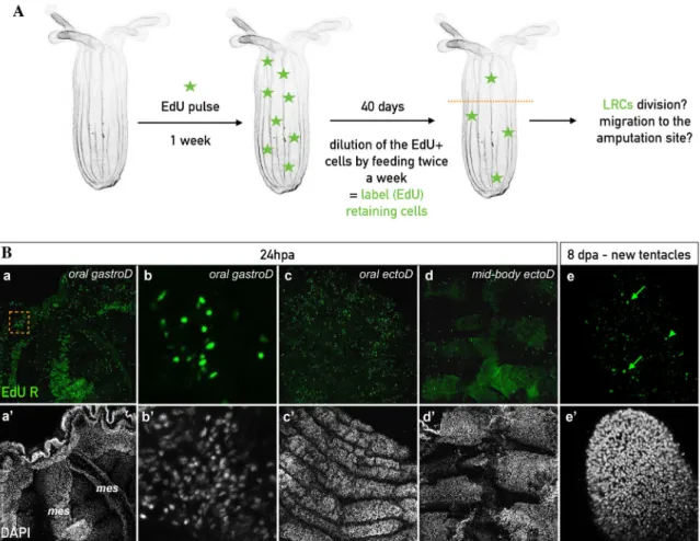
during 1h, then washed 5 times and chased for the period of interest. In order to identify 194
of the Label Retaining Cells (LRCs), animals were incubated for one week with EdU, 195
then chased up to 11 weeks for juveniles or 3 to 5 month for adults. 196
197
Irradiation
198
For irradiations experiments, animals where placed in a 10cm diameter petri dish with 199
15ml 1/3 ASW. Irradiations at 100 and 300 Grays where performed on the juvenile 200
polyps using a X-ray irradiator (CP-160 Cabinet X-Radiator, Faixitron). After irradiation, 201
animals were recovering during 4 hours in their culture medium (1/3ASW) at room 202
temperature before further cutting experiments. 203
204
Imaging
205
Live animals were imaged using a protocol described in [63]. The imaging setup was 206
composed of either with a Zeiss Stereo Discovery V8 Discovery or a Zeiss Axio Imager 207
A2 (both Carl Zeiss Microscopy GmbH, Jena, Germany) equipped with a Canon 6D 208
(Canon) or a Axiocam 506 color camera (Zeiss) digital camera, triggering two external 209
Canon Speedlite 430 EX II Flashes and controlled by the Canon Digital Photo 210
Professional software (Canon Inc, Tokyo, Japan). Images were edited using Adobe 211
Lightroom 5 and/or Photoshop CS6 software (Adobe Systems Inc, San Jose, CA, USA). 212
Labeled animals were analyzed using a Zeiss LSM Exciter confocal microscope running 213
the ZEN 2009 software (Carl Zeiss Microscopy GmbH, Jena, Germany) from the IRCAN 214
imaging platform (PICMI). Each final image was reconstituted from a stack of confocal 215
images using Z-projection (maximun intensity or standard deviation) of the ImageJ 216
software (Rasband, W.S., ImageJ, U. S. National Institutes of Health, Bethesda, 217
Maryland, USA, http://imagej.nih.gov/ij/, 1997-2014). 218
Results
220
The regenerative capacity of the physa is age dependent
221
In order to assess if all body parts of Nematostella possess similar regenerative 222
capacities and if those limits are age dependent, we dissected adult and juvenile 223
Nematostella into several isolated body parts. Following dissection, we analyzed each
224
parts capacity to regenerate the missing oral or aboral structures within seven days 225
(Figures 1, Additional file 1: Figure S1). Throughout the manuscript the isolated body 226
parts that are tracked to score regeneration are indicated as follow: [name of the isolated 227
part]. As an example, when a tentacle is isolated and tracked for it is ability to regenerate, 228
it is indicated as [tentacle]. [full body column + physa] is the equivalent of the sub-229
pharyngeal amputation experiment performed in previous studies [59,62] (Figures 1Ac,c’, 230
Bh,h’, C) and is considered as positive control in the corresponding experiments. 231
232
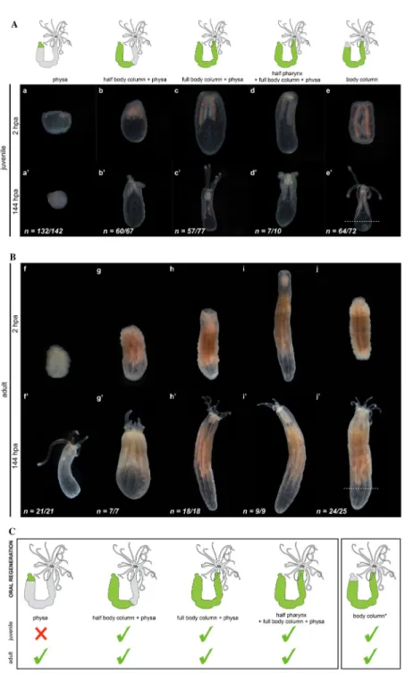
We first addressed the capacity of isolated body parts to regenerate missing oral 233
structures (oral regeneration) and we observed that most body regions isolated from 234
either juvenile or adult polyps regenerate within 144hpa. The only exception was the 235
isolated juvenile (but not adult) physa that failed to regenerate missing oral features 236
(pharynx, mouth, tentacles, Figure 1Aa-e, a’-e’, Bf-j, f’-j’, C). The capacity of body parts 237
to regenerate is correlated with the presence of massive cell proliferation at the 238
amputation site throughout regeneration (Figure 2A, B). Massive cell proliferation was 239
also observed at the amputation site (Figure 3Aa,b) and is required for regeneration of the 240
adult [physa] (Figure 3Ac,d). Interestingly, no or very little cell division was detected in 241
juvenile [physa] (Figure 3Ba,b). These observations show that for the various isolated 242
body parts the capacity to regenerate is associated to cell proliferation, suggesting similar 243
cellular mechanisms involved in this process along the different regions of the polyp. 244
They also highlight that existing age-dependent differences in the regenerative capacity 245
of Nematostella are body part specific. 246
247
A similar conclusion was obtained when analyzing the regenerative capacity of 248
isolated juvenile or adult oral body regions to reform aboral structures (aboral 249
regeneration) (Additional file 2: Figure S2). Only juvenile [tentacles + full pharynx + half 250
body column] and [body column], as well as adult [tentacles + full pharynx], [tentacles + 251
full pharynx + half body column] and [pharynx] regenerated (Additional file 1: Figure 252
S2Ad, d’, Bg-j, g’-j’, C). Intriguingly though, we did not observed massive cell
253
proliferation at the amputation site, neither for the regenerating nor the non-regenerating 254
isolated body parts (Additional file 3: Figure S3), suggesting different cellular strategies 255
involved in the oral vs aboral regeneration processes. 256
257
Taken together these experiments clearly illustrate the existence of limits in the 258
regenerative abilities of distinct Nematostella body parts (i.e. the tentacles) that is also 259
varying in an age-dependent manner (i.e. physa). While aboral regeneration seems to 260
involve mainly cell/tissue rearrangements, importantly, the capacity of body parts to 261
reform missing oral structures is tightly linked to the ability of cells to re-enter mitosis, 262
suggesting qualitative variations in the tissues that form the body of Nematostella. 263
264
Mesenteries are present in the adult but not in the juvenile physa
Intrigued by the observation that some body parts were able to perform oral 266
regeneration when isolated from adults but not from juveniles (e.g. physa, Figure 1Aa,a’, 267
Bf,f’, C), we analyzed in detail the anatomy of the physa in juvenile vs adult polyps 268
(Figure 4). DIC microscopy revealed that in addition to the size variation between 269
juvenile and adult physa, a major difference exists in the thickness and elongation of the 270
longitudinal muscle fiber extensions within the mesenteries (Figure 4a,c). While in adult 271
physa we observed opaque longitudinal extensions from the aboral end of the mesenteries 272
towards the aboral-most tips of the physa (Figure 4c), in juvenile physa, we couldn't 273
detect those longitudinal extensions (Figure 4a). We further performed confocal imaging 274
of the juvenile and adult [physa] using phalloidin to label actin microfilaments 275
(longitudinal and transversal muscle fibers as well as cell cortex) and DAPI to label the 276
nucleus (Figure 4b,b’,d,d’). We observed that in adult [physa], the longitudinal muscle 277
fibers are considerably thicker compared to the ones present in juvenile tissues (Figure 278
4b,d). Orthogonal projection of these confocal images further revealed significant 279
differences in the organization of the longitudinal muscle fibers in juvenile or adult 280
[physa] epithelia (Figure 4b’,d’). In juvenile [physa], we observed a discrete protrusion 281
from the longitudinal muscle fibers toward the gastric cavity (Figure 4b’). In contrast, the 282
longitudinal muscle fibers of the adult [physa] are thick and shaped in a characteristic 283
manner (Figure 4d’). Within the adult endoderm, they form a stalk at the base of the 284
epithelium that becomes undulated more distally (Figure 4d’). Interestingly, this 285
particular form is strikingly similar to the organization of parietal muscles that support 286
the developing mesenteries [36]. Thus, these observations show the presence of forming 287
mesenteries only in the adult physa and suggest a correlation between the presence of the 288
mesenteries and the capacity of a given tissue to regenerate. 289
290
Mesenterial tissue is required for cell proliferation and regeneration
291
While in the juvenile [physa] no or very little mitotic activity was detected in the majority 292
of cases (Figure 3Ba, a’), 24 out of 63 juvenile [physa] displayed localized cell 293
proliferation at the amputation site (Figure 3Bb-c,b’-d’). Interestingly, DIC and 294
fluorescence imaging revealed that the observed localization and intensity of cellular 295
proliferation were linked to the presence and the amount of remaining mesenterial tissues 296
in the juvenile [physa] (Figure 3Ba’-d’). This observation strongly supports the idea that 297
the presence and amount of mesenteries remaining in the isolated body part, are 298
associated to cell proliferation during regeneration. 299
300
In order to directly test our hypothesis that mesenteries are crucial for cell 301
proliferation and regeneration in Nematostella, we performed further tissue isolation 302
experiments in adults. By cutting twice transversally, we isolated a part of the mid-trunk 303
region that we named [BE + mes] (Body wall Epithelia plus mesenteries). We then 304
opened it longitudinally to separate the body wall epithelia [BE] from the mesenteries 305
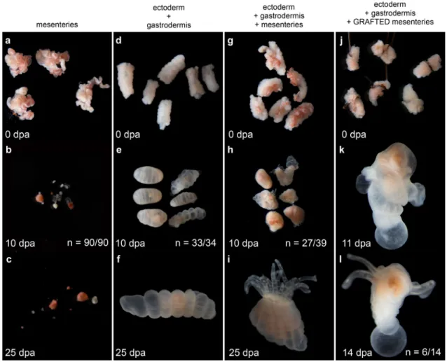
[mes], and cultured the [BE] and [mes] separately (Figure 5, Additional file 4: Figure S4). 306
We then analyzed daily if the isolated tissues [BE] or [mes] or the combination of both, 307
[BE + mes], are able to regenerate a fully functional polyp. The [mes] did not regenerate 308
(90 out of 90 cases) but instead, began to fragment one day after isolation, and 309
progressively degraded over time (Figure 5a-c). Surprisingly, while the wound healed, 310
none of the [BE] regenerated neither and they remained intact even after 25 days post 311
isolation (33 out of 34) (Figure 5d-f). Only the [BE + mes] regenerated 10 days after 312
tissue isolation (27 out of 39, Figure 5g-i) and the resulting polyps were able to feed 313
(Additional file 5: Figure S5). To further analyze the importance of the mesenteries to 314
induce regeneration in the surrounding [BE], we performed rescue experiments by 315
grafting [mes] to the endodermal component of the [BE]. Interestingly, nearly half of the 316
grafts regenerated a polyp (6 out of 14 cases, Figure 5j-l). However, regeneration was not 317
complete and none of the regenerated polyps were able to feed. After several weeks, one 318
of them lost its tentacles and all reduced in size (data not shown). 319
320
To advance our understanding of the role of the mesenteries in inducing 321
regeneration, we analyzed cell proliferation at 24, 72 and 168hpa (7dpa) in the isolated 322
tissues described above. While at 24hpa, we did not observe cellproliferation neither in 323
[BE] nor in [BE + mes], at 72 and 168hpa we detected massive and localized cell 324
proliferation only in the [BE + mes] (Figure 6a-f). At those time points, cell proliferation 325
was detected in a density gradient of EdU+ cells in the tissue, allowing us to determine 326
the site where the future tentacles will form (Figure 6e-f). Taken together, our data 327
clearly show that i) the presence of [BE] is required to maintain the integrity of the 328
mesenteries, and that importantly, ii) the mesenteries are required and possess the 329
capacity to induce cell proliferation as well as regeneration of the body wall epithelia. 330
Thus, a crosstalk between [mes] and [BE] is required for the regeneration process in 331
Nematostella, with a particularly crucial role of the mesenteries in the induction of this
332 333
Label retaining cells are present in Nematostella tissues
334
In whole body regeneration models, tissues lacking regenerative capacities have 335
been associated to the absence of undifferentiated stem cells in this compartment (e.g. 336
planarian pharynx [9-11], Hydra tentacles [12,13]). In the previous set of experiments we 337
have identified the juvenile physa that, in contrast to its adult counterpart is devoid of 338
mesenteries, as a body part lacking regenerative capacity. Thus an appealing hypothesis 339
is that the mesenteries are one compartment that acts a as a potential source of adult stem 340
cells involved in regeneration in Nematostella. 341
342
Quiescence or slow cycling is among the characteristics of vertebrate adult stem 343
cells that participates in the protection of their genomic integrity [64]. In order to identify 344
the existence of slow cycling / quiescent cells populations in Nematostella, we carried out 345
label retaining experiments used in many vertebrate models to identify tissue specific 346
stem cell populations [65-69]. After a one-week EdU pulse and extensive rinsing steps in 347
uncut juveniles and adults, the labeled animals were feed once (juveniles) or twice 348
(adults) per week during the 11 weeks (juveniles) or 39 days (adults) periods of the chase 349
(Figure 7). 350
351
Starting at seven weeks or 39 days after the EdU pulse in juveniles or adults 352
respectively, we began to distinguish isolated and randomly localized EdU+ label 353
retaining cells (LRCs, Figure 7). Those LRCs were detected as expected in the 354
mesenteries of juvenile and adult polyps (Figure 7C,D). Note that in addition to randomly 355
dispersed LRCs in adult mesenteries, we also observed dense EdU+ cells in a restricted 356
region close to the gametes, suggesting the presence of germ line precursors in this 357
specific location (Figure 7D, Additional file 6: Figure S6g,g’). 358
359
To our surprise we also detected LRCs within the body wall epithelia (Figure 360
7A,B. Interestingly, we detected LRC pairs in juveniles and adult epithelia, suggesting 361
that a subpopulation of quiescent cells are able to divide to maintain tissue homeostasis in 362
Nematostella (Figure 7Ae,e’,f,f’,Bh,h’). In adults, the density of LCRs after 39 weeks
363
seemed reduced in the endoderm compared to the ectoderm, suggesting that the cellular 364
renewal takes place faster in the endoderm than in the ectodermal (Additional file 7: 365
Figure S7a,a’,b,b’). To reinforce the idea that we have detected quiescent/slow cycling 366
cells in Nematostella, LRCs were still detected in the epithelia of the body wall (as well 367
as in the mesenteries) five months after the initial EdU pulse (Additional file 8: Figure 368
S8k,k’). However, while after 39 days of chase LRCs were still detected in the tentacles, 369
after 5 months no more LRCs were observed in this part of the body (Additional file 8: 370
Figure S8h-j,h’-j’), suggesting that the cellular turnover (causing the elimination of the 371
LRCs) in the tentacle region is increased compared to the body column. 372
373
To make sure that the LRCs we detected are not all terminally differentiated cells, 374
we analyzed the nuclear morphology and organization of those EdU+ cells. While we 375
detected differentiated cells such as cnidocytes, excretory cells and batteries of 376
nematocysts (Additional file 7: Figure S7d-f,d’-f’), we also observed a subpopulation of 377
cells with highly condensed DNA (Additional file 7: Figure S7c,c’), reminiscent of stem 378
cell nuclei in Hydractinia [22]. The presence of LRCs in the mesenteries (other than 379
potential germ line precursors) as well as in the epithelia that are able to re-enter the 380
mitotic cycle under physiological conditions, further support the presence of adult stem 381
cells in the anthozoan Nematostella. 382
383
Irradiation resistant LRCs in the mesenteries and response to the amputation stress
384
The capacity to retain EdU labelling is one feature of slow cycling/quiescent stem 385
cell populations that thus represent an increased resistance to irradiation. Another 386
characteristic is the ability to re-enter the cell cycle after a challenge (e.g. amputation 387
stress) [64]. In order to test this ability in Nematostella, we inhibited mitosis in uncut 388
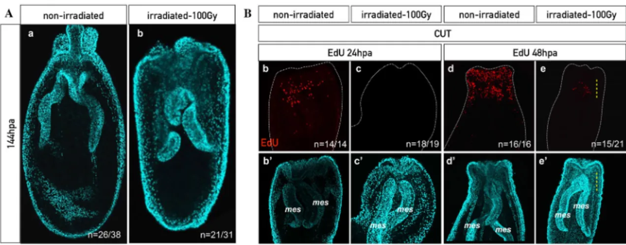
animals using X-ray irradiation. As expected, no more cell proliferation was detected four 389
hours after irradiating the animals at 100Gy (Additional file 9: Figure S9a,a’,b,b’). In 390
order to analyze if mitotic activity can re-emerge in the irradiated animals, we performed 391
EdU incorporation at various time points following irradiation on uncut polyps 392
(Additional file 9: Figure S9c-e,c’-e’). Interestingly, we observed EdU+ cells starting at 393
96hours post irradiation (hpirr) that were dispersed randomly within the mesenteries and 394
the basal part of the pharynx but not at all in the epithelia (Additional file 9: Figure 395
S9e,e’). These data clearly show that a pool of cells within the mesenteries and the basal 396
part of the pharynx is able to escape the effects of irradiation. 397
398
In order to test if this small pool of irradiation resistant quiescent/slow cycling cells 399
is sufficient to allow proper regeneration, we performed a sub-pharyngeal amputation 400
four hours after irradiation. Doing so, we observed that regeneration was impaired. More 401
precisely, the majority of irradiated animals were blocked prior to the formation of the 402
pharynx at step 2 of the sub-pharyngeal oral Nematostella regeneration staging system 403
[62] (100Gy: Figure 8A, Additional file 10: Figure S10i,i’; 300Gy: Additional file 11: 404
Figure S11b,b’, Additional file 12: Figure S12). To analyze the capacity of the 405
quiescent/slow cycling cells to respond to the amputation stress, we repeated the same 406
experiment (sub-pharyngeal amputation of irradiated polyps) that was followed by EdU 407
labeling at various time points post dissection. To our surprise, we detected the presence 408
of EdU+ cells in the most oral part of the mesenteries already at 48hpa in the amputated 409
polyps (Figure 8Be,e’). Later (120hpa), in addition to the ones in the oral tips of the 410
mesenteries, we also observed EdU+ cells in the epithelia at the amputation site (100Gy: 411
Additional file 10: Figure S10i,i’; 300Gy: Additional file 11: Figure S11b,b’). While 412
quiescent/slow cycling cells that escape irradiation are able to re-enter the cell cycle in 413
uncut as well as cut polyps, their timing of emergence and dispersion is different. They 414
are either detected 96hpa and distributed along the oral-aboral axis of the mesenteries in 415
uncut animals, or restricted to the oral most part of the mesenteries in dissected polyps 416
starting 48hpa. Latter observation suggest that quiescent/slow cycling cells are migrating 417
from the aboral region of the mesenteries towards the oral most part of those structures in 418
response to the amputation stress. 419
420
Taken together these results show that i) the quiescent/slow cycling cells of the 421
mesenteries alone are not sufficient to allow full regeneration, ii) that at least an 422
additional pool of fast cycling cells (the ones affected by irradiation) are required for 423
reformation of lost body parts and iii) that the LRCs of the mesenteries that escape the 424
effects of irradiation, are activated by the amputation stress by displaying mitotic activity 425
and by migrating towards the amputation site. Thus, we now provide a series of 426
evidences that highlight the presence of quiescent/slow cycling stem cells in Nematostella, 427
in particular, those located within the mesenteries. 428
429
LRCs are migrating from the mesenteries to the body wall epithelia
430
We have shown that a crosstalk between the mesenteries and the epithelia is 431
required for cell proliferation/regeneration and that quiescent/slow cycling stem cells are 432
potentially migrating from the mesenteries to the surrounding epithelia in response to the 433
amputation stress. To test this hypothesis, we designed a protocol that combined EdU 434
pulse and chase with grafting experiment. More precisely, we labeled LRCs of the entire 435
polyp and isolated its mesenteries, named [mesLRC] (LRC stained mesenteries). We then 436
grafted the [mesLRC] to the endodermal component of the [BE] from an unlabeled 437
animal (Figure 9A). After 14 days, 3 out of 18 grafts regenerated. Nonetheless, all 18 438
grafts were fixed in order to chase the EdU staining and determine the localization of 439
LRCs in the chimeric animals. To our surprise, we not only observed LRCs in the 440
previously unlabeled [BE] (5 out of 18 cases), but also far from the [mesLRC] source 441
(Figure 9Ba). In one of the regenerated [mesLRC] [BE] grafts, a large amount of LRCs 442
have even migrated towards the newly formed tentacles (Figure 9Bd). This clearly 443
demonstrates that LRCs are able to migrate from the mesenteries towards the epithelia, 444
and even within the epithelia to integrate specific regions. Importantly, we also observed 445
LRC pairs (in addition to single LRCs), showing that the LRCs that migrated from the 446
[mesLRC] to the [BE] were able to divide and are not post-mitotic (Figure 9Bb,c). A 447
detailed analysis of the DNA of migrating LRCs using confocal microscopy revealed that 448
i) their nucleus is round, ii) very similar in size, iii) posses a compact DNA and iv) the 449
majority is located in the endoderm of the [BE] (Figure 9C). Latter observation strongly 450
suggests that the graft re-establishes the physical contact between the endodermal 451
components of the mesenteries and the [BE]. In sum, we now have clear evidence that 452
LRCs, or their immediate progeny, are able to migrate from the mesenteries towards the 453
body wall and tentacle epithelia under stress conditions where they may participate in 454
regenerating missing body parts. 455
456
LRCs localized in the epithelia respond to the amputation stress
457
In addition to the clearly defined pool of mesenterial LRCs, we have also 458
observed LRCs within the epithelia that were however, less numerous compared to the 459
mesenterial ones (Figure 7). To perform a detailed characterization of all LRCs present in 460
Nematostella, we also analyzed the dynamics of LRCs localized in the epithelia of the
461
body wall during regeneration. We performed sub-pharyngeal amputation on LRCs 462
positive adult polyps (Figure 10A) and analyzed at various regeneration time points (24-463
192hpa) i) the localization of the LRCs, ii) their ability to divide and form clusters in 464
response to the amputation stress and iii) their becoming in the newly regenerated head 465
(Figure 10B). The LRCs of the mid-body and aboral regions remained largely unaffected. 466
However, we detected a general, although variable, accumulation of LRCs as well as the 467
presence of LRC clusters at the amputation site starting at 24hpa (Figure 10Ba-d,a’-d’). 468
This variability could be either associated i) to the different metabolic states of each 469
individual or ii) to the increased division rate at the amputation site at later stages that 470
may cause the dilution of the EdU signal or iii) both. Importantly though, we detected 471
single LRCs as well as pairs of LRCs in the newly formed tentacles 8 days post-472
amputation (192hpa) (Figure 10Be-e’’). 473
474
Taken together, our data show that the LRCs of the epithelia, although they are 475
not able to escape the effects of irradiation, i) are activated in response to the amputation 476
stress by dividing and accumulating at the amputation site and ii) participate in the newly 477
formed head (e.g. tentacles epithelia), providing clear evidence that those epithelial LRCs 478
are actively involved in tissue renewal and thus, also possess stem cell-like characteristics. 479
480
Accumulation of fast cycling cell at the amputation site during oral regeneration
481
We have seen that the mesenterial LRCs (reactivated during regeneration 482
following irradiation) alone are not sufficient to promote full regeneration. This shows 483
the necessity of a tissue crosstalk and in particular, the requirement of another pool of 484
(stem) cells localized in the body wall epithelia. During the irradiation experiment, we 485
blocked cell proliferation in cells that are undergoing mitosis including fast cycling cells 486
required for tissue homeostasis. In order to test if a population of fast dividing cells from 487
the uncut animal also migrates to the wound site during regeneration, we performed a 1h 488
EdU pulse in uncut animals followed by extensive washes to eliminate any excessive 489
EdU. This EdU pulse labels the fast dividing cells involved in tissue homeostasis (Figure 490
11Aa). We then performed a sub-pharyngeal amputation and chased the EdU+ cells 491
during the process of regeneration from 0 to 120hpa (Figure 11A,B). 492
In uncut control animals, EdU+ cells were randomly dispersed throughout the 494
body wall epithelia as well as in the mesenteries for all analyzed time points [59](Figure 495
11Aa-d, Additional file 13: Figure S13). Starting at 24 hours post-pulse, first homeostatic 496
EdU+ cell divisions are detected (still in a dispersed manner) in uncut animals, as 497
indicated by the presence of EdU+ cell pairs (Figure 11Ab-d). Interestingly, in sub-498
pharyngeally amputated polyps, we observed a strong accumulation of EdU+ cells in the 499
oral regions of the mesenteries while their remaining aboral parts progressively become 500
devoid of those cells (Figure 11B). This accumulation, indicating amputation-site-501
directed cellular migration within the mesenteries is already visible at 24hpa Figure 502
11Bi,j, while EdU+ cells are still randomly located within the epithelia (Figure 11Ae,f). 503
However, starting at 48hpa, EdU+ cells accumulate massively in a restricted region of the 504
epithelia at the amputation site and, in a few cases (48hpa: 9 out of 31), a zone depleted 505
of EdU+ cells is observed in the region below the massive accumulation of EdU+ cells 506
(Figure 11Ag,h). 507
508
Those observations are further quantitatively supported by carefully counting individual 509
Edu + cells in three different zones (Z1’, Z1’’ and Z2) of the body epithelia along the 510
oral-aboral axis of the uncut vs cut animal during regeneration (Additional file 14: Figure 511
S14). In addition to the difference in the average number of EdU+ cells in zone Z1’ in 512
uncut (sub-pharyngeal region) vs cut (amputation site), this cell counting revealed clear 513
differences in the Z1” (lower mid-body region) and Z2 (aboral end) zones for the same 514
conditions. In fact, while the average number of EdU+ cells in Z1” and Z2 is constant or 515
decreasing respectively in regenerating animals, it is increasing in both zones in uncut 516
controls (Additional file 14: Figure S14). In sum, these results show that fast cycling cells 517
(required for regeneration as eliminated during irradiation) from the region below zone 518
Z1’ of the body wall epithelia, migrate towards the amputation site and in cooperation 519
with LRCs, actively participate in the renewal of missing tissues. 520 521 522 523 524 525 526 527 528 529 530 531 532 533
Discussion
534
In the present study, we have highlighted the unexpected role of the mesenteries 535
not only as a reservoir of quiescent/slow cycling stem cells, but also as the tissue 536
harboring the signal to induce cell proliferation and regeneration in Nematostella. Long-537
term EdU pulse and chase experiments revealed the presence of quiescent/slow cycling 538
cells (LRCs) that respond to the amputation stress by reentering the mitotic cycle, 539
accumulation at the wound-site and participate in the reformation of missing structures. 540
Combining classical graft experiments with EdU pulse and chase experiments, revealed 541
cellular migration from the mesenteries to the epithelia, as well as toward the amputation 542
site in response to the amputation stress. We further showed that LRCs alone are not 543
sufficient and that homeostatically fast cycling cells are required for regeneration. In sum, 544
our present work has not only revealed the requirement of a tissue crosstalk between the 545
mesenteries and the epithelia of the body wall to initiate the cellular dynamics underlying 546
regeneration but importantly also the synergic effect of fast cycling cells and 547
slow/quiescent stem cells to enable full reformation of lost body parts. This study 548
provides also for the first time in an anthozoan cnidarian a set of strong evidences for the 549
presence of adult stem cell populations that are directly involved in the regenerative 550
response. 551
552
We thus propose a mechanistic model for head regeneration in Nematostella, in 553
which the mesenteries, once in contact with the epithelia of the amputation site (Step 1, 554
[62]) emit a regeneration-initiating signal that activates fast and slow-cycling stem cell 555
populations from the mesenteries as well as the epithelia by inducing their division and 556
migration toward the wound site. Importantly, a synergic effect between those two stem 557
cell populations is required to pursue and complete the regeneration process. The 558
regeneration inducing signal emitted by the mesenteries, the mechanism of synergic 559
cooperation between the two stem cell populations and whether de- or trans-560
differentiation processes are also involved in the tissue repair and regeneration program 561
of Nematostella, remains to be investigated. 562
563
Distinct cellular mechanisms between oral vs aboral regeneration in Cnidaria
564
By comparing the regenerative capacity of various body parts to reform missing 565
oral as well as aboral structures, we observed that regeneration of aboral tissues does not 566
involve massive cellular proliferation at the amputation site, while oral regeneration does. 567
These data suggest that aboral regeneration either i) uses a different cellular mechanism 568
than the one described for oral regeneration in order to reform the missing physa and/or 569
ii) that the requirement of regenerating aboral tissues is less urgent (and thus does not 570
require massive localized cell proliferation) than quickly reforming a missing head region 571
that is more complex and crucial for feeding and defense. A distinct cellular mechanisms 572
for oral vs aboral regeneration has been recently described in the cnidarian hydrozoan 573
Hydra and Hydractinia [20,22] supporting the idea that using different mechanism to
574
regenerate opposite part of the body in a same system seems to be a conserved feature 575
among Cnidaria. The studies performed in Hydrozoa show that similar to our 576
observations, no massive cell proliferation is detected at the aboral amputation site 577
[20,22]. In Nematostella, while we also observe a slight reduction in tentacle size during 578
aboral regeneration that could contribute to the reformation of the physa via tissue 579
reorganization, we have no evidence for a complete transformation of the body column 580
into an aboral fate prior to re-growing a fully functional polyp. However, recent work on 581
Nematostella shows a clear difference in the transcriptional response of the oral versus
582
aboral regeneration process [61]. This study also shows that the early molecular response 583
(8hpa) to the amputation stress is more similar between oral vs aboral regions, compared 584
to later time points (24 and 72hpa) [61]. Interestingly, our previous study on Nematostella 585
regeneration revealed that early oral regeneration steps (from step 0 to step 1; between 0 586
to 24hpa) are cell proliferation independent, while the later ones (from step 2 to step 4, 587
between 24 to 120hpa) are cell proliferation dependent [62]. Thus, in Nematostella, the 588
mechanisms driving the early steps of tissue regeneration such as wound healing seem 589
similar between oral versus aboral regeneration as they are both cell proliferation 590
independent. However, additional experiments are required to gain a better understanding 591
of the cellular and molecular mechanisms involved in aboral regeneration in 592
Nematostalla, in particular during later regeneration phases, when initial wound healing
593
and early response is completed. 594
595
Regenerative capacity of a given body part is age-dependent
596
It has been previously shown that oral regeneration after sub-pharyngeal 597
amputation in Nematostella, occurs at a comparable time-scale in both juveniles and 598
adults and that cellular proliferation is required in both cases [62]. Our present study 599
revealed that clear variation in the capacity to regenerate exists in one given body part 600
depending on the age of the organism. The obtained data show that the tissue composition 601
within a given body region is changing with age. In particular, the isolated physa 602
regenerates only in adults, but not in juveniles that are unable to induce sufficient cellular 603
proliferation, probably by the lack of i) stem cell population(s), ii) initiation signal(s) or 604
both. In addition, this approach was useful to identify which body part/structure of 605
Nematostella is required for the initiation of regeneration and to orient our research on
606
the potential stem cell population(s) involved in the regeneration process. 607
608
Mesenteries are required and sufficient to induce cell proliferation and regeneration
609
Taking advantage of the differential regenerative capacity of juvenile and adult 610
physa, we identified the mesenteries as crucial structures to induce cellular proliferation 611
and subsequent regeneration in the surrounding tissues. By performing tissue dissociation 612
and grafting experiments, we have shown that isolated mesenteries, grafted back to the 613
remaining [BE] of the same individual, are sufficient to induce regeneration. Extending 614
this finding to other body parts of juveniles that did not regenerate when isolated such as 615
[tentacles], [tentacles + half pharynx], [tentacles + full pharynx] and [pharynx], it seems 616
plausible that the common factor explaining the lack of regeneration in those juvenile 617
body parts are the missing or not completely formed mesenteries. [tentacles] or [tentacles 618
+ half pharynx] isolated from adults did not regenerate neither, while [tentacles + full 619
pharynx] or [pharynx] did. Tentacles clearly lack mesenterial tissues and interestingly, 620
the mesenteries are anchored to the pharynx in its aboral regions. As the half pharynx that 621
remained with the tentacles in [tentacles + half pharynx] corresponds to the oral half of 622
the pharynx, the absence of regenerative capacity in juvenile and adult [tentacles] or 623
[tentacles + half pharynx] can thus be linked to the absence of mesenteries in those body 624
parts. Fully formed mesentery anchors are present in the adult pharynx that are sufficient 625
to induce regeneration from [tentacles + full pharynx] or [pharynx]. In analogy to our 626
findings in the juvenile physa, we expect that the mesenterial anchors, or at least 627
important components of it (i.e. stem cells), in the juvenile pharynx are not fully formed 628
or are absent. This may explain the absence of regenerative capacity of those isolated 629
body parts. 630
631
One puzzling observation in the graft experiments (restoring the regenerative 632
capacity) was that the regenerated animals possessed a tentacle crown but not a mouth 633
and were therefore unable to feed. One probable explanation for this is the number of 634
mesenteries required for full regeneration. In the graft experiment performed in our 635
present study only one mesentery has been grafted into the epithelia leading to a partial 636
regeneration (head without mouth/pharynx). By testing the removal of only 7, 6 or 5 637
mesenteries on 8 total from the adult body wall epithelia, we have observed that at least 638
two mesenteries are required for proper mouth formation. In none of the cases in which 639
only one mesentery was left in contact with the body wall epithelia, the regenerated 640
polyps were able to fed, showing that only partial regeneration occurs (data not shown). 641
This evidence for the requirement of at least two mesenteries for proper pharynx 642
reformation is further enhanced by previous observations that during oral regeneration the 643
two mesenteries fuse to one to each other, and that the fused oral parts of the mesenteries 644
give rise to the vast majority of the newly formed pharynx [62]. 645
646
A tissue crosstalk is required for initiating a regenerative response
The observation that the mesenteries are required to induce regeneration in the 648
surrounding epithelia leads to three main hypotheses concerning the mechanism 649
involved: 1) A long-range diffusing “regeneration” signal emitted by injured mesenteries 650
is required to initiate cellular proliferation in the surrounding epithelia. 2) The physical 651
integrity of the tissue that links the mesenteries to the body epithelia is important to relay 652
the “regeneration” signal. 3) A pool of stem cells (or their offspring) migrates from the 653
mesenteries to the body wall epithelia to initiate the regeneration process. We tested the 654
first hypothesis by incubating the isolated body wall epithelia in close but not physical 655
contact with isolated pieces of mesenteries in the same wells. In none of the cases the 656
body wall epithelia regenerated (data not shown), suggesting that the molecular induction 657
signal (if there is any) from the mesenteries is not sufficient alone, or that it is too diluted 658
in the well, to induce regeneration of the epithelia. Alternatively, it might also be possible 659
that only endodermal cells respond to the induction signal to transform it into a 660
regenerative response. In this case, the fact that the isolated epithelia wounded and closed 661
with the endodermal cells inside prevents the signal to target the responsive cells. While 662
additional experiments are required to fully address hypothesis 1, we currently favor 663
hypothesis 2 and 3 to explain the role of the mesenteries in the regeneration process of 664
Nematostella.
665 666
The importance of physical tissue crosstalk during regeneration has been studied 667
during vertebrate intestinal epithelium regeneration in which the connective tissues have 668
been shown to be crucial for the regeneration of the epithelium [70].In addition,cell/cell 669
contact molecules such as integrins are known to be involved in the regeneration process 670
in vertebrates [71,72] [73]. The migration of stem cells to the amputation site has been 671
shown for regeneration of complex structure [74,75] and it has been proposed that 672
mechanical stress induces cell migration as in smooth muscles [76]. Our study has 673
provided clear evidences for a cellular transfer of cells from the mesenteries to the 674
epithelia as well as the existence of two populations of cells that have stem cells 675
characteristics (Hypothesis 3). The observation that the mesenteries disintegrate when 676
isolated from the body wall epithelia highlight the importance of a tight and physical 677
tissue interaction between the epithelia and the mesenteries to maintain the integrity 678
and/or homeostasis of the mesenteries (Hypothesis 2). Hypothesis 2 and 3 are further 679
reinforced by the fact that the process of regeneration is delayed in graft experiments 680
compared to [BE + mes] (14 vs 10 days, respectively). This could be explained by the 681
fact that the tissue connecting the mesentery and the body wall epithelia [62] needs to be 682
first reformed, before either the inductive signal can be relayed, or before the stem cell 683
population can migrate from the mesenteries to the body wall epithelia. 684
685
Migration of different pools of cells during regeneration
686
Our work highlights evidences that cell migration occurs during Nematostella 687
regeneration as it is the case in others invertebrate and vertebrate regeneration models 688
studied so far [1]. These evidence are: i) LRCs migrate from the mesenteries to the 689
surrounding epithelia, ii) fast cycling cells involved in tissue homeostasis migrate to the 690
amputation site, and iii) following irradiation, slow cycling EdU+ cells within the 691
mesenteries that escape irradiation accumulate the most oral part of the mesenteries, 692
while EdU+ cells are randomly dispersed in the mesenteries in irradiated uncut animals. 693
These data show that this population of cells re-entering the cell cycle in irradiated 694
animals migrate to the amputation side during regeneration. However, latter cell 695
population is not sufficient alone to fully complete regeneration as the regeneration 696
process in irradiated polyps is blocked at step 2 of the sub-pharyngeal Nematostella 697
staging system [62]. We recently showed that inhibition of cell proliferation using 698
Hydroxy Urea during Nematostella regeneration, blocked the regeneration process at step 699
1 [62], suggesting that the slow cycling, irradiation-resistant cells that re-entered the 700
mitotic cycle in response to the amputation stress are crucial to transition at least from 701
step 1 to step 2. 702
703
In Nematostella, although we were able to detect a “first wave” of migrating cells 704
only between 24-48hpa, we cannot exclude that cell migration of other cell populations 705
also occurs at earlier time points between 0 to 24hpa, as it has been shown in Hydra [20]. 706
The potential second “wave” of cell migration, reflected by the decrease in the number of 707
the EdU+ cells in zone Z2 (most aboral part of the animal) during regeneration, might 708
correspond to a replacement of stem cells populations in a zone that got depleted of it. 709
This kind of stem cell replenishment has been shown during tissue homeostasis or tissue 710
transplantation experiments in Hydra to repopulate a tissue that was i-cell deficient [77] 711
[78]. Our experiments show that fast as well as slow cycling cells are able to migrate 712
during Nematostella regeneration, however, we currently don’t know if the migrating 713
cells are the stem cells or their progeny. Further characterization of the molecular identity 714
of those migrating cells as well as the development of in vivo tools is required in 715
Nematostella to answer this question.
717
Stem cells in Anthozoa
718
Prior to the present study, cells populations with clear stem cell characteristics have not 719
been described in Nematostella or more generally in anthozoan cnidarians (sea anemones, 720
corals) [32,57,59]. Our present work highlights the existence of two populations of 721
proliferating cells that are synergistically required for regeneration: i) one pool, 722
composed of slow cycling/quiescent cells (LRCs) and ii) another pool, composed of fast 723
cycling cells involved in tissue homeostasis in the uncut animal. Both cell populations 724
possess features that are characteristic for stem cells: 1) The behaviour of the slow 725
cycling/quiescent/label retaining cells (LRCs) per se, as it is one important feature of 726
adult stem cells that is deployed in mammalian stem cells to preserve genomic integrity 727
[64,69]. These slow cycling/quiescent cells are able to escape X-ray irradiations and 728
respond to the amputation stress by migrating and accumulating towards the wound site, 729
where they actively divide and participate in the reformation of lost structures. While the 730
potency of those LRCs has not been properly addressed yet, we already observed that 731
they are able to give rise to sensory cells within the regenerated tentacles. 2) The fast 732
cycling cells, respond to the amputation stress by migrating towards the amputation site 733
where they actively divide and are required for regeneration, probably for later 734
regeneration steps (3 and 4). In the hydrozoans Hydra and Hydractinia, fast cycling stem-735
cells (i-cells and/or their progeny) also divide and migrate to the amputation site and 736
actively participate in the regeneration process [20,22]. Thus, our results suggest a 737
conserved pool of fast cycling stem cells in cnidarians that are involved in tissue 738
homeostasis and regeneration. 739
740
Stem cell/pluripotency markers in ctenophores and cnidarians are known to 741
include the germ lines markers piwi, vasa, nanos and PL10 [22,79-84] [85]. A previous 742
study in Nematostella analyzed the expression pattern of those genes (with the exception 743
of piwi) from early development to the primary polyp stage [86]. In the juvenile, Nvnos1 744
is found in some patches of cells in the ectodermal epithelia of the body wall. The authors 745
suggested that these patches of Nvnos1+ cells are “population of nematocyst precursors 746
with stem cells characteristics” [86]. However, all other analyzed germline genes NvPl10, 747
Nvvasa1, Nvvasa2 and Nvnanos2 are expressed solely in the mesenterial tissues, excluded
748
from the epithelia of the body wall and interestingly, are not detected within the physa of 749
juveniles [86]. Those data combined with our current work further strengthen the idea 750
that a specific stem cell population, able to respond to the amputation stress is located in 751
the mesenteries of Nematostella. 752
753
However, it is important to note that the germ line of anthozoans is located in the 754
mesenteries [86]. This of course raises the questions if i) the germ line cells correspond to 755
the pool of pluripotent stem cells involved in the regeneration process, or if ii) the 756
mesenteries contain in addition to the germ line, a pool of set aside stem cells that 757
respond to the amputation stress by migrating towards the wound site to reform missing 758
body parts. If the first idea would be true, then one would expect that a tradeoff between 759
sexual reproduction and regeneration might exist, as regeneration would exhaust the 760
germ-line pool for the tissue repair process. First experiments to analyze if sexual 761
reproduction is impaired after bisection, revealed that the spawning capacity of the 762
regenerated adult polyps remained unchanged (data not shown). While this suggests that 763
the germ line is different from the stem cell population in the mesenteries, additional 764
experiments are required to fully address this question. 765
766
Taken together, we provide a list of evidences that strongly support the idea that 767
there are at least two populations of stem cells in Nematostella that are actively deployed 768
and act in synergy during the regeneration process. However, further cellular (self-769
renewal, potency) as well as molecular characterization (e.g. stem cell/pluripotency 770
markers) is needed in order to characterize in details their stem cell identity. 771
772
The presence of mesenteries as specific feature of anthozoans may account for a spatial
773
dissociation of the signal-emitting and signal-receiving compartments required for
774
regeneration
775
Hydrozoans lack mesenteries and their pluripotent stem cells (i-cells) are located 776
in the epithelia of the midgastric region and are able to give rise to the germ line as well 777
as somatic cells [87-89]. The present study revealed that anthozoans possess stem cell 778
populations that are located throughout the body within the epithelia but importantly also 779
within the mesenteries. Because the oocytes (germ line) are located within the 780
mesenteries, it is highly probable that, if any pluripotent stem cells (giving rise to germ 781
and soma) exist in Nematostella they are located within the mesenteries. Taken into an 782
evolutionary context one proposition would be that hydrozoans (Hydra, Hydractinia) 783
cope the lack of mesenteries with the existence of pluripotent stem cells (at the origin of 784
germ and somatic cells) as well as a regeneration-inducing signal localized both directly 785
![Figure 2. A. Cell proliferation in juveniles at 48hpa for [half body column + physa] (a), [full body column](https://thumb-eu.123doks.com/thumbv2/123doknet/13172665.390683/51.918.144.776.105.830/figure-cell-proliferation-juveniles-half-column-physa-column.webp)
![Figure 3. Cell proliferation in adult (A) and juvenile (B) [physa]. A. In adults, cell proliferation is present](https://thumb-eu.123doks.com/thumbv2/123doknet/13172665.390683/52.918.162.764.103.814/figure-cell-proliferation-adult-juvenile-adults-proliferation-present.webp)


![Figure 9. A. Diagram of the protocol to assess cell migration in graft experiments. [epithelia EdU+] and](https://thumb-eu.123doks.com/thumbv2/123doknet/13172665.390683/58.918.243.670.102.848/figure-diagram-protocol-assess-migration-graft-experiments-epithelia.webp)