Distinct Roles of RZZ and Bub1-KNL1 in Mitotic
Checkpoint Signaling and Kinetochore Expansion
The MIT Faculty has made this article openly available.
Please share
how this access benefits you. Your story matters.
Citation
Rodriguez-Rodriguez, Jose-Antonio et al. "Distinct Roles of RZZ
and Bub1-KNL1 in Mitotic Checkpoint Signaling and Kinetochore
Expansion." Current Biology 28, 21 (November 2018): P3422-3429.e5
© 2018 Elsevier
As Published
http://dx.doi.org/10.1016/j.cub.2018.10.006
Publisher
Elsevier BV
Version
Author's final manuscript
Citable link
https://hdl.handle.net/1721.1/126113
Terms of Use
Creative Commons Attribution-NonCommercial-NoDerivs License
Distinct roles of RZZ and Bub1-KNL1 in mitotic checkpoint
signaling and kinetochore expansion
Jose-Antonio Rodriguez-Rodriguez1, Clare Lewis1, Kara L. McKinley2,3, Vitali
Sikirzhytski4,5, Jennifer Corona1, John Maciejowski1, Alexey Khodjakov4,5, Iain M. Cheeseman2,3, and Prasad V. Jallepalli1,*
1Molecular Biology Program, Sloan Kettering Institute, Memorial Sloan Kettering Cancer Center,
New York, NY, 10065, USA
2Whitehead Institute for Biomedical Research, Nine Cambridge Center, Cambridge, MA 02142,
USA
3Department of Biology, Massachusetts Institute of Technology, Cambridge, MA 02142, USA 4Wadsworth Center, New York State Department of Health, Albany, NY 12201
5Rensselaer Polytechnic Institute, Troy, NY 12180
Summary
The Mad1-Mad2 heterodimer is the catalytic hub of the spindle assembly checkpoint (SAC), which controls M phase progression through a multi-subunit anaphase inhibitor, the mitotic checkpoint complex (MCC) [1, 2]. During interphase, Mad1-Mad2 generates MCC at nuclear pores [3]. After nuclear envelope breakdown (NEBD), kinetochore-associated Mad1-Mad2 catalyzes MCC assembly until all chromosomes achieve bipolar attachment [1, 2]. Mad1-Mad2 and other factors are also incorporated into the fibrous corona, a phospho-dependent expansion of the outer kinetochore that precedes microtubule attachment [4–6]. The factor(s) involved in targeting Mad1-Mad2 to kinetochores in higher eukaryotes remain controversial [7–12], and the specific phosphorylation event(s) that trigger corona formation remain elusive [5, 13]. We used genome editing to eliminate Bub1, KNL1, and the Rod-Zw10-Zwilch (RZZ) complex in human cells. We show that RZZ’s sole role in SAC activation is to tether Mad1-Mad2 to kinetochores. Separately, Mps1 kinase triggers fibrous corona formation by phosphorylating two N-terminal sites on Rod. In contrast Bub1 and KNL1 activate kinetochore-bound Mad1-Mad2 to produce a “wait anaphase” signal, but are not required for corona formation. We also show that clonal lines isolated after BUB1 disruption recover Bub1 expression and SAC function through
nonsense-*corresponding author and lead contact: jallepap@mskcc.org.
Author Contributions
J.-A.R.-R., C.L., K.L.M., J.C., J.M., and P.V.J. performed molecular biology, cell imaging, and biochemical studies and analyzed data. V.S. performed electron microscopy and analyzed data. A.K., I.M.C., and P.V.J. planned and supervised research, analyzed data, and secured funding. J.-A.R.-R. and P.V.J. wrote the paper with input from all authors.
Publisher's Disclaimer: This is a PDF file of an unedited manuscript that has been accepted for publication. As a service to our customers we are providing this early version of the manuscript. The manuscript will undergo copyediting, typesetting, and review of the resulting proof before it is published in its final citable form. Please note that during the production process errors may be discovered which could affect the content, and all legal disclaimers that apply to the journal pertain.
HHS Public Access
Author manuscript
Curr Biol
. Author manuscript; available in PMC 2019 November 05.Published in final edited form as:
Curr Biol. 2018 November 05; 28(21): 3422–3429.e5. doi:10.1016/j.cub.2018.10.006.
A
uthor Man
uscr
ipt
A
uthor Man
uscr
ipt
A
uthor Man
uscr
ipt
A
uthor Man
uscr
ipt
associated alternative splicing (NAS). Our study reveals a fundamental division of labor in the mammalian SAC and highlights a transcriptional response to nonsense mutations that can reduce or eliminate penetrance in genome editing experiments.
eTOC
Rodriguez-Rodriguez et al. identify distinct roles for Bub1, KNL1, and RZZ in SAC signaling and fibrous corona formation. They also show that BUB1-disrupted clones re-express Bub1 and regain SAC function via nonsense-associated alternative splicing, a often-overlooked transcriptional response that can limit penetrance in genome editing experiments
Results
The RZZ complex is required to maintain SAC arrest but not to initiate it
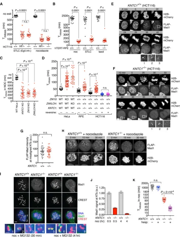
To analyze RZZ’s roles in fibrous corona assembly and SAC signaling, we used AAV and CRISPR/Cas9 to modify both alleles of KNTC1 (Rod) in HCT116 cells, a diploid human colorectal cell line (Figure S1A-C). KNTC1HF/- (hypomorph-flox) cells expressed Rod at ~20% of the wildtype level (Figure S1D) and exited mitosis prematurely when microtubule polymerization (nocodazole, 99±6 min s.e.m.) or spindle bipolarity (S-trityl-L-cysteine (STLC), 193±9 min) were inhibited. In contrast wildtype cells never exited mitosis during the 16-hour timelapse (Figure 1A). We obtained viable KNTC1–/– clones after expressing Cre recombinase, which were as SAC-defective as KNTC1HF/- cells (Figure 1A and Figure S1E). Early escape from spindle poison-induced mitotic arrest was also observed in KNTC1–/– human retinal pigment epithelial (RPE) cells and KNTC1, ZW10, and ZWILCH KO HeLa cells (Figure 1B-C and Figure S1F-I). On the other hand, untreated RZZ-null cells had longer and more heterogenous mitotic timing, suggesting frequent but transient SAC activation (Figure 1D). Consistently, inhibiting the SAC kinase Mps1 caused KNTC1–/– cells to exit mitosis with wildtype kinetics (Figure 1D). These observations suggest that RZZ maintains (but does not initiate) SAC signaling at unattached or improperly attached
kinetochores in multiple human cell types.
RZZ mediates a temporal switch in how Mad1-Mad2 is targeted to kinetochores
To understand RZZ’s impact on mitotic chromosome and SAC signaling dynamics, we expressed and imaged H2B-mCherry and FLAP (FLAG-GFP-TEV-S peptide)-Mad1 using spinning disk confocal microscopy. Mad1 first localized at kinetochores at nuclear envelope breakdown (NEBD), then dissociated as chromosomes congressed at the metaphase plate (Figure 1E and Video S1; n=10 cells). Congression was less efficient in KNTC1–/– cells, consistent with the lack of Spindly and dynein at kinetochores ([14] and Figure S1F-I), but Mad1 was still targeted to misaligned chromosomes as effectively as in wildtype cells (Figure 1F-G and Video S2; n=14 cells). We conclude that early mitotic cells can recruit Mad1-Mad2 to kinetochores and inhibit anaphase onset in the absence of the RZZ complex. Next we analyzed Mad1 dynamics in cells undergoing nocodazole-induced SAC arrest. Mad1 initially localized to kinetochores at NEBD in both wildtype and KNTC1–/– cells, but this localization was not persistently maintained in the absence of RZZ (Figure 1H). To
A
uthor Man
uscr
ipt
A
uthor Man
uscr
ipt
A
uthor Man
uscr
ipt
A
uthor Man
uscr
ipt
confirm this result for endogenous Mad1, wildtype and KNTC1–/– RPE cells were treated with nocodazole and MG132 (to block mitotic exit) for 30 min or 4 hours, then fixed and analyzed by immunofluorescence microscopy (IFM). In wildtype cells Mad1 formed large crescents that were stable over time, whereas it formed compact foci in KNTC1–/– cells that were eventually lost from kinetochores (Figure 1I-J). Suppressing early mitotic Mad1-Mad2 recruitment by treatment with Aurora B inhibitors [15, 16] eliminated the residual SAC response in KNTC1–/– cells (Tmitosis=39±10 min; Figure 1K). These results reveal a
temporal switch from RZZ-independent to RZZ-dependent recruitment of Mad1-Mad2 during chronic SAC signaling.
Mps1 promotes kinetochore expansion by phosphorylating the N-terminus of Rod
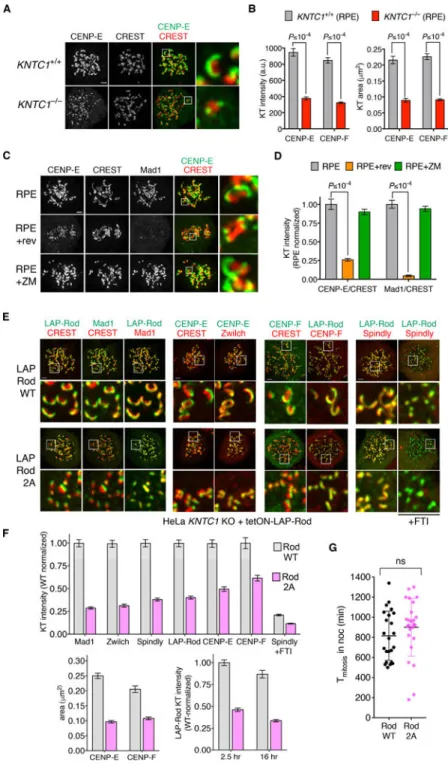
Mad1-Mad2 and RZZ localize to the fibrous corona, a phosphodependent expansion of the outermost kinetochore layer that persists until end-on microtubule attachments are formed [5, 6, 17]. Kinetochore expansion is thought to accelerate mitotic ‘search and capture’ by promoting lateral microtubule attachment [4] and to enhance SAC signaling [5]. RZZ is closely related to endomembrane coatomers that form oligomeric lattices [18, 19] and is likely a “building block” of the corona itself [13, 14, 20, 21]. Consistent with these proposals, two other corona-associated proteins (CENP-E [22] and CENP-F [23]) did not form crescents in KNTC1–/– cells (Figure 2A-B and S2A). To ensure that these results reflected loss of kinetochore expansion and not protein mislocalization, we performed correlative light-electron microscopy in cells expressing CENP-A-GFP as a centromere marker. Serial sectioning revealed circumferential expansion of trilaminar plates and fibrous material in wildtype cells (n=14 kinetochores), whereas the kinetochores of KNTC1–/– cells appeared as compact discs (n=15; Figure S2B and [13]). We conclude that the RZZ complex is required for fibrous corona formation.
In parallel we looked for mitotic kinases that might activate RZZ for kinetochore expansion. We found that CENP-E kinetochore crescents become compact after treating cells with an Mps1 inhibitor, but not after treatment with an Aurora B inhibitor (Figure 2C-D and [13]). Through global phosphoproteomic screening, we identified two Mps1-modified sites at the N-terminus of Rod (T13 and S15), upstream of its β-propeller domain [24]. To test the function of these sites, wildtype (WT) and nonphosphorylatable (2A) versions of Rod were expressed in T-Rex FLP-in HeLa cells as LAP (EGFP-TEV-S-peptide) fusions (Figure S2C). Both LAP-RodWT and LAP-Rod2A were incorporated into the full RZZ complex based on co-immunoprecipitation assays (Figure S2D). We then disrupted the KNTC1 locus in these cells using CRISPR/Cas9 and isolated transgene-complemented clones. Although LAP-RodWT and LAP-Rod2A both localized to unattached kinetochores in the absence of endogenous Rod, only LAP-RodWT formed crescents (Figure 2E-F). Thus far the only post-translational modification known to be required for crescent formation is C-terminal farnesylation of Spindly, which enables its kinetochore recruitment via interaction with Rod’s β-propeller domain [14, 20, 25, 26]. However Rod2A recruited Spindly and other corona-associated proteins in proportion to its own reduced abundance (Figure 2F). Despite having lower levels of Mad1-Mad2, the compact kinetochores in Rod2A cells sustained mitotic arrest in nocodazole as effectively as those in RodWT cells (Figure 2G). We conclude
A
uthor Man
uscr
ipt
A
uthor Man
uscr
ipt
A
uthor Man
uscr
ipt
A
uthor Man
uscr
ipt
that Rod’s N-terminal phosphorylation is required for fibrous corona formation but not SAC signaling.
Mad1-Mad2 requires a non-receptor activity of Bub1 to inhibit anaphase
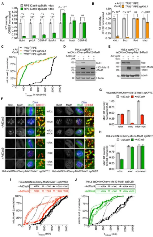
Bub1 is required for kinetochore expansion in Xenopus egg extracts [5] but its role in fibrous corona formation in human cells has not been examined. Bub1’s role in the SAC also remains controversial, with inconsistent results across studies [7–12]. To test Bub1’s
contribution to these aspects of kinetochore structure and function, we deleted BUB1 in RPE cells via doxycycline-inducible CRISPR/Cas9 [27]. BUB1–/– cells treated with nocodazole formed kinetochore crescents containing Rod, CENP-E, and Mad1, but not CENP-F [12] (Figure 3A and Figure S3A). To ensure complete depletion and avoid postmitotic arrest [27], we deleted BUB1 or its kinetochore scaffold KNL1 [1, 2] in p53-deficient RPE cells. KNL1–/– cells formed crescents with normal levels of RZZ but slightly less Mad1 (22% reduction; Figure 3B and Figure S3B). However deleting BUB1 or KNL1 decreased the period of nocodazole-induced mitotic arrest by 76% and 93% (median Tmitosis=130 min for
KNL1–/– cells and 460 min for BUB1–/– cells, versus 1935 min for control cells), indicating that that SAC signaling was functionally compromised.
Although kinetochores in BUB1–/– and KNL1–/– cells have high levels of Mad1-Mad2, we could not exclude the possibility that a small but functionally important pool was missing. Therefore we tested the consequences of deleting BUB1 or KNTC1 and simultaneously expressing a constitutively kinetochore-bound form of Mad1 (Mis12-Mad1) that is refractory to SAC silencing at metaphase [28] (Figure 3D-J). Mis12-Mad1 expression triggered a mitotic arrest in KNTC1–/– cells that was even longer (median Tmitosis=1170
min) than that observed in wildtype cells expressing Mis12-Mad1 (median Tmitosis=780 min;
Figure 3I). This hyperactive response likely reflects RZZ’s role in stripping Mad1 and other SAC mediators from metaphase kinetochores via dynein-dependent transport [29–31]. In contrast BUB1–/– cells had a much weaker response to Mis12-Mad1 kinetochore tethering (median Tmitosis=130 min; Figure 3J). We next combined Mis12-Mad1 expression with
nocodazole treatment to eliminate dynein-dependent stripping and engage upstream SAC signaling. This regimen further extended the mitotic arrest in KNTC1–/– cells (median Tmitosis=1355 min, versus 1560 min for wildtype cells) but accelerated mitotic exit in
BUB1–/– cells (median Tmitosis=240 min) relative to nocodazole treatment alone (median
Tmitosis=460 min; Figure 3I-J). We conclude that Mad1-Mad2 tethering can bypass RZZ, but
not Bub1, with respect to SAC signaling. Our findings suggest that RZZ’s crucial and likely sole function in SAC activation is to maintain Mad1-Mad2 at kinetochores, whereas RZZ-dependent corona formation is not required. In contrast Bub1 is not required for RZZ to localize at kinetochores, form the fibrous corona, or recruit Mad2. However Mad1-Mad2 still requires a non-receptor activity of Bub1 to generate a “wait anaphase” signal. BUB1-disrupted clones re-express Bub1 and regain SAC function via nonsense-associated
alternative splicing
The SAC defect we observed after acute BUB1 disruption is consistent with studies in Bub1 conditional-knockout MEFs [9, 32] but not with recent studies in BUB1-disrupted human cell clones [33–35] (Figure 4A). To understand the basis of this discrepancy, we isolated 13
A
uthor Man
uscr
ipt
A
uthor Man
uscr
ipt
A
uthor Man
uscr
ipt
A
uthor Man
uscr
ipt
clones after acute disruption of BUB1 in p53-deficient RPE cells. All clones exhibited a partial (3–30%) recovery of Bub1 expression, kinetochore localization, and H2A kinase activity as judged by IFM with antibodies that recognize Bub1’s N-terminus and T120-phosphorylated H2A (Figure 4B-E). We performed RT-PCR and sequencing on five clones (Figure 4F and Data S1). Full-length BUB1 transcripts harbored exon 4 indels that induce frameshift and early termination (Figure S4A). We also observed shorter transcripts that skipped part or all of exon 4 and/or utilized cryptic splice sites (Figure S4B-F). A number of alternatively spliced transcripts encoded BUB1 ORFs with short N-terminal deletions or insertions, thus explaining Bub1 re-expression (Figure S4C-F). Eleven of 13 clones exhibited partial or complete recovery of SAC function relative to acute deletion of BUB1 (Figure 4G). Among the five clones analyzed by RT-PCR and sequencing, clone 12 was fully SAC-proficient and had the highest rate of in-frame transcripts (6 of 36), whereas clone 21 had intermediate SAC function and a lesser rate (3 of 31). No in-frame transcripts were identified in clone 8 (0 of 18) and clone 24 (0 of 21), which were the most SAC-defective (Figure 4G). Taken together, these results suggest that nonsense-associated alternative splicing (NAS) [36] attenuates and in some cases suppresses the effects of null mutations in BUB1. Our findings demonstrate how genome editing can trigger both acute loss of function and compensatory changes in mRNA structure that result in phenocopying of unedited cells.
Discussion
In cells treated with spindle poisons, Mad1-Mad2 remains bound to kinetochores and catalyzes MCC production for 1000 min or more, thus extending mitosis at least 30-fold. How (and why) this sustained response occurs is not well understood. In yeast, Bub1 is the sole receptor for Mad1-Mad2 and required for the SAC [37], but models for Mad1-Mad2 regulation in mammalian cells differ considerably [7–12]. In our studies, acute BUB1 or KNL1 deletion led to SAC failure despite high levels of Mad1-Mad2 at kinetochores. Furthermore BUB1–/– cells were largely unresponsive to Mis12-Mad1, which is
constitutively tethered to kinetochores and cannot be silenced at metaphase. Similar results were obtained independently using a Ndc80-Mad1 fusion [35]. These data strongly suggest a non-receptor function of Bub1-KNL1 that is required for kinetochore-bound Mad1-Mad2 to inhibit anaphase. One possibility is that Bub1 functions as a co-catalyst in MCC assembly by recruiting Cdc20 to kinetochores [7, 38]. Asking if this occurs in vivo will require SAC-independent methods for synchronizing BUB1–/– and KNL1–/– cells in mitosis in sufficient quantity and purity for biochemical studies of MCC assembly [39] or tools SAC-dependent synchronization.
Our studies also shed light on RZZ’s role in the SAC. By tracking Mad1 dynamics with high temporal resolution, we demonstrate that kinetochores in early mitotic cells can recruit Mad1-Mad2 without RZZ and can delay anaphase onset by 100–300 min, thus mitigating the impact of less efficient chromosome congression in RZZ-null cells. However
kinetochores with attachment defects that persist beyond this timeframe require RZZ to recruit Mad1-Mad2 and maintain SAC arrest. Expressing Mis12-Mad1 reinstated long-term arrest in KNTC1–/– cells, suggesting that RZZ’s crucial and perhaps only role in the SAC is to tether Mad1-Mad2 to kinetochores. It has been proposed that RZZ mediates SAC signaling at unattached but not tensionless kinetochores [10]. However KNTC1–/– cells
A
uthor Man
uscr
ipt
A
uthor Man
uscr
ipt
A
uthor Man
uscr
ipt
A
uthor Man
uscr
ipt
challenged with spindle poisons that block attachment (nocodazole) or permit attachment without tension (STLC and taxol) escaped SAC arrest with similar kinetics (Figure 1B). RZZ is also implicated in formation of the fibrous corona, a structural expansion of the outer kinetochore that precedes microtubule attachment [4, 6, 17]. Kinetochore expansion depends on mitotic kinases [5, 13], but relevant phosphorylation events are not known. We identified two Mps1-regulated phosphosites just upstream of Rod’s β-propeller domain [24] that are required for kinetochore expansion but not SAC arrest. Rod and Spindly not only interact via this domain [14, 20] but also inhibit their own assembly into polymers [13, 40]. Together, these findings suggest that phosphorylation alleviates a structural barrier to Spindly-RZZ polymerization.
In this study and others [3, 27, 39, 41, 42], we used genome editing to delete or disrupt exons of genes involved in cell division. Normally this results in a “knockout” because of nonsense-mediated decay (NMD), a pathway that degrades mRNAs with premature termination codons (PTCs) [43, 44]. However PTCs can also trigger NAS, a less well-understood pathway in which splicing rules are relaxed to bypass the PTC and restore expression of near-full length ORFs [36, 45, 46]. NAS could also explain why BUB1 exon 2-disrupted HeLa cells manifest a clear SAC defect after BUB1 exon 8-specific RNAi [35]. In conclusion, NMD and NAS have opposite effects on the expressivity and penetrance of nonsense mutations, and should not be overlooked in the design, analysis, and interpretation of genome editing experiments.
STAR Methods
CONTACT FOR REAGENT AND RESOURCE SHARING
Further information and requests for resources and reagents should be directed to and will be fulfilled by the Lead Contact, Prasad Jallepalli (jallepap@mskcc.org).
EXPERIMENTAL MODEL AND SUBJECT DETAILS
Cell lines and chemicals—Cell lines used in this study are described in the Key Resource Table. HeLa (human cervical adenocarcinoma, female) and HEK293 (human embryonic kidney, female) derivatives were grown at 37°C in Dulbecco’s modifi ed eagle medium (DMEM) with 10% tetracycline free fetal bovine serum, 100 U/ml penicillin, and 100 U/ml streptomycin. hTERT-RPE (human retinal pigment epithelium, female) derivatives were grown at 37°C in a 1:1 mixture of DMEM and Ham’s F- 12 medium with 10% fetal bovine serum, 100 U/ml penicillin, and 100 U/ml streptomycin, and 2.5 mM L-glutamine. HCT116 (human colorectal adenocarcinoma, male) were grown at 37°C in McCoy’s 5A medium with 10% fetal bovine serum, 100 U/ml penicillin, and 100 U/ml streptomycin. Unless stated otherwise, nocodazole (660 nM), taxol (1 µM), S-trityl-L-cysteine (10 µM), MG132 (10 µM), hesperadin (100 nM), ZM447439 (2 µM), reversine (500 nM), and FTI-288 (10 µM) were used at the indicated concentrations.
A
uthor Man
uscr
ipt
A
uthor Man
uscr
ipt
A
uthor Man
uscr
ipt
A
uthor Man
uscr
ipt
METHOD DETAILS
Transgene expression—LAP-Rod (WT or 2A) was cloned into pcDNA5/FRT/TO. Constructs were cotransfected with pOG44 into HeLa T-Rex Flp-In cells using FuGene 6 (Roche). Integrants were selected using hygromycin (0.2 mg/ml), picked as single colonies, and induced with doxycycline (0.8 µg/ml). mCherry-Mis12-Mad1 [28] was cloned into a piggyBac vector containing a doxycycline-inducible promoter (tetON) and constitutively expressing reverse tetracycline transactivator (rtTA) and neomycin phosphotransferase (neoR) linked by the self-cleaving T2A peptide. HeLa cells were cotransfected with this construct and pSuperPiggyBac transposase (System Biosciences), selected in G418 (0.5 mg/ ml), and induced as above. For stable expression of FLAP-Mad1, EGFP-CENP-A, and H2B-mCherry, retroviral transfer plasmids were cotransfected with pVSV-G into Phoenix 293 cells. For stable expression of mRuby-CENP-A or gene-specific sgRNAs, lentiviral transfer plasmids were cotransfected with psPAX2 and pMD2.G into Lenti-X 293T cells (Clontech). 24 to 48 hr later, supernatants were filtered, mixed 1:1 with fresh medium containing polybrene (20 µg/ml), and applied to target cells for 24 hr. Transductants were selected in G418, blasticidin (5 µg/ml), or puromycin (5 to 20 µg/ml).
AAV-mediated gene targeting—5’ and 3’ homology arms encompassing KNTC1 exons 2 and 3 were amplified from human BAC clone RP11–18E11 using Pfusion DNA
polymerase. A new loxP site was added upstream of exon 2 via XbaI digest and linker ligation. The entire targeting construct was transferred to pAAV as a NotI fragment. All manipulated regions were checked by sequencing to ensure their integrity. Procedures for preparing infectious AAV particles, transducing HCT116 cells, and isolating correctly targeted clones were performed as described [48]. The FRT-neoR-FRT cassette was excised through transient expression of FLP recombinase (pCAGGS-FLPe) and limiting dilution. To delete KNTC1HF, cells were infected with AdCre (Vector Development Laboratory, Baylor College of Medicine).
CRISPR/Cas9-mediated genome editing—Zifit (http://zifit.partners.org/ZiFiT/ ChoiceMenu.aspx) and sgRNA Designer ( http://www.broadinstitute.org/rnai/public/analysis-tools/sgrna-design) were used to identify and rank candidate CRISPR/Cas9 targets for predicted on- and off-target activities. For transient expression, sequences were ordered as overlapping 60-nt oligonucleotides, annealed and extended into a 100-bp duplex using Pfusion DNA polymerase, and cloned into an AflII-digested guide RNA expression vector (Addgene 41824) by Gibson assembly. Equal amounts of human codon-optimized Cas9 (Addgene 41815) and sgRNA vectors were transfected into HCT116 cells using FuGene 6 and into RPE cells using a Nucleofector 2b device (Lonza). For stable expression, target sequences were ordered as 24-nt oligonucleotides with asymmetric 5’ overhangs, phosphorylated with T4 polynucleotide kinase, then annealed and cloned into BsmBI-digested lentiGuide-puro (Addgene 52963) or pLenti-sgRNA (Addgene 71409) using T4 DNA ligase. Lentiviral transduction was performed as described above. Gene deletion was initiated by inducing a doxycycline-regulated Cas9 transgene present in the host cell line [27] or by infection with AdCas9 (ViraQuest).
A
uthor Man
uscr
ipt
A
uthor Man
uscr
ipt
A
uthor Man
uscr
ipt
A
uthor Man
uscr
ipt
Immunofluorescence microscopy and live-cell imaging—Antibodies used in this study are listed in the Key Resource Table. Cells were fixed and permeabilized in PEMFT (20 mM PIPES, pH 6.8, 10 mM EGTA, 1 mM MgCl2, 4% paraformaldehyde, and 0.2%
Triton X-100) for 13 min, blocked in 4% BSA, and stained with primary antibodies
overnight. Species-specific secondary antibodies conjugated to Alexa Fluor 488, 564, or 647 were applied for 1 hr. Coverslips were mounted in ProLong Gold, imaged with a 100x oil objective on a DeltaVision Elite microscope (GE Life Sciences), and deconvolved in SoftWoRx using measured point spread functions. For timelapse experiments cells were grown in multiwell plates or 35 mm glass-bottom dishes (MatTek) and imaged on a Nikon Eclipse Ti microscope equipped with a stage-top incubator and CO2 delivery system, 20x
and 40x air objectives and 60x (1.4 NA) and 100x (1.45 NA) oil objectives, a Yokogawa CSU-X1 unit, and sCMOS (Andor Xyla 5.5) and EMCCD (Photometrics Evolve 512) cameras. Acquisition was performed with NIS Elements (v5.41). Epifluorescence and/or DIC images were acquired at 10-min intervals to measure mitotic arrest in response to spindle poisons, or at 2-min intervals to measure unperturbed mitotic timing. Confocal imaging of FLAP-Mad1 and H2B-mCherry was performed at 2- to 5-min intervals. Fluorescence intensities were quantified in ImageJ (v1.51) and analyzed in Prism 7.0 (GraphPad).
Correlative light-electron microscopy—Cells were fixed with 2.5% glutaraldehyde (Sigma) in PBS, pH 7.4–7.6 for 30 min, rinsed with PBS (3 × 5 min), and mounted in Rose chambers. Multimode (DIC and 3-color fluorescence) datasets were obtained on a Nikon TE2000 microscope equipped with a PlanApo 100× 1.45 NA objective lens at 53-nm XY pixels and 200-nm Z-steps. All LM images were deconvolved in SoftWoRx (v5.0) with lens-specific PSFs. Post-fixation, embedding, and sectioning were done as previously described [47]. Thin sections (70–80 nm) were imaged on a JEOL 1400 microscope operated at 80 kV using a side-mounted 4.0 megapixel XR401 sCMOS AMT camera (Advanced Microscopy Techniques Corp). Full series of images recorded at 10K magnification were used to reconstruct the volume of the cell, match orientation and superimpose this volume on the corresponding LM dataset. Higher-magnification images (40K) were then collected for individual kinetochores.
Cell lysis, immunoprecipitation, and Western blotting—Cell extracts were prepared by resuspending pellets in ice-cold buffer B (140 mM NaCl, 30 mM HEPES, pH 7.8, 5% glycerol, 10 mM sodium pyrophosphate, 5 mM sodium azide, 10 mM NaF, 10 mM PMSF, 0.3 mM sodium orthovanadate, 20 mM b-glycerophosphate, 1 mM DTT, 0.2 mM
microcystin, and 1x protease inhibitor cocktail (Sigma)) prior to nitrogen cavitation (1250 psi, 45 min; Parr Instruments) and centrifugation at 20,000 x g for 30 min. LAP-Rod was immunoprecipitated with GFP antibodies coupled to protein G-Dynabeads using
bis(sulfosuccinimidyl)suberate (BS3). Zw10 was immunoprecipitated without BS3
crosslinking. Extracts and immunoprecipitates were separated by SDS-PAGE and transferred to PVDF or nitrocellulose membranes. Membranes were blocked and probed with primary antibodies and secondary antibody-HRP conjugates in 5% nonfat dry milk in TBST (Tris-buffered saline + 0.05% Tween-20) before detecting signals via enhanced
chemiluminescence (Western Lightning Plus, PerkinElmer).
A
uthor Man
uscr
ipt
A
uthor Man
uscr
ipt
A
uthor Man
uscr
ipt
A
uthor Man
uscr
ipt
RT-PCR and sequencing—Total RNA was purified (RNeasy Mini kit, Qiagen) and reverse-transcribed with random hexamers (SuperScript IV First-Strand Synthesis System, ThermoFisher). RT reactions were diluted 100-fold and amplified with BUB1-specific primers spanning exons 2–6 or exons 3–8. PCR products were cloned into pCR4 (TOPO-TA Cloning Kit, ThermoFisher) and sequenced with M13 reverse primer. Reads were trimmed and aligned in SeqMan Pro and annotated in SeqBuilder (DNASTAR LaserGene v14).
QUANTIFICATION AND STATISTICAL ANALYSIS
Quantitative data was compiled in Excel and analyzed using Prism 7.0. Details of each statistical analysis (number of cells (n), number of experiments (N), and measures of central tendency (mean or median)) are specified in the figure legends and Results. Unless stated otherwise, error bars indicate SEM. P ≤ 0.05 (with adjustment for multiple-hypothesis testing where applicable) was used as the significance threshold.
Supplementary Material
Refer to Web version on PubMed Central for supplementary material.
Acknowledgments
We thank George Church, Didier Trono, David Sabatini, and Feng Zhang for sharing plasmids through Addgene. P.V.J. was supported by NIH grants R01GM094972 and P30CA008748.
References
1. London N, and Biggins S (2014). Signalling dynamics in the spindle checkpoint response. Nat Rev Mol Cell Biol 15, 736–747. [PubMed: 25303117]
2. Musacchio A (2015). The Molecular Biology of Spindle Assembly Checkpoint Signaling Dynamics. Curr Biol 25, R1002–1018. [PubMed: 26485365]
3. Rodriguez-Bravo V, Maciejowski J, Corona J, Buch HK, Collin P, Kanemaki MT, Shah JV, and Jallepalli PV (2014). Nuclear pores protect genome integrity by assembling a premitotic and mad1-dependent anaphase inhibitor. Cell 156, 1017–1031. [PubMed: 24581499]
4. Magidson V, Paul R, Yang N, Ault JG, O’Connell CB, Tikhonenko I, McEwen BF, Mogilner A, and Khodjakov A (2015). Adaptive changes in the kinetochore architecture facilitate proper spindle assembly. Nat Cell Biol 17, 1134–1144. [PubMed: 26258631]
5. Wynne DJ, and Funabiki H (2015). Kinetochore function is controlled by a phospho-dependent coexpansion of inner and outer components. J Cell Biol 210, 899–916. [PubMed: 26347137] 6. Hoffman DB, Pearson CG, Yen TJ, Howell BJ, and Salmon ED (2001). Microtubule-dependent
changes in assembly of microtubule motor proteins and mitotic spindle checkpoint proteins at PtK1 kinetochores. Molecular biology of the cell 12, 1995–2009. [PubMed: 11451998]
7. Vleugel M, Hoek TA, Tromer E, Sliedrecht T, Groenewold V, Omerzu M, and Kops GJ (2015). Dissecting the roles of human BUB1 in the spindle assembly checkpoint. J Cell Sci 128, 2975– 2982. [PubMed: 26148513]
8. Klebig C, Korinth D, and Meraldi P (2009). Bub1 regulates chromosome segregation in a kinetochore-independent manner. J Cell Biol 185, 841–858. [PubMed: 19487456]
9. Zhang G, Lischetti T, Hayward DG, and Nilsson J (2015). Distinct domains in Bub1 localize RZZ and BubR1 to kinetochores to regulate the checkpoint. Nature communications 6, 7162.
10. Silio V, McAinsh AD, and Millar JB (2015). KNL1-Bubs and RZZ Provide Two Separable Pathways for Checkpoint Activation at Human Kinetochores. Dev Cell 35, 600–613. [PubMed: 26651294]
A
uthor Man
uscr
ipt
A
uthor Man
uscr
ipt
A
uthor Man
uscr
ipt
A
uthor Man
uscr
ipt
11. Caldas GV, Lynch TR, Anderson R, Afreen S, Varma D, and DeLuca JG (2015). The RZZ complex requires the N-terminus of KNL1 to mediate optimal Mad1 kinetochore localization in human cells. Open biology 5.
12. Raaijmakers JA, van Heesbeen R, Blomen VA, Janssen LME, van Diemen F, Brummelkamp TR, and Medema RH (2018). BUB1 Is Essential for the Viability of Human Cells in which the Spindle Assembly Checkpoint Is Compromised. Cell reports 22, 1424–1438. [PubMed: 29425499] 13. Sacristan C, Ahmad MUD, Keller J, Fermie J, Groenewold V, Tromer E, Fish A, Melero R, Carazo
JM, Klumperman J, et al. (2018). Dynamic kinetochore size regulation promotes microtubule capture and chromosome biorientation in mitosis. Nat Cell Biol 20, 800–810. [PubMed: 29915359]
14. Gama JB, Pereira C, Simoes PA, Celestino R, Reis RM, Barbosa DJ, Pires HR, Carvalho C, Amorim J, Carvalho AX, et al. (2017). Molecular mechanism of dynein recruitment to
kinetochores by the Rod-Zw10-Zwilch complex and Spindly. J Cell Biol 216, 943–960. [PubMed: 28320824]
15. Saurin AT, van der Waal MS, Medema RH, Lens SM, and Kops GJ (2011). Aurora B potentiates Mps1 activation to ensure rapid checkpoint establishment at the onset of mitosis. Nature communications 2, 316.
16. Santaguida S, Vernieri C, Villa F, Ciliberto A, and Musacchio A (2011). Evidence that Aurora B is implicated in spindle checkpoint signalling independently of error correction. EMBO J 30, 1508– 1519. [PubMed: 21407176]
17. McEwen BF, Arena JT, Frank J, and Rieder CL (1993). Structure of the colcemid-treated PtK1 kinetochore outer plate as determined by high voltage electron microscopic tomography. J Cell Biol 120, 301–312. [PubMed: 8421050]
18. Schmitt HD (2010). Dsl1p/Zw10: common mechanisms behind tethering vesicles and microtubules. Trends Cell Biol 20, 257–268. [PubMed: 20226673]
19. Civril F, Wehenkel A, Giorgi FM, Santaguida S, Di Fonzo A, Grigorean G, Ciccarelli FD, and Musacchio A (2010). Structural analysis of the RZZ complex reveals common ancestry with multisubunit vesicle tethering machinery. Structure 18, 616–626. [PubMed: 20462495] 20. Mosalaganti S, Keller J, Altenfeld A, Winzker M, Rombaut P, Saur M, Petrovic A, Wehenkel A,
Wohlgemuth S, Muller F, et al. (2017). Structure of the RZZ complex and molecular basis of its interaction with Spindly. J Cell Biol
21. Basto R, Scaerou F, Mische S, Wojcik E, Lefebvre C, Gomes R, Hays T, and Karess R (2004). In vivo dynamics of the rough deal checkpoint protein during Drosophila mitosis. Curr Biol 14, 56– 61. [PubMed: 14711415]
22. Cooke CA, Schaar B, Yen TJ, and Earnshaw WC (1997). Localization of CENP-E in the fibrous corona and outer plate of mammalian kinetochores from prometaphase through anaphase. Chromosoma 106, 446–455. [PubMed: 9391217]
23. Rattner JB, Rao A, Fritzler MJ, Valencia DW, and Yen TJ (1993). CENP-F is a .ca 400 kDa kinetochore protein that exhibits a cell-cycle dependent localization. Cell Motil Cytoskeleton 26, 214–226. [PubMed: 7904902]
24. Maciejowski J, Drechsler H, Grundner-Culemann K, Ballister ER, Rodriguez-Rodriguez JA, Rodriguez-Bravo V, Jones MJK, Foley E, Lampson MA, Daub H, et al. (2017). Mps1 Regulates Kinetochore-Microtubule Attachment Stability via the Ska Complex to Ensure Error-Free Chromosome Segregation. Dev Cell 41, 143–156 e146. [PubMed: 28441529]
25. Holland AJ, Reis RM, Niessen S, Pereira C, Andres DA, Spielmann HP, Cleveland DW, Desai A, and Gassmann R (2015). Preventing farnesylation of the dynein adaptor Spindly contributes to the mitotic defects caused by farnesyltransferase inhibitors. Molecular biology of the cell 26, 1845– 1856. [PubMed: 25808490]
26. Moudgil DK, Westcott N, Famulski JK, Patel K, Macdonald D, Hang H, and Chan GK (2015). A novel role of farnesylation in targeting a mitotic checkpoint protein, human Spindly, to
kinetochores. J Cell Biol 208, 881–896. [PubMed: 25825516]
27. McKinley KL, and Cheeseman IM (2017). Large-Scale Analysis of CRISPR/Cas9 Cell-Cycle Knockouts Reveals the Diversity of p53-Dependent Responses to Cell-Cycle Defects. Dev Cell 40, 405–420 e402. [PubMed: 28216383]
A
uthor Man
uscr
ipt
A
uthor Man
uscr
ipt
A
uthor Man
uscr
ipt
A
uthor Man
uscr
ipt
28. Maldonado M, and Kapoor TM (2011). Constitutive Mad1 targeting to kinetochores uncouples checkpoint signalling from chromosome biorientation. Nat Cell Biol 13, 475–482. [PubMed: 21394085]
29. Gassmann R, Holland AJ, Varma D, Wan X, Civril F, Cleveland DW, Oegema K, Salmon ED, and Desai A (2010). Removal of Spindly from microtubule-attached kinetochores controls spindle checkpoint silencing in human cells. Genes Dev 24, 957–971. [PubMed: 20439434]
30. Howell BJ, McEwen BF, Canman JC, Hoffman DB, Farrar EM, Rieder CL, and Salmon ED (2001). Cytoplasmic dynein/dynactin drives kinetochore protein transport to the spindle poles and has a role in mitotic spindle checkpoint inactivation. J Cell Biol 155, 1159–1172. [PubMed: 11756470]
31. Silva PM, Reis RM, Bolanos-Garcia VM, Florindo C, Tavares AA, and Bousbaa H (2014). Dynein-dependent transport of spindle assembly checkpoint proteins off kinetochores toward spindle poles. FEBS letters 588, 3265–3273. [PubMed: 25064841]
32. Perera D, Tilston V, Hopwood JA, Barchi M, Boot-Handford RP, and Taylor SS (2007). Bub1 maintains centromeric cohesion by activation of the spindle checkpoint. Dev Cell 13, 566–579. [PubMed: 17925231]
33. Currie CE, Mora-Santos MD, Smith C, McAinsh AD, and Millar JB (2018). Bub1 is not required for the checkpoint response to unattached kinetochores in diploid human cells. bioRxiv
34. Raaijmakers JA, Tanenbaum ME, Maia AF, and Medema RH (2009). RAMA1 is a novel kinetochore protein involved in kinetochore-microtubule attachment. J Cell Sci 122, 2436–2445. [PubMed: 19549680]
35. Zhang G, Kruse T, and Nilsson J (2018). The RZZ complex facilitates Mad1 binding to Bub1 ensuring efficient checkpoint signaling. bioRxiv
36. Cartegni L, Chew SL, and Krainer AR (2002). Listening to silence and understanding nonsense: exonic mutations that affect splicing. Nature reviews. Genetics 3, 285–298.
37. London N, and Biggins S (2014). Mad1 kinetochore recruitment by Mps1-mediated phosphorylation of Bub1 signals the spindle checkpoint. Genes Dev 28, 140–152. [PubMed: 24402315]
38. Faesen AC, Thanasoula M, Maffini S, Breit C, Muller F, van Gerwen S, Bange T, and Musacchio A (2017). Basis of catalytic assembly of the mitotic checkpoint complex. Nature 542, 498–502. [PubMed: 28102834]
39. Maciejowski J, George KA, Terret ME, Zhang C, Shokat KM, and Jallepalli PV (2010). Mps1 directs the assembly of Cdc20 inhibitory complexes during interphase and mitosis to control M phase timing and spindle checkpoint signaling. J Cell Biol 190, 89–100. [PubMed: 20624902] 40. Gassmann R, Pereira C, Reis RM, Gama JB, Cheerambathur DK, and Carvalho AX (2018).
Self-assembly of the RZZ complex into filaments drives kinetochore expansion in the absence of microtubule attachment. bioRxiv
41. Tsou M-FB, Wang W-J, George KA, Uryu K, Stearns T, and Jallepalli PV (2009). Polo kinase and separase regulate the mitotic licensing of centriole duplication in human cells. Developmental Cell 17, 344–354. [PubMed: 19758559]
42. McKinley KL, Sekulic N, Guo LY, Tsinman T, Black BE, and Cheeseman IM (2015). The CENP-L-N Complex Forms a Critical Node in an Integrated Meshwork of Interactions at the Centromere-Kinetochore Interface. Mol Cell 60, 886–898. [PubMed: 26698661]
43. Popp MW, and Maquat LE (2016). Leveraging Rules of Nonsense-Mediated mRNA Decay for Genome Engineering and Personalized Medicine. Cell 165, 1319–1322. [PubMed: 27259145] 44. Lykke-Andersen S, and Jensen TH (2015). Nonsense-mediated mRNA decay: an intricate
machinery that shapes transcriptomes. Nature Reviews Molecular Cell Biology 16, 665. [PubMed: 26397022]
45. Anderson JL, Mulligan TS, Shen MC, Wang H, Scahill CM, Tan FJ, Du SJ, Busch-Nentwich EM, and Farber SA (2017). mRNA processing in mutant zebrafish lines generated by chemical and CRISPR-mediated mutagenesis produces unexpected transcripts that escape nonsense-mediated decay. PLoS Genet 13, e1007105. [PubMed: 29161261]
A
uthor Man
uscr
ipt
A
uthor Man
uscr
ipt
A
uthor Man
uscr
ipt
A
uthor Man
uscr
ipt
46. Mou H, Smith JL, Peng L, Yin H, Moore J, Zhang XO, Song CQ, Sheel A, Wu Q, Ozata DM, et al. (2017). CRISPR/Cas9-mediated genome editing induces exon skipping by alternative splicing or exon deletion. Genome biology 18, 108. [PubMed: 28615073]
47. Magidson V, He J, Ault JG, O’Connell CB, Yang N, Tikhonenko I, McEwen BF, Sui H, and Khodjakov A (2016). Unattached kinetochores rather than intrakinetochore tension arrest mitosis in taxol-treated cells. J Cell Biol 212, 307–319. [PubMed: 26833787]
48. Berdougo E, Terret M, and Jallepalli P (2009). Functional dissection of mitotic regulators through gene targeting in human somatic cells. Methods Mol Biol 545, 21–37. [PubMed: 19475380]
A
uthor Man
uscr
ipt
A
uthor Man
uscr
ipt
A
uthor Man
uscr
ipt
A
uthor Man
uscr
ipt
Highlights
• RZZ mediates a temporal switch in how Mad1-Mad2 is recruited to kinetochores
• Mps1 phosphorylates Rod’s N-terminus to trigger fibrous corona formation
• Mad1-Mad2 requires a non-receptor activity of Bub1 to inhibit anaphase
• Nonsense-associated alternative splicing can circumvent BUB1 disruption
A
uthor Man
uscr
ipt
A
uthor Man
uscr
ipt
A
uthor Man
uscr
ipt
A
uthor Man
uscr
ipt
Figure 1. A temporal switch in the requirements for Mad1-Mad2 targeting to kinetochores during SAC signaling.
(A) AAV- and CRISPR-mediated genome editing was used to modify the KNTC1 locus in HCT116 cells (Figure S1A-E). Cells expressing H2B-mCherry were treated with nocodazole or STLC and followed by epifluorescence and DIC timelapse microscopy. Images were acquired at 10-min intervals. Mitotic duration (from NEBD to chromatin decondensation) was quantified in at least 25 cells per condition per experiment (N=2). P-values were computed using Kruskal-Wallis and Dunn’s multiple comparisons tests. Error bars throughout the paper indicate s.e.m. unless stated otherwise. (B) Wildtype and KNTC1–/–
A
uthor Man
uscr
ipt
A
uthor Man
uscr
ipt
A
uthor Man
uscr
ipt
A
uthor Man
uscr
ipt
RPE1 cells (Figure S1F-I) were treated with nocodazole, STLC, or taxol and followed using DIC optics. Cell rounding (mitotic entry) and cortical blebbing and flattening (mitotic exit) were used as landmarks. (C) Clonal HeLa KNTC1, ZW10, and ZWILCH knockouts [27] were treated with nocodazole and followed as in (B). (D) Mitotic timing in unperturbed wildtype and RZZ-deficient HeLa, RPE, and HCT116 cells. Where indicated Mps1 kinase was inhibited with reversine. (E and F) Wildtype and KNTC1–/– HCT116 cells expressing H2B-mCherry and FLAP-Mad1 were filmed during unperturbed mitosis using spinning disk confocal microscopy. Insets show enlarged views of FLAP-Mad1 recruitment to and dissociation from kinetochores. Scale bars throughout the paper are 10 μm unless stated otherwise. See also Videos S1 and S2. (G) Quantification of FLAP-Mad1 at misaligned chromosomes in (E) and (F). (H) Cells in (E) and (F) were filmed in the presence of nocodazole (n=6 for wildtype and n=14 for KNTC1–/–). (I and J) Wildtype and KNTC1-null RPE cells were treated with nocodazole and MG132 for 30 min or 4 hours before fixation for IFM. Mad1/CREST fluorescence intensity ratios were determined for at least 100 kinetochores in 5 cells per condition (N=3). (K) Wildtype and KNTC1-null RPE cells were treated with nocodazole in the presence or absence of hesperadin (hesp) to inhibit Aurora B kinase. Mitotic duration was determined from 30 cells per condition. See also Figure S1.
A
uthor Man
uscr
ipt
A
uthor Man
uscr
ipt
A
uthor Man
uscr
ipt
A
uthor Man
uscr
ipt
Figure 2. Mps1 phosphorylation at the N-terminus of Rod triggers RZZ-dependent kinetochore expansion.
(A and B) Wildtype and KNTC1–/– RPE cells were treated with nocodazole and MG132 for 2.5 hours before IFM with antibodies to CENP-E (A) or CENP-F (Figure S2A). Crescent size and intensity were determined in 5 to 10 cells (N=3). (C and D) RPE cells were treated with nocodazole and MG132 for 2 hr, after which reversine (rev) or Aurora B inhibitor ZM447439 (ZM) was added or omitted for 1 hr (n=10 cells, N=3). CREST-normalized kinetochore intensities of Mad1 and CENP-E were determined by IFM. (E and F) KNTC1-null HeLa cells reconstituted with LAP-RodWT or LAP-Rod2A were treated with nocodazole
A
uthor Man
uscr
ipt
A
uthor Man
uscr
ipt
A
uthor Man
uscr
ipt
A
uthor Man
uscr
ipt
for 2.5 hr. IFM was used to visualize crescents and quantify kinetochore-associated LAP-Rod, Mad1, CENP-E, CENP-F, Spindly, and CREST (n=100 kinetochores in 5 cells each, N=2). Where indicated, cells were treated with the farnesyltransferase inhibitor FTI-288 (FTI) as a positive control for blockade of Spindly targeting to kinetochores [25, 26]. Note that kinetochores in LAP-Rod2A cells remained compact during long-term SAC arrest (16 hr nocodazole treatment). (G) The duration of mitotic arrest in nocodazole-treated LAP-RodWT and LAP-Rod2A cells was determined by DIC timelapse (n≥50 cells, N=2) and compared using the Mann-Whitney U test. See also Figure S2.
A
uthor Man
uscr
ipt
A
uthor Man
uscr
ipt
A
uthor Man
uscr
ipt
A
uthor Man
uscr
ipt
Figure 3. RZZ and Bub1-KNL1 have distinct roles in recruiting and activating Mad1-Mad2 at kinetochores during SAC signaling.
(A) RPE iCas9 cells expressing sgBUB1 were treated with or without doxycycline and analyzed by IFM after 5 days (for images see Figure S3A). (B) TP53−/− RPE cells with or without sgKNL1 were treated with AdCas9 and analyzed by IFM after three days (for images see Figure S3B). (C) AdCas9-treated cells were filmed in the presence of nocodazole for 48 hours. Cumulative frequency of mitotic exit is plotted. (D and E) HeLa cells
expressing BUB1- or KNTC1-specific sgRNA were treated with AdCas9. After three days, mCherry-Mis12-Mad1 was induced with doxycycline for 17 hr. Protein depletion or
A
uthor Man
uscr
ipt
A
uthor Man
uscr
ipt
A
uthor Man
uscr
ipt
A
uthor Man
uscr
ipt
expression was confirmed by Western blotting. (F-H) Cells in (D and E) were analyzed by IFM, either with or without a further 3-hr treatment with nocodazole. Mad1/CREST ratios were determined for ≥100 kinetochores in 5 cells (N=2). (I and J) Cells in (D and E) were treated with doxycycline and/or nocodazole and followed by DIC timelapse (n≥50 cells, N=2). See also Figure S3.
A
uthor Man
uscr
ipt
A
uthor Man
uscr
ipt
A
uthor Man
uscr
ipt
A
uthor Man
uscr
ipt
Figure 4. BUB1-disrupted clones regain Bub1 expression and SAC function through nonsense-associated alternative splicing.
(A) Structure of the BUB1 locus. Locations of guide RNAs used in this and previous studies [33–35] are shown. Regions encoding N-terminal Bub1 antibody epitope and C-terminal kinase domain are indicated. (B-E). Parental TP53–/– RPE cells (–AdCas9), acute BUB1 knockout cells (+AdCas9), and BUB1-disrupted clones (c12, c08) were treated with nocodazole and MG132 for 2 hr. Centromeric H2A phosphorylation (B and D) and kinetochore-associated Bub1 (C and E) were quantified by IFM (n=100 kinetochores in 5 cells, N=2). Where indicated, individual z-slices or maximum intensity projections of
A
uthor Man
uscr
ipt
A
uthor Man
uscr
ipt
A
uthor Man
uscr
ipt
A
uthor Man
uscr
ipt
adjacent z-slices are displayed. For all other images, maximum intensity projections of full z-series are shown. (F) RNA samples were reverse-transcribed and amplified with PCR primers spanning exons 2–6. (G) BUB1-disrupted clones were treated with nocodazole and followed by DIC timelapse. Parental TP53–/– RPE cells (–AdCas9) and acute BUB1 knockout cells (+AdCas9) were used as controls. See also Figure S4 and Data S1.
A
uthor Man
uscr
ipt
A
uthor Man
uscr
ipt
A
uthor Man
uscr
ipt
A
uthor Man
uscr
ipt
A
uthor Man
uscr
ipt
A
uthor Man
uscr
ipt
A
uthor Man
uscr
ipt
A
uthor Man
uscr
ipt
KEY RESOURCES TABLE
REAGENT or RESOURCE SOURCE IDENTIFIER
Antibodies
CREST (human) Immunovision HCT-0100; RRID: AB_2744669
Rod (mouse) Santa Cruz sc-81853; RRID: AB_2133542
Zw10 (rabbit) Abcam ab21582; RRID: AB_779030
Zwilch (mouse) A. Musacchio N/A
Mad1 (mouse) A. Musacchio clone BB3–8
Mad2 (rabbit) Bethyl A300–301A; RRID: AB_2281536
Bub1 (rabbit) Genetex GTX107497; RRID: AB_1949770
Bub1 (mouse) Abcam ab54893; RRID: AB_940664
BubR1 (mouse) BD Biosciences BD612503; RRID: AB_2066085
KNL1 (rabbit) Bethyl A300–804A; RRID: AB_577218
Spindly (rabbit) Bethyl A301–354A; RRID: AB_937753
CENP-E (mouse) Abcam ab5093; RRID: AB_304747
CENP-F (rabbit) Novus NB500–101; RRID: AB_2229328
p150glued (mouse) BD Biosciences BD612708; RRID: AB_399947
phospho-T210 H2A (rabbit) Active Motif 39391; RRID: AB_2744670
phospho-T13/S15 Rod (rabbit) Jallepalli Lab [24] N/A
EGFP (mouse) Santa Cruz sc-9996; RRID: AB_627695
EGFP (mouse) ThermoFisher A-11120 (clone 3E6); RRID: AB_221568
EGFP (rabbit) Jallepalli Lab [24] N/A
α-tubulin (mouse) Santa Cruz sc-5286; RRID: AB_628411
α-tubulin (rat) Chemicon MAB1864 (clone YL½); RRID: AB_2210391
mCherry (rabbit) ThermoFisher PA5–34974; RRID: AB_2552323
mCherry (mouse) Abcam ab125096; RRID: AB_11133266
Bacterial and Virus Strains
AdCas9 ViraQuest N/A
Ad5 CMV Cre Vector Development Lab, Baylor
College of Medicine
Lot#032415
Biological Samples
Chemicals, Peptides, and Recombinant Proteins
Nocodazole Sigma Cat#M1404; CAS:31430–18-9
A
uthor Man
uscr
ipt
A
uthor Man
uscr
ipt
A
uthor Man
uscr
ipt
A
uthor Man
uscr
ipt
REAGENT or RESOURCE SOURCE IDENTIFIER
S-Trityl-L-cysteine (STLC) ThermoFisher Cat#2191; CAS:2799–07-7
MG132 Selleckchem Cat#S2619; CAS:133407–82-6
Hesperidin Selleckchem Cat#S2309; CAS:520–26-3
ZM447439 Tocris Cat#2458; CAS:331771–20-1
Reversine Cayman Chemical Cat#10004412; CAS:656820–32-5
FTI-277 Sigma Cat#F9803; CAS:170006–73-2
G418 Corning Cat#61234-RF; CAS:108321–42-2
Blasticidin ThermoFisher Cat#A1113903; CAS:2079–00-7
Puromycine ThermoFisher Cat#A1113803; CAS: 58–58-2
Hygromycin Calbiochem Cat#400051; CAS: 31282–04-9
FuGene 6 Promega Cat#E2691
SuperScript IV First-Strand Synthesis System ThermoFisher Cat#18091050
TOPO-TA Cloning Kit ThermoFisher Cat#450071
Critical Commercial Assays
Deposited Data
Experimental Models: Cell Lines
HEK293 ATCC CCL-1573
Lenti-X 293T Clontech 632180
Phoenix-GP293 ATCC CCL-3215
HCT116 ATCC CCL-247
HCT116 KNTC1 (HF/+) this study N/A
HCT116 KNTC1 (HF/-) this study N/A
HCT116 KNTC1 (−/−) this study N/A
HeLa ATCC CCL-2
HeLa + sgKNTC1 this study N/A
HeLa TetON-mCherry-Mis12-Mad1 this study N/A
HeLa TetON-mCherry-Mis12-Mad1 + sgKNTC1 this study N/A
A
uthor Man
uscr
ipt
A
uthor Man
uscr
ipt
A
uthor Man
uscr
ipt
A
uthor Man
uscr
ipt
REAGENT or RESOURCE SOURCE IDENTIFIER
HeLa iCas9 Cheeseman Lab [27] N/A
HeLa iCas9 KNTC1 KO Cheeseman Lab [27] Subcloned after sgKNTC1 + dox
HeLa iCas9 ZW10 KO Cheeseman Lab [27] Subcloned after sgZW10 + dox
HeLa iCas9 ZWILCH KO Cheeseman Lab [27] Subcloned after sgZWILCH + dox
HeLa Flp-In T-Rex S. Taylor N/A
HeLa Flp-In T-Rex LAP-Rod WT (KNTC1 KO) this study Subcloned after sgKNTC1 + AdCas9
HeLa Flp-In T-Rex LAP-Rod 2A (KNTC1 KO) this study Subcloned after sgKNTC1 + AdCas9
RPE1 Clontech now ATCC CRL-4000
RPE1 KNTC1(−/−) this study Subcloned after cotransfection with KNTC1 gRNA
vectors (tru1 + tru5) and Cas9
RPE1 iCas9 Cheeseman Lab [27] N/A
RPE1 iCas9 + sgBUB1 Cheeseman Lab [27] N/A
RPE1 TP53(−/−) this study Subcloned after cotransfection with TP53 gRNA
vector and Cas9
RPE1 TP53(−/−) + sgBUB1 this study N/A
RPE1 TP53(−/−) + sgKNL1 this study N/A
Experimental Models: Organisms/Strains
Oligonucleotides
Recombinant DNA
pcDNA5/FRT/TO ThermoFisher V6520–20
pcDNA5/FRT/TO-LAP-Rod (WT) this study N/A
pcDNA5/FRT/TO-LAP-Rod (2A) this study N/A
pOG44 ThermoFisher V6005–20
PB-tetON-mCherry-Mis12-Mad1 this study N/A
pSuperPiggyBac transposase System Biosciences PB210PA-1
pAAV-lacZ Agilent AAV Helper-Free System (240071)
pRC Agilent AAV Helper-Free System (240071)
A
uthor Man
uscr
ipt
A
uthor Man
uscr
ipt
A
uthor Man
uscr
ipt
A
uthor Man
uscr
ipt
REAGENT or RESOURCE SOURCE IDENTIFIER
pAAV-KNTC1-HF this study N/A
pCAGGS-FLPe GeneBridges A201
pVSV-G Clontech PT3343–5
pQCXIN-FLAP-Mad1 Jallepalli Lab [3] N/A
pQCXIB-H2B-mCherry Jallepalli Lab [3] N/A
pQCXIB-GFP-CENP-A this study N/A
empty gRNA vector G. Church Addgene 41824
TP53 gRNA vector; target GGCAGCTACGGTTTCCGTC
M.-F. Tsou N/A
KNTC1.tru1 gRNA vector; target GTGGCCACTAAACACTTC
this study N/A
KNTC1.tru5 gRNA vector; target GTGGACGTTATTCCTAA
this study N/A
human codon-optimized Cas9 G. Church Addgene 41815
psPAX2 D. Trono Addgene 12260
pMD2.G D. Trono Addgene 12259
lenti-sgRNA D. Sabatini Addgene 71409
lentiguide-puro F. Zhang Addgene 52963
lentiguide-puro-sgKNL1; target TAATTTAAAGCTTCACACCGthis study N/A
lenti-sgKNTC1; target CCCGCCCAGGCAATGTACAGCheeseman Lab [27] N/A
lenti-sgBUB1; target CTTTTCTTGAACCGACACTCCheeseman Lab [27] N/A
lenti-sgZWILCH; target CTTTGCTGATCAACTGCACTCheeseman Lab [27] N/A
lenti-sgZW10; target CAAAACTTCTGTCACGAACGCheeseman Lab [27] N/A
Software and Algorithms
Zifit Zinc Finger Consortium http://zifit.partners.org
sgRNA Designer Broad Institute http://www.broadinstitute.org
SoftWoRx GE Healthcare https://www.gelifesciences.com
NIS Elements v5.41 Nikon https://www.nikoninstruments.com RRID:SCR_014329
ImageJ v1.51 NIH https://imagej.nih.gov RRID:SCR_003070
Prism 7.0 GraphPad https://www.graphpad.com RRID:SCR_002798
Lasergene v14 DNASTAR https://www.dnastar.com