Dynamics of dopamine signaling and network activity in the
striatum during learning and motivated pursuit of goals
by Mark W. Howe B.S. Biology University of Wisconsin-Madison, 2006 MASSACHUSETTS INSTITUTE
FER1
LBRA
R E
SUBMITTED TO THE DEPARTMENT OF BRAIN AND COGNITIVE SCIENCES INPARTIAL FULFILLMENT OF THE REQUIREMENTS FOR THE DEGREE OF DOCTOR OF PHILOSOPHY IN NEUROSCIENCE
AT THE
MASSACHUSETTS INSTITUTE OF TECHNOLOGY FEBRUARY 2013
02012 Mark W. Howe. All Rights Reserved. The author hereby grants to MIT permission to reproduce
and to distribute publicly paper and electronic copies of this thesis document in whole or in part in any medium
now known or hereafter created.
17
Signature of author:
Department of Brain and Cognitive Sciences September 28, 2012
Certified by:
Ann M. Graybiel Walter A. Rosenblith Professor of Neuroscience Thesis Supervisor Accepted by: _
Matthew A. Wilson Sherman Fairchild Professor of Neuroscience Director of Graduate Education for Brain and Cognitive Science
Dynamics of dopamine signaling and network activity in the striatum
during learning and motivated pursuit of goals
by Mark W. Howe
B.S. Biology
University of Wisconsin-Madison, 2006
Submitted to the Department of Brain and Cognitive Sciences on September 28, 2012 in Partial Fulfillment of the Requirements for the Degree of Doctor of Philosophy in
Neuroscience
Learning to direct behaviors towards goals is a central function of all vertebrate nervous systems. Initial learning often involves an exploratory phase, in which actions are flexible and highly variable. With repeated successful experience, behaviors may be guided by cues in the
environment that reliably predict the desired outcome, and eventually behaviors can be executed as crystallized action sequences, or "habits", which are relatively inflexible. Parallel circuits through the basal ganglia and their inputs from midbrain dopamine neurons are believed to make critical contributions to these phases of learning and behavioral execution. To explore the neural mechanisms underlying goal-directed learning and behavior, I have employed
electrophysiological and electrochemical techniques to measure neural activity and dopamine release in networks of the striatum, the principle input nucleus of the basal ganglia as rats learned to pursue rewards in mazes. The electrophysiological recordings revealed training dependent
dynamics in striatum local field potentials and coordinated neural firing that may differentially support both network rigidity and flexibility during pursuit of goals. Electrochemical
measurements of real-time dopamine signaling during maze running revealed prolonged signaling changes that may contribute to motivating or guiding behavior. Pathological over or under-expression of these network states may contribute to symptoms experienced in a range of basal ganglia disorders, from Parkinson's disease to drug addiction.
Thesis supervisor: Ann Graybiel Title: Institute Professor
Table of Contents
1
General Introduction ... 81.1 Conceptual overview ... 8
1.2 Neural Mechanisms...12
1.2.1 Parallel circuits through the basal ganglia...13
1.2.2 Segregated information processing and behavioral control in basal ganglia circuits...14
1.2.3 A role for dopamine prediction error signaling in reinforcement learning...18
1.2.4 A role for dopamine in motivating behavior...22
1.2.5 Circuit mechanisms for the influence of dopamine on motivated behavior...23
1.2.6 Reward predictive signaling in striatal circuits...25
1.2.7 Brief Summary...27
1.3 Scientific philosophy and research summary...28
2 Shifts in Local Field Potential Oscillations and Network Synchronization During Learning...32
2.1 Introduction and background...32
2.2 R esults...33
2.2.1 Beta oscillation dynamics during T-maze task performance...34 2.2.2 Changes in beta and high gamma oscillations during T-maze
2.2.3 Putative medium spiny neurons and fast-spiking interneurons are synchronized with beta and gamma oscillations...41 2.2.4 Beta bursting and gamma bursting differ in their spatial
synchrony across the striatum...44 2.3 D iscussion...48
2.3.1 Beta band oscillations occur in brief bursts, marked by
transient population synchrony...49 2.3.2 Changes in beta and high gamma oscillations during T-maze
learning...51 2.3.3 Consequences of the shift in network processing from local to
global spike-LFP synchronization... 53
2.3.4 Relationship of the oscillatory network states to reinforcement learning theory...55
2.3.5 Relationship of the beta oscillations observed here to beta observed in Parkinson's disease...57 2.3.6 Possible mechanisms of beta burst generation...57 2.4 Methods...59
3 Dopamine Dynamics in the Basal Ganglia During Motivated Pursuit of Goals..64 3.1 Introduction and background...64 3.2 R esults...70
3.2.1 Validation of dopamine signals measured with FSCV...70 3.2.2 Dopamine signals ramp from the start of maze running to goal
3.2.3 Phasic transients were present on some maze sessions and
were distinct from the ramping responses...75
3.2.4 Ramping dopamine signals displayed consistent biases towards one of the two maze arms...76
3.2.5 Ramping dopamine signals were sensitive to manipulations of reward value and scaled with changes in maze length... 81
3.2.6 Ramping dopamine signals were independent of the specific actions taken to get reward...82
3.2.7 End-arm biases in dopamine signaling were inversely correlated with beta power at task end...84
3.3 D iscussion...87
3.3.1 Sources of dopamine ramping...87
3.3.1.1 U ncertainty...87
3.3.1.2 Time, space, and path integration...90
3.3.1.3 Progressive accumulation and multiple transients...91
3.3.1.4 Pre-synaptic modulation...92
3.3.1.5 Sources of maze arm selectivity...92
3.3.2 Dopamine signal heterogeneity...93
3.3.3 Impact of ramping dopamine signals on striatal networks...95
3.3.4 Behavioral functions of the ramping dopamine signals...97
3.3.4.1 Learning in uncertain environments...97
3.3.4.2 Spatial navigation...98
spatial navigation...100
3.3.5.1 Background...101 3.3.5.2 Qualitative model...104 3.3.5.3 Predictions of the model, outstanding questions, and
future experiments...107
3.3.5.4 Implications...110
Acknowledgements
The work presented in this thesis is the product of a concerted effort of many members of the Graybiel lab and the Phillips lab, who have contributed valuable advice and technical support throughout the process. I am particularly grateful to my advisor Dr. Ann Graybiel, for her unwavering support and encouragement of this project and her scientific feedback and advice on
everything from broad conceptual direction to technical approach and data interpretation. I would also like to thank the following people: My officemates Hisham Atallah and Eric
Burguiere for the frequent valuable discussions regarding interpretation of my data, and for their candid and insightful critiques of my conclusions. I'm also grateful to Hisham for providing me access to his data (see Chapter 2). Patrick Tierney for helping me to develop and set up the FSCV experiments. Paul Phillips, Stefan Sandberg, and the rest of the Phillips lab for sharing their expertise in FSCV and allowing me to learn the technique in their lab. Kyle Smith for generously offering his encyclopedic knowledge of the literature and for setting aside significant chunks of his time to edit and critique my papers and proposals. Katy Thorn, Terra Barnes, and Hu Dan for tolerating my youthful naivete and patiently teaching me the behavioral
electrophysiology techniques. Dan Gibson and Andrew McCool for their programming and analysis assistance (especially for the data presented in Chapter 2) and general knowledge of electrophysiology. Henry Hall for teaching me the ropes in the Graybiel lab and helping me with all manner of miscellaneous (and not always pleasant) tasks from ordering equipment to dealing with animal care issues to building maze contraptions. Yasuo Kubota for painstakingly editing my papers and proposals. Ledia Fernandez for allowing me to collaborate with her on her dopamine depletion studies. Christine Keller-McGandy and other members of the histology lab for performing all the histology on my rat brains. My undergraduate research assistants: Greg Telian, Tshiamo Lechina, and Letitia Li for their hard work and willingness to learn. Alex McWhinnie for general computer and graphic design help. Kostas and Sylvester in the machine shop for their masterful technical work. Greg Hale and Josh Sarinana for helpful presentation feedback and thought provoking scientific discussions (and being great roommates). My committee members, Drs. Matt Wilson, Sebastian Seung, and Ki Ann Goosens for their helpful guidance and penetrating questions. Dr. Lew Haberly, my undergraduate advisor, for inspiring me to pursue academic research and for taking the time to teach me how to do science. My family and my patient fiance, Lisa, for encouraging me and giving me perspective along the way. And all the Graybiel lab members and members of the MIT-BCS community for being fantastic friends and scientific colleagues.
Chapter 1: Introduction
1.1 Conceptual overview
The brain possesses a multitude of computational resources that allow it to collect information from the external world and transform that information to generate internal perceptions and thoughts. These representations are then used to learn, select, and motivate actions. Brain circuits do not treat all information equally, even at the lowest levels of sensory processing, nor do they produce behavioral output at random. Neural signals are filtered and transformed with a purpose: to produce actions that lead to goals.
Some goals are simple and innate (obtain food, avoid harm, ect.), but many goals are abstract and are learned with experience (getting a Ph.D). The brain constructs predictions about how to achieve these goals based on information from the external environment, and directs and
motivates actions based on those predictions. Not all goals are created equal, so the brain utilizes mechanisms for selecting predictions and actions based on their relative values. When the
environment changes, or when goals become more or less valuable, the brain is able to adjust its predictions and shift behaviors to adapt to those changes.
How are brain circuits molded to generate the predictions that are needed to direct actions towards goals? One way is through direct experience. When an animal obtains food, for example, feedback signals in the brain could modify synaptic connections so that patterns of
trials. This process is termed reinforcement learning (Sutton 1998). Under this framework, repeated experience with goals builds crystallized representations which can be used to guide consistent behaviors. When experience with goals is limited or unpredictable, animals must engage in flexible, exploratory behavior.
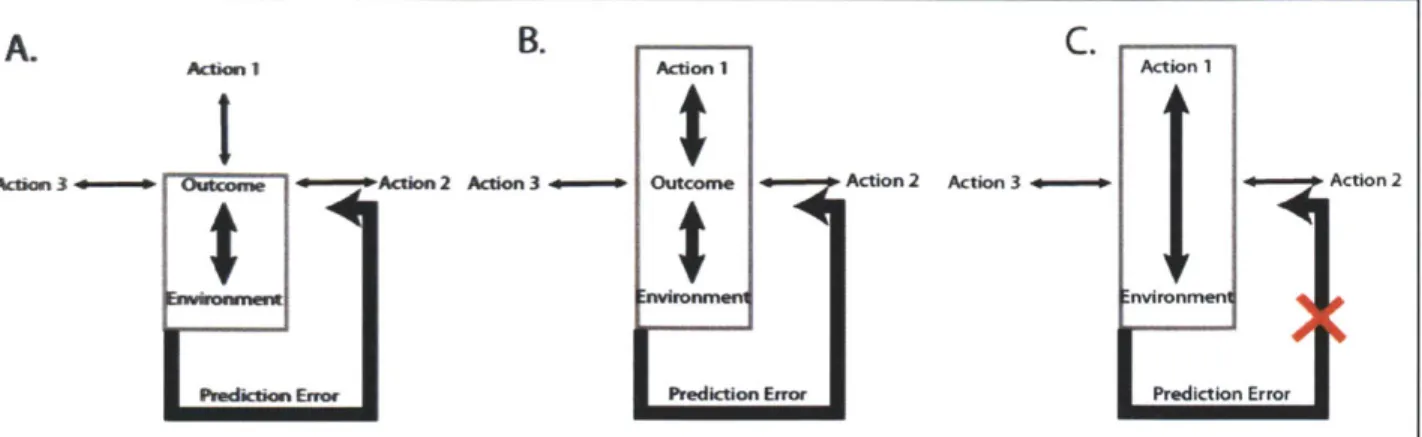
In a particular environment, an animal may choose from a variety of alternative actions at any given moment. Exploration means that the course of action chosen by the animal given a particular set of environmental conditions is highly variable. Behavioral exploitation, on the other hand, means that given a set of sensory inputs (state), an animal performs the exact same action or sequence of actions nearly every time. The degree of behavioral flexibility, by this definition, may be directly dependent on the strength of the links between the environmental representations (sensory stimuli, ect.), particular actions, and outcomes (Fig. 1.1). These links may be viewed as "predictions" that vary in a fluid manner over the course of a behavior. The strength of predictions (associative links) may determine the degree to which behavior is exploratory (highly variable) or exploitative (invariant).
The shift from flexible exploration to behavioral consistency or "exploitation" has been widely studied across a variety of animal species (Doupe, Perkel et al. 2005; Graybiel 2005; Graybiel 2008; Humphries, Khamassi et al. 2012). One elegant example is the process by which
songbirds learn how to sing. Young birds generate highly variable sequences of song syllables with the goal of matching their vocal output to the song template produced by their parents. They use the auditory feedback from their vocalizations to gradually adjust their song.
A. B. C.io
Acnion I A 1 Action 1
Action 3+ Ouom *Action 2 Action 3- Outcome Action 2 Action 3 - Action 2
wirnen nvironmer
Pediction Enor Prediction Error Prediction Error
Figure 1.1 Different predictive states that drive behavior. A. In this case, a particular rewarding outcome becomes strongly linked to a predictive sensory state but not to any specific action. The link is built by prediction errors generated by repeated experience with the outcome. Because there are no strong predictive links with any particular action, the animal may engage in variable (exploratory) behavior in this state, aimed at reaching the predictive sensory state. Violations of the predicted association between outcome and sensory state can lead to
modification of this prediction via prediction error feedback signals. B. If a particular action reliably precedes the unpredicted rewarding outcome, links may be formed between that action and the outcome, in addition to the sensory state as in A. In this case, predictive sensory states may initiate particular actions or action sequences that lead to the predicted outcome. C. With repeated predicted exposure to the outcome, action-state links may guide behavior
independently of predictions regarding the outcome. Since there is no prediction made regarding the outcome, changes in the reward may not influence the execution of the action, leading to "habitual" behavior.
is relatively inflexible to perturbation. The neural mechanisms involved in this process have been carefully studied and have yielded important insight into how reinforcement learning might occur in mammalian species (Nordeen and Nordeen 1997; Doupe, Solis et al. 2004; Doupe, Perkel et al. 2005; Fee and Scharff 2010).
A slightly different view on behavioral flexibility has come from a classic set of behavioral studies carried out by Dickinson and colleagues, which revealed that prolonged training on instrumental lever press tasks can elicit behavior that is insensitive to changes in the outcome value (Balleine and Dickinson 1998). In this paradigm, rats received injection of lithium chloride, a chemical that induces temporary sickness, after ingesting the reward inside their
home cage. Effects of this "reward devaluation" protocol on lever press behavior were tested at different stages of training and under different schedules of reinforcement on the lever press task. Rats which underwent reward devaluation after prolonged instrumental behavior continued to perform the lever press, while rats that received the reward devaluation after minimal training
stopped pressing. Insensitivity to reward devaluation has been used as an operational definition for "habitual behaviors", those that are less sensitive to changes in the outcome and thus
inflexible to additional modification (Fig. 1.1). These behaviors are believed to be guided exclusively by links between environmental stimuli and actions.
An important point regarding the above hypothesis is that strong links between an outcome and a particular action or sensory representation do not necessarily mean that the behavior is
insensitive to changes in the outcome, as in the reward devaluation studies. In fact, the opposite may be true. When rats were given reward on a variable interval schedule in the instrumental operant task described above (reward delivered at random intervals regardless of lever presses), they were actually less sensitive to outcome devaluation than when they were given reward on a fixed ratio schedule (reward delivered every x number of presses) (Balleine and Dickinson 1992; Balleine and Dickinson 1998). In this case, the link between the outcome and the lever press action was strongest in the fixed ratio case, meaning a stronger outcome "prediction" was present. Changes in the outcome violated this prediction more severely than in the variable interval case, where the prediction (the links between the action/environment and the outcome) was weak (Fig. 1.1 C). These violations of prediction are termed "prediction errors" and are a critical component of reinforcement learning theory (Schultz, Dayan et al. 1997; Waelti, Dickinson et al. 2001; Schultz 2002).
The songbird learning and the instrumental lever pressing studies illustrate important principles that may help to more precisely distinguish flexible from inflexible behaviors. However, most
behaviors performed by humans and other mammals are not single fixed action patterns (i.e. a song or a lever press), but rather are fluid movements through space and time that need to be constantly adjusted based on changing environmental conditions. The degree to which a behavior is subject to variation (exploration) may depend on the ongoing strength of the links between the representations of the environment and representations of actions or goals (Fig. 1.1).
Stronger links between the environment/actions and outcomes create stronger predictions which drive invariant behaviors but also allow that behavior to be sensitive to changes in the outcome. Not all behaviors can be learned through experience with the desired outcome. Many behaviors are aimed at goals that have never actually been experienced (as in getting a Ph.D). To direct these complex behaviors, the brain must construct abstract models that are composed of multiple predictive layers, a function that is believed to heavily rely on circuits of the neocortex (Miller and Cohen 2001). The evolution and expansion of the neocortex, particularly the prefrontal cortex, has accompanied the emergence of highly abstracted "intelligent" behavior in primates and humans. This thesis will not address the unique predictive capacities enabled by the neocortex. It is very likely, however, that abstract cortical models rely on simple predictive building blocks constructed through the conserved mechanistic principles of reinforcement learning.
1.2.1 Parallel circuits in the basal ganglia
The neural mechanisms underlying the concepts described in section 1.1 have received
increasing attention over the last decade but are still far from a complete level of understanding. There is not one single brain structure responsible for constructing predictions based on sensory experience then translating those predictions into actions - this function depends on the
coordinated action of multiple distributed areas. Converging evidence from neurological case studies in humans and brain manipulation and electrophysiological recording studies in rodents have implicated the basal ganglia as being particularly critical for learning and motivating behaviors.
The principle input nucleus of the basal ganglia, the striatum (or caudoputamen in primates) receives converging inputs from a wide range of cortical and subcortical structures, and thus is in an ideal position to integrate many types of information to form predictions or conjunctive associations between stimuli (Shepherd 2004). The projections to the striatum are anatomically segregated: sensory and motor cortical areas project to the dorsal and lateral regions of the striatum (putamen in primates), prefrontal and premotor cortices project to the medial region, and the hippocampal formation, amygdala, and prefrontal cortex project to the ventral regions of the striatum (also called the nucleus accumbens) (Voorn, Vanderschuren et al. 2004) (Fig. 1.2). The outputs of the striatum through the substatia nigra and pallidum to the thalamus remain segregated, creating loops that are believed to process information largely in parallel (Alexander, DeLong et al. 1986).
1.2.2 Segregated behavioral control and information processing in basal ganglia loops
A series of pharmacological inactivation studies in the striatum of rodents and primates have indicated that the segregated cortico-basal ganglia loops may play distinct roles in controlling behavior. Hikosaka and colleagues inactivated the posterior putamen (roughly analogous to the dorsolateral striatum in rodents) and the anterior caudate (analogous to the rodent dorsomedial striatum) in primates trained to perform new and well practiced motor sequences. They observed that manipulation of the posterior putamen disrupted the performance of well practiced
sequences while the anterior caudate inactivation disrupted performance of new sequences (Miyachi, Hikosaka et al. 1997). Neuronal recordings in the two regions were consistent with the behavioral findings (Miyachi, Hikosaka et al. 2002). Based on this evidence, they suggested that circuits in the basal ganglia learn procedures of actions in parallel based on different
coordinate frames: the anterior caudate uses spatial (or sensory state) information to guide behaviors early in training, while the posterior putamen relies on a motor coordinate frame to guide movement sequences late in training (Hikosaka, Nakahara et al. 1999).
Studies in the rodent have provided further support for the view of parallel processing of information across basal ganglia loops. Inactivation of the dorsolateral region of the striatum results in behavior that is more sensitive to changes in the outcome value, whereas inactivation of the dorsomedial region renders behavior less sensitive to outcome devaluation and also prevents animals from forming associations between actions and particular outcomes during learning (Yin, Knowlton et al. 2004; Yin, Knowlton et al. 2005; Yin, Ostlund et al. 2005; Yin, Knowlton et al. 2006).
Frontal cortex
Midline and intralarminar thalnic nuclei -IfD PFC-GrtcosMataa projections p Hea damstniatal orttcons
sassdan saaloin ompieK Hipocapasonnalian
TRENDS in Neuosciences
Figure 1.2 Topographical arrangement of projections to the rodent striatum. The schematic shows a coronal cross-section. Afferents are roughly organized along a ventromedial to dorsolateral gradient, with limbic regions projecting mainly to ventromedial and motor and sensory regions projecting to dorsolateral
striatum. Adapted from (Voorn, Vanderschuren et al. 2004)
Thus, the dorsolateral striatun may participate in guiding behaviors based on links between stimuli and responses (S-R) without incorporating information about the outcome (i.e. "habits"), while the dorsomedial striatum may participate in behavior guided by links between actions and outcomes (A-O). This is consistent with the idea that parallel basal ganglia circuits utilize different types of information to create predictive associations that can drive behavior.
The ventral medial striatum or nucleus accumbens is notoriously difficult to study using lesion or behavioral pharmacological techniques because these manipulations typically render the animal drastically void of motivation, even to eat and drink. Evidence from electrophysiological studies and specific localized manipulations suggest that this area is involved in learning and representing predictive associations between stimuli and outcomes (S-0) and using these representations to motivate behavior (Salamone and Correa 2002; Nicola, Yun et al. 2004; Atallah, Lopez-Paniagua et al. 2007; Calaminus and Hauber 2007; Humphries and Prescott 2010). The ventral striatum may play a key role in learning instrumental behavior as well (Atallah, Lopez-Paniagua et al. 2007).
Additional evidence for parallel processing of information by striatal learning circuits has come from electrophysiological recordings in behaving animals. In these studies, ensembles of striatal neurons in different subregions were recorded (sometimes simultaneously) for up to several months as rodents learned instrumental T-maze tasks (Barnes, Kubota et al. 2005; Kubota, Liu et al. 2009; Thorn, Atallah et al. 2010). Ensemble firing patterns in the ventromedial, dorsomedial, and dorsolateral striatal regions displayed dramatically different firing patterns and training dependent dynamics as learning proceeded. This data suggests that links between environmental stimuli, actions, and outcomes may be differentially represented across cortico-basal ganglia loops, depending in part on the types of inputs that each area receives. Parallel striatal circuits may also evolve at different rates during learning, depending on the dynamics and the sensory properties of the environment and the actions required to reach the desired goals. Consequently,
behavioral control may be exerted by any of these cortico-basal ganglia circuits, alone or in cooperation.
The strength of the associative links between the sensory and motor representations and the outcome may determine which basal ganglia circuits are most active in controlling behavior. The dramatic effects on motivation produced by the ventral striatum lesions
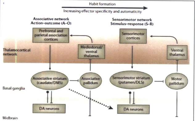
Habit formation
Increasing effector specificity and automaticity Associative network Action-outcome (A-O) Sensorimotor network Stimulus-response (S-R)
II
rimotorI
,palid(nputamenDSIII
Figure 1.3 Model by Yin and Knowlton for the progression from outcome driven behavior to habitual behavior. Basal ganglia loops involving the
dorsomedial striatum (caudate in primates) and prefrontal and premotor cortex form associative links between actions and outcomes, whereas loops involving the
dorsolateral striatum (putamen) facilitate primarily sensory state - action links. With increasing training, behavior becomes increasingly insensitive to outcomes (habitual) and is driven more strongly by the dorsolateral circuits. The circuits largely operate in parallel at the level of the basal ganglia but may interact through open loop projections to the dopamine system (dashed line). Adapted from (Yin and Knowlton 2006).
may reflect the crucial role that predictive links between sensory representations and outcomes play in the general invigoration of behavior. Unlike dorsal circuits, which may rely on specific repeatable action representations to be associated with rewarding outcomes, ventral striatal circuits may require only that a particular feature (or combination of features) of the external environment be reliably associated with a rewarding outcome, in many situations, a more
common occurrence. Many possible specific action patterns may lead an animal to food, but that food may be associated only (for example) with a specific set of predictive environmental
features. This often (but not always) can result in a progression of behavioral control, from ventromedial striatal circuits involved in sensory state dependent control over learning and behavior to dorsolateral circuits involved in action specific, outcome independent control
(Hikosaka, Nakahara et al. 1999; Yin and Knowlton 2006; Atallah, Lopez-Paniagua et al. 2007).
1.2.3 A role for dopamine prediction error signaling in reinforcement learning
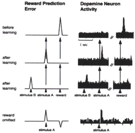
A critical element of reinforcement learning theory is a signal that carries information about deviations from established predictions, i.e. "reward prediction errors." This signal can be used to flexibly adjust predictions based on feedback from outcome experience. Wolfram Schultz and colleagues carried out a series of landmark electrophysiological studies in the primate midbrain dopaminergic nuclei, the ventral tegmental area (VTA) and substantia nigra pars compacta (SNc), which revealed firing patterns that were highly consistent with the reward prediction error signals postulated by reinforcement learning modeling studies (Fig. 1.3) (Schultz, Dayan et al.
1997; Waelti, Dickinson et al. 2001; Schultz 2002). This work was carried out using a simple classical conditioning task: head-fixed monkeys were given an auditory cue that was followed a
short interval later by delivery of ajuice reward. Dopamine neurons were initially fired vigorously in response to the unpredicted juice reward, but with extended training those same
neurons began responding to the predictive auditory cue. When the juice reward was withheld suddenly, the dopamine neurons showed a dip in their firing rates at the time the juice was typically delivered. This behavior of the dopamine neurons suggests that they signal positive prediction errors early in training which may be used to drive the construction of predictions about the environment. The firing to the auditory cue may represent a prediction about the upcoming reward (or a prediction error in the timing of the auditory cue). When rewards of different sizes were delivered, both the positive prediction errors to the reward and the learned predictive signals to the cue scaled with the reward magnitude, indicating that predictions and prediction errors can reflect relative values of outcomes (Tobler, Fiorillo et al. 2005).
Subsequent work by a variety of labs has shown that these dopamine signals also reflect reward identity, delay to reward delivery, and effort required to obtain the reward (Waelti, Dickinson et al. 2001; Morris, Nevet et al. 2006; Day, Jones et al. 2010; Gan, Walton et al. 2010).
Dopamine neurons project broadly to nearly all cortical and subcortical brain areas (Fig. 1.4), but their projections are strongest to the striatum. There, dopamine is critical for synaptic plasticity via long term potentiation (LTP) and depression (LTD). Stimulation of dopamine neurons to the striatum paired with stimulation of the afferent cortical projections to the striatum induces lasting potentiation of the glutamatergic cortical inputs (Reynolds and Wickens 2002). Altering the timing of the cortical stimulation with respect to the dopamine pulse can switch the effect on synaptic transmission from potentiation to depression (Calabresi, Maj et al. 1992; Calabresi, Picconi et al. 2007; Shen, Flajolet et al. 2008). By these Hebbian mechanisms, dopamine
impulses might strengthen co-occurring inputs to striatal neurons, allowing those inputs to drive responses more effectively on subsequent exposures to the stimuli. The creation of
Ieiard Prdion Dopenine Neuron
Error Activity
I wc
rfkg
auus 8 ulimulus A remiad a.lu 0 admuluin A rmod
Ivwd rdw
fa"us A sAa A
Figure 1.4 Reward prediction error signaling in midbrain dopamine neurons. Electrophysiological recordings were made from putative dopamine neurons in the midbrains of head fixed monkeys performing a classical conditioning task.
Histograms in the right column show responses of a
representative single unit. Initially, the monkeys were given free unpredicted reward, and the neuron fired phasically at reward delivery (top). When a predictive auditory stimulus preceded the reward delivery at a fixed interval, the neuron fired instead to the stimulus, and no longer to the reward, and when a second stimulus was introduced, the neuron fired to the first stimulus (mid). When the stimulus was delivered but the reward was withheld, the neuron displayed a decrease in firing rate at the usual reward time (bottom). Adapted from (Suri and Schultz 1998)
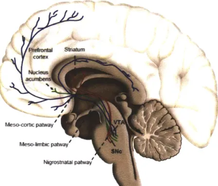
Meso c paway' Meso-inec patway'
Figure 1.5 Schematic of projections from dopamine systems of the midbrain. Dopaminergic nuclei in the midbrain can be roughly segregated on the basis of their projection patterns. The substantia nigra pars compacta projects mainly to areas of the sensorimotor caudoputamen (striatum) through the
nigrostriatal pathway, while the ventral tegmental area projects to limbic and prefrontal cortical structures including the nucleus accumbens, prefrontal cortex, and hippocampus. Image adapted from http://neuro-science.blogspot.com/201 1/10/dopamine-and-adult-neurogenesis.html
differential synaptic weight profiles in the striatum may be the neural implementation of reward predictions.
1.2.4 A role for dopamine in motivating behavior
Manipulation of dopanine signaling has provided some support for the role of prediction error signaling in forming reward predictions in the striatum based on external stimuli (Yun,
Wakabayashi et al. 2004; Zweifel, Parker et al. 2009; Flagel, Clark et al. 2011; Wang, Li et al. 2011). Mice with specific attenuation of transient dopamine signaling are impaired in acquiring appropriate responses to predictive cues (Zweifel, Parker et al. 2009; Wang, Li et al. 2011). The primary effect of global, non-specific dopamine depletion is not a simple generalized learning deficit, however, but instead is a profound impairment in motivation (Zhou and Palmiter 1995). Pharmacological blockade of dopamine signaling produces impairments in exerting effort to pursue and obtain rewards (Salamone and Correa 2002; Salamone, Correa et al. 2007). Moreover, diseases of the dopamine system, such as Parkinson's disease, produce severe
difficulties in initiating and maintaining movements. One possible interpretation of these results is that dopamine is involved in forming but also in transmitting predictions about stimuli related to goals. The prediction error signals at goals create predictions (perhaps in striatal circuits) which are themselves transmitted by dopamine neurons in the service of initiating and
maintaining behaviors. By this view, dopamine signals to environmental stimuli (such as the signal to the auditory tone in the Schultz studies) are created by reward prediction errors and represent predictions that can directly initiate and maintain actions. A form of this hypothesized role of dopamine signaling has been termed "incentive salience," and has gained increasing support in the literature (Berridge 2007; Flagel, Clark et al. 2011).
A key point regarding the incentive salience hypothesis is that dopamine prediction error and prediction signals need not be necessary for learning or executing (respectively) specific action representations (i.e. a sequence of movements) persay. Predictive dopamine impulses to environmental stimuli may have a general invigorating effect on behavior rather than initiating specific behavioral patterns (Schultz, Apicella et al. 1992; Schultz, Apicella et al. 1993; Setlow, Schoenbaum et al. 2003; Nicola, Yun et al. 2004; Berridge 2007). In other words, dopamine may not be required for establishing links between stimuli and responses, but may be important for incorporating reward feedback into predictive sensory representations (Fig. 1.1).
1.2.5 Circuit mechanisms for the influence of dopamine on motivating behavior
Behavioral pharmacology (Calaminus and Hauber 2007; Salamone, Correa et al. 2007;
Calaminus and Hauber 2009) and disease states indicate that a main site for the immediate action of predictive dopanine signaling on motivating behaviors may be the striatum itself. One effect of transient dopamine signaling in the striatum is to promote long term synaptic plasticity, as previously discussed, but dopamine signals also have powerful immediate effects on striatal network activity. Signaling through DI receptors increases and maintains the excitability of medium spiny neurons, while signaling through D2 receptors holds them in a hyperpolarized resting state (Surmeier, Ding et al. 2007; Gerfen and Surmeier 2011). Dl and D2 receptor expression is largely segregated onto different populations of medium spiny neurons (Gerfen, Engber et al. 1990; Le Moine and Bloch 1995). These distinct populations differ also in their output projections to the downstream basal ganglia nuclei. The D1 expressing neurons project to the substatia nigra pars reticulata (SNr) and the internal segment of the globus pallidus (GPi),
while the D2 expressing neurons project to the external segment of the globus pallidus (GPe) (Gerfen, Engber et al. 1990). The net effect of activation of these two pathways on thalamic neurons is believed to be opposite: Dl neuron activation, through the direct pathway, promotes thalamic excitation, while D2 neuron activation, through the indirect pathway produces net thalamic inhibition (Hikosaka and Wurtz 1985; Mink 1996). Moreover, activity in these two pathways is believed to promote opposite effects on movement: the direct pathway promotes movement, while the indirect pathway suppresses movement (Mink 1996; Kravitz, Freeze et al. 2010).
Dopanine signaling, by virtue of its opposing effects on direct and indirect pathway neurons is believed to have a net movement promoting effect.. This hypothesis has gained support from behavioral studies in transgenic mice that express light sensitive channelrhodopsin specifically in either Dl or D2 expressing neurons. Optical stimulation of direct pathway (D1) neurons
produces increased movement, while indirect pathway stimulation produces attenuated
movement (Kravitz, Freeze et al. 2010). Moreover, a prominent theory of Parkinson's disease states that dopamine degeneration produces hyperactivity in the indirect pathway neurons, resulting in movement suppression (Obeso, Marin et al. 2008). This theory has gained some support from electrophysiological studies in behaving animals which have shown increased firing rates of striatal projection neurons with dopamine depletion (Kish, Palmer et al. 1999).
The feedback interactions between the striatum and dopamine systems are not entirely
reciprocal: dopamine projections extend to larger regions of the striatum (and other brain areas) than they receive projections from (i.e. non-reciprocal open loops) (Haber, Fudge et al. 2000;
Joel and Weiner 2000; Matsuda, Furuta et al. 2009). Regional divergence of dopamine projections is present on multiple scales, from inter-regional (on the scale of striosomes and matrix) to cross-regional (ventromedial to dorsolateral divergence). One unique function of the
dopamine system then may be to broadcast predictions (and prediction errors) to regions of the striatum which do not themselves encode reward predictions. This would allow the predictive dopamine signals to generally promote ongoing actions and state representations.
Finally, dopamine signaling may be critical for modulating oscillatory network dynamics in striatal networks. Stable oscillatory network states can be created by fine tuned ionic
conductances in combination with local network interactions (Wang 2010; Buzsaki and Wang 2012). These oscillatory states have been associated with a variety of functions including memory formation, voluntary movement, and sensory binding (Engel, Fries et al. 2001; Engel and Fries 2010; Wang 2010). A large body of evidence suggests that dopamine signaling may affect the expression of these network states, both positively and negatively (Chapter 2; (Brown 2007; Berke 2009; Benchenane, Peyrache et al. 2010). Thus, the effect of dopamine on the striatal networks may not be as simple as increasing or decreasing firing rates in different populations, but it may fundamentally change the way in which the striatal networks process information.
1.2.6 Reward predictive signaling in striatal circuits
According to models for basal ganglia dependent reinforcement learning, the targets of the dopamine neuron reward prediction error signals should represent reward predictions in their
firing patterns. Electrophysiological recordings of neurons in the striatum in behaving animals have revealed some evidence for predictive representations. Single units in the ventral striatum (primarily the nucleus accumbens core) of both rodents and primates acquire predictive
responses to cues during learning of classical and instrumental conditioning tasks and these responses scale with outcome value (Hikosaka, Sakamoto et al. 1989; Schultz, Apicella et al.
1992; Schultz, Apicella et al. 1993; Setlow, Schoenbaum et al. 2003; Nicola, Yun et al. 2004; Goldstein, Barnett et al. 2012). In the dorsomedial striatum, units have been shown to represent contingencies between actions and outcomes (i.e. they fire for particular actions and scale with the outcome value) (Kimchi and Laubach 2009; Kimchi, Torregrossa et al. 2009; Stalnaker, Calhoon et al. 2010; Stalnaker, Calhoon et al. 2012). These observations are consistent with the behavioral work that has implicated the ventral striatum in representing stimulus-outcome contingencies and the dorsomedial striatum in representing action-outcome contingencies (Yin and Knowlton 2006).
The striatum sends projections directly to the midbrain which could, in principle, provide the dopamine neurons with their predictive firing properties. In the dorsal striatum, these neurons are clustered into regions called "striosomes" which are distinguished from the surrounding "matrix" neurons by their differential molecular expression patterns and afferent projections (Graybiel and Ragsdale 1978; Graybiel, Ragsdale et al. 1981; Gerfen 1985). In the ventral striatum, the distinction of striosome and matrix compartments is less clear, but the projections to dopamine nuclei (primarily the VTA) are at least as strong as the dorsal (Kalivas, Churchill et al.
1993; Humphries and Prescott 2010). The projections from the striatum to dopamine nuclei come from GABAergic medium spiny neurons, so direct synaptic connections cannot be directly
translated into the excitatory predictive responses observed in the dopamine neurons (though excitation may be achieved by local interneuron disinhibition). There are a number of indirect routes by which the striatum could communicate reward predictions to dopamine neurons. For example, neurons of the ventral striatum send inhibitory projections to the ventral pallidum, which sends direct inhibitory projections to the ventral tegmental area (Kalivas, Churchill et al. 1993; Humphries and Prescott 2010). Excitatory responses in the ventral striatum would thus generate disinhibition of dopaminergic neurons (Floresco, Todd et al. 2001; Floresco, West et al. 2003). It is likely that multiple convergent pathways from a variety of brain areas, not one particular source, contribute to the predictive responses in dopaminergic neurons (Grace, Floresco et al. 2007).
1.2.7 Brief Summary
-The basal ganglia nuclei, in particular the striatum, contribute both to learning and motivating behaviors towards goals.
-Striatal subregions receive topographically organized projections and this organization is preserved throughout the cortico-basal ganglia circuits.
-Striatal subregions make different contributions to learning and behavioral control and exhibit
distinct neuronal firing patterns during behavior.
-The control of different basal ganglia loops over behavior may be determined by the strength of associative links between motor patterns, sensory representations, and outcomes.
-Midbrain dopamine neurons transmit reward prediction error signals, which may be used to establish associative links between reward predictive stimuli, actions, and outcomes through synaptic plasticity in the striatum and elsewhere.
-Reward predictive signals related to environmental stimuli and actions emerge with experience and are represented by neuronal firing in the striatum, particularly in the ventral and medial striatum, and in dopamine neurons (in addition to other brain regions).
-Predictive signals transmitted by dopamine neurons after learning may initiate and motivate behaviors via widespread projections to the striatum and other regions.
1.3 Scientific philosophy and research summary
The objective behind the research described in this thesis is to further the fundamental understanding of the brain mechanisms that guide and motivate human behavior. A
comprehensive model would ideally incorporate a minimum set of basic principles needed to achieve general predictive power at the level of neural circuit behavior. Clearly, complex fimctions like behavioral learning, selection and motivation involve the coordinated action of multiple brain areas. Current methods do not allow us to monitor the neural activity patterns in all of these circuits simultaneously, so we must either study them in isolation (or a few at a time) or construct biologically realistic computational models of neural networks. Both approaches are currently being employed in various forms. One version of the latter relies on a comprehensive database of anatomical connectivity and molecular expression patterns and has been termed "connectomics" (Lichtman and Sanes 2008; Jain, Seung et al. 2010; Denk, Briggman et al. 2012; Reid 2012). The task of collecting this data, cataloging it, and packaging it into informative
computational models is daunting at many levels, but rapid progress is being made on all these fronts. Another similar effort to construct large scale biologically realistic models of the cortex, called the Blue Brain Project, is being carried out by Henry Markram and colleagues (Markram 2012). While computationally enormous in scope, this project does not rely on detailed
knowledge of specific synaptic connections like connectomics, but instead uses connectivity and molecular expression patterns of typical cortical microcircuits then constructs a large-scale simulation based on those basic computational units.
A second approach is to measure the electrical and chemical dynamics in neural circuits in awake, behaving animals in regions of the brain known to be critical for guiding behaviors. The idea behind this more focused method is to derive basic principles of circuit operation that might be generalized across multiple behaviors and to large neural ensembles extending across multiple brain regions. These principles would not predict circuit behavior with exact precision but would make general, qualitative predictions. Precise temporal and cell-type specific
manipulation of brain circuits, such as with genetically expressed opsins (Boyden, Zhang et al.
2005; Deisseroth, Feng et al. 2006; Witten, Steinberg et al. 2011), would allow us to test the
predictions of the models empirically. Such models would be useful not only for understanding some of the basic philosophical questions about human behavior, but would also allow us to understand how circuit operation is disrupted in neurological disorders. It is this general approach that has guided the work presented in this thesis.
In recent decades, a variety of new techniques have been developed that allow us to measure both electrical and chemical dynamics of neural circuits chronically in behaving animals with
high temporal and spatial precision (Robinson, Venton et al. 2003; Buzsaki 2004; Dombeck, Khabbaz et al. 2007). In this thesis, I will describe work using two of these techniques: multi-site tetrode recording and fast-scan cyclic voltammetry to study the electrophysiological and electrochemical dynamics respectively of the striatum in rats during learning and execution of goal-directed behaviors.
The second chapter will describe how oscillations in the local field potential and associated network synchrony in the striatum evolve over the course of learning a simple associative maze task. We have found that early stages of learning are marked by higher frequency oscillatory activity in the gamma range, which gives way to lower frequency beta oscillations over the course of learning. Spiking synchrony shifts during this period as well, from primarily local coordination during the high gamma dominated learning period, to widespread global synchrony during beta oscillations late in training. We propose that a high frequency, local network regime favors plasticity and flexibility early in learning when rewards are unpredicted, but that repeated experience with predicted rewards promotes global network synchrony that favors fixed,
inflexible behavior behaviors. I also propose a mechanism by which this transition might take place under the framework of reinforcement learning.
In the third chapter, I will describe the dynamics of dopamine signaling in the striatum of rats performing the same T-maze behavior as in Chapter 1. Dopamine is a neurotransmitter known to be critical for forming predictions and motivating actions to achieve goals. We used chronic fast-scan cyclic voltammetry, an electrochemical technique, to obtain real-time subsecond measurements of dopamine concentration in various regions of the striatum. This study revealed
that dopamine signaling in the maze was not transient in nature, as has been previously described for simpler classical conditioning tasks, but rather increased continuously during maze running and peaked at goal reaching. The signal moreover was sensitive to the maze environment, showing biases for different maze arms that could be manipulated by providing different reward sizes at the ends of the maze. We go on to show, using a combination of electrophysiology and fast scan cyclic voltammetry, that the dopamine signals display an inverse relationship to the beta-band oscillations described in Chapter 1, suggesting that they play a significant role in shaping network behavior. We postulate that the dopamine signals represent predictions about the value of upcoming rewards that vary continuously with the animals' proximity to that
reward. Such a signal suggests a mechanism for controlling goal directed behaviors in a flexible, context dependent manner. Finally, I propose a qualitative model by which these dopamine signals could be generated that makes testable predictions about the underlying circuit
mechanisms. I hope that this work will stimulate future theoretical and experimental work and contribute to a unifying description of the neural mechanisms underlying behavior.
Chapter 2: Shifts in local field potential oscillations and
network synchronization during learning
2.1 Introduction and background
Rhythmic activity in the extracellular local field potential (LFP) is thought to reflect the coordination of local neuronal networks along different spatial and temporal scales. This oscillatory coordination has been proposed to serve as an organizational tool that could, among other things, facilitate long-range communication between brain areas, provide a temporal structure for memory formation, and act as a filter for perceptual attention (Engel, Fries et al. 2001; Buzsaki and Draguhn 2004; Wang 2010). The frequency specific fluctuations in power of LFP oscillations vary widely across brain areas and are highly dependent on the ongoing
behavioral state of the animal. Thus, LFP activity in different frequency bands may serve different behavior-specific roles.
In the basal ganglia, oscillations in the LFP have likewise been measured across multiple frequency bands and have been associated with a wide range of behavioral states. Beta
oscillations (15-30Hz) in motor cortices upstream of the basal ganglia have been linked with a lack of or suppression of movement initiation (Sanes and Donoghue 1993; Baker, Olivier et al.
1997). In Parkinson's Disease, which is characterized by a degeneration of dopaminergic signaling to the basal ganglia, beta oscillations in basal ganglia nuclei are abnormally strong, indicating that hyper-synchronization of basal ganglia networks in this frequency range may
contribute to the movement difficulties observed in this disease (Brown 2007). However, beta oscillations seem to be important for normal basal ganglia functioning as well, as they have been observed in the striatum, the basal ganglia's principal input nucleus, during sequential movement tasks in primates (Courtemanche, Fujii et al. 2003) and during action selection in rodents
(Leventhal, Gage et al. 2012). These studies also indicate that the role of beta oscillations in the basal ganglia is not limited to controlling motor output exclusively.
High gamma oscillations (70-90Hz), common in cortical structures, have also been measured in basal ganglia nuclei, particularly in the striatum. Unlike beta oscillations, high gamma in the striatum has been associated with periods of active movement (Masimore, Schmitzer-Torbert et al. 2005). Moreover, coordination in high gamma between the amygdala and the limbic region of the striatum has been shown to correlate with behavioral performance during Pavlovian association learning (Popescu, Popa et al. 2009), and high gamma power is strongest during the early stages of instrumental learning and wanes with extended training (van der Meer and Redish 2009).
This evidence suggests that different oscillatory network states in the basal ganglia may actively facilitate both network plasticity and behavioral rigidity as a behavior is learned and becomes habitual with overtraining. We tested this hypothesis by recording spiking and local field potentials from the ventral striatum of rats as they learned and eventually habitually performed an associative T-maze task.
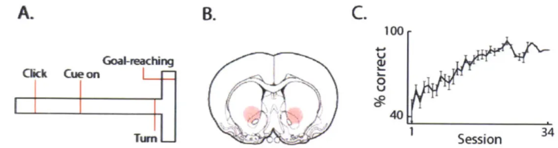
2.2.1 Beta oscillation dynamics during associative T-maze behavior A. B. C. 100-4-& Goal-reaching , Click Cue on 0 Turn 1 Session
Figure 2.1 Task description and recording sites. A. Layout of the T-maze task. B. Region sampled by tetrode recordings (red shading). C. Average learning curve for all animals (n = 7).
We recorded spike and LFP activity bilaterally from the ventromedial part of the striatum (here called VMS) in 7 rats fitted with chronically implanted headstages carrying assembles of 12 independently movable tetrodes. The rats were trained to perform a T-maze task (Barnes, Kubota et al. 2005) that required them to turn left or right in response to auditory cues in order to receive a reward of either bits of chocolate or a drop of chocolate milk at the end of the arm indicated by the corresponding auditory instruction cue (Fig. 2.1; Methods). The tetrode recordings started at the beginning of training and continued until the rats had completed at least 10 consecutive 40-trial sessions with performance levels above the learning criterion of 72.5% correct (Fig. 2.1 C). The tips of most tetrodes were confirmed by later histology to be in the VMS, and they were mainly within the core of the nucleus accumbens (Fig. 2.1B, Methods).
Spectral analysis of LFPs recorded after completion of training indicated task-related modulation of oscillatory activity in multiple frequency ranges in the session-averaged LFPs (Fig. 2.2 A and
Cueon Gacl-in
B.
21Click Cue on Turm Goal
-0.5 0 0.5 -0.5 0 05 -0.5 0 0.5 -0.5 0
5 6 :
-.5 0 0.5 -05 0 0.5 -0.5 0.5 -0.5 0
-1 0 1 2 3 4 5 6 -0.5 0 0.5 -0.5 0 0.5 .5 0 0.5 -0.5 0
Tine (s) Time (s)
Figure 2.2 Beta power in ventral striatum increases after goal-reaching on correct trials. A. Session-averaged spectrograms for correct trials (n = 22 trials, top) and for incorrect trials
(n = 18 trials, bottom) for a representative 40-trial session. B. Top: Z-score normalized beta power (15-28 Hz) averaged across all sessions and recording tetrodes for correct trials (red) and incorrect trials (blue). Red vertical lines indicate centers of task-event windows as labeled.
Middle: Average z-score normalized run-speed. Bottom: Average z-score normalized
acceleration. Shading shows standard errors of the mean (SEM) computed across sessions.
A.
- an ,60 ~40 U.2 9 5 WsB). Particularly prominent was a transient increase in power in the beta band (15-28 Hz) that occurred directly after goal-reaching (Fig. 2.2A). The increase was strong on correctly performed trials but was weak on incorrect trials (Fig. 2.2 A and B). This modulation of beta-band power did not exhibit a clear relationship to either the run speed or the acceleration of the animals (Fig. 2.213).
To investigate this beta-band activity in more detail, we examined raw and band-pass filtered
LFP traces from single A. Before delay
45 20
trials. Remarkably, this
X
analysis showed that the extended period of
0
increased beta power B. After delay
45 20
visible in the
session-averaged spectrograms W
after goal-reaching did not
200
Goal- Reward delivery Goal- Reward delivery
represent continuous beta- 'eaching reaching
Figure 2.3 Increases in beta power are not dependent on band activity, but instead, the presence of the primary reward. Reward delay control.
Session-averaged beta power for correct (left) and incorrect corresponded to brief (right) trials during a standard 40-trial session (A) and
during a control session in which reward was delayed for 2 s bursts of 2-4 cycles of after goal-reaching (B).
high-amplitude beta
activity (-100-200 ms) that occurred at slightly different times in different trials (Compare Figs. 2.2A and 2.5B).
To determine whether the transient beta-burst activity was related to reward at the end of the correct runs, we performed two control experiments in which we manipulated the primary reward that the rats received. In the first experiment (n = 2 rats over 13 sessions), we tested whether the sharp increase in beta power was tied to the animals' receiving the chocolate milk reward by delaying the chocolate milk delivery until 2 s after goal-reaching (Fig. 2.3). The increase in beta power after goal reaching did not shift forward in time, but instead, continued to occur just after goal-reaching (Fig. 2.3B). Moreover, beta power in incorrect trials, nearly absent before the delay, became stronger during the trials with reward delay (Fig. 2.3).
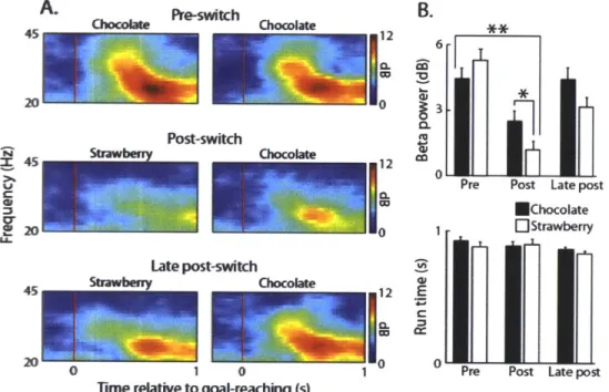
A. 45 r Pre-switch B. 6 CO 40 Post-switch X 45 Cr 1_ 2 LSL Late post-switch i (hneftnIm 0 EChocolate OStrawberry a: E CC 0 1 0
Time relative to goal-reaching (s)
Figure 2.4 Beta power is altered following a switch in reward identity. Reward-switch control. A. Top: Beta power averaged across correct trials on the left and right sides of the maze (left and right columns, respectively) during two sessions averaged just prior to switching the primary reward. Middle: Average beta power after goal reaching for 2 sessions averaged immediately after primary reward on the left arm of the maze was switched from chocolate milk to strawberry milk. Bottom: Average beta power for two sessions averaged, 5 days after the switch to the
strawberry milk as reward. B. Average beta power during the period from 0 to 0.8 s after goal-reaching (top) and average run times from turn offset to goal-reaching
(bottom) for the sessions shown in A. Error bars represent SEM. *P < 0.01; * *P <
In the second control experiment (n = 1 rat over 6 sessions), we tested whether changing the identity of the primary reward would influence the beta power after goal-reaching (Fig. 2.4). We did this by suddenly changing the reward in one end-arm of the T-maze from chocolate milk to
strawberry milk after the rat had completed ten days of overtraining on the task. Despite the fact that the rat was pre-exposed to strawberry milk in his homecage and drank it readily, we
observed a strong and significant (P < 0.01, two-tailed t-test) decrease in post-goal beta power on correct trials for both the strawberry milk and the chocolate milk end-arms (Fig. 2.4). This
decrease was significantly stronger for the arm baited with strawberry milk than for the end-arm baited with chocolate milk (P < 0.01, two-tailed t-test; Fig. 2.4B) and could not be
accounted for by changes in the run speed of the animal (Fig. 2.4B). After five consecutive days of exposure to the new reward, however, the beta power had rebounded nearly to pre-switch levels (Fig. 2.4). The results from these two control experiments indicate that post-goal beta power is not tied explicitly to the receipt of primary reward but may reflect a particular internal state that can be modified by unexpected changes in the primary reward.
2.2.2 Changes in beta and high gamma oscillations during T-maze learning
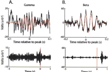
We analyzed the entire set of data recorded throughout training to determine whether the patterns of oscillatory LFP activity changed across learning. We found that both beta- and gamma-band trial-averaged activities were composed, when examined trial-by-trial, of transient bursts of oscillations lasting one to several cycles (Fig. 2.5). These bursts happened with varying probability across the T-maze task for the two different frequency bands: gamma bursting was strongest before goal reaching, while beta bursts were more prevalent after goal reaching (Fig.
A. Gamma B. Beta
20 60
-2-6
-01 00.1 -02 0 0.2
Time relative to peak (s) Time relative to peak (s)
15 60 4b 0 00 -15 T-0 3 0 1 3 0 1 Time (s) Time (s)
Figure 2.5 Beta and gamma oscillations occur in bursts and are active around different task periods.
Representative examples of single high amplitude gamma (A) and beta (B) bursts. Top Row: The raw LFP signal is shown in black, and the beta band-pass filtered signal is overlaid in red. Both signals are z-score normalized to the mean and standard deviation of each signal across the entire trial. Bottom Row: Band-pass filtered traces for the high gamma oscillations and beta oscillations. Note the different task modulation for the two frequency bands.
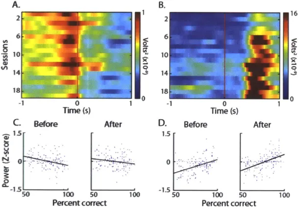
2.6). The occurrence of trial-end beta activity was highly experience-dependent and was accompanied by inversely changing high gamma-band activity (Fig. 2.6). High gamma power was strong both before and after goal-reaching during initial training and the first days of over-trining, but it diminished significantly as training progressed. This gamma activity also became more restricted in duration (Fig. 2.6; R = -0.28, P < 0.001 for before, R = -0.17 P = 0.05 for
after). In sharp contrast, the rise in beta-band power after goal-reaching, typical of correct trials at the end of training, was scarcely detectable even on correct trials at the beginning of training (Fig. 2.6B). This beta-band activity did not become prominent during the correct runs until about the time the rats reached the learning criterion, and it then continued to increase throughout the over-training period (Fig. 2.6 B and D; R = 0.53, P < 0.002). Beta power during the T-maze runs,
prior to goal-reaching, was much weaker than that after goal-reaching (Figs. 2.6B), but it also showed a significant positive correlation with the percent correct performance of the animals (Fig. 2.6D; R = 0.42, P < 0.0002). These findings demonstrate that high gamma power during T-maze performance significantly decreases with learning in the VMS while simultaneously recorded beta power during the T-maze run particularly just after goal-reaching increases with
learning. A. 2 @1 In -1 0 Time (s) Before After 100 50 100 Percent correct B. 1 40 0 UC 14 18 -1 0 1 Time (s) D. Before 16 0 After 1.5
0
-1.5 50 100 50 100 Percent correctFigure 2.6 Beta power during task performance increases with learning, whereas gamma power decreases with learning. A and B. Gamma (A) and beta (B) power around goal reaching for all sessions run by one representative rat. Each row represents the average power over 18 correct trials. C and D. Mean z-score
normalized gamma (C) and beta (D) power prior to goal-reaching (left plot) and after goal-reaching (right plot) as a function of percent correct performance for all sessions run by all rats (n= 7) combined. Power was z-score normalized in the analyzed window across all sessions for each rat individually.
C. 15r U In N a, -1.5- L 50
2.2.3 Putative medium spiny neurons and fast-spiking interneurons are synchronized with beta and gamma oscillations.
We next asked whether
A
the spike activities of
35
~30
50 S25
striatal fast spiking 820
kOAVO r 15
interneurons (FSIs) and
L
100-0 0 0 i a 01
striatal projection neurons B
100 500
recorded were modulated so400
0 20D[~
during identified high
DOk
00 ISI ntegvaI(s) 01
gamma or beta bursts. Figure 2.7 Subtypes of striatal neurons were
distinguished based on firing rate and interspike interval We separated neurons distribution. Sample waveform (left) and distribution of
interspike intervals (ISIs, right) are shown for a putative into putative subtypes medium spiny neuron (A) and a putative fast spiking
interneuron (B). Note the smaller percentage of long ISIs for based on their interspike the putative fast spiking interneuron relative to those for the
putative projection neuron. interval (ISI) distributions
and their baseline firing rates as described previously (Kubota, Liu et al. 2009) (Methods and Fig. 2.7). Nearly 50% of the FSIs (n = 163/332) were significantly modulated during high amplitude beta bursts, and a similar proportion were modulated around high amplitude gamma bursts (n = 161/332; Rayleigh's test, P < 0.05). Just over 50% of the beta-modulated FSIs
(98/163) were modulated by both beta and high gamma rhythms, reflecting the overlap in these frequencies mid-learning. Remarkably, as a population, the FSIs with transiently synchronized spiking fired most strongly near the troughs of both high amplitude gamma oscillations and high