Hyponatremia in peritoneal dialysis: excessive free water, hyperosmolarity, or malnutrition?
(
Hyponatrémie en dialyse péritonéale : surcharge d´eau libre, hyperosmolarité ou malnutrition?)Javier de Arteaga
Servicio de Nefrologia. Hospital Privado Universitario, Córdoba, Argentina.
Résumé
L’hyponatrémie est l’une des anomalies électrolytiques les plus fréquentes en pratique clinique et s’associe à une morbi-mortalité élevée. L’hyponatrémie en dialyse est elle aussi assez fréquente : 6 a 29 % des patients en hémodialyse et 11 a 26 % des patients en dialyse péritonéale (DP) ont une natrémie ≤ 135mmol/L.
Dans cette revue nous traiterons de l’hyponatrémie chez les patients en DP qui présente certaines spécificités liées à la technique de dialyse et dont en découle une prise en charge thérapeutique différente de celle proposée aux patients non dialysés avec hyponatrémie.
Les spécificités de la DP sont principalement liées à une qualité d’ultrafiltrat, à la fois en terme d’extraction sodée et de transfert d’eau libre, différente selon l’agent osmotique utilisé dans le dialysat (agent osmotique cristalloïde ou colloïde) et selon les caractéristiques propres de la membrane péritonéale (fonctionnalité des aquaporines, préservation de la conductance osmotique au glucose…). La perte de la fonction rénale résiduelle, fréquente à moyen-long terme en DP, est elle aussi fortement corrélée à l’apparition d’une hyponatrémie.
Enfin, la dénutrition, situation fréquente en dialyse et souvent associée à une hypokaliémie, peut, elle aussi, être une cause d’hyponatrémie.
Cet article a pour but d’aider à comprendre la physiopathologie de l’hyponatrémie et ses spécificités en DP pour ainsi proposer un traitement approprié.
Le
B
ulletin de la
D
ialyse à
D
omicile
journal officiel du Registr e de D ialyse Péritonéale de Langue Française RDPLF www .rdplf.or g Summary
Hyponatremia is one of the most common electrolyte abnormalities in clinical practice and is associated with higher morbidity and mortality. On dialysis, hyponatremia is also quite common: 6 to 29% of hemodialysis patients and 11 to 26% of peritoneal dialysis (PD) patients have a serum ≤ 135mmol / L.
Hyponatremia in PD has unique aspects that confer different clinical consequences and treatment needs compared to subjects with normal kidney function.
The specificities of PD are mainly related to an ultrafiltrate quality, both in terms of sodium extraction and free water transfer, different according to the osmotic agent used in the dialysate (crystalloid or colloid osmotic agent) and according to the characteristics of the peritoneal membrane (functionality of aquaporins, preservation of osmotic conductance to glucose ...).
In the short or medium term, loss of residual renal function is highly correlated with the appearance of hyponatremia. Finally, undernutrition, a frequent situation in dialysis and often associated with hypokalemia, can also be a cause of hyponatremia.
The purpose of this article is to help understand the pathophysiology of hyponatremia in PD, which is of paramount importance to ensure appropriate treatment.
Abbreviations PD : Peritoneal Dialysis
APD : Automated Peritoneal Dialysis
CAPD : Continuous Ambulatory Peritoneal Dialysis ICH : Intracellular hyperhydration
ECS : Extracellular Sector ICS : Intracellular Sector ECD : Extracellular dehydration UF: Ultrafiltration
INTRODUCTION
Hyponatremia is one of the most common fluid and electrolyte changes in hospitalized patients (1). Mechanisms for homeostasis and the water-soluble equilibrium of intra- and extracellular compartments maintain osmolarity and intravascular volume within a very narrow range. Any osmolarity imbalance leads to a rapid transfer of free water from the compartment with the lowest osmolarity to the one with the highest osmolarity. Another physiological principle of the control of the natremia is that of the different compartments electroneutrality.
Hyponatremia is defined as a plasma sodium value of less than 135 mmol/L. Its prevalence in peritoneal dia-lysis (PD) is about 10–20%. Hypokalemia is even more common and may be up to 50% (1, 4). There are few data on the incidence of hyponatremia (de novo) in PD, but it can be assumed that it is around 15–20% in the first year of PD. Its appearance is associated with a membrane with high permeability, the use of icodextrin, and malnutrition (which will most often lead to the asso-ciation of hypokalemia with hyponatremia (5)). The loss of residual renal function could also be implicated (2). For logical reasons, in this article, we will not take into account the role of the antidiuretic hormone (ADH) in the distal nephron because of this stage renal failure (stage 4 and 5 CKD, including in PD); even if diuresis exists, ADH has only minimal or no effect on free water clearance and the concentration of urine.
REMINDERS OF SOME NOTIONS OF PHYSIOLOGY AND DEFINITION Definitions
-Osmolarity: quantity of diffusible osmoles or not / L of plasma.
-Osmolality: quantity of diffusible osmoles or not / Kg of plasma water without taking into account the
possibi-lity of exerting an osmotic pressure gradient.
Effective osmolality or Tonicity: amount of
non-diffu-sible or active osmoles / L of plasma.
The tonicity of the extracellular sector (ECS) and intra-cellular sector (ICS) are equal. A change in the number of active osmoles of ECS (sodium) or ICS (potassium and organic osmolytes) and therefore responsible for a change in volume in the 2 compartments.
The variations of the natremia are therefore related to those of sodium, potassium and water, as shown by the Edelman equation:
Natremia = (Exchangeable Na+ + Exchangeable K+) / Total Water (5)
Natremia = (Exchangeable Na+ + Exchangeable K+) / Total Water (5) Notions of physiology
- Hyponatremia is determined by free water ingestion which cannot be excreted.
- Hypernatremia is determined by the loss of free water which is not compensated- Hypovolemia represents a loss of sodium and water.
Sodium is the main osmolyte in plasma. Any «true» hy-ponatremia is hypo-osmolar. This hypo-osmolality is at the origin of a movement of water from the ECS to the ICS and thus of an intracellular hyperhydration (ICH). If this is not the case, the osmolarity is normal or high. In this case we speak of pseudo-hyponatremia
(cf annex 1)
Hyponatremia associated with a plasma hypo-osmola-lity is therefore explained by a relative water content higher than the sodium stock. This sodium stock can be normal, increased (ICH + ECH = global hyperhydration) or decreased (ICH + ECD). Hyponatremia is therefore the consequence of an excess of water and not, primi-tively, a lack of sodium.
WHAT ARE THE PROTAGONISTS OF SODIUM BALANCE DISORDERS IN PD?
·
-Plasma osmolality through the mechanism of thirst, -The effective plasma volume through the mechanism of thirst,
-The «exchangeable» mass of sodium and potassium, -Total body water,
- Pituitary osmo-receptors and ADH if maintaining resi-dual renal function,
- Central and peripheral baroreceptors,
journal officiel du Registr e de D ialyse Péritonéale de Langue Française RDPLF www .rdplf.or g
-The concentration of sodium in the dialysate * (currently mar-keted dialysates have a sodium concentration of 132 to 134 mmol / L, close to the plasma sodium concentration) (10). - Gibbs-Donnan equilibrium: If on one side of the peritoneal membrane there are large ne-gatively charged molecules (proteins in the capillaries, for example), these attract positively charged ions (Na+) and repel those negatively charged (Cl-).
The mechanism at the origin of hyponatremia is then: - Either a negative balance of sodium and / or potassium ions.
- Or a positive balance of free water.
However, just as in acid-base equilibrium disorders, it is rare for a single cause to induce a change in serum sodium and it is often several mechanisms that can ex-plain it.
PRACTICAL SITUATIONS
According to Cherney and M. Halperin (1), in order to make a physiological analysis of the problem, it would suffice to ask yourself the following questions (and find answers!) :
1. Is there a significant increase in weight?
2. Is there a significant decrease in extracellular volume? 3. Is there a retention of an organic molecule whose dis-tribution is restricted to the extracellular compartment (mannitol, glucose, icodextrin)?
4. Is there a change in the concentration of bicarbonate in the plasma ?
Situation n° 1 : Positive water balance with significant weight gain (ECH)
Dans In this situation, an excess of free water is the single and leading cause of hyponatremia and may be exacerbated by a thirst disorder, a loss of residual renal function and an inappropriate UF. In the case of an anu-ric PD patient, there is a weight gain proportional to the amount of water ingested. Total body water accounts for about 60% of weight in adult men and 55% in women (Figure 1). The water ingested by the patient distributes proportionally within the different compartments (60%
intracellular and 40% extracellular), thus causing hypo-natremia. Thus, a weight gain of 4 kg represented by the consumption of 4 liters of free water corresponds to an increase of about 10% of the total volume of water. In this case, a decrease of 10% in the serum sodium will lower natremia from initial value 140mmol/L to 126 mmol/L(Figure 2).
Situation n° 2 : bilan négatif des ions sodium/potassium, soit une diminution du nombre d’osmoles actifs.
Situation n° 2 : Negative balance of sodium / potassium ions, ie a decrease in the number of active osmoles. Hyponatremia may be the result of a negative balance in sodium and / or potassium in relation to either insuf-ficient intake or excessive losses (digestive losses, UF with excessive sodium extraction).
a)Sodium salt deficiency with ECD: the osmolality of
the extracellular compartment is therefore reduced, fa-voring the passage of free water from the ECS to the ICS
journal officiel du Registr e de D ialyse Péritonéale de Langue Française RDPLF www .rdplf.or g
Fig. 1: Distribution of the different aqueous compartments according to age and gender (as a percentage of total water) according to Friis-Hansen: Body water composition in children during growth. Up to date as of 2018.
ICF
(24 L) (16 L)ECF 4 L
EFW
Expanded ECW causes NaCL excretion returning
the ECF toward normal 2.4 L 1.6 L
VP
Fig. 2: Hyponatremia with weight gain. Source: Cherney et al. (1). ICF : Intracellular fluid ; ECF : extracellular fluid; EFW : Excess Fludi Water. VP: inhibition of vasopressin secretion when there is still renal function (not effective in PD).
*note : The sodium
extrac-tion on PD is done mainly by convection and not by diffusion (more important is ultrafiltration (UF), more important is sodium extraction). Indeed the use of a PD fluid having a sodium concentration close to that of the plasma allows very little diffusion. Sodium sieving is another particular aspect of PD ex-changes that accounts for higher sodium extraction in CAPD than in APD (cf
In case of malnutrition, there is a hypokalemia by default of intake. This hypokalemia may then be accompanied by hyponatremia as described above. An intracellular potassium deficiency also causes hypo-osmolarity of the ICS causing a passage of water from the ICS to the ECS in order to maintain the osmotic balance, again favoring hyponatremia. The permanent supply of glucose, osmo-tic agent mainly used in PD, can also induce hyperinsuli-nemia resulting in hypokalemia by transfer of potassium from ECS to ICS. These different causes of hypokalemia act separately or together to induce hyponatremia. Situation n° 3 : positive balance of water with osmol gain without significant weight gain. The example asso-ciated with the use of icodextrin.
The presence of hypertonic metabolites of glucose or of icodextrin (maltose, isomaltose ...) in the ECS leads to a free water passage from the ICS, resulting in hyponatremia (Figure 4). Hyponatremia is not causing the reduction of the extracellular volume (ECD)
and intracellular edema (ICH).
Suppose, for example, a man weighing 70 kg with 400 mmol of sodium loss: The serum sodium will decrease from 140 to 130 mmol/L due to the passage of water from the ECS to the SIC following the osmotic force, causing a reduction of the extracellular volume of about 1.85 liters, and this should be clinically detectable by the doctor.
b) Deficiency of potassium salts with no change in ECS:
in the case where the patient has a pure potassium chlo-ride deficiency, the loss of potassium in the ICS will be compensated by the passage of sodium from the ECS to the ICS, thus ensuring electro-neutrality. When the elimination of potassium is accompanied by an anion coming from the ECS (chlorine or bicarbonate), there is for the ECS a net loss of 2 solutes (Na + which gains the ICS and Cl- lost with the K + ) when there is no loss for SIC (Loss of K + offset by Na + gain) (Figure 3). This situation is accompanied by metaboloc alkalosis.
journal officiel du Registr e de D ialyse Péritonéale de Langue Française RDPLF www .rdplf.or g
Figure 3 : (Source: Cherney et al. (1)): Left: Hyponatremia due to the passage of water from the ECF to the ICF in a situation of reduced extracellular volume and pure loss of sodium for a short time. Right: Sodium passes from ECF to ICF to ensure electroneutrality in case of loss of pure K. (ICF : Intracellular fluid ; ECF : extracellular fluid)
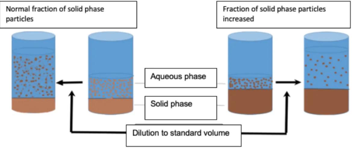
Figure 5 : Fig. 5 . Modifié d’après Turchin et al (11) : Pseudohyponatremia: The serum contains a so-called solid phase and a liquid phase. Figure 4 : Source: Cherney et al. (1) : Hyponatremia due to increased extracellular volume (ECF) water passage. ((ICF : Intra-cellular fluid ; ECF : extraIntra-cellular fluid, VP : Vasopressin)
hypo-osmolar as in the previous cases (called true hyponatremia) but hyper-osmolar. (cf annex 1). This situation has been observed since the first clinical trials with icodextrin. It causes in most patients a decrease of 2 or 3 mmol / l of natremia.
TREATMENT OF HYPONATREMIA IN PD Taking into account the principles of physiopathology previously exposed, we propose for the treatment of hy-ponatremia in DP, to follow the algorithm developed by Musso and Bargman (9), keeping in mind that the occur-rence of a Hyponatremia in PD is often multifactorial. Step 1: rule out a pseuhyponatremia linked to a do-sing artifact (Figure 5).
Step 2: assess the possibility of hyperosmolar hyponatre-mia (prescription of icodextrin, major hyperglycaehyponatre-mia). Step 3: Assess the nutritional status and look for hypoka-lemia due to deficiency of intake.
Step 4: evaluate the ECS which, if it is increased, signi-fies a gain in water then requiring the preferential use in
the dialysate of a crystalloid osmotic agent and / or of hypertonic solutions allowing a UF richer in free water than the icodextrin.
CONCLUSION
Hyponatremia is a common situation in PD. Its etiolo-gies are somewhat different from those observed in the presence of normal renal function, vasopressin having a very weak or even no effect in advanced renal failure. In PD, hyponatremia is most often the consequence of an excess of free water characterized by an increase in the weight of the patient. Malnutrition, often associated with hypokalemia, is also a cause of hyponatremia. The treatment of hyponatremia must therefore take into account an accurate determination of the patient’s dry weight as well as a thorough nutritional evaluation. DISCLOSURE
Author declared no conflict of interest
journal officiel du Registr e de D ialyse Péritonéale de Langue Française RDPLF www .rdplf.or g
ANNEXE 1 : Pseudo-hyponatremia in non-hypo-osmolar hyponatremia
1/ Normal osmolality hyponatremia = laboratory artifact caused by hyperprotidemia or hyperlipemia (increased serum solid phase at the expense of the liquid phase). In this situation the percentage of water decreases. Three methods can be used to measure natremia: flame emission spectrometry and indirect potentiometry which make a result in molarity and are therefore sensitive to changes in plasma water. Indirect potentiometry, on the other hand, renders a result in molality and is therefore insensitive to variations in plasma water (FIG. 5).
2/ Hyponatremia with increased osmolality: the presence in the plasma of an active osmole other than sodium (major hyperglycemia, mannitol, etha-nol ...) causes a transfer of water from the ICS to the ECS.
ANNEXE 2 : Ultrafiltration on peritoneal dialysis
1/ Ultrafiltration resulting from a crystalloid osmosis:
About 50% of the UF occurs early during the exchange via the ultra-small pores (aquaporins) that causes a free water transfer only. About 50% of the UF is then done for the total duration of the exchange via the small pores (water + electrolytes). This phenomenon explains the «sodium sieving coefficient» defined by the reduction of the sodium concentration which reaches its maximum at the 60th minute of an equilibrium test using a hy-pertonic bag : ultrafiltration coefficient of ultra small pores increase strongly from 5% in physiological condition up to 50% with a hyhy-pertonic bag. The passage of free water alone through the ultra small pores explains this phenomenon which is at the base of the 3 pores theory of Bengt Rippe (3). 2/ Ultrafiltration resulting from a colloid osmosis:
About 90% of the UF is, this time, only through small pores (water and electrolyte transfer). There is no water transfer via ultra small pores with a colloid agent.
3/ Influence of PD technique using crystalloid osmosis on UF:
The exchange time in APD is shorter than in CAPD, the UF can be mostly composed of free water and low sodium removal. The CAPD via longer exchange times is therefore a technique for better sodium extraction than APD
REFERENCES
1) 1) Cherney, DZ et al. A physiological analysis of hy-ponatremia: implications for patients on peritoneal dia-lysis. Perit Dial Int 2001 Jan;21 (1):7-13
2) M. Rhee,CM, et al. Hyponatremia in the Dialysis Po-pulation. Kidney Int Rep 2019;4,769–780
https://doi.org/10.1016/j.ekir.2019.02.012
3) Rippe B,Venturoli et al. Fluid and electrolyte trans-port across the peritoneal membrane during CAPD ac-cording to the three-pore model. Perit Dial Int 2004 Jan-Feb 24 (1)):10-27
4) Zanger, R. Hyponatremia and Hypokalemia in Pa-tients on Peritoneal Dialysis
Seminars in Dialysis 2010 Nov-Dec;23 (6): 575–580 https://doi.org/10.1111/j.1525-139X.2010.00789 5) Minhtri K. Nguyen and Ira Kurtz. Determinants of plasma water sodium concentration as reflected in the Edelman equation: role of osmotic and Gibbs-Donnan equilibrium
Am J Physiol Renal Physiol 2004; 286:F828–F837. 6) Dimitriadis et al : Hyponatremia in Peritoneal Dia-lysis: Epidemiology in a Single Center and Correlation with Clinical and Biochemical Parameters
Perit Dial Int 2014 May;34 (3) : 260-270 doi: 10.3747/pdi.2012.00095
7) Kim, S.: 2011 Progress in Peritoneal Dialysis: editeur R Krediet. doi: 10.5772/891
Available at : http://www.intechopen.com/books/pro-gress-in-peritoneal-dialysis
8) Spasovski,S, Vanholder,R . Clinical practice guide-line on diagnosis and treatment of hyponatraemia. Eur J Endocrinol. 2014 Feb 25;170(3):G1-47. doi: 10.1530/EJE-13-1020
9) Musso , C.· Bargman, J.: Asymptomatic hyponatre-mia in peritoneal dialysis patients: an algorithmic ap-proach: letter to editor
International Urology and Nephrology November 2014; 46 (11) : 2239–2241
10)Vrtovsnik, F. Nouveaux dialysats péritoneaux pour quel bénéfice? Bulletin de la Dialyse à Domicile. 2018; 1 (3): available at https://doi.org/10.25796/bdd.v1i3.63 11) Turchin, A et al. Mind the gap. N Engl J Med 2003; 349:1465-1469. doi 10.1056/NEJMcps031078
Received : 19/08/28
accepted after revision 19/11/29 published l9/12/15
journal officiel du Registr e de D ialyse Péritonéale de Langue Française RDPLF www .rdplf.or g
Open Access : cet article est sous licence Creative commons CC BY 4.0 : https://creativecommons.org/licenses/by/4.0/deed.fr
Vous êtes autorisé à :
Partager — copier, distribuer et communiquer le matériel par tous moyens et sous tous formats
Adapter — remixer, transformer et créer à partir du matériel pour toute utilisation, y compris commerciale. Cette licence est acceptable pour des œuvres culturelles libres.
L’Offrant ne peut retirer les autorisations concédées par la licence tant que vous appliquez les termes de cette licence. selon les conditions suivantes : Attribution — Vous devez créditer l’Œuvre, intégrer un lien vers la licence et indiquer si des modifications ont été effectuées à l’Oeuvre. Vous devez indiquer ces informations par tous les moyens raisonnables, sans toutefois suggérer que l’Offrant vous soutient ou soutient la façon dont vous avez utilisé son Oeuvre. http://creativecommons.org/licenses/by/4.0/.