HAL Id: hal-02324259
https://hal.archives-ouvertes.fr/hal-02324259
Submitted on 5 Nov 2020
HAL is a multi-disciplinary open access
archive for the deposit and dissemination of sci-entific research documents, whether they are pub-lished or not. The documents may come from teaching and research institutions in France or abroad, or from public or private research centers.
L’archive ouverte pluridisciplinaire HAL, est destinée au dépôt et à la diffusion de documents scientifiques de niveau recherche, publiés ou non, émanant des établissements d’enseignement et de recherche français ou étrangers, des laboratoires publics ou privés.
airway disease
Augustin Belkadi, Céline Dietrich, François Machavoine, Jefferson Victor,
Maria Leite-De-Moraes
To cite this version:
Augustin Belkadi, Céline Dietrich, François Machavoine, Jefferson Victor, Maria Leite-De-Moraes.
γδ T cells amplify Blomia tropicalis -induced allergic airway disease. Allergy, Wiley, 2019, 74 (2),
1
gd T cells amplify Blomia tropicalis-induced allergic airway disease 1
2
Journal of Allergy and Clinical Immunology 3
Section designation: Letter to the Editor 4 5 Augustin Belkadi, MS a, *, Céline Dietrich, MS a, *, François Machavoine, MSca , 6
Jefferson Russo Victor, PhDb,c,d
and Maria Leite-de-Moraes, PhDa
7 8
aLaboratory of Immunoregulation and Immunopathology, Institut Necker-Enfants Malades,
9
CNRS UMR 8253, INSERM UMR 1151 and Université Paris Descartes Sorbonne Paris Cité, 10
75015, Paris, France 11
b
Laboratory of Medical Investigation LIM 56, Division of Clinical Dermatology, Medical 12
School, University of Sao Paulo, Sao Paulo, Brazil 13
c
Division of Pathology, Medical School, University of Sao Paulo, Sao Paulo, Brazil 14
d
Division of Environmental Health, FMU, Laureate International Universities, Sao Paulo, 15
Brazil. 16
17
*Both authors contributed equally to this work 18
19
Corresponding author: 20
Maria Leite-de-Moraes, Laboratory of Immunoregulation and Immunopathology, Institut 21
Necker-Enfants Malades, CNRS UMR 8253, INSERM UMR 1151 and Université Paris 22
Descartes Sorbonne Paris Cité, 75015, Paris, France. 23
E-mail: [email protected] 24
25
Disclosure of potential conflict of interest: 26
The authors declare that they have no relevant conflicts of interest. 27
28
Acknowledgements: 29
We are grateful to Momtchilo Russo and Michel Dy for discussions concerning B. tropicalis 30
and allergic mechanisms and Bernhard Ryffel for giving us the first TCRd
mice to start our 31
colony. We are indebted to the technical assistance of E. Panafieu and Rachel Rignaud and to 32
the help of Clélia Comte with the histology figures. Our thanks go also to the Cytology and 33
Histology platforms of the INEM. This work was supported by grants from CNRS (Centre 34
National de la Recherche Scientifique), INSERM (Institut National de la Santé et de la 35
Recherche Médicale), Université Paris Descartes and Legs Poix (Chancellerie des universités 36 de Paris). 37 38 Authorship Contributions: 39
M.L.M conceived and designed the experiments. 40
A.B., C.D., F.M., and J.R.V. performed the research. 41
A.B., C.D., J.R.V., and M.L.M. analysed the data. 42
M.L.M. wrote the manuscript 43
44
Text Count 45
Word count in the body of manuscript: 997
46 Figures: 2 (+ 5 supplementary) 47 Tables: 1 supplementary 48 49
Key messages (2-3 independent bulleted statements) 50
• IL-4-producing Vγ1+
gd T cells promote allergic airway inflammation in a Blomia 51
tropicalis model of asthma
52
• Human peripheral blood gd T cells are activated in atopic B. tropicalis subjects 53
compared to non-atopic donors. 54
55
Key Words 56
Blomia tropicalis; gd T cells, atopy; asthma; airway hyperreactivity; lung, IL-4, patients
57 58
Short title: gd T cells and Blomia tropicalis allergic immune responses 59
60
ABBREVIATIONS 61
62
AHR: airway hyperreactivity 63
BALF: bronchoalveolar lavage fluid 64
WT: wild type 65
3
To the Editor:
66 67
Allergic asthma is a chronic and heterogeneous immunological disease characterized by 68
airway inflammation, hyper-IgE production, mucus hypersecretion and airway hyperreactivity 69
(AHR). The most studied house dust mites (HDM) respiratory allergens belong to 70
Dermatophagoides pteronyssuns and Dermatophagoides farinae mite species. However, the
71
storage mite Blomia tropicalis represents an important allergenic source and sensitization to 72
its allergens are commonly associated with typical clinical manifestations of allergy, 73
including asthma (1-3). Although these mites may coexist in tropical and subtropical regions, 74
B. tropicalis is the major allergen in some countries (4, 5). A limited number of reports have
75
focused on the allergic processes involved in the immune responses triggered by B. tropicalis 76
(3, 6, 7). Here, we used allergic asthma murine models and ex vivo analysis of B. tropicalis 77
allergic subjects to better explore the role of immune cells in allergic responses obtained in 78
the context of B. tropicalis exposure. 79
Mice received B. tropicalis allergens exclusively administered intranasally (without 80
adjuvant) for both sensitization and challenge phases (Appendix S1). This model reproduced 81
the major allergic asthma hallmarks, namely enhanced BALF cell counts accompanied by 82
airway eosinophilia and airway hyperresponsiveness (AHR) (Figure 1A and B). We 83
addressed the involvement of IL-4, as a major Th2 cytokine by comparing lung inflammation 84
in IL-4-deficient (IL-4
-/-) and wild-type (WT-/-) mice. The lack of IL-4 resulted in the absence 85
of the typical airway eosinophilia and AHR observed in WT mice following B. tropicalis 86
sensitization and challenge (Figure S1A and B). These results show that IL-4 is required for 87
B. tropicalis allergen-induced asthma in our model.
88
Distinct immune cells, such as iNKT cells and gd T cells, may be capable to secrete 89
IL-4 and favour asthmatic inflammatory Th2-cytokine-dependent responses (8, 9). As 90
previously described, iNKT cells can be divided into three major subsets, iNKT1 (IFN-g and 91
IL-4 producers), iNKT2 (IL-4 producers) and iNKT17 (IL-17 producers) (10). These subsets 92
were present in the lung of B. tropicalis treated mice (Figure S2A). No major differences 93
were observed concerning their ability to secrete 4, 13 or IFNg but the frequency of IL-94
17-producing iNKT cells was enhanced when compared to controls (Figure S2A). However, 95
this enhanced frequency was not critical for airway inflammation since iNKT cell-deficient 96
mice presented similar airway eosinophilia than WT mice (Figure S2B). 97
Concerning gd T cells, including the Vg1.1+
(hereafter mentioned as Vg1+
) gd T cell 98
subset, we found that their number were higher in the lung of B. tropicalis-treated than in 99
NaCl-treated mice (Figure S3). It is noteworthy that airway eosinophilia and AHR were 100
significantly reduced in B. tropicalis-sensitized and -challenged TCRd
mice when compared 101
to WT controls (Figure 1C, 1D). Further, IgE levels in the serum, Mucin-5 mRNA expression 102
and mucus deposition in the lung were also impaired in TCRd
compared to WT mice 103
(Figure S4). Previous reports have shown that the influence of gd T cells on allergic responses 104
may be complex (9). Here, we focused our analysis on Vg1+
and Vg1
gd T cell subsets 105
(Figure S5A). Among gd T cells, IL-4 and IL-13 were mainly produced by the Vg1+
gd T cell 106
subset in the lungs of the asthmatic mice (Figure S5B and C). Of note, the frequency of IL-4+
107
and IL-13+
among gated Vg1+
gd T cells from B. tropicalis-treated mice was higher than in 108
controls (Figure S5B). These cells also secreted higher levels of IFNg and IL-17 in asthmatic 109
mice (Figure S5B). In contrast, the percentage of IL-4+
, IL-13+ , IL-17+ and IFNg+ among 110 gated Vg1
subset was similar in mice treated with B. tropicalis and controls (Figure S5B and 111
C). These findings indicating that cytokine-producing Vg1+
gd T cells may be critical for this 112
inflammatory allergic response. 113
The enhanced frequency of IL-4-producing Vg1+
gd T cells in the lung of B. tropicalis-114
treated mice (Figure S5B) led us to the question whether airway eosinophilia and AHR could 115
5 be restored by transferring fully competent Vg1+
gd T cells to B. tropicalissensitized and -116
challenged TCRd
mice. This was the case since the adoptive transfer of 25 000 Vg1+
gd T 117
cells from WT mice one hour before the first challenge restored airway inflammation, 118
particularly for airway eosinophilia, and AHR in TCRd
mice (Figure 1E and F). In contrast, 119
adoptive transfer of Vg1+
gd T cells from IL-4
mice had no effect (Figure 1E and F). The 120
failure to develop an effective AHR response in TCRd
mice and the inability of Vg1+
gd T 121
cells from IL-4
mice to restore AHR can be ascribed to the lack of IL-4 production by Vg1+
122
gd T cells, which compromises their contribution to asthmatic symptoms. Thus, IL-4-123
producing Vg1+ gd T cells are required for airway inflammation and AHR in our B. tropicalis
124
asthma model. 125
We further addressed the relevance of gd T cells to atopic immune process observed in 126
B. tropicalis allergic subjects. We obtained blood samples from subjects clinically classified
127
as atopic or non-atopic (no allergic responses to any tested allergen) to B. tropicalis allergens 128
(Table S1). The frequency of peripheral blood gd T cells was significantly lower in B. 129
tropicalis atopic than non-atopic individuals (Figure 2A and S6). However, these cells were
130
more activated in atopic subjects, as they spontaneously produced higher levels of IFNg and 131
IL-4 than those of non-atopic donors (Figure 2B, 2C and S6). The frequency of IL-17-132
producing gd T cells was similar in atopic and non-atopic subjects (Figure 2D and S6). 133
Overall, these observations suggest that gd T cells may be involved in immune allergic 134
processes in B. tropicalis atopic patients. Further studies are required to determine the major 135
blood gd T cell subsets activated in these patients. 136
This study has limitations since the precise nature of the stimuli (B. tropicalis antigens 137
or cytokines) required to induce IL-4 production by gd T cells and the analysis of these 138
lymphocytes in BALF from subjects susceptible to B. tropicalis remain to be determined. 139
However, our findings provided many insights into how gd T cells may contribute to immune 140
allergic responses trigged by B. tropicalis. First, gd-deficient mice provided unequivocal 141
evidence that the absence of gd T cells profoundly impairs the allergic asthma symptoms 142
induced by B. tropicalis sensitization and challenge under defined experimental conditions. 143
Moreover, airway eosinophilia and AHR in response to B. tropicalis where achieved only 144
when Vg1+
gd T cells from WT, but not from IL-4
-/-, mice were adoptively transferred to gd-145
deficient mice. Consistent with this result, gd T cells were activated in B. tropicalis atopic 146
patients, as they produced higher levels of IFNg and IL-4 than non-atopic donors. In 147
conclusion, our results suggest an unanticipated role for gd T cells in allergic immune 148
responses induced by B. tropicalis. 149 150 Augustin Belkadi, MS a, * 151 Céline Dietrich, MS a, * 152 François Machavoine, MSca 153
Jefferson Russo Victor, PhDb,c,d
154 Maria Leite-de-Moraes, PhDa 155 156 From 157 a
Laboratory of Immunoregulation and Immunopathology, Institut Necker-Enfants Malades, 158
CNRS UMR 8253, INSERM UMR 1151 and Université Paris Descartes Sorbonne Paris Cité, 159
75015, Paris, France , 160
b
Laboratory of Medical Investigation LIM 56, Division of Clinical Dermatology, Medical 161
School, University of Sao Paulo, Sao Paulo, Brazil, 162
c
Division of Pathology, Medical School, University of Sao Paulo, Sao Paulo, Brazil, 163
d
Division of Environmental Health, FMU, Laureate International Universities, Sao Paulo, 164 Brazil. 165 166 E-mail: [email protected]. 167
*Both authors contributed equally to this work 168
7 Uncategorized References
169
1. Guilleminault L, Viala-Gastan C. [Blomia tropicalis: A house dust mite in the tropics]. Rev 170
Mal Respir. 2017;34:791-801.
171
2. Fernandez-Caldas E, Lockey RF. Blomia tropicalis, a mite whose time has come. Allergy. 172
2004;59:1161-1164. 173
3. Chua YL, Liong KH, Huang CH, Wong HS, Zhou Q, Ler SS, et al. Blomia tropicalis-174
Specific TCR Transgenic Th2 Cells Induce Inducible BALT and Severe Asthma in Mice 175
by an IL-4/IL-13-Dependent Mechanism. J Immunol. 2016;197:3771-3781. 176
4. Puerta L, Fernandez-Caldas E, Mercado D, Lockey RF, Caraballo LR. Sequential 177
determinations of Blomia tropicalis allergens in mattress and floor dust samples in a 178
tropical city. J Allergy Clin Immunol. 1996;97:689-691. 179
5. Arlian LG, Morgan MS, Neal JS. Dust mite allergens: ecology and distribution. Curr 180
Allergy Asthma Rep. 2002;2:401-411.
181
6. Barboza R, Camara NO, Gomes E, Sa-Nunes A, Florsheim E, Mirotti L, et al. Endotoxin 182
Exposure during Sensitization to Blomia tropicalis Allergens Shifts TH2 Immunity 183
Towards a TH17-Mediated Airway Neutrophilic Inflammation: Role of TLR4 and TLR2. 184
PLoS One. 2013;8:e67115.
185
7. Baqueiro T, Russo M, Silva VM, Meirelles T, Oliveira PR, Gomes E, et al. Respiratory 186
allergy to Blomia tropicalis: immune response in four syngeneic mouse strains and 187
assessment of a low allergen-dose, short-term experimental model. Respir Res. 2010;11:51. 188
8. Lisbonne M, Diem S, de Castro Keller A, Lefort J, Araujo LM, Hachem P, et al. Cutting 189
edge: invariant V alpha 14 NKT cells are required for allergen-induced airway 190
inflammation and hyperreactivity in an experimental asthma model. J Immunol. 191
2003;171:1637-1641. 192
9. Hahn YS, Taube C, Jin N, Sharp L, Wands JM, Aydintug MK, et al. Different potentials 193
of gamma delta T cell subsets in regulating airway responsiveness: V gamma 1+ cells, but 194
not V gamma 4+ cells, promote airway hyperreactivity, Th2 cytokines, and airway 195
inflammation. J Immunol. 2004;172:2894-2902. 196
10. Michel ML, Keller AC, Paget C, Fujio M, Trottein F, Savage PB, et al. Identification of an 197
IL-17-producing NK1.1(neg) iNKT cell population involved in airway neutrophilia. J Exp 198 Med. 2007;204:995-1001. 199 200 LEGENDS 201 202
Figure 1: gd T cells were required for asthma symptoms induced by B. tropicalis. (A, C, E) 203
The number of total cells (Total), monocyte (Mono), eosinophil (Eosino), neutrophil (Neutro) 204
and lymphocyte (Lympho) were determined in BALF of wild-type (WT) or TCRd
asthmatic 205
(B. tropicalis) or non-asthmatic (NaCl) mice (n=7 to 20). (B, D, F) Lung resistance (Rtr) was 206
measured 24h after the last B. tropicalis challenge or controls (NaCl) (n=7 to 15). (E, F) BALF 207
cell counts (E) and lung resistance (Rtr) were measured 24h after the last B. tropicalis challenge 208
of WT, TCRd
-/-, TCRd
mice adoptively transferred with sorted Vg1+
gd T cells from WT or 209 IL-4 mice (n=5 to 10). * p<0.05; **p<0.01; ****p<0.0001. 210 211
Figure 2. gd T cells were activated in B. tropicalis atopic patients. (A) Percentages of gd T cells 212
in PBMC from non-atopic versus B. tropicalis atopic subjects. (B, C, D) Frequency of 213
spontaneous IFNg+
(B), IL-4+
(C) and IL-17A+
(D) cells among gated gd T cells from non-atopic 214
versus B. tropicalis atopic subjects. 215
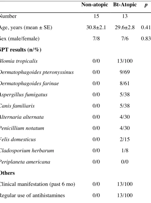
Table S1: Characteristics of the adults included in the study
Non-atopic Bt-Atopic p
Number 15 13
Age, years (mean ± SE) 30.8±2.1 29.6±2.8 0.41
Sex (male/female) 7/8 7/6 0.83 SPT results (n/%) Blomia tropicalis 0/0 13/100 Dermatophagoides pteronyssinus 0/0 9/69 Dermatophagoides farinae 0/0 8/61 Aspergillus fumigatus 0/0 5/38 Canis familiaris 0/0 5/38 Alternaria alternata 0/0 4/30 Penicillium notatum 0/0 4/30 Felis domesticus 0/0 2/15 Cladosporium herbarum 0/0 1/8 Periplaneta americana 0/0 0/0 Others
Clinical manifestation (past 6 mo) 0/0 13/100 Regular use of antihistamines 0/0 13/100 SPT - Skin prick test; mo - months
1
Augustin Belkadi, MS a,*, Céline Dietrich, MS a,*, François Machavoine, MSca,
3
Jefferson Russo Victor, PhDb,c,d and Maria Leite-de-Moraes, PhDa
4 5 On-line repository 6 7 8 METHODS 9
Mice and reagents
10
Eight to ten-week-old specific pathogen-free C57BL/6J, IL-4-/- and TCRd-/- mice were bred in
11
our facility. All animal experiments were carried out according to the guidelines for care and
12
use of animals approved by the French Institutional Committee
(APAFIS#4105-13
201511171831592).
14 15
Airway allergen sensitization and challenge model.
16
To induce allergic airway inflammation, we adapted a previously described protocol (1). In
17
brief, mice were sensitized and challenged by intranasal administrations of B. tropicalis
18
extracts (Greer laboratories, USA) at day 0 (D0) with 100µg/mouse then at D7, D9, D11, D14
19
and D16 with 50µg/mouse.
20
Twenty-four hours after the last challenge, mice were anesthetized with a mixture of
21
ketamine (150 mg/kg) and xylasine (400 µg/kg) and their tracheas were cannulated
22
(tracheostomy with ligation). FlexiVent apparatus (SCIREQ) was used to access
airway-23
specific resistance (Rn, tidal volume of 10 ml/kg at a respiratory rate of 150 breath/min in
24
response to increasing doses of aerosolized acetyl-b-methylcholine chloride (methacholine;
25
Sigma-Aldrich). Assessments were performed at least three times and the maximum R value
26
obtained after each dose of methacholine was used for the measure.
27
Airway inflammation was assessed in cytospin preparations of cells in
28
bronchoalveolar lavage fluid (BALF, 3 x 0.5 mL washes with PBS) that were stained with
29
May-Grünwald/Giemsa (Merck). For some experiments, BALF cells were also analysed by
30 flow cytometry. 31 32 Adoptive transfer 33
In some experiments, 25 x 103 electronically sorted Vg1+ or Vg1- gd T cells were adoptively
34
transferred intravenously (i.v.) to B. tropicalis immunized mice 1h before the first challenge
35
(D7). Mice were then further challenged at at D7, D9, D11, D14 and D15 with 50µg/mouse,
36
as described above.
37 38
Leucocytes from lung tissues
39
Lung tissues were cut into pieces using a GentleMACS Dissociator (Miltenyi Biotec) and
40
treated with collagenase type 4 (Thermo Fischer Scientific) plus DNAse I (Roche). The
41
lymphocyte-enriched fraction was collected at the 35-70% interface of Percoll gradients (GE
42
Healthcare). Cells were immediately stained or stimulated for 4h with 10−8 M PMA and 1 μg
43
ml-1 ionomycin, in the presence of 10 μg ml-1 brefeldin A (all from Sigma-Aldrich).
44 45
Immunofluorescence.
46
Murine cells were incubated with CD1d-PBS57-APC tetramers, anti-TCRb-APCeFluor780,
47
anti-Vg1.1-PE and anti-TCRgd-BV605 (Biolegend). Fixable viability dye was used to exclude
48
dead cells (ThermoFischer Scientific). For intracellular staining, cells were further fixed with
49
4% PFA, washed, and permeabilized with 0.5% saponin (Sigma-Aldrich), and then incubated
2
with anti-IL-17-PerCPCy5.5, anti-IL-4-PE-Cy7, anti-IFN-g-V450, anti-anti-IL-13-PE-eF610
51
or isotype control (eBiosciences). The cells were washed and fluorescence was detected using
52
a LSRFortessa (Becton Dickinson) and further analysed using the FlowJo 10.4.1 software
53 (Tree Star). 54 55 gd T cell sorting. 56
Splenocytes from WT or IL-4-/- mice, isolated as previously described (2, 3), were labelled
57
with anti-Vg1-PE and anti-TCRgd-BV605, as described above, and then sorted using a
58
FACSAria cell sorter (Becton Dickinson).
59 60
mRNA expression.
61
RNAs were extracted using the RNeasy Plus Minikit (Qiagen) including a DNase treatment.
62
Then RNA was reverse transcribed using the High Capacity RNA-to-cDNA Kit
63
(ThermoFisher Scientific), according to the manufacturer’s instructions. Primers and probes
64
for real-time PCR were provided by ThermoFisher Scientific under references: Mucin 5b
65
(Muc5b): Mm00466391_m1 and HPRT: Mm01545399_m1. All reactions were performed in
66
triplicate with TaqMan® Fast Advanced Master Mix according to the supplier’s instructions
67
for a Step One Plus apparatus (ThermoFisher Scientific). All data were normalized to the
68
internal standard, namely HPRT expression in each sample, and expressed as relative
69
expression using the DDCt method versus the reference sample.
70 71
Histology of the airways.
72
Lungs were fixed with 10% formalin via the trachea, removed and stored in 10% formalin.
73
Lung tissues were embedded into paraffin and 3µm sections were stained with periodic acid
74 Schiff (PAS). 75 76 Patient samples 77
Blood samples were collected from subjects who were previously clinically classified as
78
atopic or non-atopic individuals and voluntarily subjected to a skin prick test (SPT) to
79
confirm the atopic state. These individuals were classified into two groups: as Bt- atopic
80
individuals (clinically allergic and reactive to Blomia tropicalis extract, with co-reaction to at
81
least one allergen), and non-atopic individuals (without any clinical allergy symptoms and not
82
reactive to any tested allergen). Additional information about these individuals is shown in
83
table 1.
84
Each sample of peripheral blood mononuclear cells (PBMCs) was provided from a
85
different donor. The ethics committees at the HCor and the School of Medicine at the
86
University of São Paulo approved this study (CAAE: 15507613.4.0000.0060).
87 88
Skin prick test (SPT) and blood sample collection
89
The SPT were performed in accordance with European standards (4) with an adapted panel of
90
allergens that included the profile of Brazilian allergens (i.e., Blomia tropicalis, Canis
91
familiaris, Periplaneta americana, Aspergillus fumigatus, Penicillium notatum, Alternaria
92
alternata, Cladosporium herbarum, Dermatophagoides pteronyssinus, Dermatophagoides
93
farinae, and Felis domesticus - IPI ASAC), as previously described (5).
94
Briefly, one drop of each allergen extract or controls was applied to the volar forearm
95
and a superficial skin puncture was made using a hypodermic needle (Alko, Brazil). After 15
96
minutes, the results were considered positive when wheals measure reach a diameter of 3 mm
97
greater than that of the negative control. We excluded patients who used antihistamines,
98
glucocorticosteroids or certain other systemic drugs that can influence the SPT results within
99
15 days before the test or with severe eczema or dermographism.
100 101
3
cytokine production were performed as previously described (6, 7). Briefly, PBMC
104
separations were performed using Ficoll-Paque Plus (GE Healthcare) after centrifugation and
105
suspensions of PBMCs were phenotypic evaluated ex vivo or cultured to intracellular cytokine
106
production evaluation. PBMCs were stained with mouse anti-human gdTCR-FITC,
CD161-107
PECy5 or isotype control antibodies (BD Pharmingen).
108
To evaluate intracellular cytokine production, PBMCs were cultured in RPMI 1640
109
medium containing 10% FBS and 1 µg/mL of Brefeldin A (Sigma-Aldrich) for 24 hours
110
without additional stimulus. Cultured PBMCs were then washed, fixed with formaldehyde
111
and stained with mouse anti-human gdTCR-FITC, IL-17A-Alexa700, IL-4-PECy7,
IFN-g-112
HorizonV450 or isotype control antibodies (BD Pharmingen) in 100 µL of PBS containing
113
0.05% saponin. Thirty minutes later, PBMCs were washed, fixed, acquired using a
114
LSRFortessa cytometer (BD Biosciences, USA), and analysis was performed using FlowJo
115
software 10.1 (Tree Star).
116 117
Statistics
118
Data are expressed as means ± SEM. The AHR values were analysed with repeated- measures
119
2-way ANOVA followed by Bonferroni correction as a post-hoc test. All other values were
120
analysed with Mann-Whitney U test. Results were considered significant at a P value of 0.05
121
or less (*p<0.05; **p<0.01; ***p<0.001). Data were analyzed using GraphPad Prism version
122 6 (GraphPad Software). 123 124 125 LEGENDS 126 127
Figure S1: IL-4-producing cells were required for asthma symptoms induced by B. tropicalis.
128
(A) The number of total cells (Total), monocyte (Mono), eosinophil (Eosino), neutrophil 129
(Neutro) and lymphocyte (Lympho) were determined in BALF of wild-type (WT) or IL-4
-/-130
asthmatic (B. tropicalis) mice. (B) Lung resistance (Rtr) was measured 24h after the last B. 131
tropicalis challenge or controls (NaCl) (n=7 to 15).
132 133
Figure S2: iNKT cells were not required for asthma symptoms induced by B. tropicalis. (A)
134
A representative Facs profile showing the gating strategy used to identify iNKT (CD1d
135
tetramer+TCRb+) cells and the percentage of IFNg, IL-4, IL-13 and IL-17A positive cells, and
136
their respective isotype controls, among gated iNKT cells in the lungs of WT asthmatic (B. 137
tropicalis) or non-asthmatic (NaCl) mice.
138
(B) The number of total cells (Total), monocyte (Mono), eosinophil (Eosino), neutrophil 139
(Neutro) and lymphocyte (Lympho) were determined in BALF of wild-type (WT) or iNKT 140
(Ja18-/-)-deficient asthmatic (B. tropicalis) mice. (n=5 to 7).
141 142
Figure S3: gd T cells and the Vg1+ gd T cell subset were enhanced in the lung of B. tropicalis
143
asthmatic mice. (A) A representative Facs profile showing the gating strategy used to identify
144
TCRgd+ and Vg1+ gd T cells. (B) The number of total gd T cells and of the Vg1+ gd T cell
145
subset were determined in the lungs of wild-type asthmatic (B. tropicalis) or non-asthmatic 146
(NaCl) mice (n=5 to 10). 147
148
Figure S4: Impaired seric IgE and mucus production in the lung of TCRd-/- B. tropicalis
149
asthmatic mice. (A) Total IgE levels in the serum of WT and TCRd-/- mice following B.
150
tropicalis or NaCl administration (B) Mucin-5 mRNA expression assessed by quantitative
151
RT-PCR in the lung of WT and TCRd-/- mice following B. tropicalis or NaCl administration.
4 Data are representative of two experiments. (C) Representative PAS-stained lung histology 153
sections of controls (NaCl) and B. tropicalis-sensitized and -challenged WT and TCRd-/- mice.
154
Following B. tropicalis administration, the airways of WT mice contained more PAS-stained 155
mucus-producing cells than TCRd-/- mice.
156 157
Figure S5. Distinct cytokine production by lung Vg1+ and Vg1- gd T cell subsets. (A) Flow
158
cytometric gating strategy used to identify TCRd+Vg1+ and TCRd+Vg1- gd T cell subsets and
159
the frequency of these cells in the lung of B. tropicalis- versus NaCl-treated mice. (B, C) A
160
representative Facs profile showing the percentage of IFNg, IL-4, IL-13 and IL-17A positive
161
cells among gated TCRd+Vg1+ (B) and TCRd+Vg1- (C) gd T cell subsets and their frequency
162
in the lungs of WT asthmatic (B. tropicalis) or non-asthmatic (NaCl) mice. * p< 0.05. (D) 163
Facs profile represents the respective IL-13 versus IL-4 and IL-17A versus IFN-g isotype 164
controls. 165
166
Figure S6. gd T cells were activated in B. tropicalis atopic patients. (A) Gate strategy to 167
obtain viable lymphocytes. (B) A representative Facs profile showing the gating strategy used 168
to identify TCRgd+ cells and the percentage of IFNg, IL-4, IL-13 and IL-17A positive cells
169
among gated TCRgd+ cells.
170 171
REFERENCES
172 173
1. Zhou Q, Ho AW, Schlitzer A, Tang Y, Wong KH, Wong FH, et al. GM-CSF-licensed CD11b+ lung 174
dendritic cells orchestrate Th2 immunity to Blomia tropicalis. J Immunol. 2014;193:496-509. 175
2. Massot B, Michel ML, Diem S, Ohnmacht C, Latour S, Dy M, et al. TLR-induced cytokines promote 176
effective proinflammatory natural Th17 cell responses. J Immunol. 2014;192:5635-5642. 177
3. Michel ML, Lenoir C, Massot B, Diem S, Pasquier B, Sawa S, et al. SLAM-associated protein favors 178
the development of iNKT2 over iNKT17 cells. Eur J Immunol. 2016;46:2162-2174. 179
4. Heinzerling L, Mari A, Bergmann KC, Bresciani M, Burbach G, Darsow U, et al. The skin prick test-180
European standards. Clin Transl Allergy. 2013;3:3. 181
5. Sgnotto FDR, Oliveira MG, Lira AAL, Bento-de-Souza L, Duarte AJDS, Victor JR. Low doses of IgG 182
from atopic individuals can modulate in vitro IFN-γ production by human intra-thymic TCD4 and TCD8 cells: 183
an IVIg comparative approach. Hum Vaccin Immunother. 2017:0. 184
6. de Oliveira MG, Oliveira LM, Lira AAL, Sgnotto FDR, Duarte AJDS, Sato MN, et al. Preconception 185
allergen sensitization can induce B10 cells in offspring: a potential main role for maternal IgG. Allergy Asthma 186
Clin Immunol. 2017;13:22.
187
7. Sgnotto FDR, de Oliveira MG, Lira AAL, Inoue AHS, Titz TO, Orfali RL, et al. IgG from atopic 188
dermatitis patients induces IL-17 and IL-10 production in infant intrathymic TCD4 and TCD8 cells. Int J 189
Dermatol. 2018;57:434-440.
190 191