HAL Id: hal-01915442
https://hal-amu.archives-ouvertes.fr/hal-01915442
Submitted on 12 Nov 2018HAL is a multi-disciplinary open access archive for the deposit and dissemination of sci-entific research documents, whether they are pub-lished or not. The documents may come from teaching and research institutions in France or abroad, or from public or private research centers.
L’archive ouverte pluridisciplinaire HAL, est destinée au dépôt et à la diffusion de documents scientifiques de niveau recherche, publiés ou non, émanant des établissements d’enseignement et de recherche français ou étrangers, des laboratoires publics ou privés.
The ErpA/NfuA complex builds an oxidation-resistant
Fe-S cluster delivery pathway
Beatrice Py, Catherine Gerez, Allison Huguenot, Claude Vidaud, Marc
Fontecave, Sandrine Ollagnier de Choudens, Frédéric Barras
To cite this version:
Beatrice Py, Catherine Gerez, Allison Huguenot, Claude Vidaud, Marc Fontecave, et al.. The ErpA/NfuA complex builds an oxidation-resistant Fe-S cluster delivery pathway. Journal of Bio-logical Chemistry, American Society for Biochemistry and Molecular Biology, 2018, 293 (20), pp.7689 - 7702. �10.1074/jbc.RA118.002160�. �hal-01915442�
The ErpA/NfuA complex builds an oxidative resistant Fe-S cluster delivery pathway
Béatrice Py1,2,3*, Catherine Gerez4,5,6, Allison Huguenot1,2,3, Claude Vidaud7, Marc
Fontecave8, Sandrine Ollagnier de Choudens4,5,6, Frédéric Barras1,2,3*
From the 1 Institut de Microbiologie de la Méditerranée, Marseille, France; 2 CNRS UMR 7283, LCB, 31 Chemin Joseph Aiguier, 13009 Marseille, France; 3 Aix-Marseille Université, Marseille, France; 4 Université Grenoble Alpes, Grenoble, France; 5 CNRS UMR 5249, LCBM, Grenoble, France; 6 CEA/DRF/BIG/CBM/BioCat Grenoble, France; 7 CEA/DRF/BIAM/ Marcoule, France; 8 Laboratoire de Chimie des Processus Biologiques, UMR 8229 CNRS, UPMC Univ Paris 06, Collège de France, PSL Research University, Paris, France. Running title:
Biochemical properties of E. coli Fe-S cluster carriers
* To whom correspondence should be addressed: Béatrice Py and Frédéric Barras: Institut de Microbiologie de la Méditerranée, CNRS UMR 7283, LCB, 31 Chemin Joseph Aiguier, 13009 Marseille, France; [email protected] [email protected] Key words: iron-sulfur protein, Escherichia coli, oxidative stress, protein-protein interaction, metal ion-protein interaction _______________________________________________________________________________________________________________ ABSTRACT
Fe-S cluster containing proteins occur in most organisms wherein they assist a myriad of diverse processes from metabolism to DNA repair via gene expression and bioenergetic processes. Here we used both in vitro and in vivo methods to investigate capacity of the four Fe-S carriers, NfuA, SufA, ErpA and IscA to fulfill their targeting role under oxidative stress. Likewise, Fe-S clusters exhibited varying half-live depending on the carriers they are bound to: NfuA-bound FeS cluster was more stable (t1/2 100 min) than SufA- (t1/2 55 min), ErpA (t1/2 54 min), and IscA (t1/2 45 min). Surprisingly, presence of NfuA further enhanced stability of the ErpA-bound cluster to t1/2 100 min. Using genetic and plasmon surface resonance analyses, we showed that NfuA and ErpA interacted directly with client proteins whereas IscA/SufA did not. Moreover, NfuA and ErpA interacted one with the other. Given all these observations we propose an architecture of the the Fe-S delivery network in which ErpA is the last factor that delivers cluster directly to most if not all client proteins. NfuA is proposed to assist
ErpA under severe unfavorable conditions. Comparison with the strategy employed in yeast and eukaryotes is discussed.
___________________________________________________
Fe-S clusters rank among the oldest cofactors, and they are surmised to have been key factors for life origin and evolution (1, 2). The first Fe-S proteins identified, by their electron paramagnetic signature, were those of mitochondrial respiratory complexes (3). In the following decades biophysical and structural approaches revealed the great variety of Fe-S cluster proteins, while genomic analyses showed their widespread adoption in living organisms (4). Nowadays, in organisms Fe-S cluster proteins participate to respiration, biosynthesis of cell building blocks and cofactors, central metabolism, gene regulation, tRNA modification, DNA synthesis and repair (5).
In vitro studies revealed that, in
favorable conditions, Fe-S clusters can form spontaneously on apo-proteins clients (6, 7). However, in the 90’s, studies initiated on nitrogenase maturation revealed that in vivo Fe-S cluster assembly requires multiproteins systems : the NIF system
dedicated to nitrogenase maturation, and two general systems, ISC and SUF, which maturate most, if not all, cellular Fe-S proteins (8–10). Components of the ISC and SUF systems are conserved throughout eukaryotes and prokaryotes. In eukaryotes, the ISC and SUF Fe-S machineries are located in mitochondria and chloroplasts, respectively (11, 12).
Fe-S cluster biogenesis proceeds in two steps, assembly and delivery. The former takes place on a scaffold protein, which allows sulfur and iron to meet and combine in a Fe-S cluster. Sulfur is produced via the catalytic degradation of L-cysteine by cysteine desulfurase, whereas the molecular source of iron
remains elusive.
The delivery relies on a series of Fe-S carriers that transport scaffold-bound clusters to the apo-clients. Numerous Fe-S carriers have been identified in both prokaryotes and eukaryotes. These include so-called A-type carriers (ATC), named IscA, SufA and ErpA in prokaryotes and ISA1, ISA2 in eukaryotes (13–17). Others carriers include the P-loop NTPases, (Ind1 in mitochondria, ApbC in Salmonella) (18–21), the monothiol glutaredoxins (Grx 5 in yeast and GrxD in E. coli) (22–24) and the highly conserved NFU-type proteins that have been shown to interact with target proteins (25–33).Understanding how Fe-S clusters are delivered to the large number of functionally and structurally diverse Fe-S cluster proteins is a challenging task. Indeed, in most organisms, clients are actually a set of structurally and functionally diverse protein species and it is difficult to think of a common maturation pathway shared by all of them. Moreover, Fe-S clusters are highly sensitive to environmental changes and, in particular, can be destabilized by reactive oxygen species (ROS) depending upon their solvent accessibility on the host protein (34–36). Last, Fe-S clusters arise under different structure, i.e. 2Fe-2S, 3Fe-4S, 4Fe-4S or in more complex association with other metal and cofactors (1). How organisms keep on inserting clusters in such a great diversity of substrates and conditions and whether all Fe-S proteins get their clusters delivered by the same set of factors is a daunting issue.
Multiple parameters can be foreseen as controlling the delivery process: (i) genetically controlled level of delivery factors available in a given condition, (ii) affinity between delivery factors and apo-targets, and (iii) intrinsic biochemical features of delivery factors. Our previous study had illustrated how genetic regulation orchestrated the choice of routes Fe-S clusters take to reach essential enzymes, IspG/H, and the two transcriptional factors, IscR and NsrR, throughout fluctuating conditions (16, 37). The present study provides key findings on the two other parameters and provides us with an unprecedented view of Fe-S cluster trafficking and delivery.
We investigated the intrinsic capacity of IscA, SufA, ErpA and NfuA, to stabilize bound Fe-S cluster exposed to aerobic conditions. We proceeded by analyzing the contribution of each of them into the maturation of a series of Fe-S enzymes, including IspG and IspH, two essential Fe-S enzymes involved in the production of isoprenoids (38, 39). This provided us for the first time with a direct ranking of the Fe-S cluster carriers according to their intrinsic capacity to resist ROS potential damages. Another key finding was the observation of a privileged partnership between ErpA and NfuA, providing the cell with a new type of “hybrid” carrier, the Fe-S cluster of which exhibited the highest level of resistance to ROS. Altogether this allowed us to propose a model of the Fe-S cluster delivery network that predict ErpA to be in charge of the maturation of most if not all Fe-S enzymes. Its closer association with NfuA appears as a new strategy for the cell to meet with transiently occurring destabilizing conditions. Results NfuA is required for IspG/H maturation under oxidative stress Earlier, we found that growth of the
E. coli ΔnfuA mutant on rich medium is
severely affected in the presence of paraquat (PQ), a superoxide radical generator (25). Our previous analysis informed us that such a phenotype is likely due to an insufficient amount of
isopentenylphosphate IPP, due to a poor maturation of IspG and IspH, two Fe-S cluster containing enzymes (17). Therefore we tested whether the NfuA protein contributes to the Fe-S cluster delivery route towards maturation of IspG/H. For this, we introduced the eukaryotic IPP biosynthesis (FeS)independent pathway -referred as the mevalonate pathway (MVA)- in the ΔnfuA mutant and found that ectopic expression of this pathway was sufficient to rescue viability of the nfuA mutant (Fig. 1). This result indicated that NfuA is required for Fe-S cluster delivery to IspG/H under oxidative stress.
NfuA interacts with ErpA
To know if NfuA cooperates with the ATC, we tested the interaction of NfuA with the ATC using the bacterial two-hybrid system. NfuA/ErpA interaction was observed as the BTH101 cells synthesizing the T18-NfuA and T25-ErpA hybrid proteins (pT18-NfuA, pT25-ErpA) exhibited β-galactosidase activity (Fig. 2A). In contrast, no interaction was indicated between NfuA and IscA or between NfuA and SufA using this assay (Fig. 2A). ErpA/NfuA interaction was also tested by surface plasmon resonance (SPR) using apo-proteins. Purified NfuA was immobilized onto a Biacore sensor CM5 chip and ErpA was serially diluted and injected. The calculated dissociation constant (Kd) of the ErpA/NfuA interaction was determined to be 48.5 +/-0.5
µM (Fig. 2B). In contrast, no interaction was observed between NfuA and SufA or IscA (data not shown). Taken together these analyses revealed that NfuA and ErpA physically interact with each other, and suggest they might partner to form a discrete path within the Fe-S cluster delivery network. Unidirectional Fe-S cluster transfer from NfuA to ErpA To test if Fe-S cluster transfer occurs between ErpA and NfuA, an in vitro test was used. Holo-NfuA contains a 4Fe-4S per dimer and ErpA is able to bind 2Fe-2S cluster per monomer (17, 25, 26, 40). His-tagged holo-NfuA (1.2 +/- 0.1 Fe and 1 +/- 0.2 S/monomer; inset Fig. 3A) was incubated, under anaerobic conditions, with one
equivalent of untagged apo-ErpA, for 1 hour. After separation by chromatography onto a Ni-NTA column, iron and sulfide contents of each protein were analyzed. ErpA contained 1 +/- 0.1 Fe and 0.7 +/- 0.2 S/monomer and its visible spectrum was characteristic of a 2Fe-2S cluster protein with absorption bands at 420 nm and 320 nm. On the contrary, NfuA had lost its 420 nm Fe-S cluster absorption band and contained less than 0.1 Fe and S/monomer (Fig. 3A). In a separate experiment, holo-ErpA was incubated with apo-NfuA, and the two entities submitted to same analyses as described above after separation. No modification either in iron/sulfide content or in spectrum was observed before and after co-incubation (Fig. 3B). Together, these analyses showed that, in vitro, an unidirectional Fe-S cluster transfer occurs from NfuA to ErpA.
Stability of Fe-S cluster to oxidative damages varies with carrier identity
In vivo studies showed that ErpA and
NfuA are required for Fe-S cluster transfer under aerobiosis and oxidative stress, respectively. Therefore, we tested the stability of Fe-S clusters bound to NfuA and to ErpA when exposed to O2. Before exposure to O2, reconstituted holo-NfuA contained 1.4 +/- 0.2 Fe molecules per monomer and displayed a 4Fe-4S cluster-characteristic UV–visible spectrum in agreement with published data (40) (Fig. 4A). The degradation of the Fe-S cluster was monitored by the absorbance variation at 420nm as a function of time when diluted into oxygenated buffer containing controlled amount of O2. Under these conditions the half-life of the NfuA Fe-S cluster was 100 +/-10 minutes (Fig. 4A). Holo-ErpA exhibited a 2Fe-2S cluster-characteristic UV–visible spectrum (Fig. 4B). Upon controlled O2 exposure, half-life of its cluster was 54 +/-3 minutes, (Fig. 4B). We also tested the stability of the Fe-S cluster of two other ATC, IscA and SufA under the same experimental conditions, and found half-lives of 45 +/-4 and 55 +/-7 minutes, respectively (Fig. 4C). All together, these results showed that Fe-S cluster bound to diverse carriers exhibit different capacities to resist oxidative damages with the
decreasing stability order NfuA > ErpA = SufA > IscA.
The NfuA/ErpA-bound cluster has an increased stability
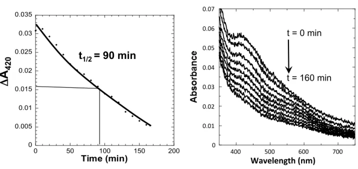
Last, we tested whether stability of the ErpA bound Fe-S cluster was modified in the presence of NfuA. Both apo-NfuA and holo-ErpA proteins were mixed and exposed to O2, as above. Half-life of the Fe-S cluster bound to ErpA reached 90 +/-6 minutes, that is, an enhancement by two-fold as compared with Fe-S bound to ErpA alone (Fig. 5). Thus, our results indicated that the NfuA somehow allowed ErpA bound Fe-S cluster to enhance its stability towards O2.
NfuA and ErpA interact directly with the client proteins IspG and IspH
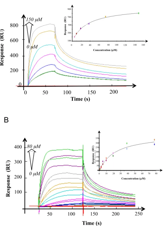
SPR was used to test the interaction between IspG/H and the Fe-S carriers. For the experiments performed with IspG, serially diluted IspG was injected into sensor chips coated with NfuA, or with ErpA. The sensorgrams showed binding of IspG to both NfuA and ErpA (Fig. 6). Global evaluation using the 1:1 Langmuir binding model yielded Kd values of 22.5 +/- 0.5 µM
for the IspG/ErpA interaction and of 30 +/- 2
µM for the IspG/NfuA interaction.
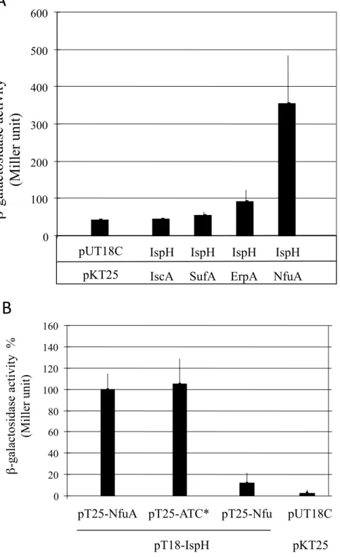
For the experiments performed with IspH, serially diluted NfuA or ErpA were injected into IspH coated sensor chips. Even though sensorgrams showed binding of NfuA and ErpA to IspH, the maximum response (Rmax), was unattainable preventing determination of a Kd value (data not shown). We also used bacterial two-hybrid approach in order to detect this interaction between NfuA, ErpA and IspH We showed that the BTH101 cells synthesizing T18-IspH and T25-NfuA hybrid proteins exhibited β-galactosidase activity, indicating an IspH/NfuA interaction (Fig. 7A). In addition, the two-hybrid system allowed us to show that the N-terminal domain of NfuA was sufficient to mediate the interaction with IspH (Fig. 7B). An interaction, albeit weak, was also observed between IspH and ErpA (Fig. 7A). In contrast, no interaction was observed between IspH and SufA or IscA (Fig. 7A).
Collectively, these results indicated that NfuA and ErpA are able to interact directly with the client IspG and IspH proteins.
Both ErpA and NfuA are required for the maturation of aconitase B and the respiratory complexes I and II
Our previous study revealed a role of NfuA in the maturation of AcnB and the respiratory complex I (Fig. S1) (40). We then asked whether ErpA participates to the maturation of these Fe-S cluster-containing proteins.
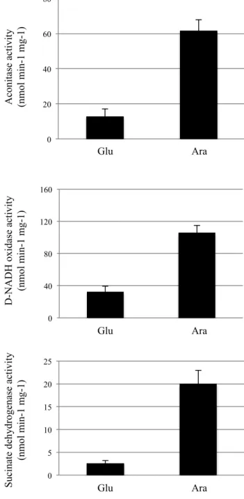
First, we assayed the AcnB activity in the conditional mutant in which the expression of the erpA gene was under arabinose induction and glucose repression referred to as LL401 (arap::erpA), in which the ΔacnA mutation has been introduced (BP721) (17). ErpA depleted cells were obtained after 2 hours growth in glucose-supplemented medium (Fig. 8). In the ErpA depleted cells, aconitase activity was decreased by 80% when compared to the ErpA replete cells grown in the presence of arabinose, while AcnB was detected at an identical level in both ErpArepleted and -depleted cells (Fig. 8). The NfuA protein level was slightly increased in ErpA-depleted cells (Fig. S2). SPR was used to investigate the interaction between ErpA and AcnB. Serially diluted ErpA (0-270 µM), was injected into sensor chips coated with AcnB (Fig. S3A). The Kd value for the ErpA/AcnB interaction was 50 +/-5 µM. We also used SPR experiments to quantify the previously shown NfuA-AcnB interaction (40). Serially diluted NfuA (0-160 µM) injected into sensor chips coated with AcnB yielded to a Kd value of 29 +/- 3 µM (Sup data, Fig. S3B).
We then tested whether ErpA was also required for the maturation of complex I. ErpA depleted cells of the LL401 strain exhibited a drastic decrease (70%) for complex I activity when compared to ErpA replete cells grown in presence of arabinose (Fig. 8).
Last, we tested the contribution of ErpA and NfuA to the maturation of the respiratory complex II (Sdh). When grown in glucose, the Sdh activity of the LL401 strain was found to be drastically reduced
(85%) (Fig. 8). In the nfuA mutant, the Sdh activity was decreased by 35% (Fig. 9).
Collectively, all these data indicate that maturation of the respiratory complexes I and II are under the assistance of both ErpA and NfuA under aerobic conditions, and that maturation of AcnB requires ErpA under aerobic conditions, and NfuA under oxidative stress conditions.
ErpA interacts with IscA and SufA
Using the Biacore experiment, we tested whether ErpA can physically interact with the ATC of the ISC and SUF machineries, IscA and SufA, respectively. ErpA was immobilized onto a Biacore sensor CM5 chip and IscA and SufA were serially diluted and injected. A dose-response curve was obtained where an increase in response units was observed with increasing concentrations of IscA (0-200 µM) and SufA (0-600 µM), indicating that ErpA interacted with IscA and SufA (Fig. 10). The sensorgrams indicated that IscA and SufA were released in a short time without the necessity of chip regeneration. The calculated dissociation constant (Kd) of the ErpA/IscA and ErpA/SufA interactions was determined to be 40 +/- 3 µM and 90 µM+/- 2 µM respectively. These results indicated that ErpA is able to interact with IscA and SufA.
Multicopy suppression of the nfuA growth defect by erpA
Results above evidenced NfuA and ErpA contribute to the maturation of the same set of enzymes. Therefore, we tested whether NfuA and ErpA are functionally redundant in vivo. For this, we made use of a multicopy-based suppression approach. A pBAD derivative plasmid expressing erpA, (pBAD-erpA), suppressed paraquat sensitivity of the nfuA mutant (Fig. 11). In contrast, increased nfuA gene dosage failed to suppress non-viability caused by erpA mutation under aerobiosis (Fig. 12). Last, pBAD-iscA and pBAD-sufA, plasmids expressing iscA and sufA, respectively, failed to suppress ΔnfuA mutant sensitivity to paraquat (Fig. 11). Altogether, these data showed that ErpA overproduction overcomes the need of NfuA for IspG/H maturation but that the converse is not true.
Discussion
Interest in Fe-S cluster biology keeps expanding as one realizes how much these cofactors are central to multiple issues, from basic knowledge to molecular medicine, antibiotic resistance and biotechnological applications (41, 42). In most organisms several dozens of structurally and functionally diverse client protein species need to acquire a Fe-S cluster for functioning. The diversity of clients, the diversity of the clusters and the fluctuating environmental conditions under which Fe-S cluster trafficking takes place make the question of the delivery a most challenging issue. Here we show that synthesizing different carriers can afford different ROS-resistance environment for transported Fe-S clusters, hence delivery circuits better adapted to sustain oxidative stress. We show that enhanced resistance to ROS can also be achieved by interaction between Fe-S carriers. We propose a model in which, right after the assembly step, Fe-S trafficking network diversifies in multiple branches, which eventually converge towards ErpA. This unprecedented view of Fe-S cluster trafficking and delivery is discussed in comparison with the eukaryotic situation.
The present study allowed us to quantitate ROS resistance capacity of a cluster bound to NfuA, ErpA, SufA and IscA carriers. Likewise, NfuA and SufA-carried clusters showed the highest resistance to ROS. This is fully consistent with the genetically based view of SufA and NfuA being stress responding factors, and predicted to be better adapted to deliver clusters under oxidative stress conditions (16, 25, 26). NfuA results from the fusion between a NFU domain, at the C-terminal region, and, at the N-terminal region, a degenerated ATC domain lacking the Fe-S cluster liganding cysteine residues (25, 26, 40). NfuA forms a dimer and binds a 4Fe-4S cluster in its NFU domain, likely at the dimer interface (25, 26, 40). Then, it is possible that the cluster stability of NfuA is related to the four cysteinyl ligations and/or shielding by the dimer formation.
An unsuspected association between NfuA and ErpA was observed both by SPR and two-hybrid based methods. This
association resulted in an enhanced stability of the ErpA bound cluster as compared when bound to ErpA alone. Next biochemical and structural studies will aim at solving the molecular basis for the enhanced ROS resistance procured by the NfuA-ErpA interaction.
Our previous phylogenetic studies permitted us to classify ATC within two different families. ErpA and IscA/SufA were classified into ATC-I and –II families, respectively (16). Members of the ATC-I family were predicted to partnership with the apo-targets, whereas the ATC-II members were thought to be connected to scaffolds (16). The present bacterial two-hybrid and SPR-based investigations fully support the phylogenetically-based functional prediction. Direct interactions were observed between apo-targets (IspG/H and AcnB) and ErpA, but not IscA and/or SufA (data not shown). Taken together with our previous genetic analysis, this observation comforts the view that scaffold-bound Fe-S clusters are transferred to ATC-II, IscA or SufA, which transfer them to ErpA, which delivers them to the apo-targets. In mitochondria, once assembled by the core biogenesis machinery, 2Fe-2S clusters reach a heteromeric platform, the ISCA1-ISCA2-Iba57 complex, which converts them into 4Fe-4S before to target them to a so-called dedicated factors, which in turn allow maturation of cellular Fe-S proteins (15, 43–45). Mammalian ISCA1/2 proteins contain 2Fe-2S clusters. Heterodimer ISCA1-ISCA2 can assemble 4Fe-4S cluster in the presence of GRX5 implying that the heterodimeric complex is the functional unit for 4Fe-4S cluster formation before their transfer to specific targets (45). ISCA1 is an ATC-II, like IscA and SufA, while ISCA2 is an ATC-I, like ErpA. In essence therefore, the complex ISCA1-ISCA2 resembles the partnership IscA/ErpA or SufA/ErpA discussed above. The difference though is that no stable bacterial ATCII-ATCI complex has ever been isolated and two-hybrid based methods failed to reveal any interaction. Note however that in mice, despite forming a complex, ISCA1 and ISCA2 are able to carry out separate tasks, presumably depending upon the substrates and the conditions (46).
Both NfuA and ErpA exhibit related affinity for Fe-S client proteins; the Kd value of the ErpA/IspG (23 µM) and NfuA/IspG (28 µM) interactions are within a similar range like were those for the interaction between AcnB and ErpA (50 µM) or NfuA (29 µM). A Kd value of 44 µM was reported for the interaction of NfuA with MiaB, involved in tRNA modification (32). Thus, like ErpA, NfuA appears to have all features required to interact directly with targets, and could be positioned at the ultimate step within the Fe-S cluster delivery process. If direct transfer between NfuA and target proteins can occur in vivo, its contribution appears very modest regarding the contribution of ErpA. Hence, lack or depletion of ErpA caused a stronger phenotype than the lack of NfuA. The erpA gene is crucial for both anaerobic and aerobic respiration as shown here by a drop in complexes I and II activities by over 70% and 85%, whereas, a nfuA mutant exhibited a less than 2-fold reduction in Complex I and Complex II activities. Similarly, aconitase B activity was down by over 80% in ErpA-depleted cells whereas it was only slightly altered under stress conditions in cells lacking NfuA. Also, erpA was able to act as multicopy suppressors of nfuA, while the reverse was not true. We thus favor the hypothesis that in E. coli, NfuA act conjointly with ErpA, by providing it a Fe-S cluster, rather being an ultimate Fe-S donor for target proteins (Fig. 13).
The yeast and human Nfu1 and the
E. coli NfuA proteins share a conserved
NFU-type C-terminal domain that binds a 4Fe-4S cluster. Interestingly, as NfuA, the yeast and human Nfu1 were found associated with the ISA proteins (33, 46). In yeast, defects exhibited by nfu1 mutation were of much lesser extent than those caused by mutations in the ISA complex (ISCA homologs in yeast) complex (13, 14, 30, 47). In fact, defects were mainly apparent under oxidative stress conditions (33, 47). Hence, the parallel with the E. coli situation is striking. Moreover, overexpression of Isa2, which we classified as an ATC-I member, was able to suppress defect of NFU1 mutant whereas overexpression of Isa1, an ATC-II member failed to do so (33). This is highly
reminiscent of what we observed here with
E. coli, wherein erpA, an ATC-I, but not iscA
or sufA, both ATC-II members, was able to act as multicopy suppressors of nfuA. This argues that in both prokaryotes and eukaryotes NFU-containing proteins are called upon in stress conditions at the delivery step, but that their contribution can be by-passed given that the level of ATC-II member (ISCA2 or ErpA) is sufficiently high, and that the proposed scenario of NFU proteins as a Fe-S refuel for ATC-I can also be envisioned at the late stage of the mitochondrial Fe-S delivery step.
In conclusion, independently of the organisms considered and of the type of Fe-S client proteins to be matured, a same overall strategy has been retained throughout evolution. Despite the multitude of apo-protein clients waiting for their clusters, cells evolved a hand-full of carriers, which they either synthesize at appropriate levels under a set of conditions, or combine in different higher-order organization, from homodimers to heterotetramers, each possibility providing a better solution to adapt to the diversity of client proteins and growth conditions. This study pinpoints how diversifying carriers, and combining different carriers, constitute two strategies the cell evolved to yield multiple delivery pathways in particular a robust ROS resistant delivery network.
Experimental procedures Strains and growth conditions
Strains used in this study are listed in Table 1. The ΔacnA::kan KEIO mutation was introduced by P1 transduction (48). E. coli strains were grown in Luria-Bertani (LB) rich medium at 37°C. Solid media contained 1.5% agar. Ampicillin was used at 50 μ g/ml. Arabinose (0.2%), and mevalonate (1 mM) were added when required.
Paraquat sensitivity Test
Paraquat sensitivity test was performed on overnight cultures that were diluted in sterile PBS and directly spotted onto LB plates containing 100µM paraquat (25). The plates were incubated overnight at 37°C before growth was scored. Plasmids construction Construction of plasmids pT18-NfuA, pT25-NfuA, pT25-ATC* was described previously. (40). Plasmids pT25-ErpA, pT25-IscA, pT25-SufA and pT18-IspH were constructed by, first, PCR amplification from the MG1655
chromosomal DNA using
2HErpAup/2HErpAdo, 2HIscAup/2HIscAdo
2HSufAup/2HSufAdo and
2HIspHup/2HIspHdo primers pairs. The
erpA-, ispH-containing PCR fragments were
digested by EcoRI PstI enzymes, and the
iscA-, sufA-containing PCR fragments were
digested by the PstI KpnI enzymes. All the constructs obtained by PCR-fragment cloning were checked by DNA sequencing.
Protein purification
Apo-NfuA, -ErpA, SufA, and -IscA, were obtained as previously described (25, 49). Aconitase B was purified as described for aconitase A (50). Purification of IspG-His was performed as described in (17) and IspH was a gift of M. Seeman (IC, Strasbourg).
Surface plasmon resonance analysis The binding studies were performed using Biacore™ T200 instrument and CM5 sensor chips. Proteins were covalently immobilized onto the chip using a standard primary amine coupling procedure. The same amine coupling procedure, but without any protein, was performed on the reference channel as a blank to subtract any non-specific binding signal. The analytes, dissolved in running Tris buffer (Tris 50 mM, NaCl 150 mM at pH 7,4), or HBS buffer (Hepes 10 mM, NaCl 150 mM, EDTA 3 mM, Tween 0,005% at pH 7,4) were serially diluted to various concentrations with the running buffer. According to their kinetics of association and dissociation, they were injected for 50 to 125 seconds during the association phase at a constant flow rate of 10 µl/min at 25°C. The dissociations were subsequently followed for 75 to 250 seconds at the same flow rate. For each cycle the sensor surface was regenerated with 10 mM Gly–HCl buffer (pH 2.0) when the data collection was finished. To calculate the rate and affinity constants the results from the sensograms were fit globally with BIAcore 3000 analysis software
(BIAevaluation Version 4.1) that propose 1:1 Langmuir binding modes.
Bacterial two-hybrid
We used the adenylate cyclase-based two-hybrid technique (51). DNA inserts encoding the proteins of interest were obtained by PCR and were cloned into pUT18C and pKT25 plasmids. After co-electroporation of the BTH101 strain with the two plasmids expressing the hybrid proteins, plates were incubated at 30°C for 2 days. Three milliliters of LB medium supplemented with antibiotics and IPTG at the recommended concentration (51) were inoculated and incubated overnight at 30°C. β-Galactosidase activity was determined as described by Miller (1992).
In vitro Fe-S reconstitution
NfuA and the three ATC were reconstituted as described (25, 52). The purified proteins were obtained primarily in the apo-form, and were reconstituted anaerobically in the glove box at 18°C as follows: 0.08 mM protein was mixed with 5 mM DTT, 10 µM IscS, 1 mM L-cysteine and 0.2 mM ferrous iron in 50 mM Tris pH 8, 50 mM KCl. After 1h30 incubation, samples were passed over a desalting column and concentrated when needed.
Cluster transfer
All the following procedures were performed anaerobically in the glove box at 18°C. Fe-S cluster transfer from holo-ErpA to apo-NfuA. Apo-NfuAhis (75 µM) was mixed for 1 h with holo-ErpA (reconstituted ErpA as described above) in buffer A (0.1 M Tris-HCl pH 8, 50 mM KCl, 5 mM DTT) in a molar ratio to give sufficient amounts of iron and sulfur per NfuA monomer. After incubation, DTT was removed from the solution on a Nap-10 column equilibrated with buffer B (0.1 M Tris-HCl pH 8, 0.1 M NaCl) prior to separation of the proteins on a Ni-NTA affinity column (1 ml) equilibrated with the same buffer. Fe-S donor proteins, which do not contain a poly histidine tag, were recovered in the flow-through and wash fractions, while NfuAhis was eluted with buffer B containing 0.2 M imidazole and desalted over a Nap-10 column to remove imidazole. Each fraction (wash and
elution fractions) was analyzed for its Fe content, and a UV-visible spectrum was recorded.
Fe-S cluster transfer from holo-NfuA to apo-ErpA. Apo-ErpA was first pre-reduced with 5 mM DTT for 10 min. DTT was removed over onto a MicroBio-Spin 6 chromatography column. Reduced ErpA (150 µM) and holo-NfuAhis were mixed together in buffer B, in a molar ratio allowing to provide one iron and one sulfur atoms per ErpA monomer. After 1 h incubation, proteins were separated on the Ni-NTA affinity column and analyzed as described above.
Protein analysis
Protein concentrations were measured by the method of Bradford using bovine serum albumin as a standard. Iron and sulfide quantifications were carried out as previously described (53, 54). UV-visible (UV-vis) spectra were recorded on a Cary Bio (Varian) spectrophotometer.
Fe-S cluster stability towards oxygen To control the amount of oxygen that would react with the reconstituted proteins Fe-S cluster, the experiment was performed using a cuvette containing 100 µL of a solution (0.1 M Tris, 0.1 M KCl at pH 8) equilibrated with air outside the glove box. At 20 °C and at 760 mmHg, this solution contains 284 µM oxygen (55). The cuvette was sealed with a septum and introduced into the glove box. Controlled amount of the reconstituted proteins were introduced into the cuvette using a Hamilton syringe to give a ratio 1/20 for (amount Fe)/O2. The kinetics of degradation of the Fe-S cluster was followed by UV-vis spectroscopy.
Enzymatic activity
Aconitase activity. Strains were grown in LB at 37°C to an OD600 of 0.8. The cells were harvested, washed with 50 ml of 50 mM Tris-HCl pH 7.5 buffer, frozen in liquid nitrogen and stored at -80°C. The cell pellets were transferred to a Coy anaerobic chamber (90% N2, 10% H2), and resuspended in anaerobic 50 mM Tris-HCl pH 7.5 (0.4% of the culture volume). Cells were lysed using a French press, and centrifuged at 16 000g for 5 min.
Supernatant was immediately frozen in liquid nitrogen. In the Coy anaerobic chamber, cell extracts containing 100 mg of protein were added to 50 mM Tris-HCl pH 7.5, 0.6 mM MnCl2, 30 mM citrate, 0.2 mM NADP+, 1.7 U of isocitrate dehydrogenase in a 1 ml final volume. Aconitase activity was assayed by following the formation of NADPH in the coupled assay as an increase in absorbance at 340 nm (56).
NADH dehydrogenase activity. NADH dehydrogenase activity was adapted from described (57). Briefly, cells were harvested by centrifugation, resuspended in 50mM phosphate buffer pH 7.5, lysed using a French press and frozen immediately in liquid nitrogen. NADH activity was assayed on the thawed samples, by immediately adding D-NADH (200 mM) as substrate and by following A340.
Succinate dehydrogenase activity. Cells were harvested by centrifugation, resuspended in buffer 50mM phosphate buffer pH 7.5 and lysed using a French
press. Following centrifugation (15 000 r.p.m. for 2h at 4°C) the supernatant was submitted to ultracentifugation (45 000 r.p.m. for 2h at 4°C). SDH activity was assayed on the pellet fraction resuspended in 50mM phosphate buffer pH 7.5. Sample were pre-incubated 30 min at 30°C in 4mM succinate, 1mM KCN, 50mM phosphate buffer pH 7,5. Assay was performed by adding DCPIP (Dichlorophenolindophenol) (100mM) and PES (Phenazine ethosulfate) (1 mM) as substrate and by following A600.
Immunoblotting procedure
Equal quantities of proteins were separated on SDS-PAGE acrylamide gels and transferred onto nitrocellulose membranes. The membrane filter was incubated with appropriate antibodies (anti-AcnB, anti-ErpA) diluted 1/2000. Immunoblots were developed by using horseradish peroxidase-conjugated goat anti-rabbit antibody, followed by chemiluminescence detection. Acknowledgements We thank all members of the FB group for fruitful discussions. Thanks are due to Béatrice Roche for construction of the arap::erpA ΔacnA strain (BP721). Thanks are due to Olivier Averseng for his advices and technical assistance for SPR experiments. This work was supported by grants from the CNRS, the ANR (FeStress), the Institut Universitaire de France, the CEA, the Aix-Marseille Université, and the Université Joseph Fourier.
Conflict of interest: The authors declare that they have no conflicts of interest with the contents
References
1.
Beinert, H. (2000) Iron-sulfur proteins: ancient structures, still full of surprises. J.
Biol. Inorg. Chem. JBIC Publ. Soc. Biol. Inorg. Chem. 5, 2–15
2.
Milner-White, E. J., and Russell, M. J. (2005) Sites for phosphates and iron-sulfur
thiolates in the first membranes: 3 to 6 residue anion-binding motifs (nests). Orig. Life
Evol. Biosphere J. Int. Soc. Study Orig. Life. 35, 19–27
3.
Beinert, H., and Sands, R. (1960) Studies on mitochondria and submitochondrial
particules by paramagnetic resonance (EPR) spectroscopy. Biochem. Biophys. Res.
Commun.
4.
Andreini, C., Rosato, A., and Banci, L. (2017) The Relationship between
Environmental Dioxygen and Iron-Sulfur Proteins Explored at the Genome Level. PloS
One. 12, e0171279
5.
Py, B., and Barras, F. (2010) Building Fe-S proteins: bacterial strategies. Nat. Rev.
Microbiol. 8, 436–446
6.
Malkin, R., and Rabinowitz, J. C. (1966) The reconstitution of clostridial
ferredoxin. Biochem. Biophys. Res. Commun. 23, 822–827
7.
Hagen, K. S., Reynolds, J. G., and Holm, R. H. (1981) Definition of reaction
sequences resulting in self-assembly of [Fe4S4(SR)4]2- clusters from simple reactants. J.
Am. Chem. Soc. 103, 4054–4063
8.
Roche, B., Aussel, L., Ezraty, B., Mandin, P., Py, B., and Barras, F. (2013)
Iron/sulfur proteins biogenesis in prokaryotes: formation, regulation and diversity.
Biochim. Biophys. Acta. 1827, 455–469
9.
Boyd, E. S., Thomas, K. M., Dai, Y., Boyd, J. M., and Outten, F. W. (2014) Interplay
between oxygen and Fe-S cluster biogenesis: insights from the Suf pathway.
Biochemistry (Mosc.). 53, 5834–5847
10.
Blanc, B., Gerez, C., and Ollagnier de Choudens, S. (2015) Assembly of Fe/S
proteins in bacterial systems: Biochemistry of the bacterial ISC system. Biochim. Biophys.
Acta. 1853, 1436–1447
11.
Braymer, J. J., and Lill, R. (2017) Iron-sulfur cluster biogenesis and trafficking in
mitochondria. J. Biol. Chem. 292, 12754–12763
12.
Balk, J., and Schaedler, T. A. (2014) Iron cofactor assembly in plants. Annu. Rev.
Plant Biol. 65, 125–153
13.
Pelzer, W., Mühlenhoff, U., Diekert, K., Siegmund, K., Kispal, G., and Lill, R. (2000)
Mitochondrial Isa2p plays a crucial role in the maturation of cellular iron-sulfur
proteins. FEBS Lett. 476, 134–139
14.
Kaut, A., Lange, H., Diekert, K., Kispal, G., and Lill, R. (2000) Isa1p is a component
of the mitochondrial machinery for maturation of cellular iron-sulfur proteins and
requires conserved cysteine residues for function. J. Biol. Chem. 275, 15955–15961
15.
Sheftel, A. D., Wilbrecht, C., Stehling, O., Niggemeyer, B., Elsässer, H.-P.,
Mühlenhoff, U., and Lill, R. (2012) The human mitochondrial ISCA1, ISCA2, and IBA57
proteins are required for [4Fe-4S] protein maturation. Mol. Biol. Cell. 23, 1157–1166
16.
Vinella, D., Brochier-Armanet, C., Loiseau, L., Talla, E., and Barras, F. (2009) Iron-sulfur (Fe/S) protein biogenesis: phylogenomic and genetic studies of A-type carriers.
PLoS Genet. 5, e1000497
17.
Loiseau, L., Gerez, C., Bekker, M., Ollagnier-de Choudens, S., Py, B., Sanakis, Y.,
Teixeira de Mattos, J., Fontecave, M., and Barras, F. (2007) ErpA, an iron sulfur (Fe S)
protein of the A-type essential for respiratory metabolism in Escherichia coli. Proc. Natl.
Acad. Sci. U. S. A. 104, 13626–13631
18.
Bych, K., Kerscher, S., Netz, D. J. A., Pierik, A. J., Zwicker, K., Huynen, M. A., Lill, R.,
Brandt, U., and Balk, J. (2008) The iron-sulphur protein Ind1 is required for effective
complex I assembly. EMBO J. 27, 1736–1746
19.
Sheftel, A. D., Stehling, O., Pierik, A. J., Netz, D. J. A., Kerscher, S., Elsässer, H.-P.,
Wittig, I., Balk, J., Brandt, U., and Lill, R. (2009) Human ind1, an iron-sulfur cluster
assembly factor for respiratory complex I. Mol. Cell. Biol. 29, 6059–6073
20.
Boyd, J. M., Pierik, A. J., Netz, D. J. A., Lill, R., and Downs, D. M. (2008) Bacterial
ApbC can bind and effectively transfer iron-sulfur clusters. Biochemistry (Mosc.). 47,
8195–8202
21.
Boyd, J. M., Lewis, J. A., Escalante-Semerena, J. C., and Downs, D. M. (2008)
Salmonella enterica requires ApbC function for growth on tricarballylate: evidence of
functional redundancy between ApbC and IscU. J. Bacteriol. 190, 4596–4602
22.
Rodríguez-Manzaneque, M. T., Tamarit, J., Bellí, G., Ros, J., and Herrero, E. (2002)
Grx5 is a mitochondrial glutaredoxin required for the activity of iron/sulfur enzymes.
Mol. Biol. Cell. 13, 1109–1121
23.
Picciocchi, A., Saguez, C., Boussac, A., Cassier-Chauvat, C., and Chauvat, F. (2007)
CGFS-type monothiol glutaredoxins from the cyanobacterium Synechocystis PCC6803
and other evolutionary distant model organisms possess a glutathione-ligated [2Fe-2S]
cluster. Biochemistry (Mosc.). 46, 15018–15026
24.
Bandyopadhyay, S., Gama, F., Molina-Navarro, M. M., Gualberto, J. M., Claxton, R.,
Naik, S. G., Huynh, B. H., Herrero, E., Jacquot, J. P., Johnson, M. K., and Rouhier, N. (2008)
Chloroplast monothiol glutaredoxins as scaffold proteins for the assembly and delivery
of [2Fe-2S] clusters. EMBO J. 27, 1122–1133
25.
Angelini, S., Gerez, C., Ollagnier-de Choudens, S., Sanakis, Y., Fontecave, M., Barras,
F., and Py, B. (2008) NfuA, a new factor required for maturing Fe/S proteins in
Escherichia coli under oxidative stress and iron starvation conditions. J. Biol. Chem. 283,
14084–14091
26.
Bandyopadhyay, S., Naik, S. G., O’Carroll, I. P., Huynh, B.-H., Dean, D. R., Johnson,
M. K., and Dos Santos, P. C. (2008) A proposed role for the Azotobacter vinelandii NfuA
protein as an intermediate iron-sulfur cluster carrier. J. Biol. Chem. 283, 14092–14099
27.
Tong, W.-H., Jameson, G. N. L., Huynh, B. H., and Rouault, T. A. (2003) Subcellular
compartmentalization of human Nfu, an iron-sulfur cluster scaffold protein, and its
ability to assemble a [4Fe-4S] cluster. Proc. Natl. Acad. Sci. U. S. A. 100, 9762–9767
28.
Léon, S., Touraine, B., Ribot, C., Briat, J.-F., and Lobréaux, S. (2003) Iron-sulphur
cluster assembly in plants: distinct NFU proteins in mitochondria and plastids from
Arabidopsis thaliana. Biochem. J. 371, 823–830
29.
Mashruwala, A. A., Pang, Y. Y., Rosario-Cruz, Z., Chahal, H. K., Benson, M. A., Mike,
L. A., Skaar, E. P., Torres, V. J., Nauseef, W. M., and Boyd, J. M. (2015) Nfu facilitates the
maturation of iron-sulfur proteins and participates in virulence in Staphylococcus
aureus. Mol. Microbiol. 95, 383–409
30.
Navarro-Sastre, A., Tort, F., Stehling, O., Uzarska, M. A., Arranz, J. A., Del Toro, M.,
Labayru, M. T., Landa, J., Font, A., Garcia-Villoria, J., Merinero, B., Ugarte, M., Gutierrez-Solana, L. G., Campistol, J., Garcia-Cazorla, A., Vaquerizo, J., Riudor, E., Briones, P., Elpeleg,
O., Ribes, A., and Lill, R. (2011) A fatal mitochondrial disease is associated with defective
NFU1 function in the maturation of a subset of mitochondrial Fe-S proteins. Am. J. Hum.
Genet. 89, 656–667
31.
Gao, H., Subramanian, S., Couturier, J., Naik, S. G., Kim, S.-K., Leustek, T., Knaff, D.
B., Wu, H.-C., Vignols, F., Huynh, B. H., Rouhier, N., and Johnson, M. K. (2013) Arabidopsis
thaliana Nfu2 accommodates [2Fe-2S] or [4Fe-4S] clusters and is competent for in vitro
maturation of chloroplast [2Fe-2S] and [4Fe-4S] cluster-containing proteins.
Biochemistry (Mosc.). 52, 6633–6645
32.
Boutigny, S., Saini, A., Baidoo, E. E. K., Yeung, N., Keasling, J. D., and Butland, G.
(2013) Physical and functional interactions of a monothiol glutaredoxin and an iron
sulfur cluster carrier protein with the sulfur-donating radical S-adenosyl-L-methionine
enzyme MiaB. J. Biol. Chem. 288, 14200–14211
33.
Melber, A., Na, U., Vashisht, A., Weiler, B. D., Lill, R., Wohlschlegel, J. A., and Winge,
D. R. (2016) Role of Nfu1 and Bol3 in iron-sulfur cluster transfer to mitochondrial
clients. eLife. 10.7554/eLife.15991
34.
Flint, D. H., Smyk-Randall, E., Tuminello, J. F., Draczynska-Lusiak, B., and Brown,
O. R. (1993) The inactivation of dihydroxy-acid dehydratase in Escherichia coli treated
with hyperbaric oxygen occurs because of the destruction of its Fe-S cluster, but the
enzyme remains in the cell in a form that can be reactivated. J. Biol. Chem. 268, 25547–
25552
35.
Alhebshi, A., Sideri, T. C., Holland, S. L., and Avery, S. V. (2012) The essential iron-sulfur protein Rli1 is an important target accounting for inhibition of cell growth by
reactive oxygen species. Mol. Biol. Cell. 23, 3582–3590
36.
Rivasseau, C., Seemann, M., Boisson, A.-M., Streb, P., Gout, E., Douce, R., Rohmer,
M., and Bligny, R. (2009) Accumulation of 2-C-methyl-D-erythritol 2,4-cyclodiphosphate
in illuminated plant leaves at supraoptimal temperatures reveals a bottleneck of the
prokaryotic methylerythritol 4-phosphate pathway of isoprenoid biosynthesis. Plant Cell
Environ. 32, 82–92
37.
Vinella, D., Loiseau, L., Ollagnier de Choudens, S., Fontecave, M., and Barras, F.
(2013) In vivo [Fe-S] cluster acquisition by IscR and NsrR, two stress regulators in
Escherichia coli. Mol. Microbiol. 87, 493–508
38.
Wolff, M., Seemann, M., Tse Sum Bui, B., Frapart, Y., Tritsch, D., Garcia Estrabot, A.,
Rodríguez-Concepción, M., Boronat, A., Marquet, A., and Rohmer, M. (2003) Isoprenoid
biosynthesis via the methylerythritol phosphate pathway: the (E)-4-hydroxy-3-methylbut-2-enyl diphosphate reductase (LytB/IspH) from Escherichia coli is a [4Fe-4S]
protein. FEBS Lett. 541, 115–120
39.
Gräwert, T., Kaiser, J., Zepeck, F., Laupitz, R., Hecht, S., Amslinger, S., Schramek, N.,
Schleicher, E., Weber, S., Haslbeck, M., Buchner, J., Rieder, C., Arigoni, D., Bacher, A.,
Eisenreich, W., and Rohdich, F. (2004) IspH protein of Escherichia coli: studies on iron-sulfur cluster implementation and catalysis. J. Am. Chem. Soc. 126, 12847–12855
40.
Py, B., Gerez, C., Angelini, S., Planel, R., Vinella, D., Loiseau, L., Talla, E., Brochier-Armanet, C., Garcia Serres, R., Latour, J.-M., Ollagnier-de Choudens, S., Fontecave, M., and
Barras, F. (2012) Molecular organization, biochemical function, cellular role and
evolution of NfuA, an atypical Fe-S carrier. Mol. Microbiol. 86, 155–171
41.
Beilschmidt, L. K., and Puccio, H. M. (2014) Mammalian Fe-S cluster biogenesis
and its implication in disease. Biochimie. 100, 48–60
42.
Ezraty, B., Vergnes, A., Banzhaf, M., Duverger, Y., Huguenot, A., Brochado, A. R., Su,
S.-Y., Espinosa, L., Loiseau, L., Py, B., Typas, A., and Barras, F. (2013) Fe-S cluster
biosynthesis controls uptake of aminoglycosides in a ROS-less death pathway. Science.
340, 1583–1587
43.
Gelling, C., Dawes, I. W., Richhardt, N., Lill, R., and Mühlenhoff, U. (2008)
Mitochondrial Iba57p is required for Fe/S cluster formation on aconitase and activation
of radical SAM enzymes. Mol. Cell. Biol. 28, 1851–1861
44.
Mühlenhoff, U., Richter, N., Pines, O., Pierik, A. J., and Lill, R. (2011) Specialized
function of yeast Isa1 and Isa2 proteins in the maturation of mitochondrial [4Fe-4S]
proteins. J. Biol. Chem. 286, 41205–41216
45.
Brancaccio, D., Gallo, A., Mikolajczyk, M., Zovo, K., Palumaa, P., Novellino, E.,
Piccioli, M., Ciofi-Baffoni, S., and Banci, L. (2014) Formation of [4Fe-4S] clusters in the
mitochondrial iron-sulfur cluster assembly machinery. J. Am. Chem. Soc. 136, 16240–
16250
46.
Beilschmidt, L. K., Ollagnier de Choudens, S., Fournier, M., Sanakis, I.,
Hograindleur, M.-A., Clémancey, M., Blondin, G., Schmucker, S., Eisenmann, A., Weiss, A.,
Koebel, P., Messaddeq, N., Puccio, H., and Martelli, A. (2017) ISCA1 is essential for
mitochondrial Fe4S4 biogenesis in vivo. Nat. Commun. 8, 15124
47.
Schilke, B., Voisine, C., Beinert, H., and Craig, E. (1999) Evidence for a conserved
system for iron metabolism in the mitochondria of Saccharomyces cerevisiae. Proc. Natl.
Acad. Sci. U. S. A. 96, 10206–10211
48.
Baba, T., Ara, T., Hasegawa, M., Takai, Y., Okumura, Y., Baba, M., Datsenko, K. A.,
Tomita, M., Wanner, B. L., and Mori, H. (2006) Construction of Escherichia coli K-12 in-frame, single-gene knockout mutants: the Keio collection. Mol. Syst. Biol. 2, 2006.0008
49.
Ollagnier-de Choudens, S., Nachin, L., Sanakis, Y., Loiseau, L., Barras, F., and
Fontecave, M. (2003) SufA from Erwinia chrysanthemi. Characterization of a scaffold
protein required for iron-sulfur cluster assembly. J. Biol. Chem. 278, 17993–18001
50.
Jordan, P. A., Tang, Y., Bradbury, A. J., Thomson, A. J., and Guest, J. R. (1999)
Biochemical and spectroscopic characterization of Escherichia coli aconitases (AcnA and
AcnB). Biochem. J. 344 Pt 3, 739–746
51.
Karimova, G., Pidoux, J., Ullmann, A., and Ladant, D. (1998) A bacterial two-hybrid
system based on a reconstituted signal transduction pathway. Proc. Natl. Acad. Sci. U. S.
A. 95, 5752–5756
52.
Ollagnier-de Choudens, S., Nachin, L., Sanakis, Y., Loiseau, L., Barras, F., and
Fontecave, M. (2003) SufA from Erwinia chrysanthemi. Characterization of a scaffold
protein required for iron-sulfur cluster assembly. J. Biol. Chem. 278, 17993–18001
53.
Beinert, H. (1983) Semi-micro methods for analysis of labile sulfide and of labile
sulfide plus sulfane sulfur in unusually stable iron-sulfur proteins. Anal. Biochem. 131,
373–378
54.
Fish, W. W. (1988) Rapid colorimetric micromethod for the quantitation of
complexed iron in biological samples. Methods Enzymol. 158, 357–364
55.
Vogel, A. (1989) Textbook of Quantitative Chemical Analysis
56.
Gardner, P. R., and Fridovich, I. (1992) Inactivation-reactivation of aconitase in
Escherichia coli. A sensitive measure of superoxide radical. J. Biol. Chem. 267, 8757–
8763
57.
Seaver, L. C., and Imlay, J. A. (2004) Are respiratory enzymes the primary sources
of intracellular hydrogen peroxide? J. Biol. Chem. 279, 48742–48750
58.
Guzman, L. M., Belin, D., Carson, M. J., and Beckwith, J. (1995) Tight regulation,
modulation, and high-level expression by vectors containing the arabinose PBAD
promoter. J. Bacteriol. 177, 4121–4130
Table 1. Strains and plasmids used in this study
Strain Relevant phenotype Reference or source
MG1655 Parental strain Laboratory collection ΔnfuA MG1655 ΔnfuA::kan cured with pCP20 (40) Wt MVA+ MG1655 derivative carrying the mevalonate (MVA) pathway genes in the chromosome (17) ΔnfuA MVA+ MG1655 ΔnfuA derivative carrying the MVA pathway genes (40)
LL401 MG1655 derivative, erpA placed under control of the pBAD
promoter of specRExBAD cassette (17) BP721 LL401 derivative carrying ΔacnA::kan This study BTH101 F- cya-99 araD139 galE15 galK16 rpsL1 (StrR) hsdR2 McrA1 McrB1 (51) Plasmid pKT25 p15A, Plac, T25 domain (CyaA 1-224 aa) (51)
pT25-IscA pKT25 expressing T25-IscA hybrid protein This study
pT25-SufA pKT25 expressing T25-SufA hybrid protein This study
pT25-ErpA pKT25 expressing T25-ErpA hybrid protein This study pT25-NfuA pKT25 expressing T25-NfuA hybrid protein (40)
pT25-ATC* pKT25 expressing the N-terminal domain of NfuA (ATC*)
fused to the T25 polypeptide (40)
pT25-Nfu pKT25 expressing the N-terminal domain of NfuA (ATC*) fused to the T25 polypeptide
(40)
pUT18C AmpR, Col E1 ori, Plac, T18 domain (CyaA 225-399 aa) (51)
pT18-NfuA pUT18C expressing T18-NfuA hybrid protein (40)
pT18-IspH pUT18C expressing T18-IspH hybrid protein This study
pBAD24 Cloning vector (58)
pBAD-nfuA pBAD derivative, nfuA placed under control of the pBAD
promoter (25)
pLAI-A
(pBAD-iscA)
pBAD derivative, iscA placed under control of the pBAD promoter
(16) pLAS-A
(pBAD-sufA) pBAD derivative, sufA placed under control of the pBAD promoter (16) pLAE-A
(pBAD-erpA)
pBAD derivative, erpA placed under control of the pBAD promoter
(16)
pUC18 Cloning vector In vitrogen
pUC-erpA pUC18 derivative carrying erpA (17)
pUC-nfuA pUC18 derivative carrying nfuA (25)
+ PQ
+ Mevalonate
+ Arabinose
LB
+ PQ
ΔnfuA
wt
wt MVA
+ΔnfuA MVA
+Figure 1. NfuA is required for IPP biosynthesis in E. coli during oxidative stress. Wild-type
(MG1655), ΔnfuA, wt MVA+ and ΔnfuA MVA+ strains were grown overnight at 37 °C in LB
medium. Cultures were diluted in sterile PBS, and 5 µL were directly spotted onto LB
medium plate, LB medium plate supplemented with 100 µM paraquat (PQ), and containing
the mevalonate (MVA) pathway inducer L-arabinose (0.2%) and the MVA substrate
(mevalonate, 1 mM). Growth was analyzed after overnight incubation at 37 °C. Each spot
represents a 10-fold serial dilution.
β
-ga
la
ct
os
ida
se
a
ct
ivi
ty
(M
il
le
r uni
t)
0 200 400 600 800 1000 1200T-
IscA SufA ErpA
pT18-NfuA
pT25-IscA pT25-SufA pT25-ErpA
pKT25
pUT18C
A
B
200
400
600
800
RU
50
100
150
Time (s)
R es p on se (R U ) Concentration (mM)Re
spons
e (RU
)
22222
270 µM 0 µM 100 50 0 150 200 250 300 0 50 100 150 200 250Figure 2. NfuA interacts with ErpA. Study of the interaction between NfuA and ErpA using
bacterial two-hybrid (A) and surface plasmon resonance (B) analyses. (A) The
β-galactosidase activity of the adenylate cyclase-deficient BTH101 strains carrying the
indicated plasmids was determined and expressed in Miller unit. (B) Serially diluted ErpA
(red: 0; green: 4.2 µM; blue: 8.44 µM; pink: 16.88 µM; cyan: 33.75 µM; yellow: 67.5 µM;
grey: 135 µM; purple: 270 µM) in HBS buffer was injected at a flow rate of 10µL/min
through flow cells with the immobilized NfuA on a CM5 chip. Inset: affinity curve of ErpA
flowing on NfuA. The experiments were run in duplicate.
0 0.1 0.2 0.3 0.4 0.5 0.6 320 400 480 560 640 720 Wavelength (nm) 0 0.1 0.2 0.3 0.4 0.5 0.6 0.7 0.8 320 400 480 560 640 720 Wavelength (nm) Wavelength (nm) Wavelength (nm) A bs orba nc e A bs orba nc e
Figure 3. Unidirectional Fe-S cluster transfer between NfuA and ErpA. (A) The apo-ErpA
(150 µM) was first pre-reduced with 5 mM DTT. After removal of the latter,
holo-NfuA-his, the Fe-S cluster donor, and the apo-ErpA proteins were mixed together in 0.1 M
Tris-HCl pH 8, 50 mM KCl in a molar ratio calculated to give 1 iron and 1 sulfur atoms per
ErpA monomer. After separation on the Ni-NTA column, the UV-vis spectra of the wash
fraction containing ErpA (solid line) and the eluate fraction (dashed line) containing
NfuA-his were recorded. (B) For transfer from holo-ErpA to apo-NfuA-his, apo-NfuA-his
(150 µM) was mixed together with the holo-ErpA in a molar ratio calculated to give 2 iron
and 2 sulfur atoms per monomer NfuA in buffer 0.1 M Tris-HCl pH 8, 50 mM KCl, 5 mM
DTT. After removal of DTT and separation onto a Ni-NTA column, the UV-vis spectra of
the wash fraction containing ErpA (solid line) and the eluate fraction (dashed line)
containing NfuA-his were recorded. The insets show holo- and apo-proteins, ErpA (solid
line) and NfuA (dashed line) before the reaction.
0 0.01 0.02 0.03 0.04 0.05 0.06 0.07 400 500 600 700
Wavelength (nm)
0 0.01 0.02 0.03 0.04 0.05 0 50 100 150 200Time (min)
0 0.01 0.02 0.03 0.04 0.05 0.06 0.07 400 500 600 700Wavelength (nm)
t = 0 min
t = 220 min
t
1/2= 100 min
t = 0 min
t = 90 min
Time (min)
t
1/2= 50 min
0 0.005 0.01 0.015 0.02 0.025 0.03 0 20 40 60 80 100B
C
A
Protein
t
1/2(min)
ErpA
rec54 +/- 4
SufA
rec55 +/- 7
IscA
rec45 +/- 3
NfuA
rec100 +/- 10
Δ
A
420Δ
A
420Figure 4. NfuA has a more stable Fe-S cluster than ErpA.
NfuA (A) and ErpA (B) were reconstituted in the glove box
and the excess of iron and sulfur taken off in a microbiospin
column. The reconstituted proteins were injected in a
cuvette containing 100 µL of oxygenated Tris buffer (28.4
nmol O
2) to give a ratio 1/20 for (amount Fe)/O
2. UV-vis
spectra at various time points (right panel) (0-220 min for
NfuA, and 0-90 min for ErpA); representation of the decay
of Abs
420as function of time (ΔAbs
420= Abs
420sample -
Abs
420blank) (left panel). (C) Half-life of the Fe-S cluster
of the indicated reconstituted proteins in oxygenated Tris
buffer. In these experiments SufA initially contained 0.9 +/-
0.1 Fe and 0.9 +/- 0.2 S/monomer and IscA contained 1 +/- 0.2Fe and 0.8 +/- 0.1 S/monomer. The experiments were run in
duplicate.
0 0.005 0.01 0.015 0.02 0.025 0.03 0.035 0 50 100 150 200 Time (min)
t
1/2= 90 min
t = 0 min
t = 160 min
00.01
0.02
0.03
0.04
0.05
0.06
0.07
400
500
600
700
Wavelength (nm)
Δ
A
420Figure 5. NfuA stabilizes the Fe-S cluster of ErpA. Equimolar amount of apo-NfuA was
added to reconstituted ErpA and the mixture was incubated for one hour inside the glove
box before being injected in a cuvette containing 100 µL of oxygenated Tris buffer (28,4
nmol O
2) to give a ratio 1/20 for (amount Fe)/O
2. UV-vis spectra every 20 min (0-160
min) (right panel); representation of the decay of Abs
416as function of time (ΔAbs
420=
Abs
420sample - Abs
420blank) (left panel).
0
0
200
400
600
800
RU
50
100
150
200
250
Time (s)
R
es
p
on
se
(R
U
)
R es p on se (R U ) Concentration (µM)22222
150 µM 0 µM 0 20 40 60 80 100 120 140 160 100 300 500 700 900 100 200 300 400 50 100 150Time (s)
200 250 R es p on se (R U )R
es
p
on
se
(R
U
)
22222
80 µM 0 µM Concentration (µM) 10 20 30 40 50 60 70 80 0 50 0 100 150 200 250 300 350A
B
Figure 6. IspG interacts with NfuA and ErpA. Surface plasmon resonance analysis of
IspG to the Fe-S clusters carrier proteins NfuA and ErpA. (A) Serially diluted IspG
(red: 0; green: 4.69 µM; blue: 9.38 µM; pink: 18.75 µM; cyan: 37.5 µM; yellow: 75
µM; grey: 150 µM) in HBS buffer was injected at a flow rate of 10µl/min through
flow cells with the immobilized NfuA on a CM5 chip. The injection of IspG (4.69
µM) was run in duplicate. (B) Serially diluted IspG (red: 0; green: 0.61 µM blue: 1.21
µM; pink: 2.44 µM; cyan: 4.88 µM; yellow: 9.75 µM; grey: 19.5 µM; purple: 39 µM;
light green 78 µM) in Tris buffer was injected at a flow rate of 10µL/min through flow
cells with the immobilized ErpA on a CM5 chip. The experiments were run in
duplicate.
β
-ga
la
ct
os
ida
se
a
ct
ivi
ty
%
(M
il
le
r uni
t)
0 20 40 60 80 100 120 140 160pT25-NfuA
pT18-IspH
pT25-ATC* pT25-Nfu
pKT25
pUT18C
B
β
-ga
la
ct
os
ida
se
a
ct
ivi
ty
(M
il
le
r uni
t)
0 100 200 300 400 500 600IspH IspH IspH
IspH
IscA SufA ErpA NfuA
A
pUT18C
pKT25
Figure 7. IspH interacts with NfuA. Study of the interaction between IspH and
NfuA using bacterial two-hybrid. (A) The β-galactosidase activity of the adenylate
cyclase-deficient BTH101 strains carrying the indicated plasmids was determined
and expressed in Miller unit. B) The N-terminal ATC* domain of NfuA interacts
with IspH. The β-galactosidase activities of the BTH101 strain carrying the
pT18-IspH plasmid together with the pT25-NfuA (full length NfuA), the pT25-ATC*
(NfuA N-terminal domain), and the pT25-Nfu (NfuA C-terminal domain) plasmids
were determined (Miller unit) and expressed as a percentage, the values obtained
with the full-length NfuA set to 100%. The pUT18C and pKT25 plasmids are
control empty vectors. The experiments were run in triplicate and the standard
errors are shown.
ErpA
AcnB
Glu AraB
0 40 80 120 160 Glu Ara D -N A D H oxi da se a ct ivi ty (nm ol m in-1 m g-1) Glu Ara S uc ina te de hydroge na se a ct ivi ty (nm ol m in-1 m g-1)A
C
D
0 20 40 60 80 Glu Ara A coni ta se a ct ivi ty (nm ol m in-1 m g-1) 0 5 10 15 20 25Figure 8. Aconitase B and respiratory complexes I and II activities are compromised
in ErpA depleted cells. Strains were grown ON in LB supplemented with arabinose.
Fresh LB medium with 0,2% of glucose (Glu) or arabinose (Ara) was inoculated.
Samples were taken and analyzed when cells reached late–exponential phase (3,5-4
hours of growth). A) Aconitase activity was assayed using the ara
p::erpA ΔacnA strain
(BP721). B) Immunoblot analysis was performed, on glucose (Glu) or arabinose (Ara)
grown cells of the BP721 strain using antibodies raised against AcnB (top panel) and
ErpA (bottom panel); identical amount of total proteins were loaded. D-NADH oxidase
activity (C) and succinate dehydrogenase (D) activities were assayed from the LL401
strain (ara
p::erpA) strains. The experiments were run in triplicate and the standard
errors are shown.
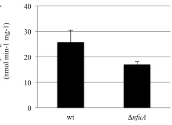
0 10 20 30 40 S uc ina te de hydroge na se a ct ivi ty (nm ol m in-1 m g-1) wt ΔnfuA
Figure 9. NfuA is required for respiratory complex II activity. Effect of a ΔnfuA
mutation on SDH activity. Succinate dehydrogenase activity was assayed from
MG1655 (wt) and MG1655 ΔnfuA. The experiments were run at least in triplicate
and the standard errors are shown.
-100 -50 0 50 100 150 200 250 -50 0 50 100 150 200 250 300 350 400 450 Adjusted sensorgram RU R es p o n se ( 0 = b as el in e) s Tim e 0 0.78 1.56 3.125 6.25 12.5 25 50 100 200