HAL Id: hal-00910117
https://hal.archives-ouvertes.fr/hal-00910117
Submitted on 27 May 2014
HAL is a multi-disciplinary open access
archive for the deposit and dissemination of
sci-entific research documents, whether they are
pub-lished or not. The documents may come from
teaching and research institutions in France or
abroad, or from public or private research centers.
L’archive ouverte pluridisciplinaire HAL, est
destinée au dépôt et à la diffusion de documents
scientifiques de niveau recherche, publiés ou non,
émanant des établissements d’enseignement et de
recherche français ou étrangers, des laboratoires
publics ou privés.
Coronary embolization of an intramyocardial hematoma
after myocardial infarction.
Raphaël P Martins, Nicolas Coquerel, Denis Amer Zabalawi, Alban-Elouen
Baruteau, Jean-Yves Gauvrit, Dominique Boulmier, Jean-Claude Daubert,
Philippe Mabo, Erwan Donal
To cite this version:
Raphaël P Martins, Nicolas Coquerel, Denis Amer Zabalawi, Alban-Elouen Baruteau, Jean-Yves
Gau-vrit, et al.. Coronary embolization of an intramyocardial hematoma after myocardial infarction..
Circulation, American Heart Association, 2010, 121 (8), pp.e220-4. �10.1161/CIR.0b013e3181d38d3c�.
�hal-00910117�
R.P. Martins, MD; N. Coquerel, MD; A. Zabalawi, MD; A.-E. Baruteau, MD; J.Y. Gauvrit, MD, PhD;
D. Boulmier, MD; J.C. Daubert, MD, PhD; P. Mabo, MD, PhD; E. Donal, MD, PhD
A
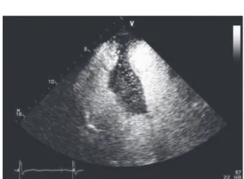
62-year-old man with a history of smoking, hypertension, and hypercholesterolemia was admitted for an inferior ST-segment elevation myocardial infarction (Figure 1). Coro-nary angiography performed 1 hour after the onset of chest pain revealed that the posterolateral artery was aneurysmal and obstructed. This blockage was treated by percutaneous coronary intervention with balloon angioplasty but without stent implanta-tion. At the end of the procedure, a small area of contrast agent stagnation was found in the pericardial area. Chest x-ray revealed no evidence of pleural effusion or pulmonary edema (Figure 2).Transthoracic echocardiography (TTE) (ViVid 7; GE Health-care, Chalfont St. Giles, UK) showed inferior wall hypokinesis with a preserved left ventricular ejection fraction of 50%. We also found a large septal mass and a small pericardial effusion. As shown in Figure 3, this mass expanded into the right ventricle and had a high echodensity. A myocardial contrast TTE revealed complete enhancement of the mass (Figure 4), suggesting a myocardial mass instead of a thrombus.
Cardiac computed tomography (GE Healthcare) with an iodinated contrast agent ruled out the presence of a tumor. However, we discovered a large myocardial hematoma of the inferior interventricular septum, measuring 41338370 mm, with active bleeding (Figure 5 and online-only Data Supplement Movie I) from the posterior descending artery. These findings suggested a septal hematoma caused by perforation of the posterior descending artery during the initial percutaneous cor-onary intervention. Given the risks of hematoma progression or septal rupture, we performed a second coronary angiography to attempt a percutaneous hemostatic procedure. Direct injection into the right coronary artery confirmed extravasation of contrast agent from the posterior descending artery, which suggested active bleeding from this coronary artery (Figure 6A). Because the extravasation disappeared after balloon inflation of the posterior descending artery, the feeding artery was embolized using 3 steel coils. Contrast agent stagnation disappeared com-pletely after coil embolization (Figure 6B).
A repeat-contrast TTE performed 1 day later showed no enhancement of the myocardial hematoma (Figure 7). The
patient underwent magnetic resonance imaging 10 days later (clinical 3-T ACHIEVA; Philips Medical Systems, Eindhoven, the Netherlands). Four-chamber imaging showed an abnormal signal area in the right side of the interventricular septum, sparing the subendocardium (Figure 8 and online-only Data Supplement Movie II). This high-signal area was localized to the inferior interventricular septum on short-axis views. The patient was discharged from the hospital on a regimen of antiplatelet agents (aspirin and clopidogrel), a b-adrenergic blocker, an angiotensin-converting enzyme inhibitor, and a statin.
The 3-month postembolization follow-up TTE revealed an impressive regression of the intramyocardial hematoma (Figure 9) with only a slight myocardial mass on the basal interventric-ular septum. Cardiac magnetic resonance imaging confirmed that the hematoma had regressed as the original area of abnormal signaling had disappeared (Figure 10). Six months after the procedure, the patient remained free from cardiovascular events. Intramyocardial hematoma is a subacute, partial rupture of the myocardium. These hematomas usually occur after myocardial infarction, chest trauma, surgery, or percutaneous coronary inter-vention, but they can also develop spontaneously.1The spontaneous
survival rate has been reported to be 10%.2Standard TTE usually
shows an echo-free or hypoechoic intramyocardial neocavity. Pre-vious reports of intramyocardial hematoma describe incomplete enhancement on contrast TTE, compared with the complete en-hancement seen with a malignant tumor.3 However, as our case
demonstrates, complete enhancement of a myocardial mass can also suggest an intramyocardial hematoma. In such instances, cardiac magnetic resonance imaging should be used to make the correct diagnosis. Intramyocardial hematoma is usually managed surgi-cally,4but conservative management has also been described. For
example, coronary coil embolization has been prescribed for coro-nary artery rupture during percutaneous corocoro-nary intervention.
To the best of our knowledge, this is the first article describing the use of coil embolization to treat intramyocardial hematoma resulting from myocardial infarction. Because our patient recov-ered uneventfully after the procedure, this treatment could be considered for intramyocardial hematoma.
From the CHU Rennes, Service de Cardiologie et Maladies Vasculaires, Rennes, France (R.P.M., A.-E.B., D.B., J.C.D., P.M., E.D.); Universite´ de Rennes 1, Rennes, France (R.P.M., A.-E.B., D.B., J.C.D., P.M., E.D.); INSERM, U642, Rennes, France (R.P.M., A.-E.B., D.B., J.C.D., P.M., E.D.); INSERM, CIC-IT 804, Rennes, France (R.P.M., A.-E.B., D.B., J.C.D., P.M., E.D.); CH Saint Brieuc, Service de Cardiologie, Saint Brieuc, France (N.C., A.Z.); CHU Rennes, De´partement de Radiologie et d’Imagerie Me´dicale, Rennes, France (J.Y.G.).
The online-only Data Supplement is available with this article at http://circ.ahajournals.org/cgi/content/full/121/8/e220/DC1.
Correspondence to Martins Raphae¨l Pedro, Service de Cardiologie et Maladies Vasculaires, CHU de Rennes, 2 rue Henri Le Guilloux, 35000 Rennes, France. E-mail [email protected]
Disclosures
None.
References
1. Harpaz D, Kriwisky M, Cohen AJ, Medalion B, Rozenman Y. Unusual form of cardiac rupture: sealed subacute left ventricular free wall rupture, evolving to intramyocardial dissecting hematoma and to pseudoaneurysm formation: a case report and review of the literature. J Am Soc
Echo-cardiogr. 2001;14:219 –227.
2. Pliam MB, Sternlieb JJ. Intramyocardial dissecting hematoma: an unusual form of subacute cardiac rupture. J Card Surg. 1993;8:628 – 637. 3. Mansencal N, Revault-d’Allonnes L, Pelage JP, Farcot JC, Lacombe P,
Dubourg O. Usefulness of contrast echocardiography for assessment of intracardiac masses. Arch Cardiovasc Dis. 2009;102:177–183. 4. Jahnke C, Hetzer R, Komoda T, Fleck E, Paetsch I. Intramural dissecting
hemorrhage of the myocardium. Circulation. 2007;115:e457– e459.
Figure 1.Standard 12-lead echocardiogram showing an ST-segment elevation in inferior leads.
Figure 2.Chest x-ray revealing no evidence of pleural effusion or pulmonary edema.
Figure 3.Transthoracic echocardiography in the apical 4-chamber view (A) and the parasternal short-axis view (B) revealing a large septal mass expanding into the right ventricle, characterized by myocardial echodensity (arrowheads).
Martins et al Coil Embolization of an Intramyocardial Hematoma e221
by guest on November 27, 2013
http://circ.ahajournals.org/
Figure 4.Contrast transthoracic echocardiography showing complete enhancement of the mass.
Figure 7.Contrast transthoracic echocardiography performed 1 day after coil embolization of the posterior descending artery, showing a lack of enhancement of the myocardial hematoma.
Figure 6.Right coronary angiography before (A) and after (B) posterior
descending artery coil embolization show-ing the active bleedshow-ing (arrowhead) and the steel coils (arrow).
Figure 8.Magnetic resonance steady-state free precession sequences per-formed in short-axis view (A) and in apical 4-chamber view (B), highlighting an abnormal signal in the right side of the inferior interventricular septum, sparing the subendocardium (arrows). Delayed enhancement sequence performed in short-axis view (C) and in apical 4-chamber view (D), revealing an area of transmural enhancement corresponding to the inferior infarcted area (arrowhead) and the interventricular septum hematoma (arrow).
Martins et al Coil Embolization of an Intramyocardial Hematoma e223
by guest on November 27, 2013
http://circ.ahajournals.org/
Figure 9.Three-month follow-up transthoracic echocardiogra-phy showing the regression of intramyocardial hematoma on an apical 4-chamber view (A) and a parasternal short-axis view (B). A small myocardial mass on the basal interventricular septum is observed on the parasternal short-axis view (B, arrow).
Figure 10.Magnetic resonance steady-state free precession sequences showing the hematoma regression on a short-axis view (A, arrow). Delayed enhancement sequence (B) revealing an area of trans-mural enhancement corresponding to the inferior wall infarct (arrow) and the hema-toma sequelae (arrowhead).