Late Infiltration of Post-orthodontic
White Spot Lesions
Spätinfiltration von White-Spot-Läsionen
nach kieferorthopädischer Behandlung
Klaus W. Neuhaus
1, Martina Graf
2, Adrian Lussi
1, Christos Katsaros
2,
Abstract
White spot lesion (WSL) infiltration has been recommended im-mediately after debonding of orthodontic brackets. It is however not clear if established inactive WSLs can also be masked through infiltration.
Orthodontic treatment of a 19-year-old patient had to be terminated prematurely due to development of multiple WSLs of varying severity. Three months after debonding, the patient pre-sented for lesion infiltration. After etching with 15% HCl gel and re-wetting of the dried surfaces it seemed that a good outcome could be expected. Lesion infiltration led to complete masking of less severe WSLs. The visual appearance of moderate and severe WSLs was improved but they were still visible after treatment.
Inactive WSLs may not represent an increased caries risk, but patients are often bothered esthetically. Infiltration by re-peated etching might be a viable approach even for inactive WSLs. Controlled clinical trials are needed to investigate the long-term performance of this technique.
Key Words: Initial caries · Enamel caries · Orthodontics · Prevention · Penetration · Masking · ICDAS
Zusammenfassung
Die Infiltration initialer kariöser Läsionen (Kreideflecke, White-Spot-Läsionen) wird unmittelbar nach der Entfernung kieferor-thopädischer Brackets empfohlen. Es ist jedoch unklar, ob auch etablierte Kreideflecken durch Infiltration maskiert werden kön-nen.
Bei einem 19-jährigen Patienten musste die festsitzende orth-odontische Phase wegen Auftretens multipler initial kariöser Läsi-onen vorzeitig beendet werden. Drei Monate nach der Entfernung der Brackets wurde der Patient zur Infiltration vorstellig. Nach dem Ätzen mit 15%igem HCl-Gel konnte durch gezieltes Wiederbefeuch-ten der getrockneWiederbefeuch-ten Zahnoberflächen das zu erwarWiederbefeuch-tende Ergebnis antizipiert werden. Die Infiltrationstechnik führte zur vollständi-gen Maskierung weniger stark ausgeprägter Kreideflecke. Stärker demineralisierte Läsionen wurden optisch verbessert, waren aber nach der Behandlung noch immer sichtbar.
Inaktive Kreideflecke sprechen nicht unbedingt für ein er-höhtes Kariesrisiko, aber Patienten stören sich häufig an der kos-metischen Einbuße. Die Infiltration dieser Läsionen mit multiplen Ätzschritten könnte auch für inaktive Kreideflecke sinnvoll sein. Es fehlen jedoch Langzeitdaten aus klinischen Untersuchungen zur Validierung dieser Methode.
Schlüsselwörter: Initialkaries · Schmelzkaries · Kiefer-orthopädie · Prävention · Penetration · Maskierung · ICDAS
1 Department of Preventive, Restorative and Pediatric Dentistry,
School of Dental Medicine, University of Bern, Bern, Switzerland,
2 Department of Orthodontics and Dentofacial Orthopedics, School
of Dental Medicine, University of Bern, Bern, Switzerland.
Received: August 25, 2010; accepted: September 27, 2010
J Orofac Orthop 2010;71:442–7 DOI 10.1007/s00056-010-1038-0
Introduction
The risk of development of white spot lesions (WSLs) dur-ing fixed orthodontic treatment is rather high. Several pre-ventive measures have been proposed to minimize the inci-dence [9]. Generally, the use of fluoride varnish [15] or other fluoride applications [6] has been recommended to avoid white spot lesion formation during fixed appliance treatment. But less is known about how to treat WSLs once they have occurred [7]. It has been shown that some “physi-ologic” regression of white spot lesions due to enamel re-mineralization is possible through saliva, although many white spot lesions may remain stable for decades [4]. Chew-ing gums have been reported to remineralize artificial white spot lesions after 21 days [11]. An enhanced remineraliza-tion effect was, however, reported for chewing gums con-taining caseinphosphopeptide-amorphous calciumphos-phate (CPP-ACP) [16]. Clinical studies comparing a fluori-dated toothpaste [5] or fluorifluori-dated toothpaste plus daily mouth rinse with a CPP-ACP remineralizing cream and 0.05% fluoridated mouth rinse [1] showed a greater regres-sion of visible WSLs due to the latter. However, this kind of treatment also requires a certain compliance of the pa-tient, as daily application of the cream is required.
Chairside methods have therefore been proposed to treat WSLs instantly regardless of the patient’s compliance. One possibility is to micro-abrade the white spot lesions us-ing HCl-erosion followed by polishus-ing with an abrasive pum-ice [2, 8]. The white spots are thus abraded and the defects subsequently filled with a resin. This efficacious method, however, sacrifices dental hard substance to an amount of approximately 360 ± 120 microns [17]. Another method in-volving surface erosion with HCl has been introduced lately [12]. Different from micro-abrasion, the aim of the infiltra-tion technique is not to remove the whitish enamel, but to micro-invasively infiltrate it with a so-called “infiltrant”, a polymerizable low viscous resin which fills out the pores of the white spot. It has been shown in vitro that infiltrants have a higher penetration coefficient than regular bonding agents and can thus penetrate the body of incipient carious lesions up to 400 microns [13]. A higher penetration coefficient al-lows a greater effect of capillary forces, which drives infil-trants deeper into white spots than regular bonding agents.
In the case of post-orthodontic WSLs it has been sug-gested that infiltration treatment be initiated as soon as pos-sible after bracket removal [14]. White spots tend to be ac-tive during and shortly after orthodontic treatment, meaning they tend to progress and thus have a higher risk of loss of surface integrity. Active enamel lesions are micro-porous and feel rough when lightly probed along the surface. Micro-porosities eventually allow a better lesion infiltration [14]. After removal of the orthodontic appliance, the micro-envi-ronment eventually changes and permits white spots to be brushed and cleaned. Under these circumstances, the WSLs change their behavior and become inactive, meaning less
Einleitung
Das Risiko, White-Spot-Läsionen (WSLs) während einer festsitzenden kieferorthopädischen Behandlung zu entwi-ckeln, ist ziemlich groß. Verschiedene präventive Maßnah-men wurden vorgeschlagen, um ihr Auftreten zu minimieren [9]. Im Allgemeinen wird der Gebrauch von Fluoridlack [15] oder anderen Fluorid-Applikationsformen [6] empfohlen, um eine Entwicklung von WSLs während der aktiven, fest-sitzenden Phase zu vermeiden. Weniger ist darüber bekannt, wie mit WSLs umzugehen ist, wenn sie einmal aufgetreten sind [7]. Es wurde gezeigt, dass durch den Speichel bis zu einem gewissen Grad eine physiologische Regression von Kreideflecken möglich ist, obwohl viele dieser Läsionen über Jahrzehnte stabil und sichtbar beiben [4]. Ferner wurde be-richtet, dass Kaugummis künstliche WSLs in situ nach 21 Tagen remineralisierten [11]. Ein verbesserter Reminera-lisationseffekt wurde darüber hinaus für Kaugummis berich-tet, die Caseinphosphospeptid-amorphes Calciumphosphat (CPP-ACP) enthielten [16]. In klinischen Studien wurde eine größere Regression sichtbarer WSLs nach regelmäßiger An-wendung einer CPP-ACP-haltigen Paste zusammen mit einer 0,05%igen Fluorid-Mundspülung beobachtet als nach bloßer Benutzung fluoridierter Zahnpasta [5] oder fluori-dierter Zahnpasta mit täglicher fluorifluori-dierter Mundspülung [1]. Diese Behandlung erfordert jedoch eine gewisse Mitar-beit auf der Patientenseite, da eine tägliche Applikation der Paste nötig ist.
Daher wurden Chairside-Methoden vorgeschlagen, um WSLs sofort und unabhängig von der Mitwirkung des Pati-enten zu behandeln. Eine Möglichkeit besteht darin, die Krei-deflecken mit Mikroabrasion zu behandeln, wobei nach Ero-sion mit HCl die Oberfläche mit einer abrasiven Bimsstein-paste poliert wird [2, 8]. Kreideflecken werden so abradiert, und eventuell vorhandene Defekte können dann mit einem fließfähigen Komposit aufgefüllt werden. Diese effiziente Methode opfert jedoch ungefähr 360 ± 120 µm Zahnhartsub-stanz [17]. Eine andere Methode, bei der ebenfalls die Zahn-oberfläche mit HCl erodiert wird, wurde kürzlich vorgestellt [12]. Anders als bei der Mikroabrasion besteht das Ziel der Infiltrationsmethode darin, den opaken Schmelz nicht zu ent-fernen, sondern ihn mikroinvasiv mit einem so genannten In-filtranten zu infiltrieren. Dieses niedrigvisköse Adhäsiv füllt die kariös erweiterten Schmelzporen auf. Es wurde in vitro gezeigt, dass Infiltranten einen höheren Penetrationskoeffizi-enten haben als reguläre Bondingmaterialien, so dass sie den Körper initialer kariöser Läsionen bis zu 400 µm weit durch-dringen können [13]. Ein höherer Penetrationskoeffizient er-möglicht den Kapillarkräften einen höheren Wirkungsgrad, so dass Infiltranten tiefer in den Kreidefleck eindringen kön-nen als herkömmliche Bondingmaterialien.
Im Fall von WSLs nach Kieferorthopädie wurde vorge-schlagen, die Infiltrationsbehandlung so bald wie möglich nach Entfernung der Brackets zu beginnen [14]. „White Spots“ sind eher während und kurz nach der
kieferorthopä-likely to progress. An inactive enamel lesion feels smooth on slight probing, and it looks shiny. The histologic correlate is a hardened surface layer of the enamel lesion. Fluoride even-tually stabilizes this surface layer but also hampers further remineralization deeper in the body of the lesion [3]. It is not clear and there is not yet histologic proof as to whether inac-tive WSLs can be successfully treated with the infiltration technique.
Case Report
This article reports a case of a patient with insufficient com-pliance leading to multiple WSL formation around fixed orthodontic appliances. The orthodontic treatment had to be terminated before achieving the desired result. The pa-tient was called up for infiltration treatment of his WSLs directly after debonding and prior to insertion of retainers. However, he postponed the treatment and was treated no sooner than 3 months after debonding. The patient gave his informed consent to infiltration treatment and was oriented that the treatment outcome might be not as good as ex-pected.
The patient (healthy male Caucasian) was 18 years old on beginning of the orthodontic treatment and 19 years old on its termination. The teeth showed a molar incisor hypomineralization with opaque hypoplastic spots but no surface discontinuities on the vestibular surfaces of ante-rior teeth and molar cusps. The patient complained about being severely disturbed by the mottled appearance of his teeth.
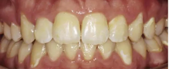
Clinical inspection showed WSLs on the labial aspects of teeth 14, 13, 22, 23, 24, 37, 36, 34, 33, 32, 42, 43, 44, and 47 (Figure 1). All WSLs except for tooth 36 were Code 2 ac-cording to the International Caries Detection and Assess-ment System (ICDAS). Only tooth 36 showed local enamel breakdown (Code 3) requiring restorative treatment. The extent of WSLs however was more pronounced on the ca-nines and premolars than on the front teeth.
dischen Behandlung aktiv. Das bedeutet, dass sie zur Progres-sion neigen und ein größeres Kavitationsrisiko aufweisen. Aktive Schmelzläsionen sind mikroporös und fühlen sich rau an, wenn man mit einer spitzen Sonde leicht über die Oberflä-che fährt. Mikroporösitäten erlauben eine bessere Infiltration [14]. Nach dem Entfernen der kieferorthopädischen Appara-turen verändert sich jedoch das Mikromilieu, indem die vor-handenen WSLs besser zu reinigen sind. Unter diesen verän-derten Umständen verändern sich die Kreideflecken und werden inaktiv, zeigen also weniger die Tendenz zur Progres-sion. Eine inaktive Schmelzläsion fühlt sich bei vorsichtiger Sondierung glatt an und sieht glänzend aus. Das histologische Korrelat ist eine wieder erhärtete Oberflächenschicht des White Spots. Fluorid stabilisiert diese Oberflächenschicht, verhindert aber auch eine weitere Remineralisierung tiefer im Läsionskörper [3]. Es ist noch unklar und der histologische Nachweis darüber fehlt, ob inaktive WSLs erfolgreich mit der Infiltrationstechnik behandelt werden können.
Kasuistik
Dieser Fallbericht handelt von einem Patienten mit ungenü-gender Compliance, die zur Entwicklung multipler initialer kariöser Läsionen um die Brackets herum führte. Die kie-ferorthopädische Behandlung musste daher vor dem Errei-chen des angestrebten Ziels abgebroErrei-chen werden. Der Pati-ent wurde für eine Infiltrationsbehandlung seiner WSLs nach Debonding und unmittelbar vor der Insertion eines Retainers aufgerufen. Wegen Terminabsage konnte die Be-handlung aber erst 3 Monate nach Debonding stattfinden. Der Patient gab sein Einverständnis zur geplanten Infiltrati-onsbehandlung und wurde darüber aufgeklärt, dass das Be-handlungsergebnis eventuell nicht so gut ausfallen könnte wie erwartet.
Der Patient (männlich, gesund, Kaukasier) war zu Be-ginn der kieferorthopädischen Behandlung 18 Jahre und am Ende der Behandlung 19 Jahre alt. Die Zähne zeigten eine Molaren-Inzisiven-Hypomineralisation mit opaken hypoplas-tischen Flecken ohne Oberflächeneinbrüche an den Labialflä-chen der Frontzähne und der Molarenhöcker. Der Patient beklagte eine starke Einschränkung durch das fleckige und gescheckt aussehende Erscheinungsbild seiner Zähne.
Die klinische Inspektion zeigte WSLs an den Labialflä-chen der Zähne 14, 13, 22, 23, 24, 37, 36, 34, 33, 32, 42, 43, 44 und 47 (Abbildung 1). Alle WSLs außer Zahn 36 entsprachen Code 2 des International Caries Detection and Assessment System (ICDAS). Nur Zahn 36 zeigte einen lokalen Schmelz-einbruch (Code 3), der eine restaurative Behandlung erfor-derte. Die Ausdehnung der WSLs war bei Eckzähnen und Prämolaren stärker ausgeprägt als bei den Frontzähnen.
Klinische Vorgehensweise
Die Behandlung wurde für den Oberkiefer und für den Un-terkiefer separat geplant. Während der ersten Behandlung wurden alle WSLs im Oberkiefer infiltriert. Die Zähne
wur-Figure 1. Situation after debonding of the brackets and insertion of a palatal retainer. Upper front teeth are dried out.
Abbildung 1. Situation nach der Entfernung der Brackets und nach Inser-tion eines palatinalen Retainers. Die oberen Frontzähne sind ausgetrock-net.
Clinical Procedure
The treatment was planned separately for the upper and lower jaws. At the first appointment, the upper WSLs were infiltrated. The teeth were isolated with rubber dam. Due to a palatal retainer from canine to canine, the split dam technique was chosen. After gluing the loose dam to the buccal and palatal gingiva with Histoacryl® (Tissue Seal,
Ann Arbor, MI, USA), additional gum isolation with Opal-dam® (Ultradent, South Jordan, UT, USA) was applied.
The teeth were cleaned first with conventional prophylactic pumice (Cleanic; KerrHawe, Bioggio, Switzerland) (Figure 2) and then rinsed and dried. The WSLs were first treated with 15% HCl-gel (Icon; DMG, Hamburg, Germany) for 2 minutes according to the manufacturer’s instructions. After thorough rinsing with air-water-spray for 30 seconds the teeth were dried with compressed air. The appearance of the pretreated labial surfaces was chalky (Figure 3). Rewet-ting with a moist micro-brush was performed to mimick the anticipated outcome. For persisting visible white spots the etching step was repeated once or twice until a favorable outcome could be mimicked (Figure 4). After applying the drying solution (Icon; DMG, Hamburg, Germany) to the surface, it was conditioned for application of the infiltrant (Icon; DMG, Hamburg, Germany). This infiltrant was ap-plied for 3 minutes and then light cured for 40 seconds. The infiltration was repeated once with application of the infil-trant for 1 minute and subsequent light curing for 40 sec-onds. After infiltration, the surplus was removed using a curette. The tooth surfaces were then polished with a brownie (Shofu, Ratingen, Germany) and with an oc-clubrush (KerrHawe, Bioggio, Switzerland) using water and light pressure (Figure 5).
den mit Kofferdam isoliert. Wegen eines Retainers im Ober-kiefer von Eckzahn bis Eckzahn wurde die so genannte Split-dam-Technik gewählt. Nachdem der lose Kofferdam am Gaumen mit Histoacryl® (Tissue Seal, Ann Arbor, MI,
USA) angeklebt wurde, erfolgte eine Isolation der noch frei liegenden Gingiva mit Opaldam® (Ultradent, South Jordan,
UT, USA). Zunächst wurden die Zähne mit einer konventi-onellen Prophylaxepaste gereinigt (Cleanic; KerrHawe, Bi-oggio, Schweiz) (Abbildung 2) und anschließend abgesprüht und getrocknet. Die WSLs wurden zunächst mit 15%igem HCl-Gel (Icon; DMG, Hamburg, Deutschland) entspre-chend den Herstellerangaben für 2 Minuten geätzt. Nach gründlichem Absprühen für 30 Sekunden mittels Dreiweg-spritze wurden die behandelten Zahnoberflächen mit Druck-luft „übertrocknet“. Das Erscheinungsbild der so vorbehan-delten Labialflächen war kreidig weiß (Abbildung 3). Ein Rewetting mit einer feuchten Microbrush erlaubte eine An-tizipation des zu erwartenden Behandlungsergebnisses. Bei persistierenden Kreideflecken wurde der Ätzschritt ein- bis zweimal wiederholt, bis das gewünschte Behandlungsziel imitiert werden konnte (Abbildung 4). Die Applikation der Trocknungslösung (Icon; DMG, Hamburg, Deutschland) konditionierte die Zahnoberfläche für die eigentliche Infilt-ration mit dem Infiltranten (Icon; DMG, Hamburg, Deutsch-land). Dieser wurde für 3 Minuten appliziert und danach 40 Sekunden lichtgehärtet. Der Infiltrationsschritt wurde da-nach einmal wiederholt, wobei der Infiltrant beim zweiten Mal vor dem Lichthärten (40 s) nur 1 Minute lang appliziert wurde. Nach der Infiltration wurden die Überschüsse mit einer Kürette entfernt. Die Vestibulärflächen wurden an-schließend mit einem Brownie (Shofu, Ratingen, Deutsch-land) und einer Occlubrush (KerrHawe, Bioggio, Schweiz)
Figure 2. Situation immediately prior to the start of infiltration 3 months after debonding.
Abbildung 2. Situation unmittelbar vor Beginn der Infiltration, 3 Monate nach Debonding.
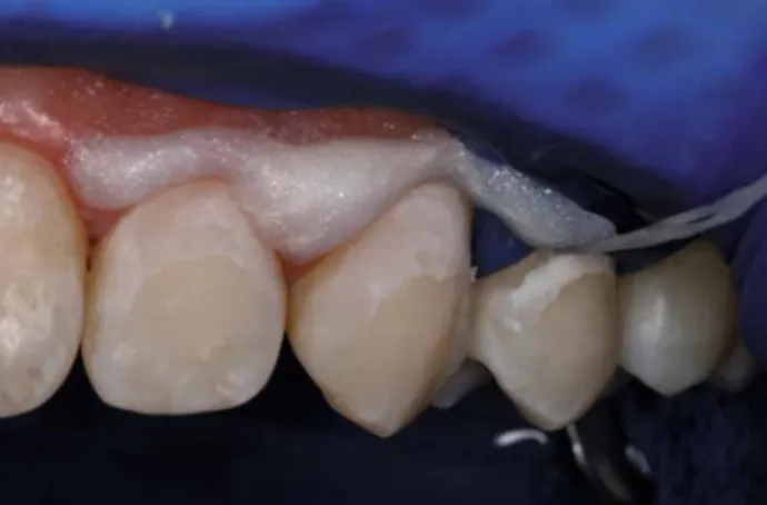
Figure 3. Situation after etching with 15% HCl-gel. The enamel looks whitish and opaque, but white spot lesions can still be discerned. Abbildung 3. Nach dem Ätzen mit 15%igem Salzsäure-Gel. Der Schmelz sieht kreidig opak aus, aber die Kreideflecken setzen sich immer noch farblich ab.
In the lower jaw, this treatment was applied accordingly. Tooth 36 received an adhesive class V filling (Ceram X mono; Dentsply, Konstanz, Germany) due to cavitation of the surface.
Discussion
Pronounced enamel lesions can be expected to have a deeper lesion body. Different lesion depths of WSLs can be expected after debonding. It was therefore interesting to compare the results of the same treatment in WSLs of dif-ferent severities. A general esthetic improvement could be observed in all WSLs. However, very severe lesions could not be completely masked indicating that the body of the lesion was not completely filled by the infiltrant. The less severe WSLs however became completely invisible or only visible after drying. Thus, in mild to moderate WSLs a shift from ICDAS II score 2 to 1 (lesion only visible after drying for 3–5 seconds with compressed air) or 0 could be achieved in this patient. It has to be noted that the infiltration of WSLs is likely to change its state of activity. In vitro, infil-trated WSLs did not progress, and in vivo infiltration led to a clinically stable situation after 10 months [14]. However, it has to be kept in mind that the method of lesion infiltra-tion has not yet been clinically validated in long-term con-trolled clinical trials. Moreover, it is noteworthy that etch-ing away the superficial layer of the white spot dissolves the most caries-resistant layer. It was shown that the superficial layer of white spot lesions is significantly more acid-resis-tant than healthy enamel [10]. Finally, etching of healthy enamel could not be totally avoided. Thus, no statement on the longevity of the treatment, including margin discolor-ation and development of secondary caries, can be given at the moment. Finally, histology of arrested white spot le-sions should be the subject of further research.
unter Wasserkühlung mit nur leichtem Druck poliert (Abbil-dung 5).
Im Unterkiefer wurden die Arbeitsschritte entsprechend wiederholt. Zahn 36 wurde wegen Kavitation der Oberfläche mit einer adhäsiven Klasse-V-Füllung versehen (Ceram X mono; Dentsply, Konstanz, Deutschland).
Diskussion
Nach Debonding können verschiedene Läsionstiefen von Kreideflecken gefunden werden, wobei deutlich sichtbare WSLs einen größeren Körper der Läsion haben und tiefer sind. Daher war es interessant, das Behandlungsergebnis un-terschiedlich stark ausgeprägter WSLs bei gleicher Behand-lung zu vergleichen. Eine generelle kosmetische Verbesse-rung konnte bei allen betroffenen Zähnen erzielt werden. Stark ausgeprägte Kreideflecken konnten jedoch nicht voll-ständig maskiert werden, was darauf hindeutet, dass der In-filtrant den Läsionskörper nicht vollständig aufgefüllt hat. Weniger stark ausgeprägte WSLs wurden jedoch vollständig maskiert oder waren nur noch nach dem Trocknen sichtbar. Leichte und mittelstark ausgeprägte WSLs konnten bei die-sem Patienten also vom ICDAS II Grad 2 zum Grad 1 (Lä-sion ist sichtbar nach 3–5 Sekunden Trocknen mit Druckluft) oder zum Grad 0 überführt werden. Es kann angenommen werden, dass die Infiltration von WSLs den Aktivitätsstatus der Läsion verändert. In vitro waren infiltrierte WSLs nicht progredient, und in vivo konnte eine klinisch stabile Situa-tion nach 10 Monaten erzielt werden [14]. Man muss jedoch beachten, dass die Publikation von Langzeitergebnissen aus kontrollierten klinischen Studien mit Infiltration von WSLs erst in näherer Zukunft erwartet werden kann. Darüber hin-aus muss festgehalten werden, dass durch die Erosion der Oberflächenzone des Kreideflecks die am stärksten kariesre-sistente Schicht entfernt wird. Es wurde gezeigt, dass die
Figure 4. Rewetting with a moist micro-brush mimicks the expected out-come.
Abbildung 4. Das Wiederbefeuchten (Rewetting) mit einer feuchten Microbrush antizipiert das zu erwartende Ergebnis.
Figure 5. Facial aspect 2 months postoperatively. Generally better appea-rance, but the severe white spot lesions are still visible.
Abbildung 5. Frontansicht nach 2 Monaten. Das Erscheinungsbild ist ge-nerell besser, aber manche Kreideflecken sind immer noch sichtbar.
However, this case showed that esthetic improvement could be achieved in all WSLs. Interestingly, infiltration worked even when performed “too late” in established, purportedly superficially remineralized WSLs.
Conclusion
Infiltration might be applicable in post-orthodontic white spot lesions even after conversion to inactive lesions. Re-peated etching could be considered as a necessary treat-ment variation. Controlled clinical trials are needed to test the longevity of the infiltration technique in inactive white spot lesions.
Acknowledgement
We would like to thank Dr. Sebastian Paris, Kiel, for valuable dis-cussions.
References
1. Andersson A, Skold-Larsson K, Hallgren A, et al. Effect of a dental cream containing amorphous cream phosphate complexes on white spot lesion regression assessed by laser fluorescence. Oral Health Prev Dent 2007;5:229–33.
2. Ardu S, Castioni NV, Benbachir N, Krejci I. Minimally invasive treat-ment of white spot enamel lesions. Quintessence Int 2007;38: 633–6.
3. Arnold WH, Cerman M, Neuhaus K, Gaengler P. Volumetric assess-ment and quantitative eleassess-ment analysis of the effect of fluoridated milk on enamel demineralization. Arch Oral Biol 2003;48:467–73. 4. Backer Dirks O. Posteruptive changes in dental enamel. J Dent Res
1966;45:503–11.
5. Bailey DL, Adams GG, Tsao CE, et al. Regression of post-orthodontic lesions by a remineralizing cream. J Dent Res 2009;88:1148–53. 6. Benson PE, Parkin N, Millett DT, et al. Fluorides for the prevention
of white spots on teeth during fixed brace treatment. Cochrane Database Syst Rev 2004:CD003809.
7. Bergstrand F, Twetman S. Evidence for the efficacy of various meth-ods of treating white-spot lesions after debonding of fixed orth-odontic appliances. J Clin Orthod 2003;37:19–21.
8. Croll TP, Cavanaugh RR. Hydrochloric acid-pumice enamel surface abrasion for color modification: results after six months. Quintes-sence Int 1986;17:335–41.
9. Derks A, Katsaros C, Frencken JE, et al. Caries-inhibiting effect of preventive measures during orthodontic treatment with fixed appli-ances. A systematic review. Caries Res 2004;38:413–20.
10. Iijima Y, Takagi O. In situ acid resistance of in vivo formed white spot lesions. Caries Res 2000;34:388–94.
11. Leach SA, Lee GT, Edgar WM. Remineralization of artificial caries-like lesions in human enamel in situ by chewing sorbitol gum. J Dent Res 1989;68:1064–8.
12. Paris S, Meyer-Lueckel H, Kielbassa AM. Resin infiltration of natural caries lesions. J Dent Res 2007;86:662–6.
13. Paris S, Meyer-Lueckel H, Colfen H, Kielbassa AM. Penetration coeffi-cients of commercially available and experimental composites intend-ed to infiltrate enamel carious lesions. Dent Mater 2007;23:742–8. 14. Paris S, Meyer-Lueckel H. Masking of labial enamel white spot
le-sions by resin infiltration – a clinical report. Quintessence Int 2009;40:713–8.
Oberflächenzone eines Kreideflecks signifikant säureresis-tenter ist als gesunder Schmelz [10]. Es konnte außerdem nicht vollständig verhindert werden, dass auch gesunde Schmelzareale mit Salzsäure angeätzt wurden. Insofern muss man festhalten, dass zum jetzigen Zeitpunkt der Langzeit-erfolg der Behandlung, einschließlich des Risikos der margi-nalen Randverfärbung oder des Auftretens von Sekundär-karies, nicht sicher ist. Abschließend sei angemerkt, dass his-tologische Untersuchungen inaktiver White-Spot-Läsio nen Gegenstand weiterer Forschung sein sollten.
Dieser Fall zeigte, dass eine kosmetische Verbesserung bei allen WSLs erreicht werden konnte. Interessanterweise funktionierte die Infiltration sogar bei „zu später“ Durchfüh-rung bei inaktiven, vermutlich oberflächlich remineralisierten WSLs.
Schlussfolgerung
Die Infiltration kann bei nach kieferorthopädischer Behand-lung aufgetretenen Kreideflecken Anwendung finden, auch wenn diese schon arretiert sind. Wiederholte Ätzschritte können bei ausgeprägten Läsionen als Behandlungsvariante ins Auge gefasst werden. Kontrollierte klinische Studien sind notwendig, um den Langzeiteffekt der Infiltrationstechnik aus kosmetischen Gründen bei inaktiven White-Spot-Läsi-onen zu validieren.
Danksagung
Dank gebührt Dr. Sebastian Paris, Kiel, für eine wertvolle Diskus-sion.
15. Stecksen-Blicks C, Renfors G, Oscarson ND, et al. Caries-preventive effectiveness of a fluoride varnish: a randomized controlled trial in adolescents with fixed orthodontic appliances. Caries Res 2007;41:455–9.
16. Sudjalim TR, Woods MG, Manton DJ, Reynolds EC. Prevention of de-mineralization around orthodontic brackets in vitro. Am J Orthod Dentofacial Orthop 2007;131:705 e1–9.
17. Tong LS, Pang MK, Mok NY, et al. The effects of etching, micro-abra-sion, and bleaching on surface enamel. J Dent Res 1993;72:67–71.
Correspondence Address
Dr. Klaus W. Neuhaus
Department of Preventive, Restorative and Pediatric Dentistry Freiburgstr. 7 3010 Bern Switzerland Phone: (+41/31) 632 49 74 e-mail: klaus.neuhaus@zmk.unibe.ch