J Neurol (2008) 255:769–771
DOI 10.1007/s00415-008-0806-x
LETTER TO THE EDITORS
JON 2806
yrs) with idiopathic late-onset
cerebellar degeneration, in whom
brain MRI demonstrated profound
atrophy of the flocculus, whereas
atrophy of nodulus and uvula was
only mild (Fig. 1). Considering the
relatively focal nature of the
cere-bellar degeneration, we asked
whether semicircular canal
(SCC)-related reflexes, which are
medi-ated mainly by the flocculus, and
OL-related reflexes, which are
mediated primarily by the nodulus,
are differentially affected in this
patient. Since he exhibited
promi-nent DBN, we also explored
whether VVOR gains were
asym-metric, i.e. lower during downward
than during upward head rotations.
The patient gave informed
written consent. The protocol was
approved by the local ethics
com-mittee. Vestibular-evoked potentials
(VEMPs) were registered from the
sternocleidomastoid muscle on
both sides [13]. The caloric
re-sponse was assessed with
electro-nystagmography (monoaural
irrigation on either side, water
temperatures 30 °C and 44 °C).
Quantitative head impulse testing
was performed as described in
[14]. Static ocular counterroll in
Sarah Marti
Alexander A. Tarnutzer
Bernhard Schuknecht
Dominik Straumann
Dissociation between
canal- and
function in cerebellar
atrophy
Received: 24 May 2007
Received in revised form: 16 October 2007 Accepted: 30 October 2007
Published online: 21 February 2008
Sirs: Vestibulo-cerebellar
degenera-tion affects the vestibulo-ocular
reflex (VOR). For instance, the
capacity to modify VOR gains (via
the flocculus [1, 2]) or to suppress
vestibular nystagmus by changing
the head re gravity (via the nodulus
[3, 4]) may be impaired. Cerebellar
patients may even demonstrate
bilateral vestibular loss [5].
Whether downbeat nystagmus
(DBN), an ocular motor sign
frequently found with
vestibulo-cerebellar degeneration, results
from asymmetric vertical VOR
(VVOR) function, remains debated
[6]. Alternatively, DBN may be
caused by loss of vertical floccular
Purkinje cells [7], asymmetries in
vertical smooth pursuit pathways
[8, 9], or dissociation of coordinate
systems for vertical saccade
gener-ation and gaze-holding [10]. DBN
is also influenced by otolith (OL)
signals [11, 12].
We report on a patient (m, 82
sustained whole-body roll
posi-tions and gravity-dependent
modu-lation of DBN along the pitch plain
were tested on a three-axis
motor-driven turntable [12].
Bilateral loss of SCC function
was documented by absent caloric
responses (not shown) and subtotal
gain reductions of the VOR along
the planes of all SCCs (Fig. 2A).
Intact OL function was
demon-strated by reproducible VEMP
(threshold at 95 dB on both sides),
preserved static ocular counterroll
(Fig. 2B), and prominent
modula-tion of DBN in the pitch plane with
maximal slow-phase eye velocity in
the prone position (Fig. 2C).
SCC- and OL-dependent reflexes
are mediated by different parts of
the vestibulo-cerebellum. The
flocculus is important for the
control of the angular VOR [1, 2],
while the nodulus and uvula are
mainly involved in the control of
OL-related reflexes [3, 4]. In our
patient, the dissociated vestibular
deficits with impaired
SCC-depen-dent, but preserved OL-dependent
reflexes, might therefore be best
explained by the pattern of the
vestibulo-cerebellar degeneration
predominantly affecting the
floccu-S. Marti, MD (쾷) · A. A. Tarnutzer · D. Straumann
Dept. of Neurology Zurich University Hospital Frauenklinikstrasse 26 8091 Zurich, Switzerland Tel.: +41-1/255-3996 Fax: +41-1/255-4380 E-Mail: [email protected] B. Schuknecht Dept. of Neuroradiology
University Hospital Zurich, Switzerland
Fig. 1 High resolution MR with a Ciss sequence covering the cerebellum (slice thickness 0.85 mm) shows marked atrophy of the flocculus (black arrow) bilaterally, while the nodulus (white arrow) is relatively spared
769_771_Marti_JON_2806.indd 769
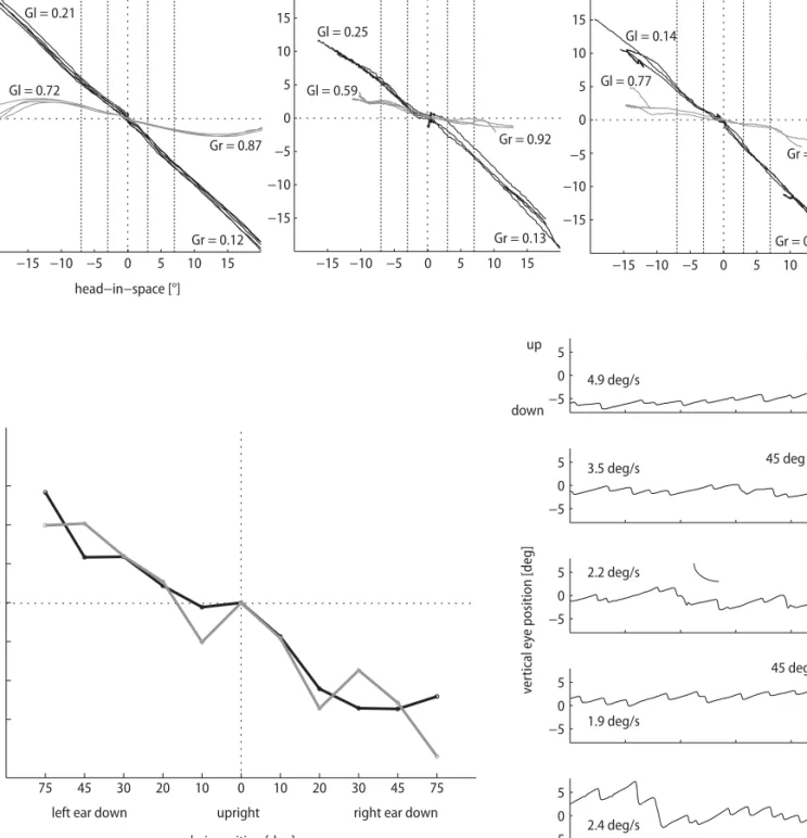
770 −5 0 5 −5 0 5 −5 0 5 −5 0 5 −5 0 5 3 4 ve rt ical ey e position [deg] supine up down 45 deg supine 45 deg prone prone upright 1 2 time [sec] 4.9 deg/s 3.5 deg/s 2.2 deg/s 1.9 deg/s 2.4 deg/s −15 −10 −5 0 5 10 15 −15 −10 −5 0 5 10 15
left anterior / right posterior right anterior / left posterior
−15 −10 −5 0 5 10 15 −15 −10 −5 0 5 10 15 −15 −10 −5 0 5 10 15 −15 −10 −5 0 5 10 15 horizontal head−in−space [°] eye−in−space [°] Gr = 0.12 Gl = 0.21 Gr = 0.13 Gr = 0.10 Gl = 0.25 Gl = 0.14 Gl = 0.72 Gr = 0.87 Gr = 0.92 Gr = 0.90 Gl = 0.59 Gl = 0.77
left ear down upright right ear down
chair position [deg]
tors eye pos [deg]
−3 −2 −1 0 1 2 3 75 45 30 20 10 0 10 20 30 45 75 a b c
Fig. 2 Data are presented in coordinates used by clinicians, i.e. eye rotations to the right, up, and clockwise from the subject’s point of view are positive. A Search-coil recordings of eye and head movements in space during head impulses in the directions of all six semicircular canals (SCC). Left panel: horizontal impulses to the right (positive) and to the left. Middle panel: vertical-torsional impulses in the plane of the right posterior (positive) and left anterior SCC. Right panel: vertical-torsional impulses in the plane of right anterior (positive) and left posterior SCC. Black traces: recordings in the patient; the vestibulo-ocular reflex (VOR) is markedly reduced in all directions. Grey traces: recordings in a healthy subject for comparison. Gr, Gl: VOR gains. B Static ocular counterroll testing on three-axis motor driven turntable (Acutronic, Jona, Switzerland): Modulation of torsional eye position in response to static whole-body roll tilt (75, 45, 30, 20, and 10° left- and right-ear-down) positions, while the subject was looking straight ahead at a laser dot. Black traces: recordings of the patient; the modulation of static ocular counterroll as a function of head roll was similar in right- and left-ear down positions. Grey traces: recordings in an age-matched healthy subject (f, 74 yrs old) for comparison. C Modulation of downbeat nystagmus slow-phase velocity as a function of whole-body position in the pitch plane. Representative sections of vertical eye position traces (duration 5 s) are shown for each of the five turntable positions. The patient directs his gaze towards a flashing laser dot straight ahead. The median vertical slow-phase eye velocity for each section is indicated. Upward ocular drift was maximal in prone and minimal in the supine position
769_771_Marti_JON_2806.indd 770
771
lus, but relatively sparing the
nodu-lus. Whether a primary
degenera-tive process of both cerebellar and
brainstem structures led to the
subtotal loss of VOR function,
re-mains unclear [5]. Alternatively,
since the cerebellar network is
par-ticularly prone to retro- and
trans-neuronal degeneration [15], the
severe reduction of VOR gains in
our patient with long-standing and
marked floccular atrophy is
proba-bly caused by a mechanism of
sec-ondary retrograde degeneration of
floccular brainstem target neurons
involved in angular VOR gain
con-trol. Clearly, this hypothesis needs
further confirmation by
histopath-ological studies. A peripheral
ori-gin of the vestibular deficits cannot
be ruled out, but seems unlikely,
because – to our best knowledge –
no peripheral vestibular disease
would cause such selective and
severe impairment of SCC
func-tion, while sparing the otolith
path-ways. Finally, the persistence of
DBN despite massively decreased
vertical SCC function in our
pa-tient speaks against the hypothesis
that DBN in general results from a
VVOR asymmetry [6], but rather
supports the theory that some
cases of DBN may be caused by loss
of vertical gaze-velocity sensitive
floccular Purkinje cells, which have
predominantly downward
directions [7].
■ Acknowledgment The Vestibulo-Oculo-motor Laboratory of the Neurology Depart-ment at Zurich University Hospital (D.S., A.T., S.M) is supported by the Swiss Na-tional Science Foundation (31-63465.00 /#3200BO-1054534), and Betty and David Koetser Foundation for Brain Research (Zurich, Switzerland). S.M. is also supported by the Bonizzi-Theler Foundation, Zurich, Switzerland.
References
1. Robinson DA (1976) Adaptive gain control of vestibuloocular reflex by the cerebellum. J Neurophysiol 39:954–969 2. Miles FA, Fuller JH, Braitman DJ, Dow
BM (1980) Long-term adaptive changes in primate vestibuloocular reflex. III. Electrophysiological obser-vations in flocculus of normal mon-keys. J Neurophysiol 43:1437–1476 3. Hain TC, Zee DS, Maria BL (1988) Tilt
suppression of vestibulo-ocular reflex in patients with cerebellar lesions. Acta Otolaryngol 105:13–20
4. Angelaki DE, Hess BJ (1995) Inertial representation of angular motion in the vestibular system of rhesus mon-keys. II. Otolith-controlled transforma-tion that depends on an intact cerebel-lar nodulus. J Neurophysiol 73: 1729–1751
5. Migliaccio AA, Halmagyi GM, McGarvie LA, Cremer PD (2004) Cerebellar ataxia with bilateral vestib-ulopathy: description of a syndrome and its characteristic clinical sign. Brain 127:280–293
6. Walker MF, Zee DS (2005) Asymmetry of the pitch vestibulo-ocular reflex in patients with cerebellar disease. Ann N Y Acad Sci 1039:349–358
7. Marti S, Straumann D, Glasauer S (2005) The origin of downbeat nystag-mus: an asymmetry in the distribution of on-directions of vertical gaze-veloc-ity Purkinje cells. Ann N Y Acad Sci 1039:548–553
8. Zee DS, Friendlich AR, Robinson DA (1974) The mechanism of downbeat nystagmus. Arch Neurol 30:227–237 9. Marti S, Bockisch CJ, Straumann D
(2005) Prolonged asymmetric smooth-pursuit stimulation leads to downbeat nystagmus in healthy human subjects. Invest Ophthalmol Vis Sci 46:143–149 10. Glasauer S, Hoshi M, Kempermann U,
Eggert T, Buttner U (2003) dimensional eye position and slow phase velocity in humans with down-beat nystagmus. J Neurophysiol 89: 338–354
11. Gresty M, Barratt H, Rudge P, Page N (1986) Analysis of downbeat nystag-mus. Otolithic vs semicircular canal influences. Arch Neurol 43:52–55 12. Marti S, Palla A, Straumann D (2002)
Gravity dependence of ocular drift in patients with cerebellar downbeat nystagmus. Ann Neurol 52:712–721 13. Colebatch JG, Halmagyi GM (2000)
Vestibular evoked myogenic potentials in humans. Acta Otolaryngol 120:112 14. Schmid-Priscoveanu A, Bohmer A,
Obzina H, Straumann D (2001) Caloric and search-coil head-impulse testing in patients after vestibular neuritis. J Assoc Res Otolaryngol 2:72–78 15. Smith MC (1975) Histological findings
after hemicerebellectomy in man: anterograde, retrograde and trans-neuronal degeneration. Brain Res 95:423–442
769_771_Marti_JON_2806.indd 771