The immune system plays a fundamental role in the matenance of a mutualistic relationship between host and in-testinal microbiota (Hooper and Macpherson, 2010). The development and maturation of the gut immune system de-pends on these microorganisms (Smith et al., 2007), and the composition of microbiota, in turn, plays a critical role in the regulation of immune system activation in the gut. For example, a lack of regulatory T (T reg) cell induction results in excessive adaptive immune responses to gut microbial
an-tigens and intestinal inflammation (Cong et al., 2002; Lodes et al., 2004). Moreover, intestinal bacteria shape host systemic immune responses by conditioning both pro- and antiinflam-matory T cell populations (Gaboriau-Routhiau et al., 2009; Ivanov et al., 2009; Atarashi et al., 2011; Round et al., 2011). Homeostatic T cell proliferation is driven by the microbial flora or their penetrant molecules (Kieper et al., 2005), and this expansion of the T cell compartment can be important in the pathogenesis of autoimmune diseases (King et al., 2004; Milner et al., 2007; Chang et al., 2008).
Hypomorphic mutations in Rag genes result in im-munodeficiency associated with autoimmune-like manifes-Omenn syndrome (OS) is caused by hypomorphic Rag mutations and characterized by a profound immunodeficiency associated with autoimmune-like manifestations. Both in humans and mice, OS is mediated by oligoclonal activated T and B cells. The role of microbial signals in disease pathogenesis is debated. Here, we show that Rag2R229Q knock-in mice developed an
inflam-matory bowel disease affecting both the small bowel and colon. Lymphocytes were sufficient for disease induction, as intesti-nal CD4 T cells with a Th1/Th17 phenotype reproduced the pathological picture when transplanted into immunocompromised hosts. Moreover, oral tolerance was impaired in Rag2R229Q mice, and transfer of wild-type (WT) regulatory T cells ameliorated
bowel inflammation. Mucosal immunoglobulin A (IgA) deficiency in the gut resulted in enhanced absorption of microbial prod-ucts and altered composition of commensal communities. The Rag2R229Q microbiota further contributed to the
immunopathol-ogy because its transplant into WT recipients promoted Th1/Th17 immune response. Consistently, long-term dosing of broad-spectrum antibiotics (ABXs) in Rag2R229Q mice ameliorated intestinal and systemic autoimmunity by diminishing the
frequency of mucosal and circulating gut-tropic CCR9+ Th1 and Th17 T cells. Remarkably, serum hyper-IgE, a hallmark of the
disease, was also normalized by ABX treatment. These results indicate that intestinal microbes may play a critical role in the distinctive immune dysregulation of OS.
Intestinal microbiota sustains inflammation and
autoimmunity induced by hypomorphic RAG defects
Rosita Rigoni,
1,2Elena Fontana,
3Simone Guglielmetti,
4Bruno Fosso,
8Anna Maria D’Erchia,
7,8Virginia Maina,
9Valentina Taverniti,
4Maria Carmina Castiello,
9Stefano Mantero,
9Giovanni Pacchiana,
1,2Silvia Musio,
10Rosetta Pedotti,
10Carlo Selmi,
2,6J. Rodrigo Mora,
11Graziano Pesole,
7,8Paolo Vezzoni,
1,2Pietro Luigi Poliani,
3Fabio Grassi,
5,12Anna Villa,
1,9and Barbara Cassani
1,21Milan Unit, Istituto di Ricerca Genetica e Biomedica, Consiglio Nazionale delle Ricerche, 20133 Milan, Italy 2Humanitas Clinical and Research Center, Rozzano, 20089 Milan, Italy
3Department of Molecular and Translational Medicine, Pathology Unit, University of Brescia School of Medicine, 25123 Brescia, Italy
4Department of Food, Environmental, and Nutritional Sciences (DeFENS), 5Istituto Nazionale Genetica Molecolare, Department of Medical Biotechnology and
Translational Medicine, and 6BIO MET RA Department, University of Milan, 20122 Milan, Italy
7Department of Biosciences, Biotechnology, and Pharmacological Sciences, University of Bari, 70121 Bari, Italy 8Institute of Biomembranes and Bioenergetics, National Research Council, 70126 Bari, Italy
9Telethon Institute for Gene Therapy, Division of Regenerative Medicine, Stem Cells and Gene Therapy, Istituto di Ricovero e Cura a Carattere Scientifico (IRC CS) San
Raffaele Scientific Institute, 20132 Milan, Italy
10Foundation IRC CS Neurological Institute, C. Besta, Neuroimmunology and Neuromuscular Disorders Unit, 20132 Milan, Italy 11Gastrointestinal Unit, Massachusetts General Hospital, Harvard Medical School, Boston, MA 02115
12Institute for Research in Biomedicine, 6500 Bellinzona, Switzerland
© 2016 Rigoni et al. This article is distributed under the terms of an Attribution–Noncommercial–Share Alike–No Mirror Sites license for the first six months after the publication date (see http ://www .rupress .org /terms). After six months it is available under a Creative Commons License (Attribution–Noncommercial– Share Alike 3.0 Unported license, as described at http ://creativecommons .org /licenses /by -nc -sa /3 .0 /). Correspondence to Barbara Cassani: [email protected]; or Anna
Villa: [email protected]
Abbreviations used: ABX, antibiotic; EAE, experimental allergic encephalomyelitis; IBD, inflammatory bowel disease; LP, lamina propria; MLN, mesenteric LN; MOG, my-elin oligodendrocyte glycoprotein; OS, Omenn syndrome; PP, Peyer’s patch; RT-PCR, real-time PCR; SI, small intestine.
The Journal of Experimental Medicine
on March 7, 2016
jem.rupress.org
tations in both humans and mice (Villa et al., 1998; Khiong et al., 2007; Marrella et al., 2007). The disease, known as Omenn syndrome (OS), is characterized by homeostatically proliferating self-reactive T and B cells with a limited receptor repertoire generated by the residual recombination activity (Rieux-Laucat et al., 1998; Signorini et al., 1999). Moreover, poor generation of thymic Foxp3+ cells and functional im-pairments in the peripheral T regulatory compartment have been reported in OS patients (Poliani et al., 2009; Cassani et al., 2010b) and in the murine model (Marrella et al., 2007), indicating that a break in immune tolerance contributes to the development of autoimmunity in OS. The symptoms are very similar to graft-versus-host disease, as inflammatory reactions particularly involve the environmental interfaces such as the skin and gut, leading to distinctive early onset erythroderma and protracted diarrhea. Infiltration in other organs such as the kidney and liver is also reported, and other features include eosinophilia, extremely elevated serum IgE levels and hypogammaglobulinaemia, susceptibility to infec-tions, and failure to thrive (Omenn, 1965; Ochs et al., 1974). The disease is rapidly fatal unless treated by allogeneic bone marrow transplantation (de la Morena and Nelson, 2014). In-terestingly, the clinical and immunological spectrum of OS presentation is extremely broad. In fact, the same mutation or different mutations affecting the same codon can manifest with different phenotypes, ranging from leaky to full-blown forms of severe combined immunodeficiency with severe au-toimmunity, even in the same family (Marrella et al., 2011). The underlying causes are largely unknown, but epigenetic and environmental factors have been considered. A role for microbial flora in the disease pathogenesis is suggested by the peculiar pathological involvement of the mucosal interfaces. However, whether chronic immune inflammation and auto-immune-like disease in OS is mediated by faults in the estab-lishment of intestinal tolerance is unknown.
We found that hypomorphic Rag2R229Q mutation is as-sociated with altered microbiota composition and defects in the gut–blood barrier, leading to enhanced systemic trans-location of microbial products. Decreasing bacterial load in Rag2R229Q mice with long-term dosing of antibiotics (ABXs) reduced local and circulating proinflammatory Th1 and Th17 T cell populations, visibly ameliorated both intestinal and sys-temic autoimmunity, and normalized serum hyper-IgE. Our results suggest that gut microbial flora play a crucial role in the pathogenesis of OS.
RES ULTS
Rag2R229Q mice develop an inflammatory bowel disease(IBD)
affecting both small and large intestines
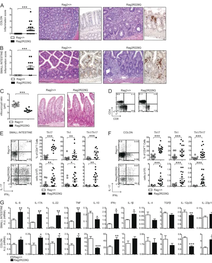
Analysis of intestinal pathology in Rag2R229Q/R229Q (herein referred to as Rag2R229Q) mice revealed different degrees of spontaneous colitis, wasting diarrhea, and rectal prolapse. The overall incidence of rectal prolapse in the Rag2R229Q colony was 5% in mice by 24 wk of age. No rectal prolapse was detected in Rag2+/+ littermates. Nonetheless, 70–80% of
all mutant mice showed substantial thickening of the colon throughout their entire length and microscopic evidence of inflammation (Fig. 1 A). Histologically, colonic inflamma-tion is characterized by crypt elongainflamma-tion, epithelial hyperpla-sia, and a large inflammatory cell infiltrate extending to the colonic lamina propria (LP), with occasional crypt abscesses. Remarkably, such morphological changes also affected the small intestine (SI) of the mutant mice with increased mu-cosal thickness and histological score and lower villus/crypt ratio (Fig. 1, B and C).
LP infiltrate consisted mainly of T lymphocytes (Fig. 1, A and B; and Fig. S1), particularly CD4+, with very few CD8+ T cells (Fig. 1 D). Correlating with mucosal thickening, abso-lute counts were three- to sixfold higher in the mutant mice than controls, in sharp contrast with the lymphoid depletion observed in the peripheral lymphoid organs. The proportion of intestinal Th17 cells was significantly higher in Rag2R229Q mice than Rag2+/+ mice, and mutants also had more IFN-γ– producing LP T cells compared with controls (Fig. 1, E and F). Furthermore, IFN-γ IL-17 double-producing T cells, fre-quently identified in the inflamed LP of patients and mu-rine models of IBD (Annunziato et al., 2007; Ahern et al., 2010), were abundant in Rag2R229Q mice (Fig. 1, E and F). To comprehensively characterize the gut inflammatory envi-ronment in the Rag2R229Q mice, we measured the expression of several cytokines. Transcript levels of IFN-γ, IL-17A, IL-6, IL-2, IL-1β, IL-22, and TNF were markedly up-regulated in the Rag2R229Q ileum and colonic tissues (Fig. 1 G). In con-trast, IL-10 and IL-12p35 levels were significantly reduced, whereas levels of IL-4, TGF-β, and IL-23p19 were not mark-edly different (Fig. 1 G). Overall, these results indicate that in Rag2R229Q mice, gut inflammation is mediated by Th1 and Th17 immune responses.
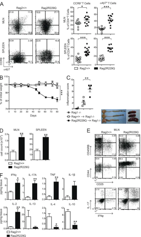
Hypomorphic CD4 T cells are sufficient to induce colitis CD4 T cells from mesenteric LNs (MLNs) of Rag2R229Q mice showed an activated memory phenotype (CD44hiCD62L−) and expressed the gut-homing receptors CCR9 and α4β7 (Mora and Von Andrian, 2006) at higher frequency com-pared with the same T cell population from Rag2+/+ mice (Fig. 2 A). Of note, we detected CCR9 and α4β7 expression in the splenic T effector cell compartment (Fig. 2 A), sug-gesting that gut inflammation resulted in the circulation of cells activated within the intestine of Rag2R229Q mice. In this line, CCR9+ T lymphocytes are increased in the circulation of IBD patients (Papadakis et al., 2001).
To determine whether Rag2R229Q CD4 T cells were sufficient to cause intestinal inflammation, total CD4+ T cells derived from either Rag2+/+ or Rag2R229Q MLNs were trans-ferred into Rag1−/− recipient mice. Analysis 10 wk afterward
revealed signs of colitis in all recipients of mutant CD4+ T cells. Rag2R229Q chimeric Rag1−/− mice showed significantly
greater weight loss, colon shortening, and inflammatory scores than Rag1−/− mice receiving Rag2+/+ CD4+ T cells (Fig. 2, B and C). Furthermore, MLNs and spleens in
on March 7, 2016
jem.rupress.org
Figure 1. Characterization of intestinal inflammation in Rag2R229Q mice. (A and B) Representative colon (A) and SI (B) sections from Rag2+/+ and
Rag2R229Q mice (8–12 wk old) stained with H&E and CD3 immunostaining. Histograms show the inflammation score in the colon (A) and SI (B) of n = 14–17
mice/group from four experiments. (C) Quantification of villous/crypt ratio of ileum from Rag2+/+ and Rag2R229Q mice (n = 10 from three experiments).
on March 7, 2016
jem.rupress.org
ients of Rag2R229Q T cells were enlarged (Fig. 2, C and D). Immunophenotype analysis revealed a marked expansion of activated T cells (CD44hi, CD45RBlow, and CD25+) in the MLN (Fig. 2 E) with a Th1/Th17 phenotype, similar to that observed in the Rag2R229Q mice at steady state (Fig. 2 E). When we examined the cytokine profile in the supernatants from cultured colonic tissue of transplanted mice, we found that the intestinal inflammatory response in the Rag2R229Q chimeric Rag1−/− mice is an exacerbated form of the disease
described in Rag2R229Q mice. Indeed, culture supernatants from the colon of Rag2R229Q chimeras contained two- to fivefold increased levels of IL-2, IFN-γ, IL-17, and TNF than supernatants of corresponding cultures from Rag1−/− mice
transferred with Rag2+/+ cells (Fig. 2 F). On the contrary, IL-4 and IL-10 levels were significantly reduced (Fig. 2 F). Overall, these results indicate that Rag2R229Q CD4 T cells are able to transfer the disease into immunodeficient hosts. Oral tolerance is impaired in Rag2R229Q mice
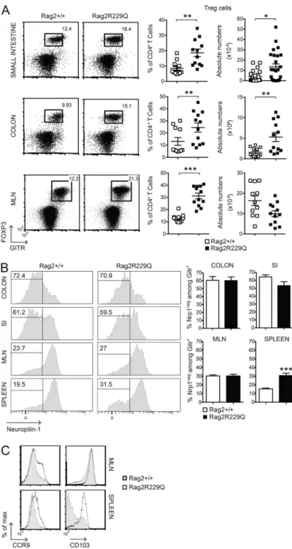
We found that Foxp3+GITR+ T reg cells were abundantly present in the inflamed intestine of Rag2R229Q mice. In fact, a significant increase in the frequency and number of T reg cells was detected in the SI and colonic LP (Fig. 3 A). In the MLNs, T reg cells were augmented only in frequency (Fig. 3 A), suggesting that T reg cell expansion is unique to the LP compartment. The gut mucosa is considered a pri-mary site for extrathymic T reg cell generation. We there-fore analyzed the expression of Neuropilin-1 (Nrp-1) as a marker of inducible T reg (iT reg) cells (Weiss et al., 2012; Yadav et al., 2012) in the gut and in secondary lymphoid organs of Rag2R229Q mice.
The percentages of Nrp-1low (iT reg) cells among GITR+ T reg cells were comparable between mutant and WT mice in both small and large intestines and higher than in MLNs (Fig. 3 B). The analysis of the spleens revealed that 30% of peripheral T reg cells in Rag2R229Q mice were constituted by iT reg cells, a significantly higher fraction than in control mice (Fig. 3 B). This result could reflect the poor generation of natural T reg cells in the thymus of mutant mice (Marrella et al., 2007). Alternatively, iT reg cell maintenance could be favored in the periphery under homeostatic conditions.
Mutant T reg cells from MLNs expressed the CD103 marker, indicative of antigen-specific expansion and differen-tiation, similar to control cells, but displayed enhanced expres-sion of the CCR9 receptor (Fig. 3 C). The analogous effector/ memory–like phenotype was observed in the peripheral T reg cell pool of Rag2R229Q but not of Rag2+/+ mice, and a major
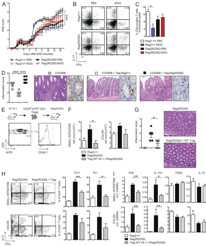
fraction (30–55% vs. 9–15% in the Rag2+/+ mice; P < 0.05) of these cells retained the CCR9 expression (Fig. 3 C), in-dicative of their gut tropism. Hence, T reg cells accumulate at the intestinal mucosal areas, although they fail to control the disease in Rag2R229Q mice. Thus, we assessed the capacity of Rag2R229Q mice to generate functionally competent T reg cells by the induction of oral tolerance (Dubois et al., 2009; Cassani et al., 2011). We used the well-established model of prevention of experimental allergic encephalomyelitis (EAE) that can be generated by orally supplementing mice with a peptide derived from myelin oligodendrocyte glycoprotein (MOG35–55; Chen et al., 1994). EAE developed in the Rag2R229Q mice supplemented orally with PBS, albeit at a lower extent than Rag2+/+ mice (Fig. 4 A). However, orally administered MOG35–55 prevented EAE only in Rag2+/+ but not in Rag2R229Q mice (Fig. 4 A). Th17 and Th1 cells are involved in EAE pathogenesis, and oral tolerance induction abrogates the generation of these pro-inflammatory T cells (Peron et al., 2010). Consistently, MOG-tolerized Rag2+/+ mice showed lower frequencies of IL-17– and IFN- γ–pro-ducing cells compared with nontolerized controls (Fig. 4 B). On the contrary, mutant mice did not show a decrease in the proportions of Th17 or Th1 cells upon oral MOG35–55 administration. In agreement, brain lymphocyte infiltration was reduced in the tolerized Rag2+/+ but not in the mutant animals (Fig. 4 C). These results suggested that Rag2R229Q iT reg cells could not control autoimmunity in this setting. To substantiate the hypothesis of functional impairment of Rag2R229Q T reg cells, we assessed their immunosuppressive potential in the severe combined immunodeficiency model of colitis induced by CD4+ CD45RBhi T cells (Powrie et al., 1994). Mice were sacrificed at week four after cell transfer, and IBD was assessed by weight loss (IBD mice: 21.1% ± 4.5; Rag2R229Q mice: 21.1% ± 2.6; WT mice: 12.6% ± 4.6; n = 6–8) and histological examination of the colon. The dis-ease severity was significantly attenuated when naive CD4 T cells were cotransferred with Rag2+/+ T reg cells (Fig. 4 D). On the contrary, Rag2R229Q T reg cells failed to protect lymphopenic mice from colitis development, despite the fact that they were readily detected in the gut tissues as in control mice (Fig. 4 D). In summary, these results demonstrate that T reg cell functionality is compromised in Rag2R229Q mice. Adoptive transfer of WT T reg cells attenuates gut inflammation in Rag2R229Q mice
To address whether the intestinal inflammation in Rag2R229Q is a consequence of T reg cell functional impairment, we
(D) Representative FACS plots showing the CD4+ and CD8+ T subsets within the CD3+ population from SI LP of Rag2+/+ and Rag2R229Q mice. (E and F)
Repre-sentative FACS plots, cumulative frequencies, and absolute numbers of SI (E) and colonic (F) LP CD4+IL-17+ (Th17), CD4+IFN-γ+ (Th1), and CD4+IL-17+IFN-γ+
(Th17/Th1) T cells from Rag2+/+ and Rag2R229Q mice. The numbers in the plots indicate the frequency of cells in each quadrant. Results are mean ± SEM.
Cumulative results of three independent experiments are shown (n = 11–20 mice/group). (G) Gene expression analysis of cytokines in the ileal and colonic tissues of mice. Target mRNA was normalized to Actb mRNA. RNA contents are shown as arbitrary units (A.U.). Data are representative results of three independent experiments with at least five mice per group. Bars, 100 µm. Values are mean ± SEM. *, P < 0.05; **, P < 0.01; ***, P < 0.001.
on March 7, 2016
jem.rupress.org
transferred sorted splenic CD25hiGITR+CD4+ T reg cells from Rag2+/+ (CD45.1) donors into Rag2R229Q (CD45.2) recipients and followed them for 4 wk. Transferred T reg cells expanded well and were readily detectable in the gut LP of Rag2R229Q mice (Fig. 4 E), indicating that T reg cells efficiently colonized the inflamed intestine. In the T reg cells transfer group, lymphocytic infiltration decreased both in the SI and colon (Fig. 4 F), resulting in a reduced inflammatory score (Fig. 4 G). Accordingly, Th1 and Th17 cell populations lowered in the gut of Rag2R229Q mice receiving Rag2+/+ T
reg cells (Fig. 4, H and I). However, production of IL-10 and TGF-β did not change (Fig. 4 I). Overall, these data confirm that impaired T reg cell function plays a role in the develop-ment of intestinal inflammation in Rag2R229Q mice.
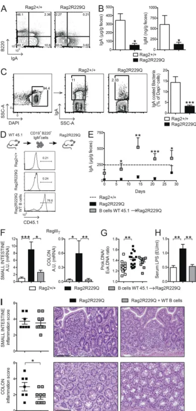
Mucosal B cell deficiency in Rag2R229Q mice causes enhanced
bacterial translocation and altered microbiota composition Intestinal B cells are poorly represented in Rag2R229Q mice. Peyer’s patches (PPs) are rudimentary, and few LP B220+ cells and IgA+ plasma cells could be detected (Fig. 5 A). In
agree-Figure 2. Rag2R229Q CD4 T cells have enhanced gut tropism and cause colitis. (A) Representative FACS
plots showing the expression of gut homing receptors, CCR9 and α4β7, on gated CD44hiCD62L−CD4+ T cells from
MLNs and spleen of Rag2+/+ and Rag2R229Q mice.
Num-bers in quadrants indicate the percentage of cells in each. Graphs show cumulative frequencies from one represen-tative experiment out of nine independent with n = 9–16 mice/group. (B) Body weight as a percentage of starting weight of Rag1−/− recipient mice after transfer of total
MLNs CD4+ T cells from Rag2+/+ or Rag2R229Q mice. (C, left)
Graph shows the colon inflammation scores of Rag1−/−
mice receiving Rag2+/+ or Rag2R229Q MLNs CD4+ T cells.
Right: Representative picture of macroscopic observation in colons and spleens. (D) Total cellularity of MLNs and spleens from Rag1−/− mice receiving Rag2+/+ or Rag2R229Q
MLN CD4+ T cells. (E) Representative FACS plots show
immunophenotype and cytokine production by CD4+ T
cells recovered from MLNs of recipient mice. (F) Cytokine profile in the supernatants from cultured colonic tissue of Rag1−/− mice adoptively transferred with Rag2+/+ or
Rag2R229Q CD4+ T cells. Data from B–F are representative
of two independent experiments with at least n = 4 an-imals. Values are mean ± SEM. *, P < 0.05; **, P < 0.01; ***, P < 0.001.
on March 7, 2016
jem.rupress.org
ment, fecal IgA and IgM levels were markedly diminished (Fig. 5 B), indicating a general deficiency in B cell function at the intestinal mucosal interface. As a consequence, a signifi-cant decrease of IgA-coated bacteria was detected in the large intestine of mutant mice (Fig. 5 C). Mucosal IgA operates immune exclusion of potentially harmful antigens and patho-gens and contains the gut microbiota (Macpherson et al., 2000; Macpherson and Uhr, 2004; Palm et al., 2014). Lack of IgA results in augmented serum LPS levels (Shulzhenko et al.,
2011). Quantification of bacteria adherent to the ileal mucosa by real-time PCR (RT-PCR; Proietti et al., 2014) showed a higher prokaryotic/eukaryotic DNA ratio in Rag2R229Q mice (Fig. 5 G) associated with increased serum LPS concentra-tions (Fig. 5 H), indicating that mucosal B cell deficiency in mutant mice resulted in enhanced mucosal colonization and peripheral translocation of bacterial products. To better evalu-ate the link between B cell deficiency and gut inflammation, we adoptively transferred naive CD45.1+B220+IgM+ WT B
Figure 3. Phenotype of T reg cells in Rag2R229Q mice.
(A) Frequency and absolute number of CD4+Foxp3+GITR+ (T reg)
T cells infiltrating the SI, colonic LP, and MLNs of Rag2+/+ and
Rag2R229Q mice (n = 11–25 from three independent experiments).
FACS plots show the coexpression of GITR+ and FOXP3+ gated on
CD4+ T cells. (B) Representative FACS analysis of Nrp-1
expres-sion by CD4+GITR+ T cells in the intestines and secondary
lym-phoid organs of Rag2+/+ and Rag2R229Q mice. Histograms show
cumulative data from two experiments (n = 7–19). The numbers in the histograms indicate the frequency of Nrp-1− cells (iT reg
cells). (C) Representative histogram plots depicting the expres-sion of CCR9 and CD103 markers on gated CD4+GITR+FOXP3+
from MLNs and spleens of Rag2+/+ and Rag2R229Q mice. Values are
mean ± SEM. *, P < 0.05; **, P < 0.01; ***, P < 0.001.
on March 7, 2016
jem.rupress.org
Figure 4. Oral tolerance is impaired in Rag2R229Q mice, and transfer of WT T reg cells attenuates gut inflammation. (A) EAE model of oral tolerance
in Rag2R229Q mice. EAE induction and progression was scored from days 0 to 26 in mice orally administered with MOG
35–55 or PBS. (B) Cytokine production
profile of CD4+ T cells. At day 26, the mice were sacrificed, and total splenocytes were stimulated ex vivo with MOG
35–55. The numbers in the plots indicate the
frequency of cells in each quadrant. (C) Frequency of CD4+ T cells infiltrating the brain of Rag2+/+ and Rag2R229Q mice. Data are shown from one representative
experiment out of two (n = 6–7 mice/group). (D) Colitis score and representative colonic sections from Rag1−/− transferred with CD45RB alone or with T reg
cells from Rag2+/+ and Rag2R229Q mice stained with H&E and CD3 immunostaining. Cumulative score results of two independent experiments are shown (n =
6–8 mice/group). (E) WT CD45.1+CD25hiGITR+CD4+ T reg cells were adoptively transferred into Rag2R229Q mice. Histogram plot show the CD45.1 expression on
GITR+CD4+ T cells in the LP of recipient Rag2R229Q mice. (F) Total cell counts from SI and colonic LP. (G) Representative colonic sections stained with H&E and
on March 7, 2016
jem.rupress.org
cells into adult Rag2R229Q recipients (Fig. 5 D). As shown in Fig. 5 E, transferred B cells generated IgA-producing plasma cells rapidly, reconstituting the secretory IgA compartment in recipient mice to the control level. Bacterial transloca-tion and intestinal inflammatransloca-tion were assessed 4 wk after the transfer. In agreement with a previous study reporting the expression of epithelial antimicrobial peptide RegIIIγ in re-sponse to mucosal IgA deficiency (Shulzhenko et al., 2011), we found that correction of IgA defect substantially reduced RegIIIγ mRNA level in the gut of Rag2R229Q recipient mice, suggesting diminished microbial stimulation of the intestinal epithelium (Fig. 5 F). Accordingly, bacterial adherence to ileal mucosa was diminished, although not significantly (Fig. 5 G). These results suggest that low-affinity T cell–independent IgA responses, such as those generated in mutant mice in the absence of T cell help (Cassani et al., 2010a), are not sufficient to contain more invasive bacteria, which tightly adhere to ep-ithelial cells in terminal ileum. Nonetheless, LPS translocation was visibly reduced in the recipients of WT B cells (Fig. 5 H). Interestingly, we found that decreased stimulation of the epi-thelium attenuated the inflammation in the colonic but not in the SI tissue (Fig. 5 I). Overall, these data suggest that B cells are required but not sufficient to maintain gut homeostasis.
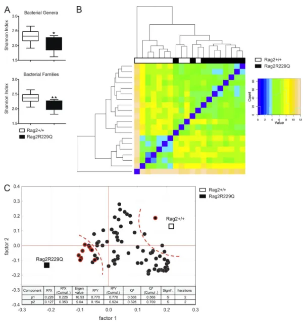
Defects in the adaptive immune compartment have pro-found impact on gut microbial ecology (Fagarasan et al., 2002; Kawamoto et al., 2014). We therefore evaluated the microbi-ota composition in Rag2R229Q mice and their cohoused WT littermates by 16S rRNA gene profiling. Overall, no marked variation was observed in the relative abundance of the dom-inant intestinal bacterial phyla (Firmicutes, Bacterioidetes, Actinobacteria, and Proteobacteria) between the two sets of mice (Fig. S2 A). However, we found that mutant mice had considerably less diverse bacterial communities at the family and genus levels compared with controls, as determined by the Shannon and Chao1 indexes of α-diversity (Fig. 6 A and not depicted). Moreover, hierarchical cluster analysis based on data of relative abundance of the bacterial families revealed that Rag2R229Q and Rag2+/+ mice formed approximate clus-tering according to their genotype (Fig. 6 B). Then, we ob-served that most of the differences in the relative abundances of single bacterial genera between Rag2R229Q and Rag2+/+ mice was due to a higher abundance in the group of WT mice (Fig. S2 B); in addition, we found that modified taxa be-longed most frequently to the phylum Proteobacteria (Fig. S2 B). Finally, to identify the bacterial genera that better describe the difference between genotypes, we performed a partial least squares (PLS1) regression analysis, which takes into con-sideration the overall composition of the microbiota, using genotype as categorical responses and the relative abundance of bacterial genera as continuous predictors. In accordance to
α-diversity analysis, PLS1 results confirmed the overall dearth of the microbiota richness in the Rag2R229Q mice compared with Rag2+/+ mice (Fig. 6 C). Furthermore, PLS1 analysis revealed that Rag2R229Q microbiota is mainly described by several genera of the phylum Proteobacteria (Fig. 6 C), pre-viously associated with chronic inflammatory condition in humans (Manichanh et al., 2012). Of note, segmented fila-mentous bacteria (Ivanov et al., 2009) were not present in our colonies, thus excluding their role in the Th17 polarizing gut environment of mutant mice (data not shown). Over-all, these data indicate that intestinal inflammation in mutant mice is associated with important alterations of the microbial communities in the gut.
Reducing intestinal bacterial load ameliorates gut inflammation in Rag2R229Q mice
To directly explore the role of microbial flora in gut
in-flammation in Rag2R229Q mice, we treated mice with
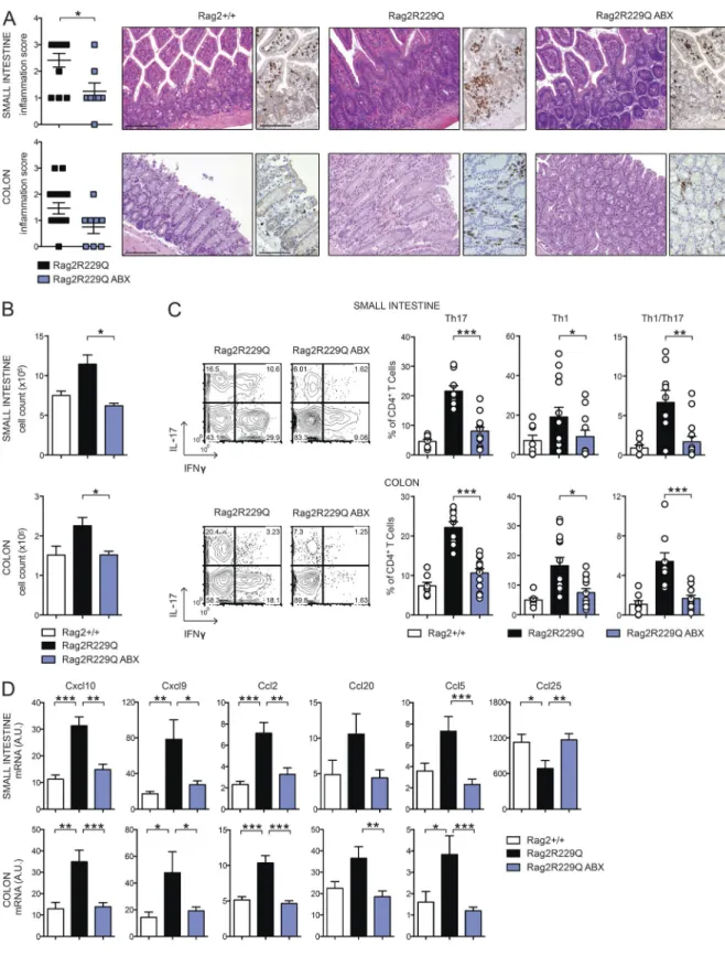
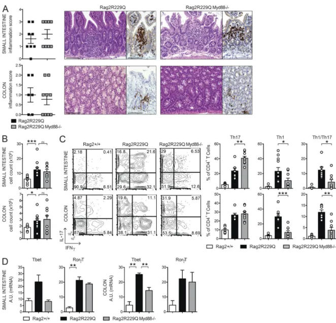
brospectrum ABXs. A regimen of three ABXs was ad-ministered to 8–12-wk-old mice for 4 wk. We did not ob-serve substantial changes in body weight between treated and untreated animals, thus ruling out potential toxicity related to the pharmacological treatment. In ABX-treated Rag2R229Q mice, we found amelioration of gut inflamma-tion associated with a resoluinflamma-tion of lymphocytic muco-sal infiltrates (Fig. 7 A) and diminished percentages and absolute numbers of IFN-γ– and IL-17–expressing CD4+ T cells in the LP and MLNs (Fig. 7, B and C; and not depicted). On the contrary, a minor reduction of these cell types was observed in the Rag2+/+-treated group (SI Rag2+/+ vs. Rag2+/+ ABX: Th17, 4.6 ± 2% vs. 3.5 ± 2.18%; Th1, 1.01 ± 1.06% vs. 1.27 ± 0.52%; Colon: Th17, 7.4 ± 2.46% vs. 3.1 ± 1.89%; Th1, 4.9 ± 1.99% vs. 3.85 ± 2.06%). Of note, concurrent deficiency of MyD88 (and consequent lack of most TLR signaling) did not protect from bowel inflammation. Rag2R229Q × MyD88−/− mice showed waste disease and analogous accumulation of in-filtrating inflammatory T cells to Rag2R229Q littermates (Fig. 8, A and B). Of note, lack of MyD88 abolished Th1 but not Th17 intestinal immune responses in Rag2R229Q mice, indicating that Th17 cell generation in mutants oc-curs independently from the TLR-MyD88–dependent sensing of commensal microbiota (Fig. 8, C and D).
Chemokines play a major role in recruiting immune cells to sites of injury. Consistent with the increased tissue infiltration and cytokine skewing, transcript levels of Th1 (Cxcl10 and Cxcl9)- and Th17 (Ccl20)-associated chemo-kines, as well as those of Ccl2 and Ccl5, were up-regulated in the bowels of Rag2R229Q mice (Fig. 7 D). However, upon ABX treatment, their expression returned to control values
colitis score in Rag2R229Q mice recipient mice and controls (n = 5 from two experiments). (H) Representative FACS plots and frequency of LP Th17 and Th1 cells
from SI and colon. (I) SI and colonic tissue expression of cytokines. RNA contents are shown as arbitrary units (A.U.). Data shown are cumulative results from two independent experiments with nine mice per group. Bars, 100 µm. Values are mean ± SEM. *, P < 0.05; **, P < 0.01; ***, P < 0.001.
on March 7, 2016
jem.rupress.org
(Fig. 7 D). Notably, analogously to Crohn’s disease (Papadakis et al., 2001), Ccl25 expression was lower in the Rag2R229Q ileum with respect to control mice (Fig. 7 D), whereas it reached normal levels in ABX Rag2R229Q mice (Fig. 7 D). Collectively, these results show that reducing microbial load beneficially affects Rag2R229Q mice.
Reducing intestinal bacterial load ameliorates systemic autoimmunity in Rag2R229Q mice
The same Th cells polarization dominating the intestinal im-mune response was evident in the spleens of mutant mice (Fig. 9 A). Hence, we examined the possibility that the anti-inflammatory effect of ABXs extended beyond the
intesti-Figure 5. In Rag2R229Q mice, mucosal B cell deficiency cor-relates with enhanced bacterial translocation. (A) Representative
FACS plots of B220+ B cells and IgA+ plasma cells infiltrating the SI
LP of Rag2+/+ and Rag2R229Q mice. Numbers indicate the frequency
of cells in each quadrant. (B) Fecal IgA and IgM were further ana-lyzed by ELI SA. Cumulative results from three different experiments with at least n = 12 mice are shown. (C) FACS staining of IgA-coated bacteria in fecal pellets of Rag2+/+ and Rag2R229Q mice. Numbers in
quadrants indicate the percentage of cells in each. Cumulative results from analysis of n = 10 mice from two experiments are reported in the bar graph. (D) Expression of CD45.1 on LP B220+ cells in recipient
Rag2R229Q mice. WT CD45.1+CD19+ B220+IgM+ cells were adoptively
transferred into Rag2R229Q mice. (E) Fecal IgA levels in recipient
Ra-g2R229Q mice and controls at the indicated time points after the cell
transfer. The dashed line indicates the mean level of IgA in Rag2+/+
mice. Cumulative results from two different experiments with n = 5–10 mice are shown. (F) Gene expression analysis of antimicrobial RegIIIγ in the ileal and colonic tissues of mice. RNA contents are shown as arbitrary units (A.U.). Cumulative results from analysis of n = 8–20 mice in two experiments are reported in the graphs. (G) Quan-titative PCR analysis of ileal adherent bacteria in Rag2+/+, Rag2R229Q,
and Rag2R229Q mice adoptively transferred with WT B cells expressed
as prokaryotic/eukaryotic DNA ratios. Cumulative results from two independent experiments with n = 10–16 mice/group are shown. (H) Serum LPS concentrations. Data shown are cumulative of two exper-iments with at least 10 mice/group. (I) Representative SI and colon sections from Rag2R229Q mice and Rag2R229Q mice adoptively
trans-ferred with WT B cells stained with H&E. Bars, 100 µm. Histograms show the SI and colonic inflammation score. Cumulative results of two experiments with n = 7–10 mice/group are reported. Values from E–I are mean ± SEM. *, P < 0.05; **, P < 0.01; ***, P < 0.001.
on March 7, 2016
jem.rupress.org
nal mucosa. We found that frequencies of Th1 and Th17 cells were considerably reduced in the spleens of ABX Rag2R229Q mice (Fig. 9 A), whereas their proportion was low and unaf-fected in ABX Rag2+/+ mice, indicating a role for microbial flora in the maintenance of circulating, other than intesti-nal, effector Rag2R229Q T cell populations. Interestingly, ABX Rag2R229Q mice also showed significantly decreased frequen-cies of systemic CCR9+ CD4 T cells (Fig. 9 B), indicating that resolution of gut inflammation normalized the circulat-ing pool of these cells.
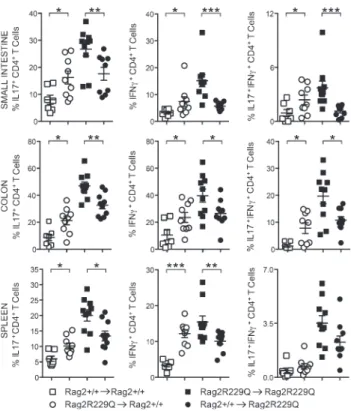
To confirm the influence of microbial flora on the inflammatory T cell phenotype of Rag2R229Q mice, inde-pendently from the host genetic susceptibility, we recolo-nized ABX-treated adult Rag2+/+ mice with fecal materials obtained from mutant mice (Rag2R229Q → Rag2+/+). Fecal transplant was repeated twice 1 wk apart, and mice were sac-rificed 3 wk later. Recolonized ABX Rag2+/+ mice showed augmented frequencies of Th1 and Th17 cells in the gut LP as well as spleens with respect to ABX Rag2+/+ mice receiving feces from Rag2+/+ mice (Rag2+/+→ Rag2+/+; Fig. 10). On
Figure 6. Rag2R229Q mice show altered microbiota composition. Adults Rag2+/+ and Rag2R229Q littermates were cohoused for 2 mo after the weaning.
Cecum fecal content was analyzed for microbiota profiling with 16s sequencing. (A) Box and whisker plots of the Shannon diversity index calculated at the bacterial genera and family level. (B) Heat map visualization at the family level, which was obtained applying the DESeq package on the taxonomic data produced by BioMaS. (C) Correlation between mouse genotype and bacterial genera in fecal samples. X and Y loading plots (factors 1 and 2) were obtained by partial least squares analysis performed by using the genotype as categorical response and relative abundance of bacterial genera as continuous predic-tors. Taxa belonging to the phylum Proteobacteria are shown with a red circle. 92% of sum of squares is explained by the first two extracted components. *, P < 0.05; **, P < 0.01.
on March 7, 2016
jem.rupress.org
Figure 7. ABX treatment dampens gut inflammation in Rag2R229Q mice. (A) Representative SI and colonic sections from controls and ABX-treated
Rag2R229Q stained with H&E and CD3 immunostaining. Bars, 100 µm. Histograms show the inflammation score in gut tissues. Data are cumulative results
of three independent experiments (n = 8–15). (B) Total cell counts from SI and colonic LP of ABX-treated Rag2R229Q and controls. (C) Representative FACS
plots and frequency of SI and colonic LP IL-17+, IFN-γ+, and IL-17+IFN-γ+ CD4 T cells. Numbers in the plots indicate the frequency of cells in each quadrant.
on March 7, 2016
jem.rupress.org
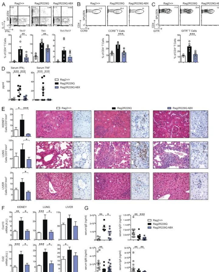
the contrary, ABX Rag2R229Q mice recolonized with Rag2+/+ feces displayed considerably fewer intestinal and peripheral Th1/Th17 cells compared with controls (Fig. 10). Like effec-tor T cells, intestinal microbiota contributes to local induc-tion of T reg cells (Round and Mazmanian, 2010; Atarashi et al., 2011), and ABX-treated mice had fewer intestinal T reg cells (Rosser et al., 2014). In agreement with these published data, we found that the proportion of GITR+ T reg cells was reduced in the LP and MLNs of ABX mice (not depicted). However, a drop of their splenic frequency was observed in the ABX Rag2R229Q but not Rag2+/+ mice (Fig. 9 C; Rag2+/+ vs. Rag2+/+ ABX: 11.7 ± 1.43% vs. 12.2 ± 5.36%), suggest-ing that most T reg cells in Rag2R229Q mice are microbiota dependent. Overall, these findings point to a substantial role of gut microenvironment and commensal bacteria in the OS pathogenesis. Next, we assessed whether the ABX regimen protected against the multisystemic autoimmune-like mani-festations of mutant mice. Indeed, Rag2R229Q mice show im-portant infiltrates of effector T cells in the skin, kidney, lung, and liver, which correlate with abnormal concentration of IFN-γ–induced CXCL10 in the serum (Cassani et al., 2010a; Marrella et al., 2012). Importantly, the decline of circulat-ing inflammatory T cells in ABX Rag2R229Q mice lowered IFN-γ and TNF serum concentrations (Fig. 9 D). We then examined the CD3+ T cell infiltrates in target organs. A de-creased number of T cells was observed in the kidney, lung, and liver of ABX Rag2R229Q mice compared with untreated controls (Fig. 9 E). In line with these results, tissue expression of inflammatory chemokines (Ccl2 and Cxcl10) was lower in ABX mutant mice (Fig. 9 F). On the contrary, skin inflam-mation was not affected by the ABX treatment (not depicted), suggesting that this district did not benefit from the reduced gut bacterial load and/or that an independent pathogenetic mechanism sustained tissue inflammation.
ABX treatment reduces hyper-IgE in Rag2R229Q mice
CD4 T cells have a pivotal role in B cell abnormalities, in-cluding plasma cell expansion and high IgE serum levels (Cassani et al., 2010a). Given that reducing microbial load in mutant mice had a clear impact on peripheral T cell pheno-type, we asked whether ABX administration could impinge on B cell defects. In Rag2+/+ mice, microbiota depletion re-duced serum IgA levels, whereas other Ig isotypes remained unchanged (mean values of Rag2+/+ vs. Rag2+/+ ABX: IgA, 8.5 × 10⁵ vs. 2.2 × 10⁵ ng/ml; IgG1, 5.4 × 10⁵ vs. 3.6 × 10⁵ ng/ml; IgM, 2.2 × 10⁶ vs. 2.6 × 10⁶ ng/ml). Remark-ably, in Rag2R229Q mice, the long-term treatment resulted in a significantly reduced serum IgE (Fig. 9 G). A similar trend was observed for IgA and IgG, but not for IgM (Fig. 9 G). Of note, frequencies and numbers of peripheral B cells were
not affected by ABX treatment. Because hyper-IgE consti-tutes a hallmark of OS, these results indicate that microbiota can constitute a pharmacological target for this pathologi-cal trait of the disease.
DIS CUS SION
Here, we investigated the role of intestinal immune responses and gut microbiota in the pathogenesis of OS. A large fraction of Rag2R229Q mice developed bowel inflammation character-ized by marked infiltration of T cells in the LP. We found that lymphocytes are sufficient for disease initiation. In fact, CD4 T cells, either isolated from colitic mice or not, were able to transfer the disease into immunodeficient hosts. Cytokine production profiling of gut infiltrating lymphocytes revealed a dominant mixed Th1/Th17 skewing, frequently implicated in the pathogenesis of IBD and experimental colitis (Powrie et al., 1994; Berg et al., 1996; Fuss et al., 1996; Ahern et al., 2010). An abundant presence of Th17 cells, also producing the IL-22 cytokine, might also contribute to the aberrant ex-pression of antimicrobial peptide RegIIIγ (Liang et al., 2006). Multiple studies revealed the importance of T reg cells for the maintenance of immune tolerance in the gastroin-testinal tract owing to the constant immune stimulation by commensal flora and food antigens (Izcue et al., 2009; Pe-dros et al., 2016). Impaired intestinal tolerance is believed to contribute to inflammation in response to microflora in IBD (Xavier and Podolsky, 2007). Several experiments in this study addressed the role of T reg cells in the gut homeostasis of Rag2R229Q mice. We showed that T reg cells accumulate in the intestine but fail to control disease in mutant mice. In the adoptive transfer model of colitis, Rag2R229Q T reg cells cotransferred with WT naive CD45RBhi CD4 T cells did not prevent disease development in Rag1−/− mice. Interestingly,
similar failure in controlling ongoing autoimmune responses has been described for Nrp-1low iT reg cells generated in inflammatory and lymphopenic environment (Yadav et al., 2012). On the contrary, the transfer of a limited number of Rag2+/+ T reg cells significantly ameliorated intestinal in-flammation by dampening Th1 and Th17 immune responses in Rag2R229Q hosts. Thus, it is conceivable that the absence of functional T reg cells plays a major role in the pathogenesis of intestinal inflammation in Rag2R229Q mice.
Distinct effector mechanisms from those operating in the Rag2R229Q T reg cells may be required to control mucosal inflammation. These may include the expression of CD62L and CCR7 (Fu et al., 2004; Schneider et al., 2007), which are necessary for homing to the inductive sites, and produc-tion of IL-10, which is essential to limiting the inducproduc-tion of pro-inflammatory Th1 and Th17 cells at the intestinal mucosa interface (Geuking et al., 2011). IL-10 mRNA levels were
(D) Expression of the indicated chemokines in the ileal and colonic tissues assessed by quantitative RT-PCR. Normalized values are indicated as arbitrary units (A.U.). Data from B–D are representative results of four independent experiments (n = 8–13 animals/group). Values are mean ± SEM. *, P < 0.05; **, P < 0.01; ***, P < 0.001.
on March 7, 2016
jem.rupress.org
significantly reduced in the intestines of Rag2R229Q mice, and IL-10 is also required for oral tolerance induction in different models (Cong et al., 2004; Navarro et al., 2011). Finally, iT reg cells derived from an oligoclonal population of CD4+ T cells may be restricted in their ability to effectively suppress autoimmune disease and maintain tolerogenicity to microbi-ota (Huter et al., 2008; Nishio et al., 2015).
Mucosal T reg cells, together with secretory IgAs, crit-ically preserve the gut barrier and control diversification of commensal species, thus ensuring host–bacterial mutualism (Kawamoto et al., 2014). Conversely, biased expansion of
cer-tain bacterial species impairs epithelial integrity and induces hyperactivation of the immune system, also promoting the generation of pro-inflammatory T cell subsets (Kawamoto et al., 2012; Kamada et al., 2013). We demonstrated that the hy-pomorphic Rag defect in B cells favors microbial access to the LP and circulation and considerably impacts on the diversity of the microbial communities in the gut. Our findings cor-relate with previous observations in mice harboring immune deficiencies affecting inflammatory responses in adaptive im-mune cells (Garrett et al., 2007; Kawamoto et al., 2014). We also showed that such altered microbiota from Rag2R229Q mice
Figure 8. Myd88 deficiency limits Th1 but not Th17 responses in the gut of Rag2R229Q mice. (A) Representative SI and colonic section from control
and Rag2R229QMyd88−/− stained with H&E and CD3 immunostaining. Bars, 100 µm. Histograms show the inflammation score in the SI and colon. Data are the
cumulative results of two independent experiments with n = 7–9. (B) Total cell counts from SI and colonic LP. (C) Representative FACS plots and frequencies of CD4+IL-17+, CD4+IFN-γ+, and CD4+IL-17+IFN-γ+ T cells from SI and colon LP. Numbers in the plots indicate the frequency of cells in each quadrant. Data
represent one of two independent experiments with 6–11 mice/group. (D) Expression of Tbet (Tbx21) and RorγT (Rorc) on sorted CD4+ T cells from SI and
colonic LP. RNA contents are shown as arbitrary units (A.U.). Data represent three independent experiments each using pooled cells from at least five mice per group. Values are mean ± SEM. *, P < 0.05; **, P < 0.01; ***, P < 0.001.
on March 7, 2016
jem.rupress.org
Figure 9. ABX treatment ameliorates systemic autoimmunity in Rag2R229Q mice. (A) Representative FACS plots and frequency of IL-17+–, IFN-γ+–,
and IL-17+IFN-γ+–producing CD4 T cells in the spleens of Rag2+/+, Rag2R229Q, and ABX-treated Rag2R229Q mice. (B) Representative FACS plots and frequency
of splenic CCR9+ T cells within the CD44+CD62L− CD4+ T cell gate. (C) Representative FACS plots and frequency of splenic CD4+GITR+ T reg cells. Data are
on March 7, 2016
jem.rupress.org
was sufficient to promote a skewed Th1/Th17 pro-inflamma-tory phenotype into Rag2+/+ mice. This observation indicates that Rag2R229Q microbiota has a pathogenic potential.
The clinical benefits deriving from long-term ABX dosing formally implicates intestinal microbes in the disease pathogenesis. We showed that both mucosal and circulating pro-inflammatory Th1 and Th17 cell subsets were signifi-cantly reduced in ABX-treated mice, establishing that skewed polarization of adaptive immunity in Rag2R229Q mice was mi-crobiota dependent. Of note, CCR9-expressing T cells were similarly affected by the treatment, consistent with resolution of gut inflammation. However, because CCR9 expression is acquired by T cells upon priming by GALT-derived dendritic cells (Mora, 2008), we propose that its presence on the vast majority of circulating T cells in Rag2R229Q mice might re-flect their original intestinal differentiation.
Although CD4+ T cells from Rag2R229Q mice are able to transfer colitis to immunodeficient hosts, it still remains to be determined whether an aberrant non–T cell population is required for the priming of dysregulated T cells. On this point, it has been shown that both innate and adaptive im-mune system stimulation by microbiota are required for driv-ing spontaneous T cell proliferation and induction of colitis in immunodeficient mice (Feng et al., 2011). Furthermore, we previously reported perturbed cell trafficking and altered lymphoid distribution of dendritic cells in Rag2R229Q mice, which tend instead to ectopically accumulate and halt at the environmental interfaces (Maina et al., 2013). It seems there-fore logical to envisage a crucial role for innate immune cells in colitis development in mutant mice.
In contrast to previous publications describing its pro-tective effects in different models of IBD induced by immune deficiencies (Rakoff-Nahoum et al., 2006; Rivas et al., 2012), we found that inhibition of MyD88-dependent pathways did not restrain intestinal inflammation in Rag2R229Q mice. Rather, Rag2R229Q × MyD88−/− mice displayed worsening of wasting syndrome and unaffected accumulation of mucosal lymphocytic infiltrates. These results, however, are consistent with the intricate effect of MyD88 deficiency on host–mi-crobial mutualism (Slack et al., 2009; Vaishnava et al., 2011). Interestingly, the absence of MyD88 signaling uncouples, in our model, the molecular requirements for generation of Th1 and Th17 cells in the gut. Despite observations that MyD88 ablation is required for the induction of both Th1 and Th17 cell responses (Fukata et al., 2008; Schenten et al., 2014), our
data showed that signaling through MyD88 is crucial for Th1 immune responses yet has a nonessential role for generation of Th17 cells. Therefore, multiple sensors may be involved in the development of these cells in the gut. On this line, it has been suggested that bacterially produced ATP can directly augment intestinal Th17 cell differentiation (Atarashi et al., 2008).
In addition to gut inflammation, reducing intestinal mi-crobial load visibly ameliorated multisystem autoimmunity in Rag2R229Q mice, confirming the critical role of gut flora in sustaining chronic immune inflammation and autoimmune disease onset/progression in genetically susceptible hosts. The evidence that this inflammatory immune phenotype is transmissible to an intact immune system in a microbiota-de-pendent manner leads us to speculate that pharmacological gut decontamination might constitute a valid strategy to limit side effects of hematopoietic stem cell transplantation in these patients (Vossen et al., 2014). In contrast, given the importance of the intestinal microbiota, interventions aimed at promoting intestinal diversity may improve immunological outcomes in OS transplant settings. Replenishment of com-mensal bacterial populations through fecal microbiota trans-plantation or targeted flora reintroduction may theoretically be effective, although these procedures still need to be care-fully evaluated in a transplant context. Alternatively, the use of dietary supplements, which may help to maintain beneficial bacteria (Gerbitz et al., 2004), might be considered to avoid episodes of dominance of harmful bacterial species during posttransplant recovery (Lane et al., 2015) and to promote healthy mucosal responses.
Importantly, our results showed that reducing the micro-bial load is beneficial for normalizing IgE levels in Rag2R229Q mice. Because our previously published data indicated that T cells in mutant mice do have a crucial role in B cell abnormal-ities (Cassani et al., 2010a), diminished IgE levels after ABXs might reflect the lower serum concentration of cytokines pro-moting class switch recombination. Interestingly, it has been recently proposed that IgE production is strongly dependent on microbial exposure during early life and that a critical level of microbial diversity is required to establish the proper immu-noregulatory networks to inhibit IgE production (Cahenzli et al., 2013). Thus, it is conceivable that reduced microbiota diversity in Rag2R229Q mice also impinges the immune reg-ulation that functions to maintain IgE levels at the baseline.
In summary, our findings might provide novel insights into the pathogenesis of OS. We showed in a murine model
resentative results of four independent experiments with at least six animals per group. (D) Levels of IFN-γ and TNF in the serum of ABX-treated Rag2R229Q
mice and controls were analyzed in three independent experiments (n = 8–13). Each serum sample was run in duplicate. (E) Representative kidney, lung, and liver sections from controls and ABX Rag2R229Q stained with H&E and CD3 immunostaining. Bars, 50 µm. Histograms show quantification of infiltrating
CD3+ T cells in the kidney, lung, and liver sections. (F) Quantitative PCR analysis of chemokine expression in the kidney, liver, and lung of Rag2+/+, Rag2R229Q,
and ABX-treated Rag2R229Q mice. RNA contents are shown as arbitrary units (A.U.). Results from two experiments with n = 4–9 mice analyzed are shown. (G)
Serum levels of IgE, IgG1, IgM, and IgA were analyzed by ELI SA in Rag2+/+, Rag2R229Q, and ABX-treated Rag2R229Q mice. Data shown are cumulative results
from four independent experiments with at least 11 mice/group. For IgE analysis, data are representative results of two independent experiments with five to eight mice/group. All values are mean ± SEM *, P < 0.05; **, P < 0.01; ***, P < 0.001.
on March 7, 2016
jem.rupress.org
that hypomorphic Rag mutation compromises gut barrier in-tegrity and alters microbiota composition. Furthermore, the loss of T cell tolerance to commensal microbiota leads to gut Th17/Th1 inflammation which, in turn, sustains dysbiosis in a well-recognized regulatory loop.
Rag2R229Q mice represent an important tool to investi-gate whether intestinal microbiota can be manipulated thera-peutically to dampen immune activation and perpetuation of inflammation. Furthermore, they may help to determine the extent to which a limited adaptive immune system also controls microbial community composition in other organ systems, such as the skin, and to clarify whether these other resident commu-nities of indigenous microorganisms shape local immune cell differentiation and similarly influence the host health. MAT ERI ALS AND MET HODS
Mice and treatments. 129Sv/C57BL/6 knock-in Rag2R229Q mice were backcrossed to C57BL/6 strain for four genera-tions. The colony was maintained onsite with heterozygous breeders, and littermates were kept in the same cages until weaning at 4 wk of age. Rag2R229QMyd88−/− double-mutant
mice and littermates controls were generated from heterozy-gous–heterozygous crossing and maintained onsite as
de-scribed for single mutants. All mouse strains were used for experiments between 8 and 13 wk of age, and, unless indi-cated otherwise, adult littermates were raised in separate cages according to the genotype after weaning. Rag1−/− (Rag1tm1Mom/J) mice were obtained from The Jackson Laboratory and C57BL/6 Ly5.1 mice from Charles River. Mice were main-tained under specific pathogen–free conditions at the Istituto Clinico Humanitas. To deplete gut flora, adult Rag2+/+ and Rag2R229Q mice, housed in separate cages, were treated daily with an ABX cocktail containing 2.5 mg metronidazole, 2.5 mg ampicillin, and 1.25 mg vancomycin in 200-µl/mouse doses by oral gavage for 4 wk. All animal procedures were performed according to protocols approved by the Istituto Clinico Humanitas and the Italian Institutional Animal Care and Use Committee.
Induction of oral tolerance and EAE. Mice were fed daily 0.25 mg MOG peptide (MOG35–55) via oral gavage for 7 d as described previously (Cassani et al., 2011). Mice were then injected s.c. in the flank with 100 µg MOG35–55 in 0.15 ml PBS emulsified in an equal volume of CFA containing 4 mg Mycobacterium tuberculosis H37 RA/ml (Difco). Pertussis toxin (200 ng/mouse/injection; List Biological Laboratories) was administered i.p. at the time of immunization and 48 h later. Animals were scored for EAE as follows: 0, no disease; 1, tail paralysis; 2, hind limb weakness; 3, hind limb paralysis; 4, hind limb plus forelimb paralysis; 5, moribund.
Induction of colitis by adoptive T cell transfer. CD4+ T cells were isolated by pooled spleens and MLNs of 10-wk-old Rag2+/+ or Rag2R229Q mice using CD4-specific magnetic beads via negative selection according to the manufacturer’s instructions (Miltenyi Biotec). Enriched CD4 T cells were subsequently sorted into naive CD4+CD25−CD45RBhi and regulatory CD4+CD45RB−CD25hi populations (>98% pu-rity) using a cell sorter (FAC SAria; BD). For colitis induction, 4 × 10⁵ WT CD4+CD25−CD45RBhi naive T cells were i.p. transferred into Rag1−/− mice alone or with 1.5 × 10⁵ CD4+CD25hi T reg cells from Rag2+/+ or Rag2R229Q mice. Recipient mice were weighted twice a week and sacrificed at week four after the transfer, when IBD was diag-nosed by severe weight loss.
Evaluation of IgA-coated bacteria by FACS. Fecal pellets were suspended in filtered PBS, homogenized, and centri-fuged at 700 g for 5 min to remove large particles from bac-teria. Then, supernatants were collected and centrifuged at 8,000 g for 10 min to remove unbound Ig’s. The bacterial pellet was suspended in PBS 1% BSA and stained with PE-la-beled anti–mouse IgA on ice for 30 min. Bacteria was sus-pended in 4% formalin overnight at 4°C for fixation and resuspended in DAPI/PBS for acquisition at a cell analyzer (FAC SCanto II; BD) using forward scatter (FSC) and side scatter parameters in logarithmic mode. Data were analyzed with FlowJo software (version 7.6.5; Tree Star).
Figure 10. Rag2R229Q gut microbiota promotes a pro-inflammatory Th1-Th17 skewing. ABX-treated adult mice were recolonized with fecal
matters from Rag2+/+ or Rag2R229Q donors. Graphs show the percentages of
intestinal and splenic CD4 T cells producing IL-17 and/or IFN-γ cytokines after fecal transplantation. Data are representative results of two indepen-dent experiments with 7–10 mice/group. Values are mean ± SEM. *, P <
0.05; **, P < 0.01; ***, P < 0.001. on March 7, 2016
jem.rupress.org
ELI SA assay. For evaluation of fecal Ig’s, feces were collected, weighed, and resuspended in 1 ml PBS containing 0.1% so-dium azide and protein inhibitors. After overnight incubation, fecal pellets were centrifuged at 10,000 g for 15 min, and supernatants were collected. Fecal suspensions were stored at −20°C. Levels of fecal IgA and IgM were measured by a mouse IgA/IgM ELI SA quantitation set (Bethyl Laboratories, Inc.) according to the manufacturer’s instructions. Serum lev-els of IgG1, IgA, and IgM were measured by a multiplex assay kit (Beadlyte Mouse Immunoglobulin Isotyping kit; EMD Millipore) according to the manufacturer’s instructions and run using a Bio-Plex reader (Bio-Rad Laboratories). Levels of IgE were measured in sera by ELI SA assay (BD). Cytokine levels in sera were measured using a specific ELI SA kit for mouse IFN-γ and TNF (R&D Systems). For cytokine deter-mination, colonic tissues were incubated at 37°C for 48 h. Cultured supernatants were collected after 48 h, and cytokine concentrations were determined by ELI SA (R&D Systems) and normalized for tissue weight.
RT-PCR. Total RNA was extracted from 1 cm of ileal and colonic tissues. Tissues from intestines, kidney, lung, and liver were homogenized in PureZOL reagent (Bio-Rad Labora-tories) using TissueLyser II (QIA GEN). RNA was extracted using the RNeasy Lipid Tissue kit (QIA GEN), and cDNA was synthetized using the High-Capacity cDNA Reverse Transcription kit (Thermo Fisher Scientific). Quantitative RT-PCR was performed on a CFX384 system (Bio-Rad Laboratories) with the SYBR green master mix (Thermo Fisher Scientific). For sorted cells, total RNA was extracted using the RNeasy Plus Micro kit (QIA GEN). RNA was retrotranscribed using the High-Capacity cDNA Reverse Transcription kit followed by a preamplification PCR using TaqMan PreAmp Master Mix (Applied Biosystems). Quanti-tative RT-PCR was performed using a RT-PCR system (ViiA 7; Thermo Fisher Scientific) with TaqMan probes (Table S1). The relative amounts of mRNAs were calculated as 2−ΔCT and expressed as arbitrary units. Each sample was analyzed in duplicate, and the relative level of expression was determined by normalization to β-actin (Actb) ribosomal RNA.
Quantification of mucosal adherent bacteria by RT-PCR. The quantification of prokaryotic/eukaryotic ratios was per-formed as described previously (Proietti et al., 2014). In brief, the latest segment of terminal ileum was collected and di-gested in lysis buffer (10 mM Tris, 100 mM NaCl, 10 mM EDTA, 0.5% SDS, and 0.4 mg/ml proteinase K). Total DNA was extracted with the isopropanol-ethanol method. The mi-crobial load was determined by RT-PCR (SYBR green) using specific primers for microbial 16s and genomic 18s. LPS measurement. Sera were collected from Rag2+/+ and Rag2R229Q mice in endotoxin-free conditions. LPS concen-tration was determined by Limulus amebocyte lysate assay (Lonza) according to the manufacturer’s instructions.
LP preparation. SIs and colons were flushed to remove con-tents, opened longitudinally, and cut into 3–5-mm pieces. PPs were removed from SIs. Tissues were washed in HBSS buffer (0.5 M EDTA, 1 mM DTT, 15 mM Hepes, and 10% FCS) under agitation for 20 min at 37°C to remove the epithelial layer. Intestine pieces were then Liberase-digested (0.15 mg/ ml; Roche) for 30 min at 37°C. Cells were washed and passed through a 100-µm cell strainer. The cell suspension obtained was loaded into a 44/67% Percoll gradient (GE Healthcare) and centrifuged at 600 g for 20 min at room temperature. Lymphoid fractions were collected at the interphase of the Percoll gradient and used for flow cytometry stainings. Cell isolation and adoptive transfer. Single-cell suspensions were prepared from pooled MLNs harvested from Rag2R229Q and Rag2+/+ mice. Total CD4 T cells were negatively selected using the CD4 T Cell Isolation kit (Miltenyi Biotec) accord-ing to the manufacturer’s instructions (purity > 95%). Puri-fied CD4+ T cells (4 × 10⁶) were then i.v. injected into Rag1−/− mice. CD25hiGITR+CD4+ T reg cells were sorted from splenic cell suspension derived from WT CD45.1 mice using the FAC SAria cell sorter. Sorted cells (5 × 10⁵) were then injected i.v. into Rag2R229Q mice. The recipient mice were analyzed 4 wk after the transfer. For B cell adoptive transfer, total CD19+ B cells were isolated from pooled spleens, MLNs, and PPs derived from WT CD45.1 mice with anti-CD19 magnetic beads via positive selection (Miltenyi Biotec). B220+IgM+IgA− B cells were then sorted from the enriched CD19+ fraction (purity > 98%) using the FAC SAria cell sorter. Rag2R229Q mice were i.v. transferred with 30 × 106 sorted B cells. Recipient mice were monitored for B cell re-constitution and were sacrificed 4 wk after the transfer. Histological analysis. 2-µm-thick formalin-fixed and paraf-fin-embedded tissue sections from liver, lung, kidney, and gut were used for routine hematoxylin and eosin (H&E) staining and immunostains. In brief, sections were dewaxed and rehy-drated, endogenous peroxidase activity was blocked by 0.3% H2O2/methanol for 20 min, and heat-induced antigen re-trieval was obtained by microwave or thermostatic bath treat-ment using EDTA buffer, pH 8.0, followed by 1-h incubation with primary antibody rabbit anti-CD3 (1:100; Dako) or rat anti-FoxP3 (1:100; eBioscience). Signal was revealed by using Real EnVision Rabbit-HRP (Dako) or Rat-on-Mouse HRP-Polymer (Biocare Medical) detection system followed by diaminobenzidine and nuclei counterstained with hema-toxylin. Digital imagines were acquired by a camera (DP70; Olympus) mounted on a microscope (Bx60; Olympus) using CellF Imaging software (Soft Imaging System GmbH). Quan-tification of CD3+ cells was performed on representative sec-tions of liver, lung, and kidney acquired by Aperio ScanScope CS Slide Scanner (Aperio Technologies). Total area of the sec-tions and number of CD3+ cells were obtained by using Ape-rio ImageScope Software, and the absolute number of CD3+ cells was expressed as the number of CD3+ cells/mm2. The
on March 7, 2016
jem.rupress.org
degree of inflammation of the gut (colon and SI) was dou-ble-blind graded using combined scores, including the grade of inflammation (grade 0, no evidence of inflammation; grade 1, low; grade 2, moderate; grade 3, high; grade 4, intense and diffuse level of inflammation), the structural changes of the glands (grade 0, absence; grade 1, focal; grade 2, partial; grade 3, diffuse level of structural changes), and the goblet cell de-pletion (grade 0, no loss; grade 1, low; grade 2, moderate; grade 3, diffuse level of goblet cell depletion). The cumulative total scores ranged from 0 to 10.
Villous/crypt ratio. Villous height and crypt depth were ran-domly measured for 16 well-oriented villi of distal ileum per mouse and averaged. Data were analyzed using commercial image analysis software (ImageJ; National Institutes of Health). Flow cytometry and cell sorting. Cell suspensions were ob-tained from intestines, spleens, and MLNs and sob-tained in PBS 2% FCS with conjugated antibodies to the following markers: TCR-β (eBioscience), TCR-γδ (eBioscience), CD3 (eBio-science), CD45 (eBio(eBio-science), CD4 (eBio(eBio-science), CD8a (eBioscience), CD62L (eBioscience), CD44 (BD), CD45R (B220; eBioscience), CD357 (GITR; eBioscience), CCR9 (eBioscience), α4β7 (BioLegend), Foxp3 (eBioscience), IgA (eBioscience), IgM (eBioscience), CD25 (eBioscience), CD103 (eBioscience), CD45RB (BioLegend), and Nrp-1 (R&D Systems). Live/dead cell discrimination was obtained using the Aqua Dead Cell Stain kit (Invitrogen). For cytokine production, cells were stimulated for 4 h with 20 ng/ml PMA (Sigma-Aldrich) and 0.5 µg/ml ionomycin (Sigma-Aldrich). Golgi Stop (1,000×; BD) was added during the last 3 h of stimulation. Cells were fixed and permeabilized using intra-cellular fixation and permeabilization buffer kit (eBioscience) and stained for conjugated anti–IL-17A (eBioscience) and anti–IFN-γ (eBioscience). Flow cytometry data were ac-quired at FAC SCanto II. Data were analyzed with FlowJo software (version 7.6.5).
Microbiota analysis. For the evaluation of intestinal microbi-ota, weaned Rag2R229Q and Rag2+/+ littermates were co-housed in the same cages until analysis. The bacterial microbiota of 20 fecal samples from 10 Rag2+/+ and 10 Rag2R229Q mice was investigated by sequencing the V5-V6 hypervariable regions of 16S rDNA gene by using the Illu-mina MiSeq platform as described previously (Manzari et al., 2015). The obtained 250 × 2 paired-end reads were taxo-nomically profiled by applying the BioMaS pipeline (Fosso et al., 2015). Paired-end reads of each sample were merged into consensus sequences using Flash (Adobe; Magoč and Salz-berg, 2011) and then dereplicated by Usearch (drive5; Edgar, 2010). The unmerged reads were trimmed of low-quality re-gions (Phred score cutoff of 25), and paired ends containing reads shorter than 50 nt were removed. Both the merged se-quences and the unmerged reads were mapped against the RDP II database (release 10.32; Cole et al., 2009) by using
Bowtie 2 (Langmead and Salzberg, 2012). The mapping data were filtered according to query coverage (≥70%) and simi-larity percentage (≥97%) and were stored in a file format suit-able for the taxonomic assignment steps. Finally, the Tango tool processed the similarity analysis data to assign the se-quences to a taxonomic clade in the NCBI taxonomy (Kim et al., 2013; Alonso-Alemany et al., 2014). Statistically signifi-cant differences of the bacterial population in Rag+/+ and Rag2R229Q mice were determined at family, genus, and species levels by using the R/Bioconductor DESeq pack-age (Anders et al., 2012).
Fecal transplantation. 10–12-wk-old adult male Rag2R229Q and Rag2+/+ mice, housed in separated cages since weaning, received long-term (30 d) dosing of ABXs. For the fecal sus-pension preparation, pellets were collected from adult female Rag2+/+ and Rag2R229Q donors, suspended in sterile PBS (three to four pellets in 2 ml PBS), and homogenized. Then, homogenized pooled fecal samples were subsequently intro-duced by gavage with a flexible plastic tube into the stomachs of each ABX-treated mouse recipient. The fecal suspension was administered twice, 1 wk apart, in 200-µl doses. Trans-plant recipients were maintained in cages dedicated to mice colonized with the same donor microbiota, Rag2R229Q or WT, until the sacrifice (n = 5–6 mice per cage per strain per donor microbiota sample per experiment). Recipient mice were an-alyzed 3 wk after the second microbiota transfer.
Statistical analysis. Results were analyzed using the nonpara-metric Mann-Whitney test, Student’s unpaired t test, and two-way ANO VA with Bonferroni post-test analysis. Results are presented as mean ± SEM. Values of P < 0.05 were con-sidered statistically significant.
Online supplemental material. Fig. S1 shows the experimental strategy for FACS staining of intestinal LP cells. Fig. S2 includes graphs showing distribution of bacterial phyla and classes and heat map showing the relative abundance of gut bacterial genera discriminating Rag2+/+ and Rag2R229Q littermates. Table S1 lists PCR primers used for the experiments in this study. Online supplemental material is available at http :// www .jem .org /cgi /content /full /jem .20151116 /DC1. ACK NOW LED GME NTS
The technical assistance of Enrica Mira Catò is acknowledged. We also thank Teresa De Filippis (University of Bari, Bari, Italy) and Caterina Manzari (Consiglio Nazio-nale delle Ricerche-Istituto di Biomembrane e Bioenergetica and Molecular Bio-diversity Lab, Lifewatch, Bari, Italy) for high-throughput sequencing of metagenomic samples.
This work was supported by Ministry of Education, University, and Research grant MIUR-FIR RBFR12I3UB and Ministry of Health Giovani Ricercatori grant GR-2011-02349759 to B. Cassani, Ministry of Education, University, and Research grant MIUR-FIRB RBAP11H2R9-004 to P. Vezzoni, Ministry of Education, University, and Research grant MIUR PON01_02589 and Consiglio Nazionale delle Ricerche Medicina Personalizzata to G. Pesole and A.M. D’Erchia, San Raffaele Telethon Institute for Gene Therapy (TIG ET) core grant A3 from the Italian Telethon Foundation to A. Villa and Fondazione Cariplo grant N-2012-0519 to A. Villa, P.L. Poliani, and F. Grassi, and
on March 7, 2016
jem.rupress.org